GIẢI CHI TIẾT ĐỀ THI OLYMPIC SINH HỌC QUỐC TẾ 2016
Bạn đang xem bản rút gọn của tài liệu. Xem và tải ngay bản đầy đủ của tài liệu tại đây (8.49 MB, 110 trang )
!
!
!
!
!
!
!
!
!
!
!
!
!
!
!
!
All IBO examination questions are published under the following Creative Commons license:
!
!
!
CC BY-NC-SA (Attribution-NonCommercial-ShareAlike) />The exam papers can be used freely for educational purposes as long as IBO is credited and
new creations are licensed under identical terms. No commercial use is allowed.
!
ANSWER KEYS FOR THEORETICAL TEST
PART B
(The final version)
Mark “✓”for True or “✕”for False statements.
PART B
Q.
B
✕
C
✓
D
✓
Q.
51
A
✕
76
A
✓
B
✓
C
✕
D
✓
52
✕
✓
✓
✕
77
✓
✕
✕
✕
53
✓
✓
✓
✕
78
✕
✓
✕
✓
54
✕
✓
✓
✓
79
✓
✕
✓
✓
55
✓
✓
✕
✓
80
✕
✓
✕
✓
56
✕
✓
✕
✓
81
✕
✓
✓
✕
57
✓
✓
✓
✕
82
✓
✓
✕
✕
58
✓
✕
✓
✓
83
✓
✓
✕
✓
59
✕
✓
✕
✓
84
✕
✕
✓
✕
60
✕
✕
✕
✓
85
✕
✕
✕
✓
61
✓
✓
✓
✕
86
✓
✕
✕
✕
62
✓
✓
✓
✓
87
✓
✓
✕
✓
63
✓
✓
✓
✓
88
✓
✕
✓
✓
64
✕
✓
✓
✕
89
✕
✓
✕
✓
65
✓
✓
✕
✓
90
✕
✕
✓
✕
66
✕
✓
✓
✕
91
✕
✕
✓
✓
67
✓
✓
✕
✓
92
✕
✕
✓
✓
68
✓
✓
✓
✕
93
✓
✕
✕
✓
69
✓
✓
✓
✓
94
✓
✕
✓
✓
70
✓
✕
✕
✓
95
✕
✓
✕
✕
71
✓
✓
✓
✓
96
✕
✓
✓
✓
72
✕
✕
✓
✓
97
✓
✓
✓
✕
73
✓
✕
✓
✕
98
✕
✕
✓
✓
74
✕
✓
✕
✕
99
✓
✓
✕
✓
75
✕
✕
✓
✓
100
✓
✓
✕
✓
THEORETICAL TEST Part B
IBO 2016. VIETNAM
Country:
Student Code;
l?"* International Biology Olympiad
17th_23rd 2016
Hanoi, Vietnam
I B ®
Hanoi
-
Vietnam
2016
THEORETICAL TEST
P A R T E
Total points: 50 points
Duration: 180 minutes
Dear Participants,
o Please write your student code in the given box.
o Write down your answers using a pen in the Answer Sheet. Only answers given in the
Answer Sheet will be evaluated,
o Part B consists of 50 questions:
• Q51-Q60: Cell Biology
• Q61-Q68: Plant Anatomy and Physiology
• Q69-Q80: Animal Anatomy and Physiology
• Q81-Q83: Ethology
• Q84-Q93: Genetics and Evolution
• Q94-Q98: Ecology
• Q99-Q100: Biosystematics
o There are two types of questions: True/False multiple choice questions and gap filling
questions.
• For each True/False multiple choice question, there are four statements. Mark "V" for
True statements and "x" for False statements in the Answer Sheet. If you need to change
an answer, you should strikethrough the wrong answer and write in the new one. See the
example below:
Statement
True
"Â
False
4
• For each gap filling question, there are four designated spaces to fill in numbers or
codes.
o Scoring for one question:
• If all four answers are correct, you will receive 1 point.
• If only three answers are correct, you will receive 0.6 point.
• If only two answers are correct, you will receive 0.2 point.
• If only one answer is correct, you will not receive any points (0).
o You can use the ruler and the calculator provided.
o Stop answering and put down your pen immediately when the bell rings at the end of the
exam. Enclose the Answer Sheet and Question Paper in the provided envelope.
Good luck!!!
2
CELL BIOLOGY
Q.51
Scientist has prepared 3 essential components for high-throughput screens of protein
kinase inhibitors. First, individual protein kinase genes are fused to the major capsid
(head) gene of T7 phage. When expressed in bacteria, the fusion proteins are assembled
into the phage capsid, with the kinases displayed on the outer surface. Second, an analog
of ATP, which can bind to the ATP-binding pocket of the kinases, is attached to
magnetic beads. Third, a bank of test compounds is prepared.
S ?o-o
test compound
> 1
sssey phage
bound to beads
%
«■
T7 phage with capsid-kinase fusion
plaque assay
magnetic beads wKh ATP analog
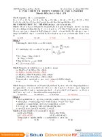
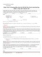
Fig.Q.51. Screening potential inhibitors of protein kinases
To measure the ability of a test compound to inhibit a kinase, phage displaying a specific
kinase is mixed with the magnetic beads in several wells of a 96-well plate. Then the test
compound is added to individual wells over a range of different concentrations. The
mixtures are incubated with gentle shaking for I hour at 25®C, the beads are pulled to the
bottom with a strong magnet, and all the free (unbound) components are washed away.
Finally, the remaining, attached phage are dissociated from the beads using an excess of
the same ATP analog that is attached to the beads, and counted by measuring the number
of plaques they form on a bacterial lawn on a Petri dish (Fig.Q.51).
Indicate in the Answer Sheet if each of the following statements is True or False.
A. When the binding process reaches equilibrium, all potential inhibitor molecules
will be bound to the kinase.
3
B. Test compounds that score well in this assay bind in the ATP-binding cleft of the
kinase.
C. Small differences in evolutionary conserved ATP binding sites on kinases allow
targeting specific kinases.
D. A strong binding test compound will yield a low count in the plaque assay.
Answer key:
A. False, B. False, C. True. D. True
Explanation:
A. False: At equilibrium, most inhibitors can bind to the kinase, but some
inhibitor molecules can dissociate from the kinase.
B. False: Some test compound could change the ATP binding site of the kinase
by binding to an allosteric region of the kinase, which could be very far away from the
binding site.
C. True: As the binding sites are similar but not identical between kinases,
molecules that are specific for one kind of kinases can be developed.
D. True: In the presence of a strongly binding test compound, most of the phage
will be attached to the test compound and will be washed away at the end of the
incubation. Thus, strongly binding test compounds will give a low count in the plaque
a s s a y.
Reference; Molecular Biolog of the cell. B. Alberts et al
Griffin JD (2005) Interaction maps for kinase inhibitors. Nat.Biotechnol. 23,308-309.
Fabian MA et al. (2005) A small molecule-kinase interaction map for clinical kinase
inhibitors. Nat. Biotechnol. 23, 329-336.
4
Q.52
You identified a gene in fission yeast, homologous to a telomerase subunit from a
protozoan. You then make a targeted deletion of one copy of the gene in a diploid strain
of the yeast and then induce sporulation to produce haploid organisms. All four spores
germinate perfectly, and you are able to grow colonies on nutrient agar plates. Every 3
days, you re-streak colonies onto fresh plates. After four such serial transfers, the
descendants of two of the original four spores grow poorly, if at all. You take cells from
the 3-, 6-, and 9-day master plates, prepare DNA from them, and cleave the samples at a
chromosomal site about 35 nucleotides away from the start of the telomere repeats. You
separate the fragments by gel electrophoresis, and hybridize them to a radioactive
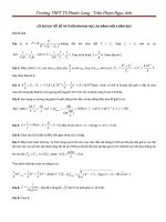
telomere-specific probe (Fig.Q.52). Assume that generation time is 6 hours.
spore 1 spore 2 spore 3 spore 4
days
369369369369S
markers
(bp)
Fig.Q.52. Analysis of telomeres from four fission-yeast spores.
WT is the normal diploid yeast
Indicate in the Answer Sheet if each of the following statements is True or False.
A. The average length of telomere in fission yeast is 280 nucleotides.
B. Spores 2 and 4 appear to lack telomerase.
C. Fission yeast telomeres lose less than 10 nucleotides per replication.
D. The fission yeasts that lose their telomeres will have normal size.
Answer key:
A. False; B. True, C. True. D. False
Explanation:
A. False: The region of intense hybridization to telomeres in the unaffected
spores (1 and 3) and normal diploid yeast extends from less than 200 nucleotides to just
5
over 300 nucleotides, averaging about 250 nucleotides. Since the cleavage site is 35
nucleotides from the beginning of the telomere repeats, the average length of telomere
repeat in fission yeast is just over 200 nucleotides.
B. True: The descendants of spores 2 and 4 show telomere shortening with time,
whereas the descendants of spores 1 and 3 remain the same size. Thus, spores 2 and 4
appear to lack telomerase
C. True: telomeres lose less than 100 nucleotides every 3 days. At four
generations per day [(24 hours/day)/(6 hours/generation)] the yeast go through about 12
generations in 3 days. Thus, they lose less than 8 nucleotides per generation (100
nucleotides/12 generations).
D. False: the majority of fission yeast that lose their telomeres stop dividing but
continue to grow in size, forming abnormally large cells.
Reference: Molecular Biolog of the cell. B. Alberts et al
Nakamura TM, Morin GB, Chapman KB, Weinrich SL, Andrews WH, Lingner J, Harley
CB & Cech TR (1997) Telomerase catalytic subunit homologs from fission yeast and
human. Science 277, 955-959.
6
Q.53
Reoxygenation after a period of lack of oxygen causes cardiomyocyte damage. One of
the most potential indices evaluating myocardial functions is mitochondrial membrane
potential, which is labeled by a cell permeant dye (positively-charged, grey color)
readily accumulating in active mitochondria due to their relative negative charge.
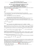
The figure below illustrates hypoxia/reoxygenation (HR)-treated single myocyte model
(1) with or without pre-hypoxic treatment of drug A. Myocyte images were captured at
time points (a, b, c).
Ti m e ( m i n )
(1)
(2)
(3)
Fig.Q.53.
Indicate in the Answer Sheet if each of the following statements is True or False.
A. As seen in Fig.Q.53.(2)a, cardiomyocytes are a type of striated muscle cells.
B. Hypoxia induces acidic pH in myocardial mitochondria.
C. Drug A pretreatment is good for cell because it prevents the collapse of
mitochondrial membrane potential in HR.
D. Captured images in drug A pretreatment group are presented in (2) and captured
images in HR treatment without pretreatment of drug A are presented in (3).
Answers key:
A. True, B. True, C. True, D. False.
Explanation:
A. True: See Fig. (2)
B. True: Hypoxia induces the accumulation of H^ in the matrix.
C. True: Drug A pretreatment is good for cell because it prevents the collapse of
mitochondrial membrane potential in HR.
- Collapse of mitochondrial membrane potential is presented with a reduction in
dye intensity level. So, drug A is good for cell.
7
D. False: Captured images in drug A pretreatment group are presented in (2) and
captured images in HR treatment without pretreatment of drug A are presented in (3).
- Capture images in drug A pretreatment group should be presented in (3) because
drug A protects cell from reoxygenation injury with more dye-label mitochondria (grey
color).
Reference
Angelos, M. G., V. K. Kutala, et al. (2006). "Hypoxic reperfusion of the ischemic heart
and oxygen radical generation." Am J Physiol Heart Circ Physiol 290(1): H341-H347.
Han, J., S.-J. Park, et al. (2013). "Effects of the novel angiotensin II receptor type I
antagonist, fimasartan on myocardial ischemia/reperfusion injury." International Journal
of Cardiology 168(3): 2851-2859.
Thu, V. T., H.-K. Kim, et al. (2012). "NecroX-5 prevents hypoxia/reoxygenation injury
by inhibiting the mitochondrial calcium uniporter." Cardiovascular Research 94(2): 342350.
8
Q.54
Antifreeze glycoproteins (AFGPs) possess the ability to inhibit the formation of ice and
are therefore essential to the survival of many marine teleost fishes that routinely
encounter sub-zero temperatures. A typical AFGP consists of repeating tripeptide units,
the alanyl-threonyl-alanyl (Ala-Thr-Ala)n unit connected to a disaccharide through a
glycosidic bond at the second hydroxyl group of the threonine residue. To identify
chemical groups which affect antifreeze activities of this glycoprotein, scientists
synthesized numerous AFGP analogues by modifying both the structure of the sugar
moieties and the peptide by replacing three groups R|, R2.R3 as shown in Fig.Q.54 with
different chemical groups and recorded the antifreeze activity.
r.
CH2OH
Fig.Q.54. The structure of a typical AFGP
The results of the study are shown in the following table.
Indicate in the Answer Sheet if each of the following statements is True or False.
A. A disaccharide bound to the threonine residue is required for antifreeze activity.
B. A mutant that has threonine residues replaced with serine residues significantly
reduces antifeeze activities.
C. AAacetyl group at the C-2 position is required for antifreeze activity.
9
D. Different numbers of repetitive motifs in AFGP genes amongst closely related
species might have been caused by DNA polymerase inaccuracy.
Answer key:
A: False, B: True, C: True, D: True
Explanation:
The purpose of the question is to test comparative skill and ability to analyze table data.
The question is also to test understanding on simple structure amino acids.
AFGPs display significant antifreeze activity if presence of the N-acetyl group at the C2
position of a disaccharide, and the methyl group of the threonine residue is not modified.
A. False. AFGP has activity when R3 is hydrogen (H), therefore the amino acid
residues binding with the monosaccharide is not required for antifreeze activity.
B. True. Serine residues can form glycosidic bonds with the disaccharide but they
do not have the hydrophobic group methyl (-CH3). In this case, R2 is FI (see the table) so
AFGP has no antifree activity.
C. True. If 7\/^acetyl is replaced with -OH or 0-acetyl, the activity of AFGP will be
lost.
D. True. Probably a slippage of DNA polymerase happened during replication and
resulted in extension of repetitive elements.
Reference
1. Jeong Kyu Bang, Jun Hyuck Lee Ravichandran N. Murugan, Sung Gu Lee,
Hackwon Do, Hye Yeon Koh, Hye-Eun Shim, Hyun-Cheol Kim and Hak Jun
Kim, 2013. Antifreeze Peptides and Glycopeptides, and Their Derivatives:
Potential Uses in Biotechnology. Marine Drugs: I, 2013-2041.
10
Q.55
Fi subunit (a peripheral membrane protein) of the ATP synthase catalyses ATP
synthesis using proton motive force responsible for the rotation of Fq subunit (integral
membrane protein complex) in one direction. F| is composed of three a and three b
subunits arranged in alternating manner around a central shaft, the y subunit.
To study the rotation, Masasuke Yoshida and his team attached a fluorescently labelled
actin filament to y and watched its movement.
fl u o r o p h o r e
a c t i n fi l a m e n t
Fig.Q.55A. Attachment of labelled actin filament to ATP synthase.
Rotating actin filaments were observed by an inverted fluorescence microscope after
addition of 2 mM ATP into a chamber containing actin-taged asbsy complex
immobilized on the bottom side) as a mirror image formed on a camera. The time
interval between images was 220 ms.Aseries of 13 images were taken and is shown in
Fig. Q.55B.
Fig.Q.55B. Sequential image of rotating filament attatched to the sunbunit in the asbsy
subcomplex. The number indicate the shot image
Indicate in the Answer Sheet if each of the following statements is True or False.
A. Hydrolysis of ATP by Fi leads to the conformational change of a and b subunits.
11
B. From the set of figures, the filament rotated anticlockwise (looking from the
membrane side).
C. Rotary rate is around 0.4 rounds per second.
D. Rotating the actin filament in the opposite direction is coupled with ATP
synthesis.
Answer key:
A: True, B: True, C: False, D: True
Explanation:
To uncover many biological processes, scientists usually analyze series of images taken
with time course. The idea in the question is to test ability to analyze a such series of
images taken with time course. Beside, the question is designed to test ability to
calculate data based on images as well as the position of ATPase on the membrane and
mechanism behind generation of motive force.
A. True. The motive force rotating y subunit results from set of conformational
changes in F1 subunits directly linked to the ATP hydrolysis, mirroring the changes
linked with the ATP synthesis.
B. True. From the set of figures, the filament rotated anticlockwise. The rotation is
obviously anticlockwise - but viewed from opposite direction, han the membrane side
and simultaneously as the mirror image. Resulting image is therefore anticlockwise.
C. False. Rotary rate is 0.375 - 0.400 rounds per second. The filament in frame 2
was at position 9 a'clock and after 9 frames (frame 12), filament was located at the same
position in the frame 2. Time for one round rotation is (220 x 9)/1000 = 1.98 s, thus the
rotary rate is 1/1.198= 0.50 rounds per second.
D. True. Rotating the actin filament clockwise is coupled with ATP synthesis. In the
experiment, hydrolysis of ATPase couples with the filament rotated anticlockwise. The
activity of ATP synthase must occur when filament rotate in the opposite direction,
clockwise.
Reference
1. Albert L. Lehninger, David L. Nelson and Michael M. Cox, 2008. Principles of
biochemistry, 5th edition. W.H. Freeman & Company.
2. Hiroyuki Noji, Ryohei yasuda, Masasuke Yoshida and kazuhiko Kinosith Jr, 1997.
Direct observation of rotation ofFl-ATPase. Nature, Vol. 386: 299-302
12
Q.56
Lactic fermented vegetables are traditional food in many Asian cuisines.
Microorganisms commonly found in the fermentation broth are lactic acid bacteria, yeast
and filamentous fungi.
Fig.Q.56 below shows the flowchart of viable cell counts (log CFU/mL) of three
different microbial groups and the pH value during the lactic fermentation course of
cabbage. Oxygen dissolved in fermentation broth decreased with time and was
completely consumed after the 22"^* day.
Fermentation time (days)
Fig.Q.56. Changes in microflora during lactic acid fermentation of cabbage.
Study Fig.Q.56 and indicate in the Answer Sheet if each of the following statements
is True or False.
A. The drop in pH value firom day 1 to day 3 was caused by only organic acids
produced by lactic acid bacteria.
B. Lactic acid produced by lactic acid bacteria favours the growth of yeast cells firom
day 10 till day 26.
C. Yeast cells shifted from fermentation to respiration after day 22.
D. Some filamentous fungi showed high tolerance at low pH.
Answer key:
A- False; B- True; C- False; D- True
Explanation:
13
A- False: Organic acid can be produce from the respiration of various organisms.
In the fermentation pile, not only lactic acid bacteria can produce organic acids but also
yeast and filamentous fungi can.
B- True: Most yeast cells have optimum pH from 4 to 4.5 for their grovvth.
Therefore, statement B is True.
C- False: After 22"'' day, dissolved oxygen in fermentation broth were depressed
so yeast cells must shift the respiration to the fermentation. Therefore, statement C is
False.
D- True: Some filamentous fungi (~ 10 CFU/mL) found in fermented cabbage at
the last stage. They are high tolerant to the low pH environment.
Reference
1- Microb Cell Factories 2011, Vol lOfSuppl 1):S5
2- Campbell R., Biology, Edition 9"*
14
Q.57
Microorganisms that live at high salt concentration (above 2M of NaCl) are exposed to
media with low water activity, and must have mechanisms to avoid water loss by
osmosis. Analyses of intracellular ionic concentration of Halobacteriales living in salt
lakes show that these microorganisms maintain extremely high salt (KCl) concentration
inside their cells. The presence of high intracellular salt concentration requires special
adaptations of the proteins and other macromolecules of the cells.
Indicate in the Answer Sheet if each of the following statements is True or False.
A. Most intracellular proteins of Halobacteriales contain a large excess of negative
charges on their outer surface.
B. Halobacteriales spend a lot of ATPs to maintain osmotic pressure.
C. Most intracellular enzymes of Halobacteriales lose their catalytic activity when
suspended in solutions containing less than 1 M NaCl.
D. Amino acids can be imported through Na'^/amino acids antiporters.
Answer key:
A. True, B. True, C. True. D. False
Explanation
A. True. The negative charges can help the protein maintain its proper conformation
required for structural stability and enzymatic activity at high concentrations of cations.
B. True. A lot of ATPs are used for maintaining extremely high salt (KCl)
concentration inside their cells and also for the extrusion of Na^ from the cell.
C. True. Most enzymes of the halobacteriales denatured when suspended in solution
containing less than 1 M NaCl.
D. False. Amino acids are imported throuth NaVamino acids symporters, the energy
for active transport of amino acids into the cell is provided by the Na gradient.
References:
Oren A. (2006). Life at high salt concentration. In: Dworkin M, Falkow S, Rosenberg E,
Schleifer K-H, Stackebrandt E (eds) The Prokaryotes. A Handbook on the Biology of
Bacteria: Ecophysiology and Biochemistry. Springer, New York. 2:263-28
15
Q.58
Influenza A genome consists of 8 separate single stranded RNA molecules, which
encode a total of 11 viral proteins. Influenza A viruses are categorized by their two
surface antigens, the hemagglutinin (H), of which there are 18 different subtypes (Hl18); and neuraminidase (N), of which there are 11 different subtypes (Nl-11)
(Fig.Q.58A). The influenza A virus life cycle is presented in Fig. Q.58B.
Fig.Q.58. Influenza A virus: (A) virus structure and (B) virus life cycle.
Indicate in the Answer Sheet if each of the following statements is True or False.
16
A. Influenza A viruses exhibit rapid evolutionary dynamics because the genome is
segmented.
B. In theory, there are 144 types of influenza A viruses.
C. Influenza A viruses exhibit high mutation rates because the genome is single
strand RNA.
D. Influenza A viruses will be active only if RNA-dependent RNA polymerase is
present in the virion.
Answer key
A. True, B. False, C. True. D. True
Explanation
A. True. A segmented genome provides a virus with the possibility of new gene
combinations, and hence a potential for more rapid evolution.
B. False. In theory, there are 198 types of Influenza A viruses.
C. True. Influenza A virus uses RNA-polymerase for its replication but the enzyme
does not possess a proof-reading-function. Hence, the error rate of viral RNApolymerase is higher than the error rate of DNA-polymerase.
D. True. RNA-dependent RNA polymerase can only synthesize by virus, if it is not
present in the virion the virus will be inactive.
References:
1. Nelson MI, Holmes EC (2007). The evolution of epidemic influenza. Nature Reviews
Genetics 8:196-205.
2. Medina RA, Garcia-Sastre A (2011). Influenza A viruses: new research developments.
Nature Reviews Microbiology 9:590-603.
17
Q.59
Phosphorylation is a major post-transiationai modification widely used in the regulation
of many cellular processes. A method to determine the phosphorylation status of proteins
is to run an electrophoresis in a modified gel with a chemical group containing metal
ions (M) that can reversibly bind phosphates and thus affects migration of
phosphorylated proteins.
Fig.Q59A. Phospho-tag poly aery lam ide gel
This technique was used to study the phosphorylation of protein p35. Three mutant
forms of this protein were generated: a serine to alanine substitution in position 8 (S8A);
a threonine to alanine substitution in position 138 (T138A) and both amino acid
substitutions (2A). Note that serine and threonine can be phosphorylated while alanine
cannot. Then two yeast strains with normal (wt) or inactive cyclin-dependent kinase 5
(CdkS) (kn) were transformed with either the wild type version of p35 gene (wt) or one
of the three mutant forms. Cell lysate of the eight resulting strains was loaded on a
Phospho-tag gel. The proteins from the gel were transferred by western-blot to a
membrane that was treated with anti-p35 antibodies. The result is shown below.
CdkS
kn
wt
Fig.Q59B. Immunoblotting with anti-p35. The arrow indicates the direction of migration
p35 bands are named Ml, M2, LI, L2, L3, and L4. L4 band corresponds to the
completely non-phosphorylated form of p35.
18
Based on the results of the experiment given above, indicate in the Answer Sheet if
each of following statements is True or False.
A. Protein p35 has only two phosphorylation sites: serine 8 and threonine 138.
B. Protein p35 can be phosphorylated by a protein kinase different from Cdk5.
C. In strain Cdk5-wt p35-S8A only a few p35 molecules are phosphorylated at TI38.
D. Phosphate groups attached to 88 are more accessible to phosphate binding groups
of the Phospho-tag gel than phosphate groups attached to T138.
Answerkey:
A: False, B: True, C: False, D: True
Explanation:
A. False. Band L3 present in lane 6 corresponds to a phosphorylated protein. As this
mutant has both 88 and T138 mutated to alanine, there should be at least one more site
that can be phosphorylated.
B. True. Presence of phosphorylated form of the protein (band L2) in the kn cells
lacking CdkS proves that protein p35 can be phosphorylated by a protein kinase diferent
from Cdk5.
C. False. As L2 corresponds to p35 phosphorylated at T138 only and LI corresponds
to p35 phosphorylated at two sites T138 and an additonal site (X) as can be seen from
comparison between lines 6 and 7 all p35 molecules in this strain are phosphorylated at
T138).
D. True. M2 band corresponds to p35 phosphorylated at 88. It might be
phosphorylated at residue X as wel. M2 has a much lower mobilty compared to both L2
(phosphorylated at T138) and LI (phosphorylated at T138 and X). The diference in
mobility given the same number of phosphates attached per molecule makes us suppose
that the mobility effect of phosphorylation of these two sites is different. As
phosphorylation of 88 has a stronger effect its phosphate should interact stronger with
the gel).
Reference
Mol Cell Proteomics. 2010 Jun; 9(6): 1133-43.
19
Q.60
Polarity, charge and moiecuiar weight of molecules can affect their rate of passive
diffusion through membranes. Amino acids and drugs like aspirin differ in both
efficiency and location of absorption. In the figure below the chemical structure the pKa
values of aspirin and arginine are represented.
2.18
^NHa—C
Aspirin
H
Arginine
Indicate in the Answer Sheet if each of following statements is True or False.
A. Aspirin diffuses through membranes mainly in the stomach because more aspirin
molecules are in deprotonated form at pH of about 1.6 in the stomach.
B. Arginine diffuses less efficiently than aspirin because of its higher molecular
weight.
C. Optimal pH range for arginine absorption by passive difusion is between 2.18 and
9.04.
D. The proton pump inhibitor, omeprazole, blocks the entry of aspirin into the blood
in the initial few minutes after oral administration.
Answer key:
A: False, B: False, C: False, D: True
Explanation:
The purpose of the question is to test understanding factors that affect membrane
transport through analysis of moiecuiar absorption in the stomach and intestine. Because
20
the pH dfiers n
i the stomach and the n
i testn
i e, absorpto
i no
l cato
i n si dfierent.
Stomach pH (pH,) =1.6, Blood pH=7.4
pKa of aspirin = 3.4., weak acid;
HA <->fr + A"
pH= pKa + Ig ([A-]/[HA)) -> Ig ((A1/[HA]) = pH-pKa
in stomach:
Ig ([A-]/[HA]) =1.6-3.4=-1.8
[A7[HA])= 0.016
[HA] = 63 [A-]
in blood:
lg([A-]/[HA]) =7.4-3.4= 4
[A-]/[HA])= 10000
[HA] = 0.0001 [A-]
[HA] from stomach to blood
A. Fasl e. . Because at stomach pH si e
l ss than the pi of asprin, the proporto
i n of
protonatedformofasprn
i si hg
iher.Themorechargedmoe
lcue
lsare,thee
lssdfu
ise
through the membrane.
B. Fasl e. Because the moe
l cua
l r weg
i ht of argn
in
i e si 174.2, smae
l r than that of
aspirin (180. 16)
C. False.Arginine is neutral (zwiterionic) between pH 9.04 and 12.48.
D. True. Aspirin is absorbed mainly in the stomach in the initial few minutes.
Omeprazoe
l causesn
i creasen
i pHn
i the stomach becauseti n
i hb
i tis proton pumpn
ig
into the stomach lumen.
Reference
1. Griaud N.M. et a.l Efect of omeprazoe
l on the bo
i avaa
li btily of unmodfii ed and
phosphop
ild
i -compe
l xed asprinn
i rats.Am
il ent PharmacolTher.199711(5);899906.
2. P.B. Mn
i er Jr, J. G. Fort^' & Y.Zhang. Intragastrci acd
i tiy and omeprazoe
l exposure
durn
i gdosn
i gwth
i eth
i erPA32540(enterci-coatedasprin325mg+m
i meda
i teree
l ase omeprazoe
l 40 mg) or enterci-coated asprin 325 mg 4 enterci-coated
omeprazoe
l 40 mg - a randomsied, phase 1, crossover study :
21
3. Wiliam D. Masonx, Nathaniel Winer. Kinetics of Aspirin, Salicylic Acid, and
SalicyiuricAcid foliowing OralAdministration ofAspirin as a Tablet and Two
Buffered Solution
22
PLANT ANATOMY AND PHYSIOLOGY
Tô"u
tdyh
teee
fcst ofcadmuim(Cd)onrootdeveolpmen,tw
t oexpem
ri enst onmazie
seedn
il gs wtih 6-cm-o
l ng root were conducted. In the f.rst experm
i ent, seedn
il gs were
growneth
iern
i meda
i suppe
lmentedwth
i 5pMCd(Cd5)orwth
ioutCd(CdO).Inthe
second experm
i ent, seedn
il gs were grown etihern
i twoa
l yers of agar wtihout Cd (CdOCdO) or una
li terayl to 100 pM Cd (CdO-CdO
l O). Four daysa
l ter,root growth was
recorded and cross-sections of roots were stained to visualize suberin lamilae in
endodermis.
Fg
i.Q61-.lDsitance(%)fromrootp
ti torootbasesi shownonthee
lft.Thepresenceof
subern
ia
l ma
li en
i the roots are shown as sod
il and dashedn
il esn
i the center figures.
White arrows in A and B indicate suberin lamellae in the endodermis.
23