Phasing in crystallography a modern perspective (iucr texts on crystallography) by carmelo giacovazzo
Bạn đang xem bản rút gọn của tài liệu. Xem và tải ngay bản đầy đủ của tài liệu tại đây (5.52 MB, 433 trang )
I N T E R N AT I O N A L U N I O N O F C RY S TA L L O G R A P H Y
BOOK SERIES
I U Cr B O O K S E R I E S C O M M I T T E E
J. Bernstein, Israel
P. Colman, Australia
J. R. Helliwell, UK
K. A. Kantardjieff, USA
T. Mak, China
P. Müller, USA
Y. Ohashi, Japan
P. Paufler, Germany
H. Schenk, The Netherlands
D. Viterbo (Chairman), Italy
IUCr Monographs on Crystallography
1
2
3
4
5
6
7
8
9
10
11
12
13
14
15
16
17
Accurate molecular structures
A. Domenicano, I. Hargittai, editors
P.P. Ewald and his dynamical theory of X-ray diffraction
D.W.J. Cruickshank, H.J. Juretschke, N. Kato, editors
Electron diffraction techniques, Vol. 1
J.M. Cowley, editor
Electron diffraction techniques, Vol. 2
J.M. Cowley, editor
The Rietveld method
R.A. Young, editor
Introduction to crystallographic statistics
U. Shmueli, G.H. Weiss
Crystallographic instrumentation
L.A. Aslanov, G.V. Fetisov, J.A.K. Howard
Direct phasing in crystallography
C. Giacovazzo
The weak hydrogen bond
G.R. Desiraju, T. Steiner
Defect and microstructure analysis by diffraction
R.L. Snyder, J. Fiala, H.J. Bunge
Dynamical theory of X-ray diffraction
A. Authier
The chemical bond in inorganic chemistry
I.D. Brown
Structure determination from powder diffraction data
W.I.F. David, K. Shankland, L.B. McCusker, Ch. Baerlocher, editors
Polymorphism in molecular crystals
J. Bernstein
Crystallography of modular materials
G. Ferraris, E. Makovicky, S. Merlino
Diffuse X-ray scattering and models of disorder
T.R. Welberry
Crystallography of the polymethylene chain: an inquiry into the structure of waxes
D.L. Dorset
18
19
20
21
22
23
24
25
Crystalline molecular complexes and compounds: structure and principles
F.H. Herbstein
Molecular aggregation: structure analysis and molecular simulation of crystals
and liquids
A. Gavezzotti
Aperiodic crystals: from modulated phases to quasicrystals
T. Janssen, G. Chapuis, M. de Boissieu
Incommensurate crystallography
S. van Smaalen
Structural crystallography of inorganic oxysalts
S.V. Krivovichev
The nature of the hydrogen bond: outline of a comprehensive hydrogen bond theory
G. Gilli, P. Gilli
Macromolecular crystallization and crystal perfection
N.E. Chayen, J.R. Helliwell, E.H. Snell
Neutron protein crystallography: hydrogen, protons, and hydration in
bio-macromolecules
N. Niimura, A. Podjarny
IUCr Texts on Crystallography
1
4
8
9
10
11
12
13
14
15
16
17
18
19
20
The solid state
A. Guinier, R. Julien
X-ray charge densities and chemical bonding
P. Coppens
Crystal structure refinement: a crystallographer’s guide to SHELXL
P. Müller, editor
Theories and techniques of crystal structure determination
U. Shmueli
Advanced structural inorganic chemistry
Wai-Kee Li, Gong-Du Zhou, Thomas Mak
Diffuse scattering and defect structure simulations: a cook book using the program
DISCUS
R.B. Neder, T. Proffen
The basics of crystallography and diffraction, third edition
C. Hammond
Crystal structure analysis: principles and practice, second edition
W. Clegg, editor
Crystal structure analysis: a primer, third edition
J.P. Glusker, K.N. Trueblood
Fundamentals of crystallography, third edition
C. Giacovazzo, editor
Electron crystallography: electron microscopy and electron diffraction
X. Zou, S. Hovmöller, P. Oleynikov
Symmetry in crystallography: understanding the International Tables
P.G. Radaelli
Symmetry relationships between crystal structures: applications
of crystallographic group theory in crystal chemistry
U. Müller
Small angle X-ray and neutron scattering from biomacromolecular solutions
D.I. Svergun, M.H.J. Koch, P.A. Timmins, R.P. May
Phasing in crystallography: a modern perspective
C. Giacovazzo
Phasing in
Crystallography
A Modern Perspective
CARMELO GIACOVAZZO
Professor of Crystallography,
University of Bari, Italy
Institute of Crystallography, CNR, Bari, Italy
3
3
Great Clarendon Street, Oxford, OX2 6DP,
United Kingdom
Oxford University Press is a department of the University of Oxford.
It furthers the University’s objective of excellence in research, scholarship,
and education by publishing worldwide. Oxford is a registered trade mark of
Oxford University Press in the UK and in certain other countries
c Carmelo Giacovazzo 2014
The moral rights of the author have been asserted
First Edition published in 2014
Impression: 1
All rights reserved. No part of this publication may be reproduced, stored in
a retrieval system, or transmitted, in any form or by any means, without the
prior permission in writing of Oxford University Press, or as expressly permitted
by law, by licence or under terms agreed with the appropriate reprographics
rights organization. Enquiries concerning reproduction outside the scope of the
above should be sent to the Rights Department, Oxford University Press, at the
address above
You must not circulate this work in any other form
and you must impose this same condition on any acquirer
Published in the United States of America by Oxford University Press
198 Madison Avenue, New York, NY 10016, United States of America
British Library Cataloguing in Publication Data
Data available
Library of Congress Control Number: 2013943731
ISBN 978–0–19–968699–5
Printed in Great Britain by
Clays Ltd, St Ives plc
Links to third party websites are provided by Oxford in good faith and for information
only. Oxford disclaims any responsibility for the materials contained in any third party
website referenced in this work.
Dedication
To my mother,
to my wife Angela,
my sons Giuseppe and Stefania,
to my grandchildren Agostino, Stefano and Andrea Morris
Acknowledgements
I acknowledge the following colleagues and friends for their generous help:
Caterina Chiarella, for general secretarial management of the book and for her
assistance with the drawings;
Angela Altomare, Benedetta Carrozzini, Corrado Cuocci, Giovanni Luca
Cascarano, Annamaria Mazzone, Anna Grazia Moliterni, and Rosanna
Rizzi for their kind support, helpful discussions, and critical reading of the
manuscript. Corrado Cuocci also took care of the cover figure.
Facilities provided by the Istituto di Cristallografia, CNR, Bari, are gratefully
acknowledged.
Preface
A short analysis of the historical evolution of phasing methods may be a useful
introduction to this book because it will allow us to better understand efforts
and results, the birth and death of scientific paradigms, and it will also explain
the general organization of this volume. This analysis is very personal, and
arises through the author’s direct interactions with colleagues active in the
field; readers interested in such aspects may find a more extensive exposition
in Rend. Fis. Acc. Lincei (2013), 24(1), pp. 71–76.
In a historical sense, crystallographic phasing methods may be subdivided
into two main streams: the small and medium-sized molecule stream, and the
macro-molecule stream; these were substantially independent from each other
up until the 1990s. Let us briefly consider their achievements and the results of
their subsequent confluence.
Small and medium-sized molecule stream
The Patterson (1934) function was the first general phasing tool, particularly
effective for heavy-atom structures (e.g. this property met the requirements
of the earth sciences, the first users of early crystallography). Even though
subsequently computerized, it was soon relegated to a niche by direct methods,
since these were also able to solve light-atom structures (a relevant property
towards the development of organic chemistry).
Direct methods were introduced, in their modern probabilistic guise, by
Hauptman and Karle (1953) and Cochran (1955); corresponding phasing procedures were automated by Woolfson and co-workers, making the crystal
structure solution of small molecules more straightforward. Efforts were carried out exclusively in reciprocal space (first paradigm of direct methods);
the paradigm was systematized by the neighbourhood (Hauptman, 1975) and
representation theories (Giacovazzo, 1977, 1980). Structures up to 150 nonhydrogen (non-H) atoms in the asymmetric unit were routinely able to be
solved.
The complete success of this stream may be deduced from the huge numbers of structures deposited in appropriate data banks. Consequently, western
national research agencies no longer supported any further research in the
small to medium-sized molecule area (the work was done!); research groups
working on methods moved instead to powder crystallography, electron crystallography, or to proteins, all areas of technological interest for which phasing
was still a challenge. Direct space approaches were soon developed, which
enhanced our capacity to solve structures, even from low quality diffraction
data.
viii
Preface
The macromolecule stream
Since the 1950s, efforts were confined to isomorphous replacement (SIR, MIR;
Green et al., 1954), molecular replacement (MR; Rossmann and Blow, 1962),
and anomalous dispersion techniques (SAD-MAD; Okaya and Pepinsky,
1956; Hoppe and Jakubowski, 1975). Ab initio approaches, the main techniques of interest for the small and medium-sized molecule streams, were
neglected as being unrealistic; indeed, they are less demanding in terms of
prior information but are very demanding in terms of data resolution.
The popularity of protein phasing techniques changed dramatically over the
years. At the very beginning, SIR-MIR was the most popular method, but soon
MR started to play a more major role as good structural models became progressively more readily available. About 75% of structures today are solved
using MR. The simultaneous technological progress in synchrotron radiation
and its wide availability have increased the appeal of SAD-MAD techniques.
The achievements obtained within the macromolecular stream have been
impressive. A huge number of protein structures has been deposited in the
Protein Data Bank, and the solution of protein structures is no longer confined
to just an elite group of scientists, it is performed in many laboratories spread
over four continents, often by young scientists. Crucial to this has been the role
of the CCP4 project, for the coordination of new methods and new computer
programs.
The synergy of the two streams
It is the opinion of the author that synergy between the two streams originated due to a common interest in EDM (electron density modification)
techniques. This approach, first proposed by Hoppe and Gassman (1968) for
small molecules, was later extensively modified to be useful for both streams.
Confluence of the two streams began in the 1990s (even if contacts were begun
in the 1980s), when EDM techniques were used to improve the efficiency of
direct methods. That was the beautiful innovation of shake and bake (Weeks
et al., 1994); both direct and reciprocal space were explored to increase phasing efficiency (this was the second paradigm of direct methods). It was soon
possible to solve ab initio structures with up to 2000 non-hydrogen atoms in the
asymmetric unit, provided data at atomic or quasi-atomic resolution are available. As a consequence, the ab initio approach for proteins started to attract
greater attention. A secondary effect of the EDM procedures was the recent
discovery of new ab initio techniques, such as charge flipping and VLD (vive
la difference), and the newly formulated Patterson techniques.
The real revolution in the macromolecular area occurred when probabilistic
methods, already widely used in small and medium-sized molecules, erupted
into the protein field. Joint probability distributions and maximum likelihood
approaches were tailored to deal with large structures, imperfect isomorphism,
and errors in experimental data; and they were applied to SAD-MAD, MR, and
SIR-MIR cases. For example, protein substructures with around 200 atoms in
the asymmetric unit, an impossible challenge for traditional techniques, could
easily be solved by the new approaches.
Preface
ix
High-throughput crystallography is now a reality: protein structures,
50 years ago solvable only over months or years, can now be solved in hours
or days; also due to technological advances in computer sciences.
The above considerations have been the basic reason for reconsidering the
material and the general guidelines given in my textbook Direct Phasing in
Crystallography, originally published in 1998. This was essentially a description of the mathematical bases of direct methods and of their historical
evolution, with some references to applicative aspects and ancillary techniques.
The above described explosion in new phasing techniques and the improved
efficiency of the revisited old methods made impellent the need for a new textbook, mainly addressing the phasing approaches which are alive today, that
is those which are applicable to today’s routine work. On the other hand,
the wide variety of new methods and their intricate relationship with the old
methods requires a new rational classification: methods similar regarding the
type of prior information exploited, mathematical technique, or simply their
mission, are didactically correlated, in such a way as to offer an organized
overview of the current and of the old approaches. This is the main aim of
this volume, which should not therefore simply be considered as the second
edition of Direct Phasing in Crystallography, but as a new book with different
guidelines, different treated material, and a different purpose.
Attention will be focused on both the theoretical and the applicative aspects,
in order to provide a friendly companion for our daily work. To emphasize
the new design the title has been changed to Phasing in Crystallography, with
the subtitle, A Modern Perspective. In order to make the volume more useful,
historical developments of phasing approaches that are not in use today, are
simply skipped, and readers interested in these are referred to Direct Phasing
in Crystallography.
This volume also aims at being a tool to inspire new approaches. On the
one hand, we have tried to give, in the main text, descriptions of the various
methods that are as simple as possible, so that undergraduate and graduate
students may understand their general purpose and their applicative aspects.
On the other hand, we did not shrink from providing the interested reader with
mathematical details and/or demonstrations (these are necessary for any book
dealing specifically with methods). These are confined in suitable appendices
to the various chapters, and aimed at the trained crystallographer. At the end
of the book, we have collected together mathematical appendices of a general
character, appendices denoted by the letter M for mathematics and devoted to
the bases of the methods (e.g. probability theory, basic crystallography, concepts of analysis and linear algebra, specific mathematical techniques, etc.),
thus offering material of interest for professional crystallographers.
A necessary condition for an understanding of the content of the book is a
knowledge of the fundamentals of crystallography. Thus, in Chapter 1 we have
synthesized the essential elements of the general crystallography and we have
also formulated the basic postulate of structural crystallography; the entire
book is based on its validity.
In Chapter 2, the statistics of structure factors is described simply: it will be
the elementary basis of most of the methods described throughout the volume.
x
Preface
Chapter 3 is a simplified description of the concepts of structure invariant
and seminvariant, and of the related origin problem.
In Chapter 4, we have synthesized the methods of joint probability distributions and neighbourhoods–representation theories. The application of
these methods to three-phase and four-phase structure invariants are described
in Chapter 5. The probabilistic estimation of structure seminvariants has
been skipped owing to their marginal role in modern phasing techniques.
In Chapter 6, we discuss direct methods and the most traditional phasing
approaches.
Chapter 7 is dedicated to joint probability distribution functions when a
model is available, with specific attention to two- and to three-phase invariants.
The most popular Fourier syntheses are described in the same chapter and their
potential discussed in relation with the above probability distributions.
Chapter 8 is dedicated to phase improvement and extension via electron
density modification techniques, Chapter 9 to two new phasing approaches,
charge flipping and VLD (vive la difference), and Chapter 10, to Patterson
techniques. Their recent revision has made them one of the most powerful
techniques for ab initio phasing and particularly useful for proteins.
X-rays are not always the most suitable radiation for performing a diffraction experiment. Indeed, neutron diffraction may provide information
complementary to that provided by X-ray data, electron diffraction becoming necessary when only nanocrystals are available. In Chapter 11 phasing
procedures useful for this new scenario are described.
Often single crystals of sufficient size and quality are not available, but
microcrystals can be grown. In this case powder data are collected; diffraction
techniques imply a loss of experimental information, and therefore phasing via
such data requires significant modifications to the standard methods. These are
described in Chapter 12.
Chapters 13 to 15 are dedicated to the most effective and popular methods
used in macromolecular crystallography: the non-ab initio methods, Molecular
Replacement (MR), Isomorphous Replacement (SIR-MIR), and Anomalous
Dispersion (SAD-MAD) techniques.
The reader should not think that the book has been partitioned into two
parts, the first devoted to small and medium-sized molecules, the second to
macromolecules. Indeed in the first twelve chapters, most of the mathematical
tools necessary to face the challenges of macromolecular crystallography are
described, together with the main algorithms used in this area and the fundamentals of the probabilistic approaches employed in macromolecular phasing.
This design allows us to provide, in the last three chapters, simpler descriptions
of MR, SIR-MIR, and SAD-MAD approaches.
Contents
Symbols and notation
1 Fundamentals of crystallography
1.1
1.2
1.3
1.4
1.5
1.6
1.7
Introduction
Crystals and crystallographic symmetry in direct space
The reciprocal space
The structure factor
Symmetry in reciprocal space
1.5.1 Friedel law
1.5.2 Effects of symmetry operators in reciprocal space
1.5.3 Determination of reflections with restricted phase values
1.5.4 Systematic absences
The basic postulate of structural crystallography
The legacy of crystallography
2 Wilson statistics
2.1 Introduction
2.2 Statistics of the structure factor: general considerations
2.3 Structure factor statistics in P1 and P1¯
2.4 The P(z) distributions
2.5 Cumulative distributions
2.6 Space group identification
2.7 The centric or acentric nature of crystals: Wilson statistical analysis
2.8 Absolute scaling of intensities: the Wilson plot
2.9 Shape of the Wilson plot
2.10 Unit cell content
Appendix 2.A Statistical calculations in P1 and P1¯
2.A.1 Structure factor statistics in P1
2.A.2 Structure factor statistics in P1¯
Appendix 2.B Statistical calculations in any space group
2.B.1 The algebraic form of the structure factor
2.B.2 Structure factor statistics for centric and acentric space groups
Appendix 2.C The Debye formula
3 The origin problem, invariants, and seminvariants
3.1
3.2
Introduction
Origin, phases, and symmetry operators
xvii
1
1
1
5
11
12
12
12
13
15
17
24
27
27
28
29
35
35
36
42
43
47
49
50
50
52
53
53
55
58
60
60
61
xii
Contents
3.3
3.4
3.5
3.6
3.7
The concept of structure invariant
Allowed or permissible origins in primitive space groups
The concept of structure seminvariant
Allowed or permissible origins in centred cells
Origin definition by phase assignment
4 The method of joint probability distribution
functions, neighbourhoods, and representations
4.1
4.2
4.3
4.4
Introduction
Neighbourhoods and representations
Representations of structure seminvariants
Representation theory for structure invariants extended to
isomorphous data
Appendix 4.A The method of structure factor joint probability
distribution functions
4.A.1 Introduction
4.A.2 Multivariate distributions in centrosymmetric structures:
the case of independent random variables
4.A.3 Multivariate distributions in non-centrosymmetric
structures: the case of independent random variables
4.A.4 Simplified joint probability density functions in the
absence of prior information
4.A.5 The joint probability density function when some prior
information is available
4.A.6 The calculation of P(E) in the absence of prior
information
5 The probabilistic estimation of triplet
and quartet invariants
5.1 Introduction
5.2 Estimation of the triplet structure invariant via its first
representation: the P1 and the P1¯ case
5.3 About triplet invariant reliability
5.4 The estimation of triplet phases via their second representation
5.5 Introduction to quartets
5.6 The estimation of quartet invariants in P1 and P1¯ via their
first representation: Hauptman approach
5.7 The estimation of quartet invariants in P1 and P1¯ via their
first representation: Giacovazzo approach
5.8 About quartet reliability
Appendix 5.A The probabilistic estimation of the triplet
invariants in P1
Appendix 5.B Symmetry inconsistent triplets
Appendix 5.C The P10 formula
Appendix 5.D The use of symmetry in quartet estimation
63
65
69
76
81
83
83
87
89
91
93
93
94
97
99
102
103
104
104
104
108
110
112
112
115
116
117
120
121
123
Contents
6 Traditional direct phasing procedures
7
xiii
125
6.1 Introduction
6.2 The tangent formula
6.3 Procedure for phase determination via traditional direct
methods
6.3.1 Set-up of phase relationships
6.3.2 Assignment of starting phases
6.3.3 Phase determination
6.3.4 Finding the correct solution
6.3.5 E-map interpretation
6.3.6 Phase extension and refinement: reciprocal space techniques
6.3.7 The limits of the tangent formula
6.4 Third generation direct methods programs
6.4.1 The shake and bake approach
6.4.2 The half-bake approach
6.4.3 The SIR2000-N approach
Appendix 6.A Finding quartets
130
131
134
136
137
138
140
141
144
144
147
148
149
Joint probability distribution functions when
a model is available: Fourier syntheses
151
7.1 Introduction
7.2 Estimation of the two-phase structure invariant (φ h − φph )
7.3 Electron density maps
7.3.1 The ideal Fourier synthesis and its properties
7.3.2 The observed Fourier synthesis
7.3.3 The difference Fourier synthesis
7.3.4 Hybrid Fourier syntheses
7.4 Variance and covariance for electron density maps
7.5 Triplet phase estimate when a model is available
Appendix 7.A Estimation of σA
Appendix 7.B Variance and covariance expressions for electron
density maps
Appendix 7.C Some marginal and conditional
probabilities of P(R, Rp , φ, φp )
8 Phase improvement and extension
8.1 Introduction
8.2 Phase extension and refinement via direct space procedures:
EDM techniques
8.3 Automatic model building
8.4 Applications
Appendix 8.A Solvent content, envelope definition, and solvent modelling
8.A.1 Solvent content according to Matthews
8.A.2 Envelope definition
8.A.3 Models for the bulk solvent
Appendix 8.B Histogram matching
Appendix 8.C A brief outline of the ARP/wARP procedure
125
128
151
152
155
156
162
164
166
168
170
173
174
176
177
177
177
184
188
190
190
191
192
193
196
xiv
Contents
9 Charge flipping and VLD (vive la difference)
198
9.1 Introduction
9.2 The charge flipping algorithm
9.3 The VLD phasing method
9.3.1 The algorithm
9.3.2 VLD and hybrid Fourier syntheses
9.3.3 VLD applications to ab initio phasing
Appendix 9.A About VLD joint probability distributions
9.A.1 The VLD algorithm based on difference Fourier synthesis
9.A.2 The VLD algorithm based on hybrid Fourier syntheses
Appendix 9.B The RELAX algorithm
198
199
201
201
205
205
206
206
211
212
10 Patterson methods and direct space properties
214
10.1 Introduction
10.2 The Patterson function
10.2.1 Mathematical background
10.2.2 About interatomic vectors
10.2.3 About Patterson symmetry
10.3 Deconvolution of Patterson functions
10.3.1 The traditional heavy-atom method
10.3.2 Heavy-atom search by translation functions
10.3.3 The method of implication transformations
10.3.4 Patterson superposition methods
10.3.5 The C-map and superposition methods
10.4 Applications of Patterson techniques
Appendix 10.A Electron density and phase relationships
Appendix 10.B Patterson features and phase relationships
214
215
215
216
217
218
219
220
221
223
225
227
230
232
11 Phasing via electron and neutron diffraction data
11.1
11.2
11.3
11.4
11.5
Introduction
Electron scattering
Electron diffraction amplitudes
Non-kinematical character of electron diffraction amplitudes
A traditional experimental procedure for electron
diffraction studies
11.6 Electron microscopy, image processing, and phasing methods
11.7 New experimental approaches: precession and rotation cameras
11.8 Neutron scattering
11.9 Violation of the positivity postulate
Appendix 11.A About the elastic scattering of electrons: the
kinematical approximation
12 Phasing methods for powder data
12.1
12.2
Introduction
About the diffraction pattern: peak overlapping
234
234
235
236
237
239
241
244
245
247
249
252
252
253
Contents
12.3 Modelling the diffraction pattern
12.4 Recovering |Fhkl |2 from powder patterns
12.5 The amount of information in a powder diagram
12.6 Indexing of diffraction patterns
12.7 Space group identification
12.8 Ab initio phasing methods
12.9 Non-ab initio phasing methods
Appendix 12.A Minimizing texture effects
13 Molecular replacement
13.1
13.2
13.3
13.4
13.5
13.6
13.7
13.8
13.9
Introduction
About the search model
About the six-dimensional search
The algebraic bases of vector search techniques
Rotation functions
Practical aspects of the rotation function
The translation functions
About stochastic approaches to MR
Combining MR with ‘trivial’ prior information: the
ARCIMBOLDO approach
13.10 Applications
Appendix 13.A Calculation of the rotation function in
orthogonalized crystal axes
13.A.1 The orthogonalization matrix
13.A.2 Rotation in Cartesian space
13.A.3 Conversion to fractional coordinates
13.A.4 Symmetry and the rotation function
Appendix 13.B Non-crystallographic symmetry
13.B.1 NCS symmetry operators
13.B.2 Finding NCS operators
13.B.3 The translational NCS
Appendix 13.C Algebraic forms for the rotation and translation functions
14 Isomorphous replacement techniques
14.1 Introduction
14.2 Protein soaking and co-crystallization
14.3 The algebraic bases of SIR techniques
14.4 The algebraic bases of MIR techniques
14.5 Scaling of experimental data
14.6 The probabilistic approach for the SIR case
14.7 The probabilistic approach for the MIR case
14.8 Applications
Appendix 14.A The SIR case for centric reflections
Appendix 14.B The SIR case: the one-step procedure
Appendix 14.C About methods for estimating the scattering
power of the heavy-atom substructure
xv
258
260
263
264
266
267
270
272
275
275
277
279
280
282
284
286
289
289
291
294
294
295
297
299
304
304
305
308
311
314
314
315
317
320
322
323
327
329
330
331
333
xvi
Contents
15 Anomalous dispersion techniques
15.1 Introduction
15.2 Violation of the Friedel law as basis of the phasing method
15.3 Selection of dispersive atoms and wavelengths
15.4 Phasing via SAD techniques: the algebraic approach
15.5 The SIRAS algebraic bases
15.6 The MAD algebraic bases
15.7 The probabilistic approach for the SAD-MAD case
15.8 The probabilistic approach for the SIRAS-MIRAS case
15.9 Anomalous dispersion and powder crystallography
15.10 Applications
Appendix 15.A A probabilistic formula for the SAD case
Appendix 15.B Structure refinement for MAD data
Appendix 15.C About protein phase estimation in the SIRAS case
Appendices
Appendix M.A Some basic results in probability theory
M.A.1 Probability distribution functions
M.A.2 Moments of a distribution
M.A.3 The characteristic function
M.A.4 Cumulants of a distribution
M.A.5 The normal or Gaussian distribution
M.A.6 The central limit theorem
M.A.7 Multivariate distributions
M.A.8 Evaluation of the moments in structure factor distributions
M.A.9 Joint probability distributions of the signs of the
structure factors
M.A.10 Some measures of location and dispersion in the
statistics of directional data
Appendix M.B Moments of the P(Z) distributions
Appendix M.C The gamma function
Appendix M.D The Hermite and Laguerre polynomials
Appendix M.E Some results in the theory of Bessel functions
M.E.1 Bessel functions
M.E.2 Generalized hypergeometric functions
Appendix M.F Some definite integrals and formulas of frequent application
References
Index
335
335
337
340
344
347
352
354
360
363
364
365
366
368
370
370
370
371
371
373
374
375
375
377
379
380
382
382
383
385
385
389
390
394
412
Symbols and notation
The following symbols and conventions will be used throughout the full text.
The bold character is used for denoting vectors and matrices.
h·r
a∧b
¯
A
s.f.
n.s.f.
s.i.
s.s.
cs.
n.cs.
RES
CORR
the dot indicates the scalar product of the two vectors h and r
cross-product of the two vectors a and b
the bar indicates the transpose of the matrix A
structure factor
normalized structure factor
structure invariant
structure seminvariant
centrosymmetric
non-centrosymmetric
experimental data resolution (in Å)
correlation between the electron density map of the target
structure (the one we want to solve) and that of a model map
Rcryst =
SIR-MIR
SAD-MAD
MR
h
||Fobs |−|Fcalc ||
h |Fobs |
crystallographic residual
single–multiple isomorphous replacement
single–multiple anomalous dispersion
molecular replacement
1
Fundamentals
of crystallography
1.1 Introduction
In this chapter we summarize the basic concepts, formulas and tables which
constitute the essence of general crystallography. In Sections 1.2 to 1.5 we
recall, without examples, definitions for unit cells, lattices, crystals, space
groups, diffraction conditions, etc. and their main properties: reading these
may constitute a useful reminder and support for daily work. In Section 1.6
we establish and discuss the basic postulate of structural crystallography: this
was never formulated, but during any practical phasing process it is simply
assumed to be true by default. We will also consider the consequences of such
a postulate and the caution necessary in its use.
1.2 Crystals and crystallographic symmetry
in direct space
We recall the main concepts and definitions concerning crystals and crystallographic symmetry.
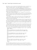
Crystal. This is the periodic repetition of a motif (e.g. a collection of molecules,
see Fig. 1.1). An equivalent mathematical definition is: the crystal is the convolution between a lattice and the unit cell content (for this definition see
(1.4) below in this section).
Unit cell. This is the parallelepiped containing the motif periodically repeated
in the crystal. It is defined by the unit vectors a, b, c, or, by the six scalar
parameters a, b, c, α, β, γ (see Fig. 1.1). The generic point into the unit cell is
defined by the vector
r = x a + y b + z c,
where x, y, z are fractional coordinates (dimensionless and lying between
0 and 1). The volume of the unit cell is given by (see Fig. 1.2)
V = a ∧ b · c = b ∧ c · a = c ∧ a · b.
(1.1)
2
Fundamentals of crystallography
unit cell
molecule
crystal
C
c
A
B
α
Fig. 1.1
The motif, the unit cell, the crystal.
aÙb
β
b
γ
a
Dirac delta function. In a three-dimensional space the Dirac delta function
δ(r − r0 ) is defined by the following properties:
δ=0
δ=∞
for (r = r0 ),
δ(r − r0 )dr = 1,
where S is the full r space. The function δ is highly discontinuous and is
qualitatively represented in Fig. 1.3 as a straight line.
Crystal lattice. This describes the repetition geometry of the unit cell (see
Fig. 1.4). An equivalent mathematical definition is the following: a crystal
lattice is represented by the lattice function L(r), where
h
γ
for (r = r0 ),
S
c
b
a
L(r) =
Fig. 1.2
The vector a ∧ b is perpendicular to the
plane (a, b): its modulus |ab sin γ | is
equal to the shaded area on the base. The
volume of the unit cell is the product of
the base area and h, the projection of
c over the direction perpendicular to the
plane (a, b). Accordingly, V = (a ∧ b) · c.
+∞
u,v,w=−∞
∂(r − ru,v,w );
(1.2)
where ∂(r − ru,v,w ) is the Dirac delta function centred on ru,v,w = ua + vb + wc
and u,v,w are integer numbers.
Convolution. The convolution of two functions ρ(r) and g(r) (this will be
denoted as ρ(r) ⊗ g(r)) is the integral
C(u) = ρ(r) ⊗ g(r) =
ρ(r)g(u − r)dr.
(1.3)
S
The reader will notice that the function g is translated by the vector u and
inverted before being integrated.
The convolution of the function ρ(r), describing the unit cell content, with
a lattice function centred in r0 , is equivalent to shifting ρ(r) by the vector r0 .
Indeed
δ
δ(r − r0 ) ⊗ ρ(r) = ρ(r − r0 ).
xo
x
Fig. 1.3
Schematic representation of the Dirac
function δ(x − xo ).
Accordingly, the convolution of ρ(r) with the lattice function L(r) describes the
periodic repetition of the unit cell content, and therefore describes the crystal
(see Fig. 1.5):
L(r) ⊗ ρ(r) =
+∞
u,v,w=−∞
∂(r − ru,v,w ) ⊗ ρ(r) =
+∞
u,v,w=−∞
ρ(r − ru,v,w ).
(1.4)
Crystals and crystallographic symmetry in direct space
Primitive and centred cells. A cell is primitive if it contains only one lattice point and centered if it contains more lattice points. The cells useful in
crystallography are listed in Table 1.1: for each cell the multiplicity, that is
the number of lattice points belonging to the unit cell, and their positions are
emphasized.
Symmetry operators. These relate symmetry equivalent positions. Two positions r and r are symmetry equivalent if they are related by the symmetry
operator C = (R, T), where R is the rotational component and T the translational component. More explicitly,
x
y
z
R11 R12 R13
x
= R21 R22 R23
R31 R32 R33
f(x)
O a
Fig. 1.4
The unit cell (bold lines) and the corresponding lattice.
T1
y + T2 ,
z
T3
g(x)
x
3
(1.5)
f(x)Äg(x)
a
x
a
x
Fig. 1.5
The convolution of the motif f with a
delta function is represented in the first
line. In the second line f is still the motif,
g is a one-dimensional lattice, f (x) ⊗ g(x)
is a one-dimensional crystal. In the third
line, a two-dimensional motif and lattice
are used.
O a
f(x,y)
Table 1.1 The conventional types of unit cell and corresponding lattice multiplicity
Symbol
Type
Positions of additional
lattice points
Number of lattice
points per cell
P
I
A
B
C
F
Primitive
body-centred
A-face centred
B-face centred
C-face centred
All faces centred
1
2
2
2
2
4
R
Rhombohedrally centred
(description with
‘hexagonal axes’)
—
(1/2, 1/2, 1,2)
(0, 1/2, 1/2)
(1/2, 0, 1/2)
(1/2, 1/2, 0)
(1/2, 1/2, 0), (1/2, 0, 1/2)
(0, 1/2, 1/2)
(1/3, 2/3, 2/3),
(2/3, 1/3, 1/3)
3
4
Fundamentals of crystallography
where (x ,y ,z ) and (x,y,z) are the coordinates of r and r respectively. In a
vectorial form,
r = Rr + T.
If the determinant |R| = 1 the symmetry operator is proper and refers to objects
directly congruent; if |R| = −1 the symmetry operator is improper and refers
to enantiomorph objects. The type of symmetry operator may be identified
according to Table 1.2:
Table 1.2 Trace and determinant of the rotation matrix for crystallographic symmetry
operators
Element
trace
determinant
1
3
1
2
1¯
1
3
0
1
4
1
1
6
2
1
1¯
3¯
1¯
2¯
1
1¯
3¯
0
1¯
4¯
1¯
1¯
6¯
2¯
1¯
Point group symmetry. This is a compatible combination of symmetry operators, proper or improper, without translational components, and intersecting at
one point. The number of crystallographic point groups is 32 and their symbols are shown in Table 1.3. Most of the physical properties depend on the
point group symmetry of the crystal (they show a symmetry equal to or larger
than the point group symmetry: Neumann principle).
Crystal systems. Crystals belonging to point groups with common features
can be described by unit cells of the same type. For example, crystals with
only three twofold axes, no matter if proper or improper, can be described
by an orthogonal cell. These crystals then belong to the same crystal system,
the orthorhombic system. The relations between crystal system-point groups
are shown in Table 1.4. For each system the allowed Bravais lattices, the
characterizing symmetry, and the type of unit cell parameters are reported.
Table 1.3 List of the 32 crystal point groups, Laue groups, and lattice point groups
Crystal
systems
Point groups
Laue classes
Lattice point groups
Non-centrosymmetric
Centrosymmetric
Triclinic
1
1¯
1¯
1¯
Monoclinic
2
m
2/m
2/m
2/m
Orthorhombic
222
mm2
mmm
mmm
mmm
Tetragonal
4
422
4¯
¯
4mm, 42m
4/m
4/mmm
4/m
4/mmm
4/mmm
Trigonal
3
32
3m
3¯
¯
3m
3¯
¯
3m
¯
3m
Hexagonal
6
622
6¯
¯
6mm, 62m
6/m
6/mmm
6/m
6/mmm
6/mmm
Cubic
23
432
¯
43m
m3¯
¯
m3m
m3¯
¯
m3m
¯
m3m
The reciprocal space
Table 1.4 Crystal systems, characterizing symmetry and unit cell parameters
Crystal system
Bravais
type(s)
Characterizing symmetry
Unit cell properties
Triclinic
Monoclinic
Orthorhombic
Tetragonal
Trigonal
Hexagonal
Cubic
P
P, C
P, I, F
P, I
P, R
P
P, F, I
None
Only one 2-fold axis
Only three perpendicular 2-fold axes
Only one 4-fold axis
Only one 3-fold axis
Only one 6-fold axis
Four 3-fold axes
a, b, c, α, β, γ
a, b, c, 90◦ , β, 90◦
a, b, c, 90◦ , 90◦ , 90◦
a, a, c, 90◦ , 90◦ , 90◦
a, a, c, 90◦ , 90◦ , 120◦
a, a, c, 90◦ , 90◦ , 120◦
a, a, a, 90◦ , 90◦ , 90◦
Space groups. Three-dimensional crystals show a symmetry belonging to one
of the 230 space groups reported in Table 1.5. The space group is a set of
symmetry operators which take a three- dimensional periodic object (say a
crystal) into itself. In other words, the crystal is invariant under the symmetry
operators of the space group.
The space group symmetry defines the asymmetric unit: this is the smallest
part of the unit cell applying to which the symmetry operators, the full content of the unit cell, and then the full crystal, are obtained. This last statement
implies that the space group also contains the information on the repetition
geometry (this is the first letter in the space group symbol, and describes the
type of unit cell).
1.3 The reciprocal space
We recall the main concepts and definitions concerning crystal reciprocal
space.
Reciprocal space. In a scattering experiment, the amplitude of the wave (say
F(r∗ ), in Thomson units) scattered by an object represented by the function
ρ(r), is the Fourier transform of ρ(r):
F(r∗ ) = T[ρ(r)] =
ρ(r) exp(2π ir∗ · r)dr,
(1.6)
S
where T is the symbol of the Fourier transform, S is the full space where the
scattering object is immersed, r∗ = s − s0 is the difference between the unit
vector s, oriented along the direction in which we observe the radiation, and the
unit vector s0 along which the incident radiation comes (see Fig. 1.6). We recall
that |r∗ | = 2 sin θ/λ, where 2θ is the angle between the direction of incident
radiation and the direction along which the scattered radiation is observed, and
λ is the wavelength. We will refer to r∗ as to the generic point of the reciprocal
space S∗ , the space of the Fourier transform.
F(r∗ ) is a complex function, say F(r∗ ) = A(r∗ ) + iB(r∗ ). It may be shown
that, for two enantiomorphous objects, the corresponding F(r∗ ) are the complex conjugates of each other: they therefore have the same modulus |F(r∗ )|.
As a consequence, for a centrosymmetrical object, F(r∗ ) is real.
5
6
Fundamentals of crystallography
Table 1.5 The 230 three-dimensional space groups arranged by crystal systems and point
groups. Point groups not containing improper symmetry operators are in a square box (the corresponding space groups are the only ones in which proteins may crystallize). Space groups
(and enantiomorphous pairs) that are uniquely determinable from the symmetry of the diffraction
pattern and from systematic absences (see Section 1.5) are shown in bold type
Crystal system
Point group
Space groups
Triclinic
1
1¯
P1
P1¯
Monoclinic
2
m
2/m
P2, P21 , C2
Pm, Pc, Cm, Cc
P2/m, P21 /m, C2/m, P2/c, P21 /c, C2/c
Orthorhombic
222
P222, P2221 , P21 21 2, P21 21 21 , C2221 , C222, F222,
I222, I21 21 21
Pmm2, Pmc21 , Pcc2, Pma21 , Pca21 , Pnc21 , Pmn21 ,
Pba2, Pna21 , Pnn2, Cmm2, Cmc21 , Ccc2, Amm2,
Abm2, Ama2, Aba2, Fmm2, Fdd2, Imm2, Iba2, Ima2
Pmmm, Pnnn, Pccm, Pban, Pmma, Pnna, Pmna, Pcca,
Pbam, Pccn, Pbcm, Pnnm, Pmmn, Pbcn, Pbca, Pnma,
Cmcm, Cmca, Cmmm, Cccm, Cmma, Ccca, Fmmm,
Fddd, Immm, Ibam, Ibca, Imma
mm2
mmm
Tetragonal
4
4¯
4/m
422
4mm
¯
4m
4/mmm
P4, P41 , P42 , P43 , I4, I41
¯ I4¯
P4,
P4/m, P42 /m, P4/n, P42 /n, I4/m, I41 /a
P422, P421 2, P41 22, P41 21 2, P42 22, P42 21 2, P43 22,
P43 21 2, I422, I41 22
P4mm, P4bm, P42 cm, P42 nm, P4cc, P4nc, P42 mc,
P42 bc, I4mm, I4cm, I41 md, I41 cd
¯ 1 c, P4m2,
¯
¯
¯ 1 m, P42
¯
¯
¯
¯
P42m,
P42c,
P42
P4c2,
P4b2,
P4n2,
¯
¯
¯
¯
I4m2,
I4c2,
I42m,
I42d
P4/mmm, P4/mcc, P4/nbm, P4/nnc, P4/mbm, P4/mnc,
P4/nmm, P4/ncc, P42 /mmc, P42 /mcm, P42 /nbc,
P42 /nnm, P42 /mbc, P42 mnm, P42 /nmc, P42 /ncm,
I4/mmm, I4/mcm, I41 /amd, I41 /acd
Trigonal–hexagonal
3
3¯
32
3m
¯
3m
6
6¯
6/m
622
6mm
¯
6m
6/mmm
P3, P31 , P32 , R3
¯ R3¯
P3,
P312, P321, P31 12, P31 21, P32 12, P32 21, R32
P3m1, P31m, P3c1, P31c, R3m, R3c
¯
¯
¯
¯
¯ R3c
¯
P31m,
P31c,
P3m1,
P3c1,
R3m,
P6, P61 , P65 , P63 , P62 , P64
P6¯
P6/m, P63 /m
P622, P61 22, P65 22, P62 22, P64 22, P63 22
P6mm, P6cc, P63 cm, P63 mc
¯
¯ P62m,
¯
¯
P6m2,
P6c2,
P62c
P6/mmm, P6/mcc, P63 /mcm, P63 /mmc
Cubic
23
m3¯
432
¯
43m
¯
m3m
P23, F23, I23, P21 3, I21 3
¯ Fm3,
¯ Im3,
¯ Ia3¯
¯ Pn3,
¯ Fd3,
¯ Pa3,
Pm3,
P432, P42 32, F432, F41 32, I432, P43 32, P41 32, I41 32
¯
¯
¯
¯
P43m,
F43m, I43m,
P43n, F43c,
I43d
¯ Pm3n,
¯ Fm3m,
¯ Fd3c,
¯
¯ Pn3n,
¯ Pn3m,
¯ Fm3c,
¯ Fd3m,
Pm3m,
¯
¯ Ia3d
Im3m,