CHAPTER 12 – BACTERIAL MULTIDRUG RESISTANCE MEDIATED BY ABC TRANSPORTERS
Bạn đang xem bản rút gọn của tài liệu. Xem và tải ngay bản đầy đủ của tài liệu tại đây (3.33 MB, 20 trang )
243
BACTERIAL MULTIDRUG
RESISTANCE MEDIATED BY ABC
TRANSPORTERS
GERRIT J. POELARENDS,
CATHERINE VIGANO,
JEAN-MARIE RUYSSCHAERT AND
WIL N. KONINGS
INTRODUCTION
Microorganisms are confronted daily with
numerous environmental toxins. The spectrum
of these toxins ranges from naturally produced
compounds (e.g. plant alkaloids), peptides (e.g.
bacteriocins) and noxious metabolic products
(e.g. bile salts and fatty acids in the case of
enteric bacteria) to industrially produced chemicals such as organic solvents and antibiotics.
Microorganisms have developed various mechanisms to resist the toxic effects of antimicrobial
agents, and drug-resistant pathogens are on the
rise (Cohen, 1992; Culliton, 1992; Hayes and
Wolf, 1990; Nikaido, 1994). One of the resistance
mechanisms involves the active extrusion of
antimicrobials from the cell by drug transport
systems. Some transporters, such as the tetracycline efflux proteins (Roberts, 1996), are dedicated systems which mediate the extrusion of
a given drug or class of drugs. In contrast to
these specific drug resistance (SDR) transporters,
the so-called multidrug resistance (MDR) transporters can handle a wide variety of structurally
unrelated compounds. On the basis of bioenergetic and structural criteria, multidrug transporters can be divided into two major classes:
(i) secondary transporters, which are driven by a
proton or sodium motive force, and (ii) ATPbinding cassette (ABC) primary transporters,
which use the hydrolysis of ATP to fuel transport
(for a recent review, see Putman et al., 2000b).
ABC Proteins: From Bacteria to Man
ISBN 0-12-352551-9
12
CHAPTER
Most bacterial multidrug transporters known
to date are secondary antiport systems that
remove drugs from the cell in a coupled
exchange with protons or sodium ions. On the
basis of size and similarities in secondary structure, these transporters are classified into four
major groups: the major facilitator superfamily
(MFS), the small multidrug resistance (SMR)
family, the resistance nodulation cell division
(RND) family, and the multidrug and toxic compound extrusion (MATE) family (Putman et al.,
2000b). Besides these secondary multidrug
transporters, a number of ATP-dependent primary drug transporters have also been identified (e.g. Barrasa et al., 1995; Linton et al.,
1994; Olano et al., 1995; Podlesek et al., 1995;
Rodríguez et al., 1993; Ross et al., 1990). These
primary drug transporters all belong to the ABC
transporter superfamily, and most of them are
SDR transporters. A well-known example is
DrrAB, an SDR transporter of Streptomyces
peucetius, which confers self-resistance to its secondary metabolites daunorubicin and doxorubicin (Guilfoile and Hutchinson, 1991).
In the Gram-positive bacterium Lactococcus
lactis, an organism used in food manufacturing
(Figure 12.1), two distinct MDR transporters
mediate resistance to toxic hydrophobic cations
and antibiotics. One system, designated LmrP,
is a proton/drug antiport system (Figure 12.2).
It belongs to the major facilitator superfamily,
and is inhibited by ionophores that dissipate
Copyright 2003 Elsevier Science Ltd
All rights of reproduction in any form reserved
ABC PROTEINS: FROM BACTERIA TO MAN
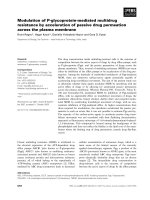
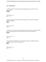
Figure 12.1. The Gram-positive lactic acid bacterium Lactococcus lactis (left picture) is used in starter
cultures for cheese production.
D
LmrA
Out
LmrA
244
LmrP
D
In
ATP
Hϩ
ADP ϩ Pi
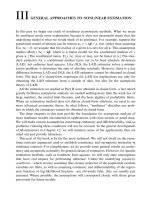
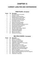
Figure 12.2. Schematic representation of two
multidrug transporters found in Lactococcus
lactis. The ABC-type primary multidrug
transporter LmrA and the secondary multidrug
transporter LmrP exemplify the two major classes
of multidrug transporters found in bacteria.
Rectangles represent the transmembrane domains
of LmrA and LmrP. Circles represent the
nucleotide-binding domains of LmrA.
the proton motive force (Bolhuis et al., 1995).
The other MDR system is an ATP-dependent
primary transporter, designated LmrA
(Figure 12.2) (van Veen et al., 1996). The role of
this chromosomally encoded primary efflux
pump in multidrug resistance was first
observed in an ethidium-resistant mutant of
L. lactis subsp. lactis MG1363. Ethidium efflux
in this mutant was inhibited by ortho-vanadate,
an inhibitor of ABC transporters and P-type
ATPases, but not upon dissipation of the
proton motive force (Bolhuis et al., 1994). Isolated membrane vesicles and proteoliposomes,
in which purified LmrA was reconstituted,
were employed to prove that transport of multiple drugs was LmrA- and ATP-dependent
(Margolles et al., 1999; van Veen et al., 1996).
Interestingly, this lactococcal LmrA protein
was the first ABC-type multidrug transporter
identified in bacteria.
Another ABC-type multidrug resistance
pump (HorA) was discovered in Lactobacillus
brevis, a major contaminant of spoiled beer
(Sami et al., 1997, 1998). This Gram-positive
lactic acid bacterium can grow in beer in spite
of the presence of antibacterial compounds
(iso-␣-acids) derived from the flowers of the
hop plant Humulus lupulus L. The hop resistance of Lb. brevis is, at least in part, dependent
on the expression of the horA gene, which is
located on a 15 kb plasmid termed pRH45
(Sami et al., 1997). The role of HorA in hop
resistance was first suggested by a spontaneous mutant lacking the pRH45 plasmid,
which displayed sensitivity to the presence of
hop compounds. Reintroduction of pRH45 into
this segregation mutant restored hop resistance
(Sami et al., 1998). These complementation
studies, as well as the heterologous expression
of the horA gene in L. lactis, demonstrated that
HorA is involved in resistance to hop compounds. Moreover, almost all lactobacilli isolated as beer-spoilage strains possess horA
homologues (Sami et al., 1997). In addition to
conferring hop resistance, HorA confers resistance to the structurally unrelated drugs novobiocin and ethidium bromide (Sami et al., 1997).
Drug transport studies in L. lactis cells and
membrane vesicles and in proteoliposomes in
which purified HorA was reconstituted identified this protein as a new member of the ABC
family of multidrug transporters (Sakamoto
et al., 2001).
Here we summarize the existing data on the
two bacterial ABC-type multidrug transporters
LmrA and HorA, and analyze structural and
mechanistic aspects of multidrug recognition
and transport. In addition, the chapter will
describe how attenuated total reflection Fourier
transform infrared (ATR-FTIR) spectroscopy
BACTERIAL MULTIDRUG RESISTANCE MEDIATED BY ABC TRANSPORTERS
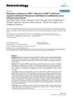
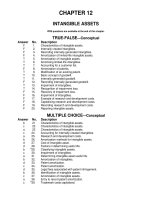
Figure 12.3. Topology model for LmrA. The LmrA protein is predicted to contain a transmembrane
domain (TMD) with six transmembrane ␣-helices, and a nucleotide-binding domain (NBD) with
the ABC signature and Walker A/B sequences. A similar model is envisaged for HorA of Lb. brevis.
has provided important information about
LmrA structure and the dynamic changes occurring during its catalytic cycle.
PROPERTIES OF LMRA
AND HORA
STRUCTURAL ASPECTS
All ABC transporters described so far show
a four-domain organization, which consists of
two transmembrane domains (TMDs), which
are thought to perform the transport function,
and two nucleotide-binding domains (NBDs),
which provide the energy for the transport
process (Higgins, 1992). The four domains may
be organized either in a multifunctional, single
polypeptide or as separate proteins. For example, in human P-glycoprotein (MDR1), like
many eukaryotic ABC transporters, the four
domains are found in one single polypeptide
chain arranged as TMD1-NBD1-TMD2-NBD2.
As derived from the DNA sequences, bacterial
LmrA is composed of 590 amino acids (calculated molecular mass of 64.6 kDa) and HorA of
583 amino acids (calculated molecular mass of
64.2 kDa). Hydropathy analysis, as shown in
Figure 12.3, suggests a putative topology for
both proteins of six membrane-spanning regions
(putative ␣-helices) in the amino-terminal
hydrophobic domain, followed by a large
hydrophilic domain containing the ATP-binding
site (Sami et al., 1997; van Veen et al., 1996).
There is now experimental evidence that
the membrane-spanning regions of LmrA are
indeed ␣-helices (Grimard et al., 2001). Based
on the topology predictions, both the aminoterminal end and the large carboxy-terminal
half are located in the cytoplasm. In addition to
the NBD, there are two putative large cytoplasmic loops (Figure 12.3) (see also Chapter 11,
HlyB). The predicted membrane topologies of
LmrA and HorA still await experimental confirmation. The NBDs of both these bacterial
transporters contain features diagnostic of an
245
246
ABC PROTEINS: FROM BACTERIA TO MAN
ABC-type ATPase, such as the ABC signature
sequence, and the Walker A and B motifs
(Figure 12.4). The sequence homology between
full-length LmrA and HorA is around 53%.
Sequence comparisons with other ABC transporters revealed that these bacterial proteins
share significant overall sequence similarity
with members of the subfamily of multidrug
resistance P-glycoproteins, most notably the
human P-glycoprotein (MDR1) (Sami et al.,
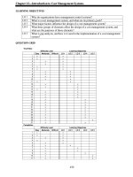
1997; van Veen et al., 1996). For example, LmrA
and each half of MDR1 share 34% identical
residues and an additional 16% of conservative
substitutions (Figure 12.4). The ABC domain of
LmrA and the ABC1 and ABC2 domains of
MDR1 are 48% and 43% identical, respectively,
whereas the identity between the TMD of
LmrA and the amino- and carboxy-terminal
TMDs of MDR1 is 23% and 27%, respectively.
The sequence conservation in the TMD of
LmrA includes particular regions (e.g. the region
comprising transmembrane helices 5 and 6),
which have been implicated as being involved
in drug binding by MDR1 (Loo and Clarke,
2000). Functionally important residues in this
region of LmrA are now being identified.
Interestingly, LmrA shares 28% overall sequence
identity with the lipid flippase MsbA from
Escherichia coli (Figure 12.4), the structure of
which was recently determined by X-ray crystallography to a resolution of 4.5 Å (Chang and
Roth, 2001). The overall sequence similarity
between LmrA and bacterial members of other
subfamilies of the ABC transporter superfamily
is less than 28% and is mostly confined to the
hydrophilic ABC domains.
In view of the general organization of ABC
transporters, LmrA and HorA are considered to
be half transporters (with the two domains
arranged in TMD-NBD manner) that have to
form homodimers in order to function as full
four-domain transporters. Recent studies on
LmrA provided evidence that this is indeed the
case. First, two covalently linked wild-type
LmrA monomers expressed from an engineered
gene yields a functional transporter, whereas the
covalent linkage of a wild-type monomer and
an inactive mutant monomer (harboring the
K388M mutation in the Walker A region) yields
an inactive transporter (van Veen et al., 2000).
The latter covalently linked dimer had also lost
all ATPase activity, demonstrating that both catalytic sites must be functional to allow ATP
hydrolysis and drug transport. Second, LmrA
solubilized from membrane vesicles prepared
from LmrA-overproducing cells behaves like
a dimer on native gels (our unpublished
data). Third, electron microscopy analysis of
purified and reconstituted LmrA revealed small,
uniform particles with a diameter of 8.5 by
5 nm, similar to those previously observed
for monomeric P-glycoprotein (S. Scheuring,
A. Margolles, H.W. van Veen, W.N. Konings and
A. Engel, unpublished data). Probably, the most
convincing evidence for the dimeric nature of
LmrA comes from co-reconstitution experiments into proteoliposomes of the cysteine-less
wild-type LmrA and a mutant form of LmrA in
which the N-ethylmaleimide (NEM)-reactive
glycine to cysteine mutation (G386C) was introduced (van Veen et al., 2000). The G386C mutant
displays wild-type transport activity but is
completely inactivated upon incubation with
NEM, whereas wild-type LmrA activity is not
affected by NEM. The transport inhibition patterns obtained with proteoliposomes, containing different ratios of wild-type and mutant
proteins, upon reaction with NEM suggest
strongly that the functional unit of LmrA is a
dimer and not a monomer, trimer or tetramer.
Taking all these data together, it is clear that the
dimeric state of LmrA is a prerequisite for function, and that functional crosstalk between two
monomers is essential for transport.
SUBSTRATE SPECIFICITY
The notion that inactivation of the secondary
multidrug transporter LmrP increases drug
extrusion mediated by the primary transporter
LmrA points to the physiological importance
of these multidrug transporters in L. lactis
(Bolhuis et al., 1995). However, except for the
observation that LmrA might act as a lipid
translocase (Margolles et al., 1999), its cellular
function is still under debate. The natural
substrates of LmrA might be found amongst the
hydrophobic compounds excreted by plants, the
natural habitat of lactococci. Indeed, LmrA can
extrude a wide variety of amphiphilic toxic
compounds, and its classification as a multidrug
transporter is evident from its currently known
spectrum of substrates. LmrA substrates include
anticancer drugs such as vinca alkaloids (vinblastine, vincristine) and anthracyclines (daunomycin, doxorubicin), or cytotoxic agents such
as antimicrotubule drugs (colchicine) and
DNA intercalators (ethidium bromide), or toxic
peptides (valinomycin, nigericin), fluorescent
membrane probes (Hoechst 33342, diphenylhexatriene), and fluorescent dyes such as
BACTERIAL MULTIDRUG RESISTANCE MEDIATED BY ABC TRANSPORTERS
Figure 12.4. Amino acid sequence alignment. The amino acid sequence of LmrA is shown with HorA from
Lb. brevis, MsbA from E. coli, and the amino- and carboxy-terminal halves of human MDR1. Residues conserved
throughout all sequences are indicated by an asterisk. Residues conserved between LmrA and MDR1 are shaded
red. Dashes represent residues absent in other sequences. Putative transmembrane regions are boxed. The Walker
A/B motifs and the ABC signature motif regions are labeled by Walker A, Walker B and ABC, respectively.
247
248
ABC PROTEINS: FROM BACTERIA TO MAN
rhodamine 6G and rhodamine 123 (Margolles
et al., 1999; van Veen et al., 1996, 1998; see more
detailed discussion in Chapter 5). LmrA modulators (i.e. compounds that reverse LmrA-mediated multidrug resistance) are also structurally
unrelated to each other and include the calcium
channel blockers verapamil and CP100-356
(analogue of verapamil), 1,4-dihydropyridines
such as nicardipine, indolizine sulfones such
as SR33557, antimalarials such as quinine
and quinidine, immunosuppressants such as
cyclosporin A, and the Rauwolfia alkaloid reserpine (van Veen et al., 1999). This broad drug
and modulator specificity is not only confined
to LmrA. A similar range of compounds was
previously found to interact with other ABC
transporters, including yeast Pdr5p (Bauer
et al., 2000; Kolaczkowski et al., 1996) and
human P-glycoprotein (Ueda et al., 1997).
The overlapping substrate and modulator
specificities of bacterial LmrA and human
P-glycoprotein reveal a functional similarity
between both proteins. Expression studies of
LmrA in insect and human lung fibroblast cells
demonstrated that LmrA was indeed able to
functionally complement P-glycoprotein (van
Veen et al., 1998). Surprisingly, LmrA was targeted to the plasma membrane and conferred
typical multidrug resistance on the human cells.
The pharmacological characteristics of LmrA
and P-glycoprotein expressed in lung fibroblast
cells were very similar, and reversal agents of
P-glycoprotein-mediated multidrug resistance
also blocked multidrug resistance mediated by
LmrA. Furthermore, the affinities of both proteins for vinblastine and ATP were indistinguishable. Finally, kinetic analysis of drug
dissociation from LmrA expressed in plasma
membranes of insect cells revealed the presence
of two allosterically coupled drug-binding sites,
indistinguishable from those of P-glycoprotein
(van Veen et al., 1998; Chapter 5). This remarkable conservation of function between these two
ABC-type multidrug transporters implies a common overall structure and transport mechanism.
L. lactis is a GRAS (generally regarded as
safe) organism, that is, an organism considered
to be non-pathogenic and safe to use in starter
cultures for cheese production (Figure 12.1)
(Gasser, 1994). In view of this, it is important to
know whether the substrate spectrum of LmrA
also includes clinically relevant antibiotics. The
antibiotic specificity of LmrA was studied in
cytotoxicity assays, in which the antibiotic susceptibilities of E. coli CS1562 cells overexpressing the transporter are compared with those of
control CS1562 cells not expressing LmrA.
Strain CS1562 (tolC6 :: Tn10) was used in these
assays because it is hypersensitive to drugs
owing to a deficiency in the TolC protein, resulting in an impaired barrier function of the outer
membrane (Austin et al., 1990). LmrA expression in CS1562 cells resulted in an increased
resistance to 17 out of 21 clinically most used
antibiotics, including broad-spectrum antibiotics belonging to the classes of aminoglycosides, lincosamides, macrolides, quinolones,
streptogramins and tetracyclines (Table 12.1)
TABLE 12.1. EFFECT OF LMRA
EXPRESSION IN E. COLI CS1562 ON
THE RELATIVE RESISTANCE TO
ANTIBIOTICS
Class
Antibiotic
Relative
resistancea
(fold)
Aminoglycosides
-Lactams
Glycopeptides
Lincosamides
Macrolides
Quinolones
Streptogramins
Tetracyclines
Others
Gentamicin
Kanamycin
Ampicillin
Ceftazidime
Meropenem
Penicillin
Vancomycin
Clindamycin
Azithromycin
Clarithromycin
Dirithromycin
Erythromycin
Roxithromycin
Spiramycin
Ciprofloxacin
Ofloxacin
Dalfopristin
Quinupristin
RP59500
Chlortetracycline
Demeclocycline
Minocycline
Oxytetracycline
Tetracycline
Chloramphenicol
Trimethoprim
2
3
2
3
1
4
1
14
33
23
264
53
35
35
2
4
163
31
55
28
12
138
8
14
11
1
a
Relative resistances were determined by dividing the
IC50 (the antibiotic concentration required to inhibit the
growth rate by 50%) for cells harboring pGKLmrA
by the IC50 for control cells harboring pGK13. For
example, the latter IC50 values varied between 0.3 and
2 M for kanamycin, ampicillin, erythromycin,
ofloxacin, dalfopristin, and minocycline. Data obtained
from Putman et al. (2000a) with permission.
BACTERIAL MULTIDRUG RESISTANCE MEDIATED BY ABC TRANSPORTERS
(Putman et al., 2000a). The secondary multidrug
transporter LmrP also confers resistance to
antibiotics, although its substrate range is
smaller than that of LmrA (Putman et al., 2001).
The antibiotic specificity of HorA is currently
being analyzed. The exceptionally broad antibiotic specificity of LmrA, the possible transfer of
the lmrA gene to other bacteria in food or the
digestive tract, and the presence of lmrA homologues in pathogenic microorganisms (van Veen
and Konings, 1998) provide a serious threat to
the efficacy of valuable antibiotics. It will be
interesting to find out whether P-glycoprotein is
involved in antibiotic export in human cells.
Using a fluorescence quenching technique,
it has recently been demonstrated that purified and reconstituted LmrA can also transport
phospholipids (Margolles et al., 1999). In
this study, extrusion of fluorescent (C6-NBDlabeled) phosphatidylethanolamine from the
outer leaflet of proteoliposomes by inwardfacing LmrA molecules (nucleotide-binding
domain exposed to the external surface) was
detected in the presence of ATP, with nonhydrolyzable ATP analogues being ineffective.
Phospholipid extrusion from the membrane
was inhibited by vinblastine, a high-affinity
substrate of LmrA. The specificity of LmrA
with respect to lipid headgroup and acyl chain
is now being studied, possibly leading to the
identification of potential physiological lipid
substrates. Several other ABC multidrug transporters have also been found to possess lipid
translocation activity, including P-glycoprotein
(for a recent review, see Borst et al., 2000 and
Chapter 22 of this volume).
SUBSTRATE RECOGNITION AND
TRANSPORT MODELS
Aqueous pore versus hydrophobic
vacuum cleaner and flippase models
Despite the remarkable conservation of functional properties between ABC-type multidrug
transporters, there is still a considerable controversy about the mechanisms by which these
proteins pump drugs from the interior of the cell
to the external medium. Several transport models have been postulated for P-glycoprotein
pump function (Figure 12.5). These include (i)
the conventional aqueous pore model, in which
substrate is transported from the cytoplasm to
the exterior (Altenberg et al., 1994), (ii) the
hydrophobic vacuum cleaner model, in which
Aqueous
pore
Hydrophobic
vacuum cleaner Flippase
In
C
M
D
R
Out
M
D
R
M
D
R
B
A
Figure 12.5. Possible mechanisms of drug transport
across the cytoplasmic membrane. Drugs may be
expelled from the cell by extrusion from the internal
water phase to the external water phase (aqueous
pore model) or by extrusion from the membrane to
the exterior (hydrophobic vacuum cleaner and
flippase models). Importantly, the hydrophobic
vacuum cleaner model predicts that hydrophobic
compounds are translocated by the MDR pump
from the inner leaflet of the membrane to the
external water phase, whereas the flippase model
predicts extrusion from the inner leaflet to the outer
leaflet of the membrane. A, Drug molecules
reaching the cell rapidly insert into the outer leaflet
of the plasma membrane. B, Flipping of the drug to
the inner leaflet of the membrane is relatively slow
and the rate-limiting step in entry. C, Membrane
release of drug molecules.
substrate is transported from the lipid bilayer
to the exterior (Raviv et al., 1990), and (iii) the
flippase model, a variation on the hydrophobic
vacuum cleaner model, in which substrate is
transported from the inner leaflet to the outer
leaflet of the lipid bilayer, after which the substrate molecules will diffuse into the external
medium (Higgins and Gottesman, 1992). The
latter two models take into account that most
drugs that interact with multidrug transporters
such as P-glycoprotein and LmrA readily intercalate into the lipid bilayer due to their high
hydrophobicity and amphiphilic nature. Drug
extrusion from the membrane is supported by
substantial experimental evidence, including
the following important observations. First, the
non-fluorescent compound BCECF-AM (an
acetoxymethyl ester derivative of 2Ј,7Ј-bis(2-carboxyethyl)-5-(and-6-)-carboxyfluorescein)
is excreted from P-glycoprotein- and LmrAproducing cells prior to hydrolysis into the
fluorescent cellular indicator BCECF by intracellular esterases (Bolhuis et al., 1996; Homolya
et al., 1993). Thus, LmrA and P-glycoprotein
prevent the accumulation of the fluorescent
indicator BCECF in the cytosol, despite the
fact that BCECF-AM is rapidly cleaved by
intracellular esterases and the resulting BCECF
is not a substrate for LmrA and P-glycoprotein.
249
ABC PROTEINS: FROM BACTERIA TO MAN
These observations strongly suggest that
BCECF-AM is extruded from the membrane.
Second, photoaffinity analogues of substrates
of P-glycoprotein only label the two transmembrane domains of P-glycoprotein and not its
hydrophilic ABC domains (e.g. Greenberger,
1993). Third, the affinity of binding of drugs to
purified and reconstituted P-glycoprotein is
modulated by their ability to intercalate into
the membrane (Romsicki and Sharom, 1999).
Fourth, cysteine-scanning mutagenesis, in combination with reaction with the thiol-reactive
substrate dibromobimane, of all predicted transmembrane segments of P-glycoprotein indicates
that the drug-binding domain of P-glycoprotein
consists of residues in transmembrane segments
4, 5, 6, 10, 11 and 12 (Loo and Clarke, 2000).
Taking these data together, it seems likely
that these transporters recognize most, if not
all, of their substrates within the membrane
(hydrophobic vacuum cleaner and flippase
models) and not from the cytoplasm (aqueous
pore model). However, these observations do
not discriminate between the vacuum cleaner
model and the flippase model.
Evidence for drug efflux from the inner
leaflet of the lipid bilayer to the exterior
The most convincing evidence for drug efflux
from the membrane to the aqueous phase is
provided by the kinetics of ATP-dependent
transport of TMA-DPH by LmrA (Bolhuis et al.,
1996) and of Hoechst 33342 by P-glycoprotein
(Shapiro and Ling, 1997a). The amphiphilic
character and the high lipid–water partition
coefficients result in partitioning of these compounds into the lipid bilayer. Conveniently,
these hydrophobic probes are strongly fluorescent when partitioned into the membrane but
essentially non-fluorescent in an aqueous environment. Since, therefore, the fluorescence
detected reflects the concentration of probe in
the membrane, these properties make it possible to follow fluorimetrically the partitioning
of these compounds into the lipid bilayer. The
increase in fluorescence intensity due to the
partitioning of TMA-DPH into the phospholipid bilayer was found to be a biphasic process
(Figure 12.6) (Bolhuis et al., 1996). This biphasic
behavior reflects the fast entry (1–2 seconds)
of TMA-DPH into the outer leaflet of the phospholipid bilayer (phase 1 in Figure 12.6),
followed by a slower (several minutes) transbilayer movement from the outer to the inner
TMA-DPH fluorescence (a.u.)
250
2
A
B
C
1
0
10
20
30
40
50
Time (min)
Figure 12.6. Time course of the rate of energydependent TMA-DPH extrusion. A washed cell
suspension of L. lactis strain MG1363 (EthR),
a mutant strain in which extrusion of TMA-DPH
is LmrA-dependent, was energized with 25 mM of
glucose, at 5 (A), 15 (B), and 40 min (C) after the
addition of 100 nM of TMA-DPH. Data obtained
from Bolhuis et al. (1996).
leaflet of the membrane (phase 2 in Figure 12.6).
When LmrA was energized in intact cells by
the addition of glucose, it was observed that
the initial rate of extrusion of TMA-DPH, monitored as a decrease in fluorescence over time,
increased with an increasing concentration of
TMA-DPH in the inner leaflet of the membrane
(Figure 12.6) (Bolhuis et al., 1996). The extent of
extrusion never exceeded the amount of TMADPH present in the inner leaflet (Figure 12.6),
indicating that the probe cannot be extruded
from the outer leaflet of the cytoplasmic membrane. When similar experiments were done
with inside-out membrane vesicles with the
inner leaflet now immediately accessible to
drug molecules, the situation was significantly
different. Upon addition of TMA-DPH to the
membrane vesicle suspension, TMA-DPH
rapidly intercalates into the exposed leaflet of
the membrane, resulting in a maximum concentration of TMA-DPH in this leaflet. Upon
energization of LmrA by the addition of ATP
(the NBD of LmrA is exposed to the exterior of
these vesicles), maximal rates of TMA-DPH
extrusion were observed at any moment after
addition of TMA-DPH and the extent of extrusion, in contrast to intact cells, now exceeded
the amount of TMA-DPH present in the internal leaflet of inside-out vesicles. These observations strongly indicate that TMA-DPH is
recognized as a substrate only after partitioning into the normal inner leaflet of the cellular
BACTERIAL MULTIDRUG RESISTANCE MEDIATED BY ABC TRANSPORTERS
Hydrophobic vacuum cleaner model
versus flippase model
It is important to note that the results presented
strongly favor a vacuum cleaner mechanism of
transport by the MDR pumps and are inconsistent with a flippase mechanism as proposed by
Higgins and Gottesman (1992). According to
the flippase mechanism the hydrophobic compounds are translocated by the MDR pump
from the inner leaflet of the membrane to the
outer leaflet followed by diffusion into the
external medium (Figure 12.5). The observation that the fluorescence of TMA-DPH or
Hoechst 33342 falls rapidly upon energization
of LmrA or P-glycoprotein indicates that these
compounds do not stay in the lipid bilayer but
are moved into the water phase.
Physiological implications of drug transport
from the inner leaflet of the membrane
This mechanism of transport of hydrophobic
drugs from the inner leaflet of the phospholipid bilayer to the exterior, as illustrated in
Figure 12.7, may have several physiological
implications. First, transport from the cytoplasmic leaflet of the membrane appears to be the
most efficient way in which MDR transporters
can prevent toxic compounds from entering
the cytoplasm. As already pointed out, drug
molecules reaching the cell rapidly insert into
the outer leaflet of the membrane, but flipping
of the drug molecules from the outer to inner
leaflet is slow and the rate-limiting step in
entry (Figure 12.7). LmrA is able to transport
drug molecules from the inner leaflet back into
the external medium, counteracting the ratelimiting step in drug entry. If the transporter
were to transport drugs from the outer leaflet of
the membrane, it would probably not be able to
compete with the high rate at which drug molecules enter this leaflet. Drug molecules would
Cytosol
Membrane
release
(slow)
n
Flip-flop
(slow)
Membrane
insertion
(fast)
Extr
u s io
membrane, and is directly transported to the
aqueous environment as observed by the
decrease in fluorescence. A similar relationship
between the initial rate of transport and the
concentration of substrate in the inner leaflet of
the cellular membrane was observed for other
multidrug transporters, including secondary
transporters (Putman et al., 2000b). Thus, secondary and ABC multidrug transporters appear
to use the same mechanism of transport for
hydrophobic drugs.
Medium
Figure 12.7. Proposed mechanism of
LmrA-mediated TMA-DPH extrusion from a cell.
The initial rate of LmrA-dependent TMA-DPH
transport correlates with the amount of TMA-DPH
in the inner membrane leaflet of whole cells, and
with the amount of TMA-DPH in the outer leaflet
of inside-out vesicles. Since the outer membrane
leaflet of inside-out vesicles corresponds to
the inner membrane leaflet of whole cells, both
observations rule out the external leaflet of whole
cells as a possible site of drug binding to LmrA.
‘escape’ into the inner leaflet and subsequently
enter the cytoplasm. Second, extrusion from the
membrane may partially explain the lack of
structural specificity and the consequent broad
substrate range of multidrug transporters. The
transmembrane domains of multidrug transporters are thought to form a pathway across
the membrane through which solutes move,
a prediction supported by structural data of
P-glycoprotein (Rosenberg et al., 2001) and MsbA
(Chang and Roth, 2001). Assuming that the
translocation pathway is only accessible from
the membrane, but not from the aqueous phase,
a drug molecule must be able to intercalate into
the membrane in order to be recognized by
the transporter. Thus, the ability to intercalate
into the membrane may pre-select compounds
to be transported from those which should not be
transported (e.g. hydrophilic cytoplasmic components). The subsequent interaction between
the intercalated substrate and the transporter
would be a second determinant of specificity.
This would allow the transporter to have (a) relatively non-selective substrate-binding site(s).
NUMBER OF SUBSTRATE-BINDING SITES
Studies on the kinetics of drug dissociation
have revealed the presence of two distinct, but
251
ABC PROTEINS: FROM BACTERIA TO MAN
1.5
Drug binding (nmol/mg of protein)
252
LmrA
1.0
0.5
Control
0.0
0
50
100
150
200
Drug concentration (nM)
Figure 12.8. Vinblastine equilibrium binding to
LmrA. Specific binding of [3H]vinblastine to
inside-out membrane vesicles without LmrA
(control) or with LmrA, as a function of the free
vinblastine concentration. Superimposed on the
data are the best-fit curves obtained for a
single-site binding model (hyperbolic, dotted curve)
and the cooperative two-site binding model
(sigmoidal, solid curve). Data obtained from
van Veen et al. (2000) with permission from
Oxford University Press.
allosterically linked, drug-binding sites in the
LmrA transporter (van Veen et al., 1998, 2000;
Chapter 5). The presence of these two drugbinding sites in LmrA is strongly supported by
the finding that vinblastine equilibrium binding can best be fitted by a model in which two
vinblastine-binding sites in the LmrA transporter interact cooperatively (Figure 12.8) (van
Veen et al., 2000). In this model, an initial vinblastine-binding event with low affinity initiates a second vinblastine-binding event with
high affinity. Based on the model, the dissociation constants for the two vinblastine-binding
sites were estimated to be approximately 150
and 30 nM vinblastine, respectively. Moreover,
a direct determination of the LmrA/vinblastine stoichiometry revealed that each homodimer of LmrA binds two vinblastine molecules
(van Veen et al., 2000), providing convincing
evidence for the presence of two sites.
Importantly, these drug-binding sites seem to
be directly related to drug transport as shown
by the reciprocal stimulation of LmrA-mediated
vinblastine and Hoechst 33342 transport at
low drug concentrations, and reciprocal inhibition at high drug concentrations (van Veen
et al., 2000). Most probably, one of the drugbinding sites interacts preferentially with
vinblastine and the other preferentially with
Hoechst 33342. At lower concentrations, vinblastine binds primarily to one site and
enhances transport of Hoechst 33342 bound at
the other site. At higher concentrations, vinblastine is able to compete with Hoechst 33342
for binding to the same site, and therefore
inhibits Hoechst 33342 transport, or vice versa.
Taken together, the results strongly suggest
that LmrA contains at least two distinct drugbinding sites, presumably located in the TMD,
with different but overlapping specificities
which interact in drug transport in a positively
cooperative manner. Support for the presence
of at least two positively cooperative sites for
drug transport in P-glycoprotein has also been
presented (e.g. Shapiro and Ling, 1997b; Sharom
et al., 1996). Thus, it appears that the transport
process of ABC-type multidrug transporters
such as LmrA and P-glycoprotein involves two
general transport-competent drug-binding sites,
which may be composed of multiple drug interaction sites to account for the wide range of
compounds that are transported. In addition to
the transport-competent drug-binding sites,
LmrA and P-glycoprotein contain regulatory
sites, which may reside outside of the transportcompetent drug-binding sites, to which allosteric
modulators bind, but are not transported
(Martin et al., 1997; van Veen et al., 1998).
STRUCTURAL CHANGES
INDUCED BY
NUCLEOTIDE BINDING
OR HYDROLYSIS
DETECTED BY ATRFTIR SPECTROSCOPY
Although the topology of LmrA in the
lipid membrane has been deduced from its
primary structure (Figure 12.3), its secondary
and tertiary structures are unknown since a
high-resolution structure of LmrA has not yet
been obtained. Such a structure would supply
extremely valuable information about the overall domain organization and the interacting
sites. However, such a structure would also be
inherently static and would not necessarily
BACTERIAL MULTIDRUG RESISTANCE MEDIATED BY ABC TRANSPORTERS
BOX 12.1. ATR-FTIR SPECTROSCOPY: EXPERIMENTAL PROCEDURE AND
SAMPLE PREPARATION
Infrared spectroscopy is based on the absorption of electromagnetic radiation by matter owing to different vibrational
modes of the chemical bonds. The infrared beam is directed into a high refractive index medium (internal reflection
element or IRE), which is transparent for the IR radiation of interest. The most usual design of an IRE is the trapezoidal
plate, which allows the orientation of protein secondary structures to be determined by means of linear dichroism.
Below a critical angle c, which depends on the refractive index of the IRE (n1) and of the external medium (n2)
according to
c ϭ sinϪ1 n21
(1)
where n21 ϭ n2/n1, the light beam is completely reflected when it impinges on the surface of the IRE. Several internal
total reflections occur within the IRE until the beam reaches the end. Interestingly, an electromagnetic disturbance exists
in the rarer medium beyond the reflecting interface. This so-called evanescent wave is characterized by its amplitude,
which falls off exponentially with the distance from the interface according to
E ϭ E0 и eϪz/dp
(2)
where E0 is the time-averaged electric field intensity at the interface, E is the time-averaged field intensity at a distance z
from the interface in the rarer medium, and dp is the penetration depth of the evanescent field. It is the presence of the
evanescent field which makes the interaction possible between infrared light and the sample present on the surface
of the IRE, within approximately the penetration depth of the field. Indeed, samples which are deposited on the IRE
absorb electromagnetic radiation of the evanescent wave, and thereby reduce the intensity of the reflected light. Hence,
the technique is referred to as ‘attenuated total reflection-IR spectroscopy’. Since the sample has to be in close contact
with the IRE, films of proteins or membranes have to be used.
The simplest sample preparation for ATR-FTIR spectroscopy has been used, in which a drop of the sample, typically
20 l of proteoliposomes containing ϳ20 g of reconstituted LmrA, is spread on the IRE surface. The solvent is slowly
evaporated under a gentle N2 flow, while a Teflon bar or pipette tip is used to spread the liquid over the useful surface
of the IRE in order to make the film as uniform as possible. While evaporating, capillary forces flatten the membranes,
which spontaneously form oriented multibilayer arrangements. During film preparation, many water molecules
remain associated with the proteins and lipids. Consequently, protein structure is not affected by the low hydration
state of the newly formed film. Moreover, the ATPase activity of reconstituted LmrA was measured after resuspension
of the film. No loss of ATPase activity was observed, confirming that film preparation does not alter LmrA
conformation and activity. Thus, the technique is very convenient for studying proteins inserted into lipid
membranes since common reconstituted vesicles (e.g. liposomes) can be used.
represent the structure of LmrA in its native
lipid environment. In view of the difficulties in
obtaining high-resolution structures of membrane proteins, lower-resolution techniques
such as IR (infrared) and ATR-FTIR spectroscopy can be employed to obtain global,
structural information of membrane proteins.
ATR-FTIR spectroscopy is particularly useful
since this permits the monitoring of structural
changes of membrane proteins in their native
lipid environment in response to physiological
modifications (Box 12.1).
ANALYSIS OF THE SECONDARY
STRUCTURE OF LMRA IN THE ABSENCE
AND PRESENCE OF NUCLEOTIDES
In order to mediate drug transport, LmrA
must couple ATP hydrolysis to conformational
changes, which alter drug-binding affinity
and/or accessibility of the transport-competent
drug-binding sites. To investigate the nature of
the conformational changes induced during
the transport cycle, ATR-FTIR spectra of LmrA,
reconstituted into liposomes, were recorded
in the absence and presence of different nucleotides. A typical spectrum of LmrA before
and after deuteration is shown in Figures
12.9A and 12.9B, respectively. The bands at
ϳ3300 cmϪ1 and ϳ2500 cmϪ1 correspond to the
O–H and O–D stretching of H2O and D2O,
respectively. In the 1800–1700 cmϪ1 region, the
band corresponding to the CϭO stretching of
the lipids is detected in both cases. Most importantly for the study of LmrA are the amide I
(1700–1600 cmϪ1 region) and the amide II
(1570–1500 cmϪ1 region) bands. The amide I
band is assigned to the (CϭO) of the peptide
253
Lipids (C –
– Ostretching)
ABC PROTEINS: FROM BACTERIA TO MAN
D2O
400
300
Amide I
Amide II
350
B
H2O
254
250
200
150
A
100
50
4000
3500
3000
2500
2000
1500
1000
Figure 12.9. ATR-FTIR spectrum in the 4000–400 cmϪ1 region of LmrA actively reconstituted into lipids
before (A) and after (B) deuteration. Thin films were obtained by slowly evaporating a sample containing
20 g of LmrA on a Ge-attenuated total reflection element. The film was then rehydrated under
a D2O-saturated N2 flow. Data obtained from Vigano et al. (2000) with permission from the American
Society for Biochemistry and Molecular Biology.
bond, while the amide II band is characteristic
of the ␦(N–H). The amide I band is by far
the most sensitive indicator of the secondary
structure (Fringeli and Günthard, 1981) and is
located in a region of the spectrum which is
often free of other bands and is composed of
80% pure CϭO vibration. The secondary structure of LmrA was determined by Fourier
deconvolution and a curve-fitting analysis on
the amide I region of a deuterated sample
(Vigano et al., 2000). H/D exchange permits
differentiation of the ␣-helical secondary structure from random secondary structure, whose
absorption band shifts from about 1655 cmϪ1 to
about 1642 cmϪ1 (Goormaghtigh et al., 1994).
The percentages of ␣-helix, -sheet, -turn
and random coil structures of LmrA, in the presence or absence of nucleotides, are presented in
Table 12.2. MgATP␥S, a non-hydrolyzable analogue of MgATP, was used to discriminate
between the influence of nucleotide binding and
nucleotide hydrolysis on the structure of LmrA.
In the presence of MgADP/Pi and MgADP, the
structure represents the situation after ATP
hydrolysis. Significant differences detected in
the amide I region demonstrate that LmrA
adopts two different conformations depending
TABLE 12.2. SECONDARY STRUCTURE
OF LMRA DETERMINED IN THE
ABSENCE AND PRESENCE OF
NUCLEOTIDESa
Substratesb
None
MgADP
MgATP
MgATP␥S
MgADP/Pi
Secondary structure
␣-Helix
(%)
-Sheet
(%)
-Turn
(%)
Random
(%)
35 Ϯ 2
35 Ϯ 1
34 Ϯ 2
35 Ϯ 2
34 Ϯ 1
24 Ϯ 2
24 Ϯ 1
36 Ϯ 2
33 Ϯ 1
35 Ϯ 1
28 Ϯ 1
31 Ϯ 1
16 Ϯ 2
18 Ϯ 1
17 Ϯ 1
13 Ϯ 1
10 Ϯ 1
14 Ϯ 2
14 Ϯ 1
14 Ϯ 1
a
Data obtained from Vigano et al. (2000) with
permission from the American Society for
Biochemistry and Molecular Biology.
b
LmrA/nucleotide molar ratio ϭ 1/5.
on the nature of the nucleotide bound to the
protein (Figure 12.10). First, LmrA alone or in
the presence of MgADP contains 35% ␣-helix,
24% -sheet, 28% -turn and 13% random coil
(Table 12.2). The proportion of ␣-helices is
higher than the proportion expected when only
BACTERIAL MULTIDRUG RESISTANCE MEDIATED BY ABC TRANSPORTERS
Figure 12.10. ATR-FTIR spectra between 1700 and 1600 cmϪ1 of deuterated LmrA actively reconstituted
into lipids. Dotted line, in the absence of nucleotide or in the presence of MgADP (LmrA/MgADP molar
ratio ϭ 1/5). Solid line, in the presence of MgATP, MgADP/Pi or MgATP␥S (LmrA/nucleotide molar
ratio ϭ 1/5). Data obtained from Vigano et al. (2000) with permission from the American Society for
Biochemistry and Molecular Biology. The results clearly show a shift indicating more -sheet in the presence
of non-hydrolyzable ATP or ADP/Pi compared with the ground state with no nucleotide or just ADP.
six transmembrane segments are in an ␣-helical
conformation (20%) (Grimard et al., 2001).
Therefore, ␣-helices, external to the membrane,
have to be present, as confirmed in the recently
reported crystal structure of the homologous
transporter MsbA (Chang and Roth, 2001). In
the presence of MgATP, MgATP␥S or
MgADP/Pi, the structure becomes enriched in
-sheet (35% ␣-helix, 34% -sheet, 17% -turn
and 14% random coil) concomitantly with a
loss of -turn structure. Therefore, LmrA is
clearly stabilized in a different secondary structure after ATP (i.e. ATP␥S) binding. However,
the protein returns to its initial secondary
structure after Pi release, when ADP is still
bound to LmrA.
The drug-binding sites of LmrA are predicted to reside within the membrane domain,
which is composed of transmembrane ␣-helices
(Grimard et al., 2001). Since the ␣-helical content of LmrA does not change in the presence of
nucleotides (Table 12.2), it seems that the ATP
binding-induced change of secondary structure
is not related to a reorientation of the transportcompetent drug-binding sites. However, it has
recently been proposed for P-glycoprotein that
ATP binding, not hydrolysis, drives the major
conformational change associated with drug
translocation, and that the reorientation of the
drug-binding sites may depend on rotation
and/or ‘tilting’ of transmembrane ␣-helices
within the membrane (Higgins and Linton,
2001; Rosenberg et al., 2001; Chapter 4).
Although it is not known whether ATP binding
to LmrA also results in loss of drug-binding
affinity, we can not exclude the possibility that
similar reorganizations, which obviously do
not affect the ␣-helical content, occur in LmrA
upon binding of ATP.
Interestingly, an ATP binding-induced enrichment in -sheet, as observed for LmrA, was
not observed for other ABC-type multidrug
transporters (P-glycoprotein and MRP1) studied so far by ATR-FTIR (Manciu et al., 2000;
Sonveaux et al., 1996). Since the ATPase and
transport activities of P-glycoprotein and
LmrA are very similar, it seems that the secondary structure change observed for LmrA
after ATP binding is related to a behavior
unique to this protein. Since LmrA must form a
homodimer to be active (see earlier section on
structural aspects), this raises the interesting
possibility that ATP binding could mediate the
secondary structural change accompanying the
assembly of LmrA into its homodimeric form.
After ATP hydrolysis and Pi release, the protein
recovers its initial secondary structure and possibly its monomeric form.
255
256
ABC PROTEINS: FROM BACTERIA TO MAN
AMIDE HYDROGEN/DEUTERIUM
EXCHANGE KINETICS OF LMRA
To further investigate the effect of ATP binding
and hydrolysis on the structure of LmrA, the
kinetics of deuteration of reconstituted protein
was monitored in the presence and absence
of different nucleotides (MgATP, MgATP␥S,
MgADP, MgADP/Pi) (Vigano et al., 2000). The
rate of amide hydrogen exchange by deuterium
is related to the solvent accessibility of the NH
amide groups. Amide hydrogen exchange was
followed by using ATR-FTIR spectroscopy to
monitor the amide II absorption peak as a function of the time of exposure to D2O-saturated
N2. The decrease of the amide II band is proportional to the number of hydrogens that have
been exchanged by deuterium and provides a
sensitive measure of LmrA structure and conformational changes. In the absence of ligands,
approximately 31% of the amide hydrogen
exchanged for deuterium within 10 s of exposure to D2O, and an additional 15% exchanged
after 2 min. The remaining 54% did not experience any exchange within 8 h of exposure to
D2O, and these protons represent the very inaccessible regions of LmrA. In the presence of
MgATP and MgADP/Pi, the fraction of slowly
exchanging amide protons decreases concomitant with an increase of intermediate exchanging amide protons. In the presence of MgATP␥S
and MgADP, the fraction of slowly exchanging amide protons is almost identical to that
observed for LmrA alone. However, approximately 31% of the amide hydrogens are
exchanged within 10 s in the protein alone,
whereas it takes 2 min to exchange 31% of the
amide hydrogens in the presence of MgADP or
MgATP␥S.
These H/D exchange measurements provide
evidence that a large portion (54%) of LmrA
is poorly accessible to the aqueous medium.
The presence of a large amide population characterized by a very low exchange rate could
be due, in part, to the shielding effect of the
membrane on a large number of residues. To
investigate this possibility, Grimard et al. (2001)
developed a new approach (monitoring infrared
linear dichroism spectra in the course of H/D
exchange), which enables the recording of
exchange rates of the membrane-embedded
region of the protein only. This approach
revealed that after 20 min 60% of the transmembrane-oriented helix amide groups have
been exchanged, i.e. an unexpectedly fast
exchange for the transmembrane region.
In contrast, only 37% seem almost inaccessible
to solvent and do not experience any exchange
within 33 h of exposure to D2O. Since the
predicted transmembrane domains of LmrA
account for only 20% of the total amino acids,
these results demonstrate that a significant proportion of the slowly exchanging amino acids
must be located outside the membrane, where
they likely form highly structured domains.
The kinetics of deuteration of P-glycoprotein
also showed a large inaccessible fraction (53%)
of the protein, where similarly only 20% of the
total amino acids of the protein are predicted
to be located inside the membrane (Sonveaux
et al., 1996).
The -turn, -sheet secondary structural
change, observed in the presence of MgATP,
MgATP␥S and MgADP/Pi (Table 12.2), is not
responsible for the change in the global accessibility of LmrA towards the external medium, as
detected by the exchange of amide protons.
Indeed, LmrA in the presence of MgATP␥S
shows no variation in the level of inaccessible
amino acids, when compared with the situation in the absence of ligand. The main changes
in the accessibility of LmrA take place in the
presence of MgATP, i.e. when normal hydrolysis is permitted. The inaccessible amino acids
decrease from 318 to 260 in the presence of
MgATP. In the presence of MgADP/Pi, the
inaccessible amino acids only decrease to 289,
while no changes are observed with MgADP or
MgATP␥S. Since the changes in accessibility
observed in the presence of MgATP and
MgADP/Pi are not related to the change of secondary structure, they could be correlated to
different tertiary structures of LmrA.
In summary, ATR-FTIR studies reveal that
LmrA undergoes a secondary structure change
and passes through three different conformational states during its catalytic cycle.
After binding of ATP, the protein structure
becomes enriched in -sheet, unlike that of
P-glycoprotein. When ATP hydrolysis takes
place, perhaps the tertiary structure of the protein changes and the protein adopts a more
accessible conformation. A third conformation
is reached after ATP hydrolysis, when Pi is still
associated to the protein. In this conformation,
the accessibility of LmrA is intermediate to the
closed conformation, observed in the absence
of nucleotides, and the opened conformation,
observed in the presence of ATP. After Pi release,
the protein recovers its initial secondary and
tertiary structures. These observed conformational changes might reflect the conformational
BACTERIAL MULTIDRUG RESISTANCE MEDIATED BY ABC TRANSPORTERS
coupling of ATP hydrolysis to drug transport,
as described in the following section.
COUPLING OF ATP
HYDROLYSIS TO DRUG
TRANSPORT
1.2
1.2
1.0
1.0
Relative drug binding
Relative drug binding
Understanding the mechanism by which ABCtype multidrug transporters couple the hydrolysis of ATP to the translocation of drugs across
the membrane is a major goal. Translocation
apparently involves alternate action of the two
ATP-binding sites, and both catalytic sites must
be functional to allow sustained drug transport
(Senior et al., 1995). As already pointed out,
LmrA contains a low-affinity drug-binding site
that is allosterically coupled to a high-affinity
drug-binding site, and these two sites appear
to be directly related to drug transport (see
also Chapter 5). Persuasive data showing the
obligatory link between the drug-binding and
catalytic cycles has come from vanadatetrapping experiments (van Veen et al., 2000).
First, as shown in Figure 12.11, heterologous
displacement by the drug CP100-356 of vinblastine from the vanadate-trapped LmrA
transporter suggested the presence of only a
single population of vinblastine-binding sites,
with binding properties similar to those of the
low-affinity vinblastine-binding site in the nontrapped transporter (compare Figures 12.11A
and 12.11B). Second, direct determination of
the vinblastine/transporter stoichiometry in
the vanadate-trapped transporter revealed a
stoichiometry of close to one, in contrast to the
value of two determined for the non-trapped
transporter. These experiments demonstrated
that of the two vinblastine-binding sites accessible in the LmrA transporter, only the lowaffinity vinblastine-binding site is accessible in
the vanadate-trapped transition state conformation of LmrA. Finally, specific photoaffinity
labeling of the vanadate-trapped LmrA transporter with [3H]-APDA, a drug that can be
transported by LmrA, was obtained in rightside-out membrane vesicles, but not in insideout membrane vesicles, demonstrating that the
low-affinity drug-binding site is exposed to the
outside (extracellular) surface of the cell membrane. The vanadate-trapped conformation of
LmrA, with a single low-affinity drug-binding
site exposed to the extracellular surface, is consistent with the hypothesis that an ATP hydrolysis-induced conformational change moves a
high-affinity drug-binding site from the inside
of the membrane to the outside with a concomitant change to a low-affinity site (van Veen
et al., 2000). Indeed, conformational changes
in LmrA upon hydrolysis of ATP have been
detected by ATR-FTIR spectroscopy (see section
above).
0.8
0.6
0.4
0.6
0.4
0.2
0.2
0.0
1eϪ12 1eϪ10 1eϪ8 1eϪ6
(A)
Drug concentration (M)
0.8
0.0
1eϪ12 1eϪ10
1eϪ4
(B)
1eϪ8
1eϪ6
1eϪ4
Drug concentration (M)
Figure 12.11. Heterologous displacement of vinblastine from LmrA by CP100-356. Non-trapped LmrA (A) and
vanadate-trapped LmrA (B) were saturated with [3H]vinblastine, and vinblastine displacement from LmrA
by CP100-356 was measured at increasing concentrations of CP100-356. For the non-trapped transporter,
the data were fitted best by a cooperative two-site drug-binding model, assuming direct competition by
CP100-356 for binding to each of the two vinblastine-binding sites in the LmrA transporter. In contrast, in
the vanadate-trapped transporter, the data suggest the presence of a single vinblastine-binding site with
binding characteristics similar to those of the low-affinity site in the non-trapped protein. Data obtained
from van Veen et al. (2000) with permission from Oxford University Press.
257
258
ABC PROTEINS: FROM BACTERIA TO MAN
The substrate-binding data obtained with
(vanadate-trapped) LmrA, when combined
with the alternating catalytic site model, in which
the ABC domains of P-glycoprotein act alternately to hydrolyze ATP (Senior et al., 1995),
led to the proposition of an alternating two-site
transport model (van Veen et al., 2000). In this
transport model (Figure 12.12; also discussed
in more detail in Chapter 5), the hydrolysis of
ATP by the ABC domain of one monomer of
the LmrA transporter is coupled to drug efflux
via its TMD. This is achieved through the
movement of a liganded, inside-facing, highaffinity drug-binding site (which binds a drug
ADP-P
ADP ATP
1
ATP
ADP
ADP-P
2
In
Out
Figure 12.12. Alternating two-site transport model.
Rectangles represent the transmembrane domains
of LmrA. Circles, squares and hexagons represent
different conformations of the nucleotide-binding
domains. Although it is not known yet whether
ATP binding, rather than hydrolysis, results in a
change in drug-binding affinity, it is assumed that
the ATP-bound (circle) state is associated with a
high-affinity drug-binding site on the inside of the
transporter. The ADP-bound (square) state is
associated with a low-affinity drug-binding site
on the outside of the transporter. The ADP-Pi
(hexagonal) state is associated with an occluded
drug-binding site, and represents the
ADP/vanadate-trapped form of the ABC domain.
According to the model, the transporter oscillates
between two configurations, each containing a
high-affinity, inside-facing, transport-competent
drug-binding site, and a low-affinity, outside-facing
drug-release site. The ATP-dependent
interconversion of one configuration into the other
proceeds via a catalytic transition state
conformation in which the transport-competent
site is occluded. The model is adapted from
van Veen et al. (2000) with permission from
Oxford University Press.
molecule in the inner leaflet of the membrane)
to the outside of the membrane, with a concomitant change to low affinity. This last step
results in release of the drug molecule into the
extracellular medium. The whole process
occurs via a catalytic transition state intermediate (which can be trapped with vanadate), in
which the transport-competent drug-binding
site is inaccessible. Importantly, ATP hydrolysis
by the ABC domain of one half of the transporter is not only coupled to drug efflux via its
TMD, but also must facilitate the return of an
unliganded, outside-facing low-affinity site, at
the membrane domain of the other halfmolecule, to an inside-facing high-affinity site.
The latter process should not be confused with
an ATP hydrolysis-induced resetting step of a
single drug-binding site in the dimeric LmrA
transporter, which alternates between highand low-affinity conformations exposed at the
inner and outer membrane surfaces (recently
reviewed by van Veen et al., 2001). Thus, in a
complete drug transport cycle, each monomer
of the LmrA dimer alternates its drug-binding
site from high affinity to occluded state to low
affinity and back to high affinity. The affinities
of the binding sites in the monomers alternate:
when the binding site in one monomer is in the
high-affinity state the binding site in the other
monomer is in a low-affinity state and vice
versa. Hence, this process is called an alternating two-site mechanism. Such a scenario implies
that both halves of the apparently symmetric
LmrA transporter are able to act asymmetrically. However, it is presently not clear whether
the binding sites are present in separate transmembrane domains or at the interface between
transmembrane domains.
CONCLUSIONS AND
PERSPECTIVES
Despite the large diversity of substrates for
ABC transporters, the specificity of each system is relatively high and only a few members
belonging to the ABC transporter superfamily
mediate multidrug resistance. Most of them are
of eukaryotic origin, such as the P-glycoproteins,
and only two characterized systems, LmrA
and HorA, are of bacterial origin. Studies on
bacterial multidrug efflux pumps are relevant
because in the last few years it has been shown
BACTERIAL MULTIDRUG RESISTANCE MEDIATED BY ABC TRANSPORTERS
that pumping activities are involved in the
ongoing emergence of antibiotic resistance in
pathogenic bacteria. In addition, LmrA is able
to complement the human multidrug resistance P-glycoprotein, supporting the clinical
and academic value of studying these bacterial
proteins.
The increasing availability of different
microbial genomes has revealed the presence
of putative ABC-type multidrug transporters,
often structurally similar to LmrA and HorA,
in all pathogenic microorganisms analyzed so
far (see list of prokaryotic genomes on the
TIGR database: The
physiological functions and substrate specificities of these pathogenic multidrug transporters
are unknown, as are the conditions in which
they are expressed. However, if these efflux
pumps are expressed in clinical isolates, due to
induction by antibiotic exposure or by up regulatory mutations, this may result in a multidrug
resistance phenotype and further selection of
such strains by antibiotic pressure. If this is
indeed the case, broad-spectrum multidrug
transporters are a serious threat to antibiotic
therapy. Insight into the incidence of (over)
expression of multidrug resistance genes in clinical strains of bacterial pathogens, the substrate
selectivities of the putative efflux pumps, and the
regulation of their expression in response to different antibiotics is therefore urgently needed.
The progress that has been achieved in
recent years to understand the functional properties of ABC-type multidrug efflux pumps is
impressive. One of the challenges that lies
ahead is to understand the structural basis of
how these fascinating and important proteins
recognize and transport such a wide range of
structurally diverse compounds. Current structures of ABC multidrug efflux pumps are of
low resolution. For a detailed understanding
of the mechanism of multiple drug binding
and translocation, high-resolution structures of
intact ABC-type multidrug transporters, both
in the presence and absence of drug and
nucleotide ligands, are required.
ACKNOWLEDGMENTS
The authors thank the present and previous
members of the Department of Microbiology
and of the Labaratoire de Chimie Physique
des Macromolécules aux Interfaces for their
valuable contribution to the research presented
in this chapter. We thank H. Bolhuis and J. Kok
for kindly providing some of the figures.
REFERENCES
Altenberg, G.A., Vanoye, C.G., Horton, J.K.
and Reuss, L. (1994) Unidirectional fluxes of
rhodamine 123 in multidrug resistant cells:
evidence against direct extrusion from the
plasma membrane. Proc. Natl Acad. Sci.
USA 91, 4654–4657.
Austin, E.A., Graves, J.F., Hite, L.A., Parker, C.T.
and Schnaitman, C.A. (1990) Genetic analysis
of lipopolysaccharide core biosynthesis by
Escherichia coli K12: insertion mutagenesis of
the rfa locus. J. Bacteriol. 172, 5312–5325.
Barrasa, M.I., Tercero, J.A., Lacalle, R.A. and
Jimenez, A. (1995) The ard1 gene from
Streptomyces capreolus encodes a polypeptide
of the ABC-transporters superfamily which
confers resistance to the aminonucleoside
antibiotic A201A. Eur. J. Biochem. 228,
562–569.
Bauer, B.E., Wolfger, H. and Kuchler, K. (2000)
Inventory and function of yeast ABC proteins: about sex, stress, pleiotropic drug and
heavy metal resistance. Biochim. Biophys.
Acta 1461, 217–236.
Bolhuis, H., Molenaar, D., Poelarends, G., van
Veen, H.W., Poolman, B., Driessen, A.J.M.
and Konings, W.N. (1994) Proton motive
force-driven and ATP-dependent drug
extrusion systems in multidrug-resistant
Lactococcus lactis. J. Bacteriol. 176, 6957–6964.
Bolhuis, H., Poelarends, G., van Veen, H.W.,
Poolman, B., Driessen, A.J.M. and
Konings, W.N. (1995) The lactococcal lmrP
gene encodes a proton motive forcedependent drug transporter. J. Biol. Chem.
270, 26092–26098.
Bolhuis, H., van Veen, H.W., Molenaar, D.,
Poolman, B., Driessen, A.J.M. and
Konings, W.N. (1996) Multidrug resistance
in Lactococcus lactis: Evidence for ATPdependent drug extrusion from the inner
leaflet of the cytoplasmic membrane.
EMBO J. 15, 4239–4245.
Borst, P., Zelcer, N. and van Helvoort, A.
(2000) ABC transporters in lipid transport.
Biochim. Biophys. Acta 1486, 128–144.
Chang, G. and Roth, C.B. (2001) Structure
of MsbA from E. coli: a homolog of the
259
260
ABC PROTEINS: FROM BACTERIA TO MAN
multidrug resistance ATP binding cassette
(ABC) transporters. Science 293, 1793–1800.
Cohen, M.L. (1992) Epidemiology of drug
resistance: implications for a postantimicrobial era. Science 257, 1050–1055.
Culliton, B.J. (1992) Drug-resistant TB may
bring epidemic. Nature 356, 473.
Fringeli, U. and Günthard, H. (1981) In: Membrane Spectroscopy (ed. E. Grell), pp.
270–332. Berlin, Heidelberg, New York:
Springer.
Gasser, F. (1994) Safety of lactic acid bacteria
and their occurrence in human clinical infections. Bull. Inst. Pasteur 92, 45–67.
Goormaghtigh, E., Cabiaux, V. and
Ruysschaert, J.M. (1994) Determination of
soluble and membrane protein structure by
Fourier transform infrared spectroscopy. III.
Secondary structures. Subcell. Biochem. 23,
405–450.
Greenberger, L.M. (1993) Major photoaffinity
drug labelling sites for iodoaryl azidoprazosin in P-glycoprotein are within, or immediately C-terminal to, transmembrane domains
6 and 12. J. Biol. Chem. 268, 11417–11425.
Grimard, V., Vigano, C., Margolles, A.,
Wattiez, R., van Veen, H.W., Konings, W.N.,
Ruysschaert, J.-M. and Goormaghtigh, E.
(2001) Structure and dynamics of the
membrane-embedded domain of LmrA
investigated by coupling polarized ATRFTIR spectroscopy and 1H/2H exchange.
Biochemistry 40, 11876–11886.
Guilfoile, P.G. and Hutchinson, C.R. (1991)
A bacterial analog of the mdr gene of mammalian tumor cells is present in Streptomyces
peucetius, the producer of daunorubicin and
doxorubicin. Proc. Natl Acad. Sci. USA 88,
8553–8557.
Hayes, J.D. and Wolf, C.R. (1990) Molecular
mechanisms of drug resistance. Biochem. J.
272, 281–295.
Higgins, C.F. (1992) ABC transporters: from
microorganisms to man. Annu. Rev. Cell Biol.
8, 67–113.
Higgins, C.F. and Gottesman, M.M. (1992)
Is the multidrug transporter a flippase?
Trends Biochem. Sci. 17, 18–21.
Higgins, C.F. and Linton, K.J. (2001) The xyz of
ABC transporters. Science 293, 1782–1784.
Homolya, L., Hollô, Z., Germann, U.A.,
Pastan, I., Gottesman, M.M. and Sarkadi, B.
(1993) Fluorescent cellular indicators are
extruded by the multidrug resistance protein. J. Biol. Chem. 268, 21493–21496.
Kolaczkowski, M., van der Rest, M.,
Cybularz-Kolaczkowski, A., Soumillion,
J.-P., Konings, W.N. and Goffeau, A. (1996)
Anticancer drugs, ionophoric peptides, and
steroids as substrates of the yeast multidrug
transporter Pdr5p. J. Biol. Chem. 271,
31543–31548.
Linton, K.J., Cooper, H.N., Hunter, I.S. and
Leadleay, P.F. (1994) An ABC-transporter
from Streptomyces longisporoflavus confers
resistance to the polyether-ionophore antibiotic tetronasin. Mol. Microbiol. 11, 777–785.
Loo, T.W. and Clarke, D.M. (2000) Identification of residues within the drug-binding
domain of the human multidrug resistance
P-glycoprotein by cysteine-scanning mutagenesis and reaction with dibromobimane.
J. Biol. Chem. 275, 39272–39278.
Manciu, L., Chang, X.B., Riordan, J.R. and
Ruysschaert, J.M. (2000) Multidrug resistance protein MRP1 reconstituted into lipid
vesicles: secondary structure and nucleotide-induced tertiary structure changes.
Biochemistry 39, 13026–13033.
Margolles, A., Putman, M., van Veen, H.W.
and Konings, W.N. (1999) The purified and
functionally reconstituted multidrug transporter LmrA of Lactococcus lactis mediates
the transbilayer movement of specific fluorescent phospholipids. Biochemistry 38,
16298–16306.
Martin, C., Berridge, G., Higgins, C.F. and
Callaghan, R. (1997) The multidrug resistance reversal agent SR33557 and modulation
of vinca alkaloid binding to P-glycoprotein
by an allosteric interaction. Br. J. Pharmacol.
122, 765–771.
Nikaido, H. (1994) Prevention of drug access to
bacterial targets: permeability barriers and
active efflux. Science 264, 382–388.
Olano, C., Rodríguez, A.M., Méndez, C. and
Salas, J.A. (1995) A second ABC transporter
is involved in oleandomycin resistance and
its secretion by Streptomyces antibioticus.
Mol. Microbiol. 16, 333–343.
Podlesek, Z., Comino, A., Herzog-Velikonja, B.,
Zgur-Bertok, D., Komel, R. and Grabnar, M.
(1995) Bacillus licheniformis bacitracinresistance ABC transporter: relationship
to mammalian multidrug resistance. Mol.
Microbiol. 16, 969–976.
Putman, M., van Veen, H.W., Degener, J.E.
and Konings, W.N. (2000a) Antibiotic
resistance: era of the multidrug pump.
Mol. Microbiol. 36, 772–774.
BACTERIAL MULTIDRUG RESISTANCE MEDIATED BY ABC TRANSPORTERS
Putman, M., van Veen, H.W. and
Konings, W.N. (2000b) Molecular properties of bacterial multidrug transporters.
Microbiol. Mol. Biol. Rev. 64, 672–693.
Putman, M., van Veen, H.W., Degener, J.E.
and Konings, W.N. (2001) The lactococcal
secondary multidrug transporter LmrP
confers
resistance
to
lincosamides,
macrolides, streptogramins and tetracyclines. Microbiology 147, 2873–2880.
Raviv, Y., Pollard, H.B., Bruggeman, E.P.,
Pastan, I. and Gottesman, M.M. (1990)
Photosensitized labeling of a functional multidrug transporter in living drug-resistant
tumor cells. J. Biol. Chem. 265, 3975–3980.
Roberts, M.C. (1996) Tetracycline resistance
determinants: mechanisms of action, regulation of expression, genetic mobility, and distribution. FEMS Microbiol. Rev. 19, 1–24.
Rodríguez, A.M., Olano, C., Vilches, C.,
Méndez, C. and Salas, J.A. (1993)
Streptomyces antibioticus contains at least
three oleandomycin-resistance determinants,
one of which shows similarity with proteins
of the ABC-transporter superfamily. Mol.
Microbiol. 8, 571–582.
Romsicki, Y. and Sharom, F.J. (1999) The membrane lipid environment modulates drug
interactions with the P-glycoprotein multidrug transporter. Biochemistry 38, 6887–6896.
Rosenberg, M.F., Velarde, G., Ford, R.C.,
Martin, C., Berridge, G., Kerr, I.D., et al.
(2001) Repacking of the transmembrane
domains of P-glycoprotein during the transport ATPase cycle. EMBO J. 20, 5615–5625.
Ross, J.I., Eady, E.A., Cove, J.H., Cunliffe, W.J.,
Baumberg, S. and Wootton, J.C. (1990)
Inducible erythromycin resistance in
staphylococci is encoded by a member of
the ATP-binding transport super-gene family. Mol. Microbiol. 4, 1207–1214.
Sakamoto, K., Margolles, A., van Veen, H.W.
and Konings, W.N. (2001) Hop resistance in
the beer spoilage bacterium Lactobacillus
brevis is mediated by the ATP-binding
cassette multidrug transporter HorA. J.
Bacteriol. 183, 5371–5375.
Sami, M., Yamahita, H., Hirono, T.,
Kadokura, H., Kitamoto, K., Yoda, K.
and Yamasaki, M. (1997) Hop-resistant
Lactobacillus brevis contains a novel plasmid
harboring a multidrug resistance-like gene.
J. Ferment. Bioeng. 84, 1–6.
Sami, M., Suzuki, K., Sakamoto, K.,
Kadokura, H., Kitamoto, K. and Yoda, K.
(1998) A plasmid pRH45 of Lactobacillus
brevis confers hop resistance. J. Gen. Appl.
Microbiol. 44, 361–363.
Senior, A.E., Al-Shawi, M.K. and Urbatsch, I.L.
(1995) The catalytic cycle of P-glycoprotein.
FEBS Lett. 377, 285–289.
Shapiro, A.B. and Ling, V. (1997a) Extraction
of Hoechst 33342 from the cytoplasmic leaflet of the plasma membrane by P-glycoprotein. Eur. J. Biochem. 250, 122–129.
Shapiro, A.B. and Ling, V. (1997b) Positively
cooperative sites for drug transport by
P-glycoprotein with distinct drug specificities. Eur. J. Biochem. 250, 130–137.
Sharom, F.J., Yu, X., Didiodato, G. and
Chu, J.W.K. (1996) Synthetic hydrophobic
peptides are substrates for P-glycoprotein
and stimulate drug transport. Biochem. J.
320, 421–428.
Sonveaux, N., Shapiro, A.B., Goormaghtigh, E.,
Ling, V. and Ruysschaert, J.M. (1996)
Secondary and tertiary structure changes
of reconstituted P-glycoprotein. A Fourier
transform attenuated total reflection infrared
spectroscopy analysis. J. Biol. Chem. 271,
24617–24624.
Ueda, K., Taguchi, Y. and Morishima, M. (1997)
How does P-glycoprotein recognize its substrates? Semin. Cancer Biol. 8, 151–159.
van Veen, H.W. and Konings, W.N. (1998) The
ABC family of multidrug transporters in
microorganisms. Biochim. Biophys. Acta
1365, 31–36.
van Veen, H.W., Venema, K., Bolhuis, H.,
Oussenko, I., Kok, J., Poolman, B.,
Driessen, A.J.M. and Konings, W.N. (1996)
Multidrug resistance mediated by a bacterial homolog of the human drug transporter
MDR1. Proc. Natl Acad. Sci. USA 93,
10668–10672.
van Veen, H.W., Callaghan, R., Soceneantu, L.,
Sardini, A., Konings, W.N. and Higgins, C.F.
(1998) A bacterial antibiotic-resistance gene
that complements the human multidrugresistance P-glycoprotein gene. Nature 391,
291–295.
van Veen, H.W., Putman, M., Margolles, A.,
Sakamoto, K. and Konings, W.N. (1999)
Structure-function analysis of multidrug
transporters in Lactococcus lactis. Biochim.
Biophys. Acta 1461, 201–206.
van Veen, H.W., Margolles, A., Müller, M.,
Higgins, C.F. and Konings, W.N. (2000) The
homodimeric ATP-binding cassette transporter LmrA mediates multidrug transport
261
262
ABC PROTEINS: FROM BACTERIA TO MAN
by an alternating two-site (two-cylinder
engine) mechanism. EMBO J. 19, 2503–2514.
van Veen, H.W., Higgins, C.F. and Konings,
W.N. (2001) Multidrug transport by ATP
binding cassette transporters: a proposed
two-cylinder engine mechanism. Res.
Microbiol. 152, 365–374.
Vigano, C., Margolles, A., van Veen, H.W.,
Konings, W.N. and Ruysschaert, J.M.
(2000) Secondary and tertiary structure
changes of reconstituted LmrA induced
by nucleotide binding or hydrolysis.
J. Biol. Chem. 275, 10962–10967.