DSpace at VNU: Determination of free and total valproic acid in human plasma by capillary electrophoresis with contactless conductivity detection
Bạn đang xem bản rút gọn của tài liệu. Xem và tải ngay bản đầy đủ của tài liệu tại đây (450.86 KB, 5 trang )
Journal of Chromatography B, 907 (2012) 74–78
Contents lists available at SciVerse ScienceDirect
Journal of Chromatography B
journal homepage: www.elsevier.com/locate/chromb
Determination of free and total valproic acid in human plasma by capillary
electrophoresis with contactless conductivity detection
Thi Thanh Thuy Pham a,b , Hong Heng See a,c,∗ , Réjane Morand d , Stephan Krähenbühl d ,
Peter C. Hauser a,∗∗
a
Department of Chemistry, University of Basel, Spitalstrasse 51, 4056 Basel, Switzerland
Centre for Environmental Technology and Sustainable Development, Hanoi University of Science, Nguyen Trai Street 334, Hanoi, Viet Nam
c
Ibnu Sina Institute for Fundamental Science Studies, Universiti Teknologi Malaysia, 81310 UTM Skudai, Johor, Malaysia
d
Division of Clinical Pharmacology & Toxicology, University Hospital Basel, Hebelstrasse 20, 4031 Basel, Switzerland
b
a r t i c l e
i n f o
Article history:
Received 10 July 2012
Accepted 29 August 2012
Available online 5 September 2012
Keywords:
Dispersive liquid–liquid microextraction
Capillary electrophoresis
Contactless conductivity detection
Valproic acid
Human plasma
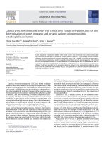
a b s t r a c t
A new approach for the determination of free and total valproic acid in small samples of 140 L human
plasma based on capillary electrophoresis with contactless conductivity detection is proposed. A dispersive liquid–liquid microextraction technique was employed in order to remove biological matrices
prior to instrumental analysis. The free valproic acid was determined by isolating free valproic acid
from protein-bound valproic acid by ultrafiltration under centrifugation of 100 L sample. The filtrate
was acidified to turn valproic acid into its protonated neutral form and then extracted. The determination of total valproic acid was carried out by acidifying 40 L untreated plasma to release the
protein-bound valproic acid prior to extraction. A solution consisting of 10 mM histidine, 10 mM 3-(Nmorpholino)propanesulfonic acid and 10 M hexadecyltrimethylammonium bromide of pH 6.5 was used
as background electrolyte for the electrophoretic separation. The method showed good linearity in the
range of 0.4–300 g/mL with a correlation coefficient of 0.9996. The limit of detection was 0.08 g/mL,
and the reproducibility of the peak area was excellent (RSD = 0.7–3.5%, n = 3, for the concentration range
from 1 to 150 g/mL). The results for the free and total valproic acid concentration in human plasma were
found to be comparable to those obtained with a standard immunoassay. The corresponding correlation
coefficients were 0.9847 for free and 0.9521 for total valproic acid.
© 2012 Elsevier B.V. All rights reserved.
1. Introduction
Valproic acid (2-propylvaleric acid, VPA) is an eight-carbon
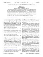
branched-chain fatty acid. Its structure is shown in Fig. 1 together
with that of caproic acid which was used as internal standard.
Valproic acid is used widely as an anticonvulsant [1] and as a moodstabilizing drug in patients with bipolar disorder [2]. Although the
mechanisms of action of valproic acid in epilepsy and bipolar disorder are currently not fully understood, the most widely accepted
processes for its antiepileptic activity involve an increase in the
concentration of the inhibitory neurotransmitter ␥-aminobutyric
∗ Corresponding author at: Department of Chemistry, University of Basel,
Spitalstrasse 51, 4056 Basel, Switzerland. Tel.: +41 61 267 10 53;
fax: +41 61 267 10 13.
∗∗ Corresponding author. Tel.: +41 61 267 10 03; fax: +41 61 267 10 13.
E-mail addresses: (H.H. See),
(P.C. Hauser).
1570-0232/$ – see front matter © 2012 Elsevier B.V. All rights reserved.
/>
acid (GABA) in certain brain regions and an inhibition of voltagedependent sodium channels [3].
Taking into account the pKa of VPA of 4.6, most valproate in
serum is deprotonated under physiological conditions. Since VPA
is highly bound to albumin (approximately 80–95%), only a small
fraction of VPA exists in the free, pharmacologically active form
[4,5]. The therapeutic range reported for total VPA in human plasma
is 50–100 g/mL [6]. Therapeutic drug monitoring (TDM) of VPA is
commonly performed for guiding therapy as there is only a poor
correlation between dose and steady state serum concentrations
between patients [7] and the difficulty to monitor the clinical effect
of valproic acid, since seizures are usually rare events. Detailed discussions are available regarding TDM of VPA in the treatment of
epilepsy [7,8] and bipolar disorders [9].
Several methods have been published for the determination of
free and total VPA in biological matrices. For the determination of
the total concentration, VPA is usually released from proteins by
acidification [10–12], which converts it into its protonated form. An
alternative method of destroying the protein-binding is precipitation of the serum proteins, e.g. by addition of an organic solvent (see
T.T.T. Pham et al. / J. Chromatogr. B 907 (2012) 74–78
Fig. 1. Structures of valproic acid (VPA) and caproic acid (CPA) used as internal
standard (IS).
for example [13]). For the determination of free VPA in the presence
of serum proteins and protein-bound VPA, free VPA is removed by a
separation step such as dialysis, ultrafiltration, ultracentrifugation
or gel filtration [14–16].
In both approaches for the quantification step the most commonly used methods are enzyme immunoassays [17,18]. This
technique is simple and reliable, but relatively expensive. A
number of chromatographic techniques such as gas chromatography (GC) [19] and liquid chromatography (LC) [20–23] have
also been reported, and have been used in conjunction with
various sample pretreatment steps. Commonly used pretreatments are, for instance, liquid–liquid extraction (LLE) [24], solid
phase extraction (SPE) [10], solid-phase microextraction (SPME)
[25], liquid-phase microextraction (LPME) [12] and dispersive
liquid–liquid microextraction (DLLME) [11]. A major drawback of
the reported chromatographic approaches is the requirement of
prior derivatization of VPA to either render it volatile or suitable
for UV-detection.
More recently, capillary electrophoresis coupled with contactless conductivity detection (CE-C4 D) has become an attractive
alternative analytical method due to its universal characteristics in
detecting any charged species without requiring a chromophore.
A further distinct advantage is the ability to carry out an analysis
in very small sample volumes. Several recent general review articles on CE-C4 D are available [26–28]. A series of applications of the
method for clinical analysis of diverse biological samples have been
reported [29–40]. Recent reviews on the applications of CE-C4 D in
pharmaceutical analysis [41,42] can also be found.
The potential usefulness of CE-C4 D for the determination of VPA
in clinical samples has been shown by Belin et al. [13]. However,
in these investigations, no distinction between free and proteinbound VPA was made and the amount of biological sample used was
too high for monitoring pediatric patients. We therefore improved
this method by reducing the plasma sample size needed and by
making the method suitable for the determination of both free and
total VPA.
2. Experimental
2.1. Reagents and materials
All chemicals were at least of analytical grade and purchased
from Aldrich or Fluka (both Buchs, Switzerland). Ultrapure deionized water was produced using a Nano-Pure water purification
system (Barnstead, IA, USA). Separation buffers were prepared
daily. Stock solutions of VPA sodium salt and caproic acid sodium
salt (CPA) as internal standard (IS) at the concentration of
1000 g/mL were prepared in deionized water and kept at 4 ◦ C.
Working standard solutions of lower concentrations were prepared
by dilution with deionized water.
2.2. Plasma samples
Blank and VPA containing plasma samples were obtained
from the Clinical Pharmacology and Toxicology Laboratory of the
75
University Hospital of Basel, Switzerland. All plasma samples were
kept at −20 ◦ C in a freezer until the experiments. The reference
values for free and total VPA content in the collected plasma
samples were measured using standard protocols adopted at the
Clinical Chemistry Laboratory of the University Hospital of Basel.
The total VPA concentration was determined using a homogenous enzyme immunoassay in a Cobas 6000 analyzer (Roche
Diagnostics, GmbH, Mannheim, Germany) using reagents from
Roche Diagnostics (Basel, Switzerland) instrument. The free VPA
was determined by first carrying out ultracentrifugation for isolation of the free VPA followed by a fluorescence polarization
immunoassay on a TDx analyzer (Abbott Laboratories, Abbott Park,
IL, USA).
2.3. Sample pretreatment procedure
For the determination of free VPA, 100 L of plasma sample was
pretreated by ultracentrifugation using Amicon ultracentrifugal filters (cut off >10,000 Da) (Millipore Corporation, Billerica, MA, USA)
for 15 min at 14,000 × g. After ultrafiltration, 40 L of the filtrate,
which contained free VPA, was placed into a 1.5 mL conical bottom
polypropylene tube. Subsequently, 10 L of a solution containing
25 g/mL CPA (internal standard resulting in a final concentration
of 5 g/mL) was added and the sample acidified with 10 L of 1 M
HNO3 to protonate VPA. The mixture was vortexed for 30 s and VPA
extracted as described below. For the determination of total VPA,
10 L internal standard and 10 L 1 M HNO3 were added directly
to 40 L of the raw plasma sample.
The optimization of the extraction step was carried out by
using blank plasma samples into which VPA was spiked at the
same level as the internal standard. For the extraction a mixture of extraction and dispersive solvent was rapidly injected
into the sample tube, the solution vortexed for 30 s and finally
centrifuged for 10 min at 6000 × g at room temperature. After
centrifugation, the lower (organic) phase was withdrawn using a
100 L microsyringe and transferred to a 200 L polypropylene
bullet tip tube. 20 L of triethylamine (TEA) solution of different
concentrations (see Section 3) was then added to the collected
organic phase, vortexed for 30 s, and centrifuged for 10 min at
6000 × g. The target analyte was back-extracted into the diluted
TEA solution and the supernatant was injected into the CE-C4 D
system.
2.4. CE-C4 D analysis
The capillary electrophoresis instrument was purpose-built and
utilized a commercial high voltage power supply module (CZE
2000R, Spellman, Pulborough, UK). The C4 D detector was built-inhouse, details can be found elsewhere [43]. The detector signals
were recorded with an e-corder data acquisition system (eDAQ,
Denistone East, NSW, Australia). A bare fused silica capillary of
50 m I.D. and 363 m O.D. (Polymicro Technologies, Phoenix,
AZ, USA) with a total length of 50 cm and effective length of
45 cm was employed. The new capillary was conditioned by first
flushing with 0.1 M NaOH for 15 min and followed by water for
10 min. The pre-conditioned capillary was then rinsed with the
separation buffer for 30 min. The running buffer employed was
slightly modified from the previous work [13] and consisted of
10 mM 3-(N-morpholino)propanesulphonic acid (MOPS), 10 mM
histidine (His), and 10 M hexadecyltrimethylammonium bromide
(CTAB) (pH 6.5). After each injection, the capillary was rinsed
with separation buffer for 3 min to maintain the reproducibility of the analysis. Injections were performed by siphoning at
18 cm height difference for 10 s. The separation voltage was set
at −16.5 kV.
76
T.T.T. Pham et al. / J. Chromatogr. B 907 (2012) 74–78
3. Results and discussion
3.1. Optimization of the dispersive liquid–liquid microextraction
First tests were carried out using direct injection of plasma
samples into the CE system as reported previously [13]. It was
found however, that some samples showed overlaps with peaks of
unknown origin. Therefore an extraction procedure was adopted
in order to consistently obtain electropherograms free of undesired matrix elements. Dispersive liquid–liquid microextraction
(DLLME) allows efficient extraction of small samples. In this procedure a mixture of two solvents, one soluble in water, the other
not, is rapidly injected into an aqueous sample. This leads to the
formation of finely dispersed droplets into which the extraction of
the analytes occurs. Subsequently, phase separation is performed
and the enriched analyte can then be determined in the sedimented
phase [44,45]. Several factors affecting the extraction efficiency of
DLLME were comprehensively examined to seek for optimum conditions. For these tests, valproate and caproate as internal standard
were added to blank plasma samples (both at a final concentration
of 5 g/mL) and these were acidified in order to protonate, and thus
neutralize, analyte and internal standard. Caproic acid (CPA) has a
molecular structure which is very similar to that of valproic acid
(VPA) (see Fig. 1).
3.1.1. Selection of extraction and dispersive solvents
An ideal extraction solvent in DLLME should demonstrate characteristics such as higher density than water, high extraction
capability for analytes of interest, low solubility in water, and low
volatility [44,46]. On the other hand, the dispersive solvent should
be miscible with the extraction solvent as well as the sample solution to enlarge the contact area between the extraction solvent and
the sample solution. Based on these requirements, 3 extraction solvents namely tetrachloroethylene (C2 Cl4 ), chloroform (CHCl3 ) and
carbon disulfide (CS2 ) were studied in combination with 4 dispersive solvents, i.e. acetonitrile (MeCN), methanol (MeOH), acetone
(Ace), and 2-propanol (IPA). It was found that CHCl3 hardly formed
an emulsified solution when added to plasma regardless of the dispersive solvent being used. When CS2 was employed, emulsified
solutions were observed, but clear phase separation could not be
achieved after centrifugation. Nevertheless, mixtures of C2 Cl4 with
various dispersive solvents studied were found to be able to form
satisfactory emulsified solutions and phase separation was instantaneously achieved after the vortex and centrifugation processes.
Hence, C2 Cl4 was selected as extraction solvent and its performance
with various dispersive solvents was evaluated. In order to maintain consistency, 13 L of each dispersive solvent with 87 L of
C2 Cl4 was always added to the 40 L of the blank plasma to which
acid as well as VPA and CPA had been added. As can be seen in
Fig. 2, the highest VPA peak area response was obtained when IPA
was used as dispersive solvent. The same result was obtained for
CPA.
3.1.2. Effect of extraction/dispersive solvent ratio and volume of
solvent mixture
Different ratios of C2 Cl4 :IPA solvent mixtures were studied to
seek for optimum extraction conditions. The volume of the solvent
mixture was fixed at 100 L and this was again added to the 40 L
of the blank plasma which had then been acidified and spiked with
VPA and CPA. As can be seen from Fig. 3, the peak area response
for the VPA extract increased according to the increase of C2 Cl4
percentage in the mixture. A significant increase of VPA responses
was observed from 20% of C2 Cl4 to 50% and ultimately reached its
maximum at 87% of C2 Cl4 . When the percentage of C2 Cl4 was further increased, no significant further enhancement of VPA and CPA
Fig. 2. Effect of dispersive solvents on the peak area response of VPA (n = 3). Extraction conditions: sample volume, 50 L; extraction solvent, 87 L C2 Cl4 ; dispersive
solvent, 13 L; concentration of VPA, 5 g/mL.
response was observed. Hence, the C2 Cl4 :IPA ratio of 87:13 was
adopted.
To consider the effect of the solvent volume on extraction efficiency, different volumes of C2 Cl4 :IPA mixtures with the optimum
ratio of 87:13 were tested. The volumes ranged from 50 to 175 L.
It was found that when even smaller volumes were employed
(<50 L), the organic droplets were not properly formed and not
well-dispersed in the relatively viscous plasma sample. As can be
seen from Fig. 4, the amount of VPA detected increased significantly by increasing the solvent volume from 50 to 125 L and then
reached a maximum in the range from 125 to 175 L. Although
the total solvent volume used in this study is relatively high compared to the amounts used in most of the studies reported, the
Fig. 3. Effect of the volume ratio of C2 Cl4 :IPA on the peak area response of VPA
(n = 3). Extraction conditions: extraction solvent, C2 Cl4 ; dispersive solvent, IPA; total
solvent volume, 100 L. Other conditions as for Fig. 2.
T.T.T. Pham et al. / J. Chromatogr. B 907 (2012) 74–78
Fig. 4. Effect of the volume of solvent mixture on the peak area response of VPA
(n = 3). Extraction conditions: ratio of extraction solvent (C2 Cl4 ):dispersive solvent
(IPA), 87:13. Other conditions as for Fig. 2.
new approach involves an additional back-extraction procedure
that effectively transfers the analyte into only 20 L of TEA solution prior to CE-C4 D analysis. In order to ensure a high consistency
of extraction performance, the solvent volume was fixed to 150 L
for the subsequent experiments.
3.1.3. Optimization of triethylamine percentage for back
extraction
As mentioned previously, the VPA enriched in the organic phase
was back-extracted into a diluted aqueous solution of TEA [24],
which was compatible with the subsequent CE-C4 D analysis. 20 L
of this solution was used as this was the minimum volume which
could be handled reliably with the CE-system employed. Concentrations of 0.05%, 0.1%, 0.25%, 0.5%, 1% and 2.5% were tested for
their suitability. For TEA solutions of 0.05% and 0.1%, the extraction recoveries for VPA were generally unsatisfactory with values
of 47–62%. An increase of the TEA percentage to 0.25% and 0.5%
resulted in improved extraction recoveries of 86%. The result for
CPA was identical. For higher concentrations, poor baseline stabilities resulted in the CE-C4 D analysis. Hence, a percentage of 0.5%
of TEA was adopted for the back-extraction solution for the subsequent CE-C4 D analyses.
77
Fig. 5. Electropherogram for (a) blank plasma spiked with CPA (5 g/mL, as internal
standard, IS) and (b) blank plasma spiked with VPA (5.4 g/mL) and CPA (5 g/mL).
CE conditions: buffer 10 mM MOPS/10 mM His, pH 6.5, CTAB 10 M, siphoning injection at 18 cm height difference for 10 s, separation voltage −16.5 kV.
3.2. Method validation
The optimum DLLME parameters finally arrived at were as follows: 40 L of plasma sample acidified with 10 L 1 M HNO3 , 10 L
of 25 g/mL CPA internal standard solution (5 g/mL final concentration), 150 L of 87% C2 Cl4 :13% IPA as solvent mixture, and 20 L
of 0.5% TEA solution as back-extraction medium. Normalization
of the peak areas obtained for VPA with the peak areas for CPA
resulted in a good linearity for VPA with a correlation coefficient
of 0.9996 in the concentration range from 0.4 to 300 g/mL (note
that these tests were carried out for unfiltered plasma). This linear
range covered the entire therapeutic range of VPA in human plasma
which is 5–10 g/mL for free and 50–100 g/mL for total valproate.
The limit of detection (LOD) and limit of quantification (LOQ) were
determined as 0.08 g/mL and 0.24 g/mL, respectively (calculated
for signal-to-noise ratios of 3 and 10 from a comparison of peak
heights with the maximum amplitude of the short term baseline
deviations). The reproducibilities for peak area were found to be
between 0.7% and 3.5% (RSD, n = 3) for the concentration range from
1 to 150 g/mL. For illustration, electropherograms for an extract
of blank plasma spiked with CPA and for an extract of blank plasma
spiked with CPA and VPA are shown in Fig. 5.
Table 1
Quantitative results for free and total VPA in human plasma samples.
Sample ID
Free VPA (g/mL)
4
a
DLLME–CE-C D
BVS11
EH4
AP10
JS09
GP14
EH5
a
16.2
5.4
4.8
5.4
7.0
4.6
Errors are standard deviations (n = 3).
±
±
±
±
±
±
0.3
0.1
0.2
0.6
1.3
0.2
Total VPA (g/mL)
Immunoassay
DLLME–CE-C4 Da
14.8
3.7
3.6
5.3
6.0
3.6
116.2
56.5
37.2
61.1
71.9
43.5
±
±
±
±
±
±
1.0
1.6
0.7
4.2
10.7
5.3
Immunoassay
99.5
56.5
33.8
63.0
75.7
44.2
78
T.T.T. Pham et al. / J. Chromatogr. B 907 (2012) 74–78
3.3. Analysis of human plasma samples
A total of 6 human plasma samples had been collected in a
clinical study conducted at the University Hospital of Basel. The
plasma samples were first tested using standard protocols based on
enzyme immunoassay techniques employed in the Clinical Chemistry Laboratory of the University Hospital of Basel (see Section
2.3 for details), followed by measurement using the developed
DLLME–CE-C4 D approach. Note that the appearance of the electropherograms of these samples containing VPA is very similar from
those of blank plasma to which VPA had been spiked as shown in
Fig. 5. The results for free (for the filtered sample) and total VPA (for
the not filtered sample) are summarized in Table 1. It is observed
that the overall results obtained using DLLME–CE-C4 D are comparable to the results obtained employing the standard enzyme
immunoassay. The correlation coefficients, r, for the two pairs of
data were determined as 0.9847 for free VPA and 0.9521 for total
VPA, indicating an acceptable relationship.
4. Conclusion
In this work, the determination of free and total VPA in a total
volume of only 140 L of human plasma employing CE-C4 D for
quantification was developed. The method requires a filtration step
in order to be able to distinguish between free and bound analyte
and an extraction procedure to avoid potential peak overlaps, but
no chemical or enzymatic conversion steps, as needed for the established methods, are required to make the analyte amenable for
quantification. The method is deemed suitable for the routine therapeutic drug monitoring (TDM), in particular for pediatric patients
for whom the available sample volumes are limited.
[4]
[5]
[6]
[7]
[8]
[9]
[10]
[11]
[12]
[13]
[14]
[15]
[16]
[17]
[18]
[19]
[20]
[21]
[22]
[23]
[24]
[25]
[26]
[27]
[28]
[29]
[30]
[31]
[32]
[33]
[34]
[35]
Acknowledgements
The authors would like to thank the Swiss Federal Commission for Scholarships for Foreign Students (Thi Thanh Thuy Pham),
the Universiti Teknologi Malaysia (Hong Heng See) and the Swiss
National Science Foundation (grant numbers 200021-129721/1
and 200020-137676/1) for financial support.
References
[1] C.U. Johannessen, S.I. Johannessen, CNS Drug Rev. 9 (2003) 199.
[2] D.J. Bond, R.W. Lam, L.N. Yatham, J. Affect. Disord. 124 (2010) 228.
[3] C.U. Johannessen, Neurochem. Int. 37 (2000) 103.
[36]
[37]
[38]
[39]
[40]
[41]
[42]
[43]
[44]
[45]
[46]
C. DeVane, Psychopharmacol. Bull. 37 (2003) 25.
E. Perucca, CNS Drugs 16 (2002) 695.
T. Tatsuhara, H. Muro, Y. Matsuda, Y. Imai, J. Chromatogr. A 399 (1987) 183.
E. Yukawa, Clin. Pharmacokinet. 31 (1996) 120.
E. Kozer, D. Scolnik, W.M. Agamata, S.K. Weiss, Z.H. Verjee, G. Koren, Ther. Drug
Monit. 25 (2003) 17.
J. Fleming, M. Chetty, Clin. Neuropharmacol. 29 (2006) 350.
S. Gao, H. Miao, X. Tao, B. Jiang, Y. Xiao, F. Cai, Y. Yun, J. Li, W. Chen, J. Chromatogr.
B 879 (2011) 1939.
H.R. Sobhi, A. Kashtiaray, H. Farahani, F. Abrahimpour, A. Esrafili, Drug Test.
Anal. 2 (2010) 362.
P. Shahdousti, A. Mohammadi, N. Alizadeh, J. Chromatogr. B 850 (2007) 128.
G.K. Belin, S. Krähenbühl, P.C. Hauser, J. Chromatogr. B 847 (2007) 205.
H. Kurz, H. Trunk, B. Weitz, Arzneimittelforschung 27 (1977) 1373.
T.C. Kwong, Clin. Chim. Acta 151 (1985) 193.
A.C. Metha, TrAC-Trends Anal. Chem. 8 (1989) 107.
S. Cooreman, E. Cuypers, M. De Doncker, P. Van Hee, W. Uyttenbroeck, H. Neels,
Immuno-Anal. Biol. Spec. 23 (2008) 240.
A.A. Elyas, V.D. Goldberg, N. Ratnaraj, P.T. Lascelles, Ann. Clin. Biochem. 17
(1980) 307.
J. Darius, J. Chromatogr. B 682 (1996) 67.
C. Lucarelli, P. Villa, E. Lombaradi, P. Prandini, A. Brega, Chromatographia 33
(1992) 37.
M. Nakamura, K. Kondo, R. Nishioka, S. Kawai, J. Chromatogr. B 310 (1984) 450.
J.H. Wolf, L. Veenma-van der Duin, J. Korf, J. Chromatogr. B 487 (1989) 496.
M.-C. Lin, H.-S. Kou, C.-C. Chen, S.-M. Wu, H.-L. Wu, J. Chromatogr. B 810 (2004)
169.
H. Amini, M. Javan, A. Ahmadiani, J. Chromatogr. B 830 (2006) 368.
M. Krogh, K. Johansen, F. Tønnesen, K.E. Rasmussen, J. Chromatogr. B 673 (1995)
299.
W.K.T. Coltro, R.S. Lima, T.P. Segato, E. Carrilho, D.P. de Jesus, C.L. do Lago, J.A.F.
da Silva, Anal. Methods 4 (2012) 25.
ˇ P.C. Hauser, Electrophoresis 32 (2011) 30.
P. Kubán,
ˇ P.C. Hauser, Electrophoresis 30 (2009) 3305.
P. Kubán,
ˇ A. Scholer, P.C. Hauser, J. Chromatogr. A 1213 (2008) 100.
X.Y. Gong, P. Kubán,
ˇ
˚
P. Tuma,
K. Málková, E. Samcová, K. Stulík,
J. Sep. Sci. 33 (2010) 2394.
ˇ
˚
P. Tuma,
K. Málková, Z. Wedellová, E. Samcová, K. Stulík,
Electrophoresis 31
(2010) 2037.
˚
P. Tuma,
E. Samcová, Chem. Listy 101 (2007) 200.
˚
P. Tuma,
E. Samcová, F. Duˇska, J. Sep. Sci. 31 (2008) 2260.
ˇ
˚
P. Tuma,
E. Samcová, K. Stulík,
Anal. Chim. Acta 685 (2011) 84.
ˇ J. Tanyanyiwa, A. Rainelli, P.C. Hauser, Anal. Chim. Acta 525
Q.J. Wan, P. Kubán,
(2004) 11.
ˇ P.C. Hauser, Lab Chip 8 (2008) 1829.
P. Kubán,
ˇ L.L. Yuan, J.H. Zhao, S.F.Y. Li, P.C. Hauser, Electrophoresis 27
W.S. Law, P. Kubán,
(2006) 1932.
W. Pormsila, S. Krähenbühl, P.C. Hauser, Anal. Chim. Acta 636 (2009) 224.
W. Pormsila, R. Morand, S. Krähenbühl, P.C. Hauser, J. Chromatogr. B 879 (2011)
921.
W. Pormsila, R. Morand, S. Krähenbühl, P.C. Hauser, Electrophoresis 32 (2011)
884.
L. Suntornsuk, Anal. Bioanal. Chem. 398 (2010) 29.
A.A. Elbashir, H.Y. Aboul-Enein, Biomed. Chromatogr. 24 (2010) 1038.
ˇ P.C. Hauser, Meas. Sci. Technol. 17 (2006) 3317.
L. Zhang, S.S. Khaloo, P. Kubán,
X.-H. Zang, Q.-H. Wu, M.-Y. Zhang, G.-H. Xi, Z. Wang, Chin. J. Anal. Chem. 37
(2009) 161.
M. Rezaee, Y. Yamini, M. Faraji, J. Chromatogr. A 1217 (2010) 2342.
A.R. Zarei, F. Gholamian, Anal. Biochem. 412 (2011) 224.