DSpace at VNU: New phenolic compounds from the lichen Parmotrema praesorediosum (Nyl.) Hale (Parmeliaceae)
Bạn đang xem bản rút gọn của tài liệu. Xem và tải ngay bản đầy đủ của tài liệu tại đây (909.74 KB, 7 trang )
Letter - spectral assignments
Received: 15 March 2015
Revised: 14 July 2015
Accepted: 21 July 2015
Published online in Wiley Online Library: 25 August 2015
(wileyonlinelibrary.com) DOI 10.1002/mrc.4316
New phenolic compounds from the lichen
Parmotrema praesorediosum (Nyl.)
Hale (Parmeliaceae)
Bui Linh Chi Huynh,a Duy Hoang Le,b Yukiko Takenaka,b Takao Tanahashib
and Kim Phi Phung Nguyenc*
Introduction
Lichens, symbiotic combination of fungi and algae, comprise more
than 20 000 species that are found in most of the environmental
habitats from the tropics to polar regions. Lichens produce a great
variety of metabolites, most of them occur only in lichens and the
others are also present in fungi and higher plants. Characteristic
secondary metabolites of lichens are depsides, depsidones,
diphenyl ethers, benzofuran, usnic acid, and anthraquinone
derivatives.[1–3]
Parmotrema is one of the largest genera of the family
Parmeliaceae. This genus of foliose lichen is widely distributed in
tropical regions and composed of c. 350 species worldwide.[4] Previous phytochemical examination on Parmotrema praesorediosum
(Nyl.) Hale, collected on a betel nut tree, in southern Thailand, revealed that this species contained some lactone fatty acids.[5] Sample collected in south Korea contained atranorin, chloroatranorin,
and fatty acids, i.e. protopraesorediosic acid and praesorediosic
acid.[4]
In the course of our systematic research on lichen substances
from the Vietnamese flora, we have examined P. praesorediosum
(Nyl.) Hale that is distributed in the south of Vietnam. In our previous
study on this species, nine compounds, including methyl
haematommate, butyl haematommate, methyl chlorohaematommate,
methyl β-orsellinate, methyl divaricatinate, atranol, atranorin, (+)-(12R)isousnic acid, and (+)-(12R)-usnic acid were isolated.[6] Further
chemical investigation on this species by using modern separation
techniques has led to the isolation of seven novel phenolic compounds, four diphenyl ethers (1–4) and three phthalide derivatives
(5–7). In this paper, we report the isolation and structure elucidation
of these compounds.
carbonyl (1730 cmÀ1), and aromatic (1645, 1455 cmÀ1) functionalities. Its 1H NMR spectrum showed one hydrogen bonded hydroxyl
group at δ 12.06 (1H, s), a formyl group at δ 10.39 (1H, d, J = 1.0 Hz),
an aromatic proton at δ 6.55 (1H, brs), two meta-coupled aromatic
protons at δ 6.31 (1H, d, J = 2.5 Hz) and 6.17 (1H, brd, J = 2.5 Hz), a
methoxy group at δ 3.50 (3H, s), and two methyl groups at δ 2.22
(3H, d, J = 0.5 Hz) and 2.00 (3H, brs). The combination of 13C and
DEPT NMR spectra of 1 (Table 1) revealed 17 carbons including a
formyl group (δ 193.6), a methoxycarbonyl [δ 167.0 (―COO), 52.4
(―OCH3)], 12 aromatic carbons (δ 102–164), five of which were oxygenated, and two methyl groups (δ 20.8 and 17.0).
Detailed analysis of HSQC, HMBC, and NOESY spectra identified
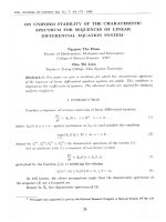
two aryl units, A-ring and B-ring. The HMBC spectrum (Fig. 2)
showed the correlations from the chelated hydroxyl group (δ
12.06, 4-OH) to carbon signals at δ 110.6 (C-3), 164.0 (C-4), and
113.9 (C-5) and from the aldehydic proton (δ 10.38) to C-3 and C4. The aromatic singlet proton (δ 6.55, H-5) showed cross-peaks
with C-1 (δ 114.9), C-3, C-4, and C-9 (δ 20.8), and three protons of
the methyl group (δ 2.22, H3-9), in turn, were correlated with C-1,
C-5, and C-6 (δ 147.7). The methoxy signal showed HMBC crosspeak with the carboxyl carbon at δ 167.0 and NOESY correlation
with H3-9, implying that the methoxycarbonyl group was situated
at C-1. Furthermore, C-2 was supposed to be oxygenated from its
chemical shift (δ 158.1) in the 13C NMR spectrum. Therefore, the A-ring
of 1 was established as 3-formyl-4-hydroxy-1-methoxycarbonyl-6methyl-2-oxygenated benzene.
The second aryl unit, B-ring, was a tetrasubstituted benzene core.
The HMBC spectrum of 1 showed correlations from a meta-coupled
aromatic proton at δ 6.17 (H-1′) to carbon signals at δ 153.3 (C-2′),
102.1 (C-3′), 136.3 (C-5′), and 17.0 (C-7′), from another proton at δ
6.31 (H-3′) to C-1′ (δ 109.5), C-2′, C-4′ (δ 148.8), and C-5′, and from
methyl protons (H3-7′) to C-1′, C-5′, and C-6′ (δ 131.1). Therefore,
Results and discussion
Magn. Reson. Chem. 2016, 54, 81–87
* Correspondence to: Nguyen Kim Phi Phung, Department of Organic Chemistry,
University of Science, National University—Ho Chi Minh City, 227 Nguyen Van
Cu Str., Dist. 5, Ho Chi Minh City, Vietnam. E-mail:
a Department of Science, Dong Nai University, Vietnam
b Department of Organic Chemistry, Kobe Pharmaceutical University, Japan
c Department of Organic Chemistry, University of Science, National University—Ho
Chi Minh City, Vietnam
Copyright © 2015 John Wiley & Sons, Ltd.
81
The thalli of P. praesorediosum were extracted with MeOH at room
temperature. A combination of chromatographic fractionation
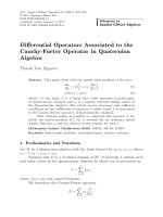
of the extract led to the isolation of seven phenolic compounds
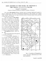
(1–7) (Fig. 1). Their structures were elucidated as the following.
Compound 1 was obtained as a yellow solid. Its molecular formula was established as C17H16O7 through the protonated molecule at m/z 333.0970 [M + H]+ in the HR-ESI-MS spectrum. IR
absorptions implied the presence of hydroxyl (3383 cmÀ1), ester
B. L. C. Huynh et al.
Figure 1. Structures of isolated compounds 1–7.
82
the B-ring of 1 was established as 2′,4′,5′-trioxygenated benzene
bearing a methyl group at 6′.
Comparison of the chemical shifts of C-1′–C-6′ of 2,4-dihydroxy6-methylphenoxy part with those of related compounds suggested
that the A-ring was linked to the B-ring through an ether linkage
between C-2 and C-5′.[7,8] This connection was well supported by
the key NOESY correlation between H-8 of the A-ring and H3-7′ of
the B-ring. Thus, compound 1 was characterized as methyl 2-(2,4dihydroxy-6-methylphenoxy)-3-formyl-4-hydroxy-6methylbenzoate and named praesorether A.
Compound 2 was also obtained as a yellow solid. Its molecular
formula was determined as C27H26O11 from the HR-ESI-MS spectrum. The 1H NMR spectral data of 2 were similar to those of 1
(Table 1) except for the absence of two meta-coupled aromatic
protons of B-ring and the presence of two newly appeared singlets
of aromatic protons and some additional signals, i.e. one methoxy
group (δ 3.88, 3H, s), one methylene group (δ 3.98 and 3.97, each
1H, br s), and one methyl group (δ 2.58, 3H, s). The 13C-NMR spectrum of 2 exhibited, besides the signals because of the same A
and B rings as 1, signals for one methoxycarbonyl group (δ 172.4,
52.2), six aromatic carbons including two oxygenated carbons
[δC 161.5 (C-2″), 160.0 (C-4″)], three quaternary carbons [δC 142.5
(C-6″), 119.5 (C-5″), 109.4 (C-1″)], and one CH [δC 101.5 (C-3″)], one
methylene carbon (δ 20.7) and one methyl carbon (δ 19.1). These
data indicated the presence of a third aromatic C-ring linked to
the B-ring in 2. This C-ring was established as the 2″,4″-dihydroxy1″-methoxycarbonyl-6″-methylphenyl moiety with a methylene
group at C-5″ by the analysis of its HMBC spectrum, which showed
the correlations from the methyl protons (δ 2.58, H3-8″) to C-1″, C-5″,
and C-6″, from the methylene (H2-8″) to C-4″, C-5″, C-6″, from an aromatic singlet proton (δ 6.33, H-3″) to C-1″, C-2″, C-4″, and C-5″, and
wileyonlinelibrary.com/journal/mrc
from the hydroxyl (δ 10.56, 2″-OH) to C-1″, C-2″, and C-3″. The substitution pattern of the ring C was further supported by the ROESY
correlation between H3-8″ and H2-8′.
The HMBC experiments showed that these methylene protons
(H2-8′) correlated to aromatic carbons of B-ring at δ 153.3 (C-2′),
113.1 (C-3′), and 149.2 (C-4′). From these findings, the methylene
carbon of the C-ring was linked to the B-ring at C-3′ (Fig. 2). Consequently, compound 2 was established as a new diphenyl ether derivative and named praesorether B.
Compound 3 was isolated as a yellow solid. Its molecular formula
was determined as C29H26O14 from the HR-ESI-MS spectrum that
meant compound 3 contained two carbon and three oxygen atoms
more than that of 2. The 1H NMR spectral data (Table 1) of 3 closely
resembled those of 2, suggesting that they had the same basic
framework except for the lack of one aromatic methine proton
and one methyl group and the appearance of an acetalic proton
at δ 5.20 and a methoxy group at δ 3.13. The comparison of 13C
NMR data of these two compounds showed that 3 also possessed
three aromatic rings connected thorough an ether linkage and a
methylene bridge as in 2, but it lacked one methyl group and
contained three more carbon atoms including one carboxyl carbon (δ 169.6), one acetalic carbon (δ 101.6), and one methoxy
carbon (δ 56.8), implying the presence of a lactone ring in 3. Although the HMBC spectrum could not afford further information
relating to the position of this lactone ring, the ROESY correlations of the methoxy protons (7′-OCH3) with the acetalic proton
(H-7′) and also with the formyl proton at δ 10.12 (H-8) of the Aring indicated the γ-lactone ring was fused to the B-ring at C-1′
and C-6′ (Fig. 3). Complete analysis of the 2D NMR data for 3 resulted in its formulation as shown, and it was named
praesorether C.
Copyright © 2015 John Wiley & Sons, Ltd.
Magn. Reson. Chem. 2016, 54, 81–87
New phenolic compounds from the Parmotrema praesorediosum
Table 1. NMR data for compounds 1À4
1a
No.
2b
3a
4b
Moiety a
δH
1
2
3
4
5
6
7
8
9
4-OH
7-OCH3
1′
2′
3′
4′
5′
6′
7′
8′
6.55
J (Hz)
δC
brs
114.9
158.1
110.6
164.0
113.9
147.7
167.0
193.6
20.8
10.39
2.22
12.06
3.50
6.17
d (1.0)
d (0.5)
s
s
brd (2.5)
6.31
d (2.5)
2.00
brs
9′
7′-OCH3
1″
2″
3″
4″
5″
6″
7″
8″
2″-OH
7″-OCH3
52.4
109.5
153.3
102.1
148.8
136.3
131.1
17.0
δH
J (Hz)
6.52 d (0.5)
10.44
2.15
12.11
3.18
6.24
brs
s
s
s
s
1.99
3.97
3.98
s
brs
brs
6.33
2.58
10.56
3.88
s
s
s
s
δC
115.5
158.9
111.0
164.3
112.9
148.2
166.6
195.6
20.5
52.0
108.9
153.3
113.1
149.2
135.8
129.9
16.7
20.7
109.4
161.5
101.5
160.0
119.5
142.5
172.4
19.1
52.2
δH
J (Hz)
δC
6.71
s
10.12
2.31
11.92
3.23
s
s
brs
s
116.5
156.4
111.8
163.6
116.2
148.1
165.8
193.7
21.2
5.20
4.05
s
brs
3.13
s
52.2
103.4
153.1
117.6
151.7
134.2
129.2
101.6
20.0
2.61
s
169.6
56.8
107.5
162.4
102.0
159.5
117.1
142.6
172.0
19.3
3.91
s
52.0
6.39
s
δH
Moiety b
J (Hz)
δC
6.48
s
10.45
2.11
12.09
3.00
6.22
s
s
brs
s
s
116.0
159.2
111.1
164.1
112.4
148.0
167.0
195.7
20.5
2.08
3.81
s
s
51.9
108.9
152.7
114.9
149.1
136.3
130.5
16.6
19.3
brs
brs
152.9
112.5
156.5
119.4
137.8
109.0
20.8
6.24
2.38
δH
J (Hz)
δC
6.47
s
10.43
2.13
12.09
3.00
6.16
s
s
brs
s
s
115.6
159.6
111.0
164.2
112.7
148.2
166.8
195.7
20.5
1.97
3.88
3.89
s
brs
brs
51.8
108.2
153.6
114.1
150.0
135.8
129.4
16.7
21.6
a
CDCl3
acetone-d6
b
Magn. Reson. Chem. 2016, 54, 81–87
(C-3′a), 149.1 (C-4′a), and 152.7 (C-2′a) of the Ba-ring and to carbon
signals at δ 112.5 (C-2″), and 152.9 (C-1″) of the C-ring, as well as correlations of the second methylene protons (δ 3.88 and 3.89, H2-8′b)
to carbon signals at δ 114.1 (C-3′b), 150.0 (C-4′b), and 153.6 (C-2′b) of
the Bb-ring and to signals at δ 119.4 (C-4″), 137.8 (C-5″) and 156.5
(C-3″) of the C-ring. Thus, the first methylene carbon linked the
C-ring to the Ba-ring at C-2″ and C-3′a and the second methylene
carbon linked the C-ring to the Bb-ring at C-4″ and C-3′b. This was
further supported by the ROESY cross peak between H2-8′b
(δ 3.88 and 3.89) and H3-7″ (δ 2.38) (Fig. 3). These information
fully established the chemical structure of compound 4 as
shown and it was named praesorether D.
Compound 5 was obtained as a white amorphous solid. Its molecular formula C12H14O6 was deduced from the protonated molecule [M + H]+ at m/z 255.0862 in the HR-ESI-MS spectrum. The IR
spectrum showed characteristic absorptions for a hydroxyl group
(3243 cmÀ1), a lactone group (1785 cmÀ1) and substituted aromatic
system (1630 and 1363 cmÀ1). The 1H NMR spectrum of 5 displayed
signals of three methoxy groups at δ 3.53, 3.62, and 3.86 (each 3H,
Copyright © 2015 John Wiley & Sons, Ltd.
wileyonlinelibrary.com/journal/mrc
83
Compound 4 was isolated as a yellow solid. Its molecular formula
was determined as C43H40O16 from the HR-ESI-MS. The 1H NMR
spectrum of 4 (Table 1) was similar to that of 1; however, all signals
ascribable to A and B-rings appeared in duplicate, and furthermore
some additional signals of one aromatic methine proton (δ 6.24, 1H,
s), two methylene protons (δ 3.89 and 3.88, each 1H, s; 3.81, 2H, s),
and protons of a methyl group (δ 2.38, 3H, s) were observed. The
same observation was also recorded for the 13C and DEPT NMR
spectra of 4 with the appearance of six more aromatic carbons (δ
156.5, 152.9, 137.8, 119.4, 112.5, and 109.0), two methylenes (δ
21.6, 19.3), and a methyl (δ 20.8).
These spectral features suggested that 4 composed of two sets of
praesorether A (1) basic skeleton (parts a and b) and a fifth aromatic
ring C. The position of functional groups in Aa, Ba, Ab, Bb, and
C-rings was confirmed by analysis of HSQC, HMBC, and ROESY
correlations. The connection of the two units, Ba and Bb-rings of 1
with the fifth aromatic ring C through two methylene groups, was
elucidated by the HMBC spectrum with the correlations of the first
methylene protons (δ 3.81, H2-8′a) to carbon signals at δ 114.9
B. L. C. Huynh et al.
Figure 2. HMBC and NOESY/ROESY correlations of 1 and 2.
s), two methylene protons at δ 4.84 and 4.88 (each 1H, d, J = 14.0 Hz,
H2-8), an aromatic proton at δ 6.88 (1H, s, H-7), an acetalic methine
proton at δ 6.33 (1H, s, H-3), and a phenolic hydroxyl proton at δ
9.08 (1H, s, 4-OH). The combination of 13C and DEPT NMR spectra
of 5 revealed 12 carbons including one carboxyl carbon (δ 168.8),
one acetalic methine carbon (δ 102.2), five aromatic quaternary carbons (δ 159.7, 153.3, 128.6, 124.5, and 116.1), one aromatic CH carbon (δ 97.6), one oxymethylene carbon (δ 70.0), and three methoxy
groups (δ 59.3, 56.3, and 56.1) (Table 2).
These spectral data suggested that 5 could be a 3-oxyphthalide
with three substituents on the benzene ring (Fig. 4). The HMBC experiments showed the correlations from the methoxy protons at δ
3.53 to a methylene group (δ 70.0, C-8) and from the methylene
protons (H2-8) to two oxygenated aromatic carbons (δ 159.7, C-6;
153.3, C-4), one aromatic quaternary carbon (δ 116.1, C-5), and also
to this methoxy carbon (δ 59.3), indicating the methoxymethyl
group at C-5. The sole aromatic proton at δ 6.88 was located at C7 by its HMBC correlations with the carboxyl carbon (δ 168.8, C-1)
and other aromatic carbons (δ 159.7, C-6; 124.5, C-3a; and 116.1,
C-5). The other substituents, methoxy and phenolic hydroxyl
groups were located at C-6 and C-4, respectively, by the analysis
of 2D NMR spectra (HMBC and NOESY). Finally, the HMBC correlations of the acetalic methine proton at δ 6.33 with the carboxyl carbon (C-1) and a methoxy carbon (δ 56.3) confirmed the methoxy
group at C-3 on the five-member ring lactone. The absolute configuration of C-3 was not determined. Thus, compound 5 was
assigned as 4-hydroxy-3,6-dimethoxy-5-methoxymethylphthalide
and named praesalide A.
Compounds 6 and 7, designated praesalides B and C, were
phthalide derivatives closely related to 5. The HR-MS measurements of 6 and 7 established the molecular formulas of C13H16O6
and C14H18O6. The 1H and 13C NMR data of 6 and 7 (Table 2) were
similar to those of 5 except for the presence of an ethoxy group
at C-8 in 6 and two ethoxy groups at C-3 and C-8 in 7 instead of
methoxy groups in 5. This was supported by the analysis of their
2D NMR (COSY, HSQC, HMBC, and NOESY) spectral features. These
results suggested the structures of 6 and 7 as 3-ethoxy-4hydroxy-6-methoxy-5-methoxymethylphthalide and 3-ethoxy-5ethoxymethyl-4-hydroxy-6-methoxyphthalide, respectively.
Experimental
General experimental procedures
The NMR spectra were measured on a Varian NMR System-500 or
INOVA-500 spectrometer, at 500 MHz for 1H NMR and 125 MHz for
13
C NMR. The HR-ESI-MS were recorded on an Exactive mass spectrometer (Thermo Fisher Scientific). The optical rotations were measured on a Jasco DIP-370 digital polarimeter. The IR spectra were
measured on Shimadzu FTIR-8200 infrared spectrophotometer.
TLC was carried out on silica gel 60F254 or silica gel 60 RP-18 F254S
(Merck) and spots were visualized by spraying with a solution of
5% vanillin in ethanol, followed by heating at 100 °C. Gravity column chromatography was performed with silica gel 60 (0.040–
0.063 mm, Merck).
84
Figure 3. HMBC and ROESY correlations of 3 and 4.
wileyonlinelibrary.com/journal/mrc
Copyright © 2015 John Wiley & Sons, Ltd.
Magn. Reson. Chem. 2016, 54, 81–87
New phenolic compounds from the Parmotrema praesorediosum
Table 2. NMR data for compounds 5–7 (CDCl3)
No.
1
3
3a
4
5
6
7
7a
8
9
1′
2′
1″
2″
4-OH
5
δH
J (Hz)
6.33
s
6.88
s
4.84
4.88
3.86
3.62
d (14.0)
d (14.0)
s
s
3.53
s
9.08
s
6
δC
168.8
102.2
124.5
153.3
116.1
159.7
97.6
128.6
70.0
56.1
56.3
59.3
δH
J (Hz)
6.40
s
6.87
s
4.83
4.88
3.85
3.86
3.94
1.33
3.53
9.03
d (14.0)
d (14.0)
s
dq (9.5, 7.0)
dq (9.5, 7.0)
t (7.0)
s
7
δC
169.0
101.5
124.9
153.3
116.0
159.6
97.5
128.6
70.0
56.1
65.3
15.1
59.3
brs
δH
J (Hz)
6.40
s
6.86
s
4.86
4.92
3.85
3.86
3.94
1.33
3.70
1.32
9.33
d (14.0)
d (14.0)
s
dq (9.5, 7.0)
dq (9.5, 7.0)
t (7.0)
q (7.0)
t (7.0)
s
δC
169.0
101.5
124.8
153.3
116.2
159.5
97.4
128.5
68.1
56.1
65.3
15.2
67.6
15.0
Plant material
The lichen thalli of P. praesorediosum were collected on the bark of
Dipterocarpus sp. at Tan Phu forest, Dong Nai province, Vietnam in
June 2009. The geographical location where the lichen was collected is at an altitude of 110 m, 11°20′–11°50′ N and 107°09′–107°
35′ E. The botanical species of P. praesorediosum (Nyl.) Hale (synonym of Parmelia praesorediosa Nyl.) was identified by MSc. Vo Thi
Phi Giao, Faculty of Biology, University of Science, National University—Ho Chi Minh City. A voucher specimen (No US-B020) was deposited in the Herbarium of The Department of Organic Chemistry,
Faculty of Chemistry, University of Science, National University—Ho
Chi Minh City, Vietnam.
Figure 4. HMBC and NOESY correlations of 5.
Magn. Reson. Chem. 2016, 54, 81–87
Extraction and isolation
The thallus material (5.0 kg) was washed under flow of tap water
and then was air-dried at ambient temp. to obviate thermally induced decomposition prior to be ground into a fine powder. The
ground powder sample (3.0 kg) was macerated by methanol at
room temperature to afford a crude methanol extract (450 g). This
crude one (450 g) was applied to silica gel solid phase extraction,
successively eluted with the following solvents: petroleum ether
(60–90 °C) (PE), chloroform (C), ethyl acetate (EA), acetone (A), and
methanol (M) to afford corresponding extracts: extract PE (40 g), extract C (105 g), extract EA (50 g), extract A (45 g), and extract M
(37 g).
The chloroform extract (105 g) was subjected to silica gel column
chromatography, eluted by the solvent system of petroleum ether–
ethyl acetate with increasing ethyl acetate to give 23 fractions from
C1 to C23. Fraction C19 (6.1 g) was applied on silica gel column and
eluted with a gradient solvent system of chloroform–acetone (95:5)
to give three fractions (C19a, C19b, and C19c). Fraction C19a (1.0 g)
was silica gel rechromatographed, eluted with chloroform–acetone
(98:2) and subjected to pre TLC using chloroform–methanol (9:1
and 95:5) as eluent to afford 1 (5.0 mg). Fraction C19b (3.2 g) was silica gel rechromatographed, eluted with chloroform–acetone (98:2)
to give six fractions (C19ba to C19bf). Fraction C19bc (454.3 mg)
Copyright © 2015 John Wiley & Sons, Ltd.
wileyonlinelibrary.com/journal/mrc
85
All NMR experiments were acquired at ambient temperature.
Chemical shifts are expressed in ppm with reference to the internal
TMS (0.000).
1
H and 13C NMR spectra were obtained using a Varian NMR
System-500 or INOVA-500 spectrometer. 1H spectra: On a Varian
NMR System-500 spectrometer, spectral width (SW) 8012.8 Hz, acquisition time (AT) 2.045 s, number of data points (NP) 32 768 K, filter band width (FB) 4000 Hz, block size (BS) 32, steady-state
transients (SS) 0, relaxation delay (D1) 1 s, spectrometer frequency
(SF) 499.73 MHz, pulse 90 width (PW) 7.9 μs, temperature (TE)
25 °C, line broadening (LB) not use; On a INOVA-500, SW
7996.8 Hz, AT 4.097 s, NP 65 530 K, FB 4000 Hz, BS 32, SS 1, D1 1 s,
SF 499.83 MHz, PW 11.5 μs, TE 25 °C, LB not use. 13C spectra: On a
Varian NMR System-500 spectrometer, SW 31 250.0 Hz, AT 1.049 s,
NP 65 536 K, FB 17 000 Hz, BS 32, D1 1 s, SF 125.671 MHz, PW
9.0 μs, TE 25 °C, LB 0.5 Hz; On a INOVA-500, SW 30 165.9 Hz, AT
1.3 s, NP 78 460 K, FB 17 000 Hz, BS 32, D1 1.7 s, SF 125.694 MHz,
PW 17.5 μs, TE 25 °C, LB 0.5 Hz.
HSQC spectra were done using the INOVA-500: D1 1.301 s, AQ
0.199 s, width 7303.9 Hz, 2D width 30 165.9 Hz, TE 25 °C, FT size
4096 × 2048. HMBC was done using the INOVA-500: D1 1.000 s,
AQ 0.128 s, width 7383.5 Hz, 2D width 30 165.9 Hz, TE 25 °C, FT size
2048 × 2048. ROESY: D1 1.000 s, AQ 0.140 s, width 7292.6 Hz, 2D
width 7292.6 Hz, TE 25 °C, FT size 2048 × 2048.
B. L. C. Huynh et al.
was subjected to pre TLC (chloroform–methanol, 95:5) to afford 2
(28.1 mg). Fraction C19ba (169.6 mg) was subjected to pre TLC
(chloroform–methanol, 95:5, 9:1 and n-hexane–diethyl ether, 5:5)
to afford 3 (18.7 mg) and 4 (7.0 mg). Fraction C20 (23.9 g) was repeatedly subjected to silica gel column chromatography, eluted
with chloroform–methanol (10:0–9:1) to obtain eight fractions
(from C20a to C20h). The fraction C20c (5.8 g) was subjected to silica
gel chromatography, eluting with solvent of chloroform–methanol to
get six fractions (from C20ca to C20cf). Fractions C20cb (979.3 mg)
was silica gel rechromatographed, eluted with n-hexane–diethyl
ether and continuously subjected to pre TLC (n-hexane–diethyl ether
(2:8) and chloroform–methanol (98:2)) to afford three compounds 5
(8.0 mg), 6 (71.7 mg), and 7 (6.2 mg).
Praesorether A (1) [methyl 2-(2,4-dihydroxy-6-methylphenoxy)-3-formyl-4-hydroxy-6-methylbenzoate]
Yellow solid. IR (KBr) νmax cmÀ1: 3383, 1730, 1645, 1455, 1265. HRESI-MS m/z 333.0970 [M + H]+, (Calcd. for C17H16O7 + H, 333.0975)
and m/z 355.0789 [M + Na]+, (Calcd. for C17H16O7 + Na, 355.0794).
1
H and13C NMR (CDCl3) data see Table 1. HMBC and NOESY see
Fig. 2.
Praesorether B (2)
Yellow solid. IR (KBr) νmax cmÀ1: 3371, 1730, 1706, 1646, 1465, 1263.
HR-ESI-MS m/z 527.1544 [M + H]+, (Calcd. for C27H26O11 + H,
527.1554) and m/z 549.1363 [M + Na]+, (Calcd. for C27H26O11 + Na,
549.1373). 1H and 13C NMR (acetone-d6) data see Table 1. HMBC
and ROESY see Fig. 2.
Praesorether C (3)
Yellow solid. [α]D23 + 3.5 (c 0.68, CHCl3). IR (KBr) νmax cmÀ1: 3394,
1732, 1651, 1455, 1276. HR-ESI-MS m/z 599.1396 [M + H]+, (Calcd.
for C29H26O14 + H, 599.1402) and m/z 621.1213 [M + Na]+ (Calcd.
for C29H26O14 + Na, 621.1220). 1H and 13CNMR (CDCl3) data see
Table 1. HMBC and ROESY see Fig. 3.
Conclusions
From the chloroform soluble fraction of the methanol extract of the
lichen P. praesorediosum (Nyl.) Hale seven novel compounds are isolated, including four diphenyl ethers praesorether A (1),
praesorether B (2), praesorether C (3), and praesorether D (4), as
well as three stable phthalides praesalide A (5), praesalide B (6),
and praesalide C (7). Their chemical structures were established primarily by NMR and MS spectroscopy.
Diphenyl ethers connected with benzyl moiety such as 2–4 are
quite unique and have not been isolated from the lichens with exception of a depsidone furfuric acid of Pseudevernia furfuracea.[9]
The one-pot synthesis of furfuric acid by the acid-catalysed alkylation led to the proposal that this compound is an artifact formed
during isolation procedure.[10] Nevertheless, it is not excluded compounds 2–4 could be genuine lichen compounds. Therefore, it is of
great interest to study the mechanism of their formation.
A literature search revealed that just four natural phthalide derivatives have been reported from lichens: Buellolide and canesolide
from Buellia canescens; 5,7-dihydroxy-6-methylphthalide from
Anamylopsora
pulcherrima;
and
7-hydroxy-5-methoxy-6methylphthalide from Usnea aciculifera.[11–13] However, this type
of compound was often found from fungi, such as rubralide C
closely related to 5, which has been isolated from the marine
sediment-derived fungus Penicillium pinophilum SD-272.[14] The
majority of secondary metabolites found in lichens are produced
by the fungal partner. However, most of secondary metabolites
known from lichens, so-called lichen substances such as depsides,
depsidones, xanthones, diphenyl ethers, and pulvinic acid are
unique to these organisms and related to the symbiosis as a small
minority occur in other fungi or higher plants.[2,15] The isolation of
the phthalides of unusual compound classes could offer some insights to the secondary metabolites of the lichens, and lichens
could be a potent source for searching unusual compounds.
Acknowledgements
Praesorether D (4)
Yellow solid. IR (KBr) νmax cmÀ1: 3366, 1706, 1645, 1458, 1268. HRESI-MS m/z 813.2394 [M + H]+, (Calcd. for C43H40O16 + H, 813.2396)
and m/z 835.2169 [M + Na]+, (Calcd. for C43H40O16 + Na, 835.2214).
1
H and 13C NMR (acetone-d6) data see Table 1. HMBC and NOESY
see Fig. 3.
Praesalide A (5) (4-hydroxy-3,6-dimethoxy-5-methoxymethylphthalide)
White amorphous solid. [α]D26 + 24.7 (c 0.23, CHCl3). IR (KBr) νmax
cmÀ1: 3243, 1785, 1630, 1363. HR-ESI-MS m/z 255.0862 [M + H]+,
(Calcd. for C12H14O6 + H, 255.0869) and m/z 277.0680 [M + Na]+,
(Calcd. for C12H14O6 + Na, 277.0688). 1H and 13C NMR (CDCl3) data
see Table 2. HMBC and NOESY see Fig. 4.
We are grateful to the Government of Vietnam (Project 322, MOET)
for the fellowship to B.L.C.H. We are grateful to MSc. Vo Thi Phi Giao
for identification of the Parmotrema specimens. Thanks are also due
to Dr. C. Tode (Kobe Pharmaceutical University) for 1H and 13C NMR
spectra, and to Dr. A. Takeuchi (Kobe Pharmaceutical University) for
mass spectral measurements. This research was financially supported by Vietnam’s National Foundation for Science and Technology Development (NAFOSTED) grant #104.01-2013.17.
Conflict of interest
The authors have declared that there is no conflict of interest.
Praesalide B (6) (3-ethoxy-4-hydroxy-6-methoxy-5-methoxymethylphthalide)
White amorphous solid. [α]D25 À2.0 (c 1.33, CHCl3). IR (KBr) νmax
cmÀ1: 3240, 1769, 1625, 1341. HR-ESI-MS m/z 269.1018 [M + H]+,
(Calcd. for C13H16O6 + H, 269.1025) and m/z 291.0837 [M + Na]+
(Calcd. for C13H16O6 + Na, 291.0845).1H and 13C NMR (CDCl3) data
see Table 2.
Praesalide C (7) (3-ethoxy-5-ethoxymethyl-4-hydroxy-6-methoxyphthalide)
86
White amorphous solid. [α]D26 + 9.5 (c 0.61, CHCl3). IR (KBr) νmax cmÀ1:
3235, 1766, 1624, 1339. HR-ESI-MS m/z 283.1174 [M + H]+ (Calcd. for
C14H18O6 + H, 283.1182) and m/z 305.0992 [M + Na]+ (Calcd. for
C14H18O6 + Na, 305.1001). 1H and 13C NMR (CDCl3) data see Table 2.
wileyonlinelibrary.com/journal/mrc
References
[1] V. Ahmadjian, M. E. Hale, The Lichens, Academic Press, New York and
London, 1973.
[2] T. H. Nash III, Lichen Biology, 2nd edn, Cambridge Univ. Press,
Cambridge, New York, 2008.
[3] S. Huneck, I. Yoshimura, Identification of Lichen Substances, SpringerVerlag Berlin Heidelberg, New York, 1996.
[4] U. Jayalal, P. K. Divakar, S. Joshi, S.-O. Oh, Y. J. Koh, J.-S. Hur. Mycobiology
2013, 41, 25–36.
[5] F. David, J. A. Elix, M. W. D. Samsudin. Aust. J. Chem. 1990, 43,
1297–1300.
Copyright © 2015 John Wiley & Sons, Ltd.
Magn. Reson. Chem. 2016, 54, 81–87
New phenolic compounds from the Parmotrema praesorediosum
[6] B. L. C. Huynh, T. H. Duong, T. Tanahashi, K. P. P. Nguyen. Vietnam J.
Chem. 2010, 48, 332–337.
[7] P. Chomcheon, S. Wiyakrutta, N. Sriubolmass, N. Ngamrojanavanich,
S. Kengtong, C. Mahidol, S. Ruchirawat, P. Kittakoop. Phytochemistry
2009, 70, 407–413.
[8] F. B. C. Okoye, S. Lu, C. S. Nworu, C. O. Esimone, P. Proksch, A. Chadli,
A. Debbab. Tetrahedron Lett. 2013, 54, 4210–4214.
[9] J. Gunzinger, R. Tabacchi, Helv. Chim. Acta 1985, 68, 1936–1939.
[10] J. A. Elix, J. E. Evans, J. L. Parker. Aust. J. Chem. 1987, 40, 2129–2131.
[11] T. Sala, M. V. Sargent, J. A. Elix. J. Chem. Soc. Chem. Commun. 1978,
1041–1042.
[12] S. Huneck, J. A. Elix. Herzogia 1993, 9, 647–651.
[13] L. T. Tuong, T. N. Vo, T. H. Duong, B. L. C. Huynh, K. P. P. Nguyen. Nat.
Prod. Commun. 2014, 9, 1179–1180.
[14] Y. Kimura, T. Yoshinari, H. Koshino, S. Fujioka, K. Okada, A. Shimada.
Biosci. Biotechnol. Biochem. 2007, 71, 1896–1901.
[15] V. Shukla, G. P. Joshi, M. S. M. Rawat. Phytochem. Rev. 2010, 9, 303–314.
Supporting information
Additional supporting information may be found in the online version of this article at the publisher’s website.
87
Magn. Reson. Chem. 2016, 54, 81–87
Copyright © 2015 John Wiley & Sons, Ltd.
wileyonlinelibrary.com/journal/mrc