DSpace at VNU: Preparation of Platinum Nanoparticles in Solution of Polyvinyl Pyrrolydone (PVP) by Laser Ablation Method
Bạn đang xem bản rút gọn của tài liệu. Xem và tải ngay bản đầy đủ của tài liệu tại đây (537.6 KB, 7 trang )
VNU Journal of Science: Mathematics – Physics, Vol. 30, No. 2 (2014) 18-24
Preparation of Platinum Nanoparticles in Solution of
Polyvinyl Pyrrolydone (PVP) by Laser Ablation Method
Nguyen The Binh*, Nguyen Dinh Thanh, Nguyen Quang Dong, Nguyen Thi Trinh
Faculty of Physics, VNU University of Science, 334 Nguyen Trai, Thanh Xuan, Hanoi, Vietnam
Received 25 April 2014
Revised 20 May 2014; Accepted 18 June 2014
Abstract: Using Nd:YAG laser we studied to produce platinum nanoparticles (PtNPs) in solution
of polyvinyl pyrrolidone (C6H9NO)n (PVP) by laser ablation method. The influence of average
laser power, laser irradiation time, laser wave length and concentration of PVP solution in water
on morphology, size distribution of PtNPs was investigated. The mean diameter of the PtNPs in
0.01M PVP solution was of 9 nm. The results on an optimum laser ablation process for preparation
of PtNPs in PVP solution were discussed in this paper.
Keywords: Laser ablation, plasmon resonance, nanoparticle, second harmonic.
1. Introduction*
Noble metal nanoparticles in liquid have become a promising material for variety of applications
such as nonlinear optical devices, optical recording media, biosensing and bioimaging applications [13]. Several chemical and physical or electrochemical methods have been developed to produce metal
nanoparticles such as chemical reduction [4], gamma irradiation [5], electron irradiation [6],
photochemical method [7], microwave processing [8] and sonoelectrochemical method [9]. In recent
years, pulsed laser ablation method was employed for preparation of several metal and semiconductor
materials in different media. One of the advantages of laser ablation method in comparison with other
conventional methods is the possible easy synthesis of nanoparticles in arbitrary liquids including
organic liquids and biological-friendly environments without contamination by reducing agents [10].
Recently, we develop laser ablation method and successfully produced Au, Ag and Cu nanoparticles in
different solutions and pure liquids [11-12]
In this work, we prepared PtNP in PVP (C6H9NO)n solution by using laser ablation method.
Factors including laser irradiation time, laser wavelength and PVP concentration that affect
morphology, size distribution of PtNPs were investigated to find out an optimal ablation procedure.
The results of the investigation are also discussed and presented.
_______
*
Corresponding author. Tel.: 84-904229007
Email:
18
N.T. Binh et al. /VNU Journal of Science: Mathematics – Physics, Vol. 30, No. 2 (2014) 18-24
19
2. Experimental
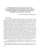
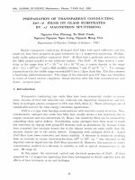
The experimental set-up of laser ablation is shown in Fig 1. Laser beam from a Nd: YAG laser
(Quanta Ray Pro 230-USA) was focused on a platinum plate (99.9% in purity) placed in a glass vessel
filed with 10 ml of PVP solution by a lens having focal length of 150 mm. The laser was set in Qswitch mode to give laser pulse duration of 8 ns, repetition rate of 10Hz.
(a)
(b)
Fig.1. Experiment set-up (a) and picture of Nd:YAG laser (b)
1. Holder 2. Prism 3. Lens 4. Glass vessel 5. Pt plate 6. Rotary system
The PVP solution vessel was placed on a support which executed repetitive circular motions at a
constant speed to prevent agglomeration of particles. PVP solutions in distilled water were prepared in
different concentrations. The solution becomes colored under action of the laser beam. A small
amount of the colored solution was extracted for absorption measurement and TEM observation. The
absorption spectrum was measured on a Shimadzu UV-Vis 2450 spectrometer. The TEM micrograph
was done on a JEM 1010-JEOL. The size of nanoparticles was determined by ImagieJ 1.37V software
of Wayne Rasband (National institutes of Health, USA). The size distribution was obtained by
measuring the diameter of more than 500 particles and using Origin 7.5 software. PVP solutions in
distilled water were prepared in mol concentrations of 0.005 M, 0.010 M, 0.015 M and 0.020 M. The
PVP solutions play the role of the surfactant in the preparation of PtNPs [13]. The ablation of Pt plate
can be carry out by the 1064 nm fundamental wavelength, 532nm second harmonic or 355 nm third
harmonic of Nd:YAG laser.
3. Results and discussion
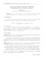
Using 532 nm wavelength of Nd:YAG laser with average power of 400mW, irradiation time of 15
min, PtNPs in PVP solution with concentrations of 0.010 M . XRD patterns of the PtNPs are shown in
Fig.2.
20
N.T. Binh et al. / VNU Journal of Science: Mathematics – Physics, Vol. 30, No. 2 (2014) 18-24
111
Intensity (a.u.)
100
200
50
220
0
30
35
40
45
50
55
60
65
70
2θ (deg rees )
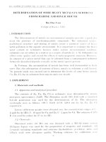
Fig.2. XRD patterns of the Pt NPs produced in PVP solution.
In the XRD patterns there are 3 peaks at 2θ = 39.8o , 46.3o and 67.4o .These peaks correspond to the
characteristic diffraction peaks of the FCC crystalline structure of Pt. The (111) peak is the strongest.
The UV-Vis absorption spectra of Pt nanoparticle colloids produced in PVP solution are shown in
Fig.3. The characteristic plasmon resonance absorption peak of Pt nanoparticle colloids was observed
at 233 nm.
Absorptance(a.u.)
2.0
233
Pt - PVP - 0.01M
1.5
1.0
0.5
0.0
300
400
500
600
Wavelength (nm)
Fig. 3. UV-Vis absorption spectrum of Pt PNs produced in PVP solutions of 0.01M.
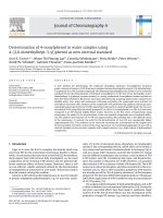
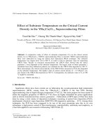
Fig.4 shows a TEM image and the size distribution of the PtNPs produced in 0.01M PVP solution
using 532nm wavelength with an average laser power of 400mW and laser irradiation time of 15 min.
From this figure it is seen that the diameter of Pt nanoparticles ranges from 2 to 20 nm. The average
size of Pt nanoparticles is 9 nm
N.T. Binh et al. /VNU Journal of Science: Mathematics – Physics, Vol. 30, No. 2 (2014) 18-24
(a)
21
(b)
Fig.4. TEM image (a) and size distribution (b) of the Pt NPs produced in 0.01M PVP solution.
To study the role of laser influence and laser irradiation time in the ablation procedure we have
prepared PtNPs with different average laser powers and laser irradiation times. The absorption spectra
of PtNPs prepared in 0.01M PVP solution using 1064 nm wavelength with different average laser
powers ranging from 200mW to 550mW and laser irradiation time of 15 minutes are plotted in Fig.5.
2.0
Absorptance(a.u.)
231.5
1.5
231.5
1.0
235
a. Pt -PVP - 200mW
b. Pt -PVP - 250mW
c. Pt -PVP - 350mW
d. Pt -PVP - 450mW
e. Pt -PVP - 550mW
d
e
c
0.5
238
b
242
a
0.0
300
400
500
600
Wavelength (nm)
Fig.5. UV-Vis absorption spectra of Pt NPs prepared by different average laser powers,
From Fig. 5, one can see that with the increase of the average laser power, from 200 mW to 550
mW, the absorptance increases and the absorption peak is shifted to the shorter wavelength (blue
shift). According to the Mie theory for sphere nanoparticles [14-17], the blue shift of the resonance
plasmon absorption peak proves the decrease of the nanoparticle size.
Fig.6 shows UV-Vis absorption spectra of PtNPs produced in 0.01M PVP solution by 400 mW
average laser power with different laser exposure time.
22
N.T. Binh et al. / VNU Journal of Science: Mathematics – Physics, Vol. 30, No. 2 (2014) 18-24
2.5
Absorptance(a.u.)
2.0
1.5
234.5
a. Pt - PVP - 10 min
b. Pt - PVP - 15 min
c. Pt - PVP - 20 min
232.5
c
231
b
a
1.0
0.5
0.0
300
400
500
600
Wavelength (nm)
Fig.6. UV-Vis absorption spectra of Pt NPs prepared with different laser exposure times.
As seen from Fig.6, the abundance of Pt PNs increases and the absorption peak is shifted to longer
wavelength (red shift) with the increase of laser exposure time from 10 to 20 min. Using 1064 nm
fundamental wavelength and 532nm, 355nm harmonics of Nd:YAG laser, we investigated the
influence of different laser wavelengths in the process. The absorption spectra of Pt NPs prepared in
0.01 PVP solution by average laser power of 250mW with different laser wavelengths are shown in Fig.7.
0.8
230nm
a. Pt - PVP - 355nm
b. Pt - PVP - 532nm
c. Pt - PVP - 1064nm
Absorptance(a.u.)
0.6
238nm
0.4
b
c
0.2
242nm
a
0.0
300
400
500
600
Wavelength (nm)
Fig.7. UV-Vis absorption spectra of Pt NPs prepared by different laser wavelengths, average laser
power of 250mW, laser exposure time of 10 min.
The obtained result in Fig.7 shows that the absorption peak corresponding to the wavelength of
532 nm is highest, meanwhile the one corresponding to the wavelength of 355 nm is lowest. This
means that the laser ablation efficiency is lowest at the laser wavelength of 355 nm in the experimental
conditions. It is contrast to the case of ablation in air where the shorter wavelength (355 nm) or higher
photon energy ablates metal more strongly than the longer wavelength (1064 nm). The low laser
ablation efficiency can be explained by the absorption effect of PtNP colloid on laser beam of 355 nm
wavelength which is near the resonance Plasmon absorption peak of Pt NPs. Meanwhile, the 1064nm
and 532nm wavelengths are far from the resonance absorption peak (Fig. 7) and the shorter
N.T. Binh et al. /VNU Journal of Science: Mathematics – Physics, Vol. 30, No. 2 (2014) 18-24
23
wavelength (532nm) with higher photon energy ablates metal more strongly than the longer
wavelength (1064 nm). The 532 nm wavelength gives highest laser ablation efficiency in these
experimental conditions.
The influence of PVP concentration on PtNPs size was also investigated. Fig. 8 presents the
absorption spectra of PtNPs prepared in different concentrations of PVP solution by average laser
power of 300mW, laser exposure time of 15 minutes.
233
236
234.5
233.5
Absorptance(a.u.)
2.0
1.5
e
d
c
b
230.5
a
b
c
d
e
a
PVP.001M
PVP.005M
PVP.010M
PVP.015M
PVP.020M
1.0
0.5
300
400
500
Wavelength (nm)
Fig. 8. The UV-Vis absorption spectra of Pt NPs prepared in different concentrations of PVP solution by
1064nm wavelengths, average laser power of 400mW, laser exposure time of 15 min.
With the increase of the PVP concentration, from 0.001M to 0.01M, the absorption profiles
become narrower and the characteristic plasmon resonance absorption peaks of PtNPs are shifted from
230.5 nm to 236 nm. When the PVP concentration increases from 0.001M to 0.01M, the plasmon
resonance absorption peak is shifted from 230.5 to 236nm (red shift). When the PVP
concentration increases from 0.01M to 0.02M, the plasmon resonance absorption peak is shifted
from 236 nm to 233 nm respectively (blue shift).
The fact that the absorption spectrum profile is narrower proves that the decrease of the dispersion
of size distribution can be explained by the presence of PVP. Indeed, in the presence of PVP (C6H9NO)n , the C=O group of the polymer interacts with metal atoms on the surface of nanoparticles.
The oxygen atoms of C=O group are attached to the metal atoms and create local surface state that
protect metal nanoparticles against growth and aggregation [18].
4. Conclusion
With the aim to find out an optimum laser ablation process for preparation of PtNPs in PVP
solution, the affect of laser power, laser wavelength, laser exposure time and concentration of PVP on
morphology, size distribution and optical properties of Pt NPs was investigated. Among 3 wavelengths
of Nd:YAG laser used (namely 355, 532 and 1064 nm), the 532nm wavelength gave the highest laser
ablation efficiency. The role of PVP solution as a surfactant for Pt NPs was elucidated. With increase
of the PVP concentration, from 0.001M to 0.02M, the UV-Vis absorption spectrum profiles become
N.T. Binh et al. / VNU Journal of Science: Mathematics – Physics, Vol. 30, No. 2 (2014) 18-24
24
narrower and characteristic plasmon resonance absorption peak of PtNPs is shifted. The obtained
results showed that the size of PtNPs can be controlled by changing PVP concentration and laser
parameters.
Acknowledgments
This research was supported by the Project QGTĐ.12.01, VNU Hanoi.
References
[1] Zhang Y J, Huang R, Zhu X F, et al. Synthesis, properties, and optical applications of noble metal nanoparticlebiomolecule conjugates. Chin Sci Bull, 57 (2012) 238-246.
[2] Haes A J, Chang L, Klein W L, et al. Detection of a biomarker for alzheimer's disease from synthetic and clinical
samples using a nanoscale optical biosensor. J Am Chem Soc, 127 (2005) 2264–2271.
[3] Endo T, Kerman K, Nagatani N, et al. Excitation of localized surface plasmon resonance using a core–shell
structured nanoparticle layer substrate and its application for label-free detection of biomolecular interactions. J
Phys: Condens Matter 19 (2007) 215201.
[4] M. G. Guzman, J. Dille, S. Godet, Synthesis of silver nanoparticles by chemical reduction method and their
antibacterial activity. Int. J. Chem.Bio. Eng. 2 (2009) 104-111.
[5] D. Long, G. Wu, S. Chen, Preparation of oligochitosan stabilized silver nanoparticles by gamma irradiation,
Rad.Phys. Chem 76 (2007) 1126-113.
[6] K. A. Bogle, S. D. Dhole, V. N. Bhoraskar, Silver nanoparticles: synthesis and size control by electron
irradiation, Nanotechnology 17(2006) 3204.
[7] K. Mallick, M. J. Witcomb, M. S. Scurrell, Polymer stabilized silver nanoparticles: A photochemical synthesis
route, J. Mater. Sci. 39 (2004) 4459-4463.
[8] H. Yin, T. Yamamoto, Y. Wada, S. Yanagida, Large scale and size-controlled synthesis of silver nanoparticles
under microwave iiradiation, Mater. Chem. Phys. 83 (2004) 66-70.
[9] J. Zhu, S. Liu, O. Palchik, Y. Koltypin, A. Gedanken, Shape-Controlled Synthesis of Silver Nanoparticles by
Pulse Sonoelectrochemical Methods,Langmuir 16 (2000) 6396-6399.
[10] F. Mafune, J. Kohno, Y. Takeda, T. Kondow, Structure and Stability of Silver Nanoparticles in Aqueous Solution
Produced by Laser Ablation J. Phys. Chem. B, 104 (2000) 8333-8337.
[11] [11] The Binh Nguyen et all, Preparation of copper nanoparticles in water and acetone by laser ablation, VNU
Journal of Science Mathematics-Physics 27,No IS (2011) 51-56.
[12] The Binh Nguyen et all, Preparation of metal nanoparticles for surface enhanced Raman scattering by laser
ablation method , IOP Science, Avd. Nat. Sci . Nanosci. Nanotechnol. 3 (2012) 025016.
[13] Milton J. Rosen (2004), Surfactants and interfacial phenomena, Wiley-Interscience, United States of America.
[14] Cowley A and Woodward, A Healthy Future: Platinum in Medical Applications, B 2011 Platinum Metals Rev. 55 98–10.
[15] Athanassiou E K, Grass R N and Stark W J, Large scale production of carbon coated copper nanoparticles,
Nanotechnolgy, 17 (2006) 1668-1673.
[16] Mafune F, Kohno J and Takeda Y Dissociation and aggregation of gold nanoparticles under laser irradiation, 105
(2001) J. Phys. Chem. B 9050–9056
[17] Mafune F, Kohno J, Takeda Y and Kondow T Formation of stable platinum nanoparticles by laser ablation in
water, 107(2003) J. Phys. Chem. B 4218–4223
[18] Takeshi Tsuji, D.-H. Thang, Yuuki Okazaki, Masataka Nakanishi, Yasuyuki Tsuboi, Masaharu Tsuji Preparation
of silver nanoparticles by laser ablation in polyvinylpyrrolidone solutions (2008) Applied Surface Science
254(16):5224-5230.