Ebook Human anatomy physiology (9th edition) Part 2
Bạn đang xem bản rút gọn của tài liệu. Xem và tải ngay bản đầy đủ của tài liệu tại đây (30.04 MB, 605 trang )
17
Blood
Overview: Blood Composition
and Functions (pp. 632–633)
Components (p. 632)
Physical Characteristics and Volume (p. 632)
Functions (pp. 632–633)
Blood Plasma (p. 633)
Formed Elements (pp. 634–646)
Erythrocytes (Red Blood Cells) (pp. 634–640)
Leukocytes (White Blood Cells)
(pp. 640–645)
Platelets (pp. 645–646)
Hemostasis (pp. 646–651)
Step 1: Vascular Spasm (p. 646)
Step 2: Platelet Plug Formation
(pp. 646–647)
Step 3: Coagulation (pp. 647–649)
Clot Retraction and Fibrinolysis (p. 649)
Factors Limiting Clot Growth or
Formation (p. 649)
Disorders of Hemostasis (pp. 650–651)
Transfusion and Blood Replacement
(pp. 651–653)
Transfusing Red Blood Cells (pp. 651–653)
Restoring Blood Volume (p. 653)
Diagnostic Blood Tests (pp. 653–654)
Developmental Aspects of Blood (p. 654)
B
lood is the river of life that surges within us, transporting nearly
everything that must be carried from one place to another. Long before modern
medicine, blood was viewed as magical—an elixir that held the mystical force of
life—because when it drained from the body, life departed as well. Today, blood still has
enormous importance in the practice of medicine. Clinicians examine it more often than
any other tissue when trying to determine the cause of disease in their patients.
In this chapter, we describe the composition and functions of this life-sustaining fluid
that serves as a transport “vehicle” for the organs of the cardiovascular system (cardio 5
heart, vasc 5 blood vessels). To get started, we need a brief overview of blood circulation,
which is initiated by the pumping action of the heart. Blood exits the heart via arteries,
which branch repeatedly until they become tiny capillaries. By diffusing across the capillary walls, oxygen and nutrients leave the blood and enter the body tissues, and carbon
dioxide and wastes move from the tissues to the bloodstream. As oxygen-deficient blood
leaves the capillary beds, it flows into veins, which return it to the heart. The returning
631
# 105016 Cust: Benjamin Cummings/CA Au: Marieb Pg. No. 631
Title: Anatomy & Physiology Server: S4C
C/M/Y/K
Short / Normal
DESIGN SERVICES OF
carlisle
Publishing Services
632
Unit 4 Maintenance of the Body
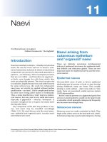
1 Withdraw blood
and place in tube.
Plasma
• 55% of whole blood
• Least dense component
Buffy coat
• Leukocytes and platelets
• <1% of whole blood
Erythrocytes
• 45% of whole blood
(hematocrit)
• Most dense component
2 Centrifuge the
blood sample.
Formed
elements
Figure 17.1 The major components of whole blood.
blood then flows from the heart to the lungs, where it picks up
oxygen and then returns to the heart to be pumped throughout
the body once again. Now let us look more closely at the nature
of blood.
Overview: Blood Composition
and Functions
Describe the composition and physical characteristics of
whole blood. Explain why it is classified as a connective
tissue.
List eight functions of blood.
Components
17
Blood is the only fluid tissue in the body. It appears to be a thick,
homogeneous liquid, but the microscope reveals that it has both
cellular and liquid components. Blood is a specialized connective tissue in which living blood cells, called the formed elements, are suspended in a nonliving fluid matrix called plasma
(plaz9mah). Blood lacks the collagen and elastic fibers typical of
other connective tissues, but dissolved fibrous proteins become
visible as fibrin strands during blood clotting.
If we spin a sample of blood in a centrifuge, centrifugal force
packs down the heavier formed elements and the less dense
plasma remains at the top (Figure 17.1). Most of the reddish
mass at the bottom of the tube is erythrocytes (ĕ-rith9ro-sīts;
erythro 5 red), the red blood cells that transport oxygen. A thin,
whitish layer called the buffy coat is present at the erythrocyteplasma junction. This layer contains leukocytes (leuko 5 white),
the white blood cells that act in various ways to protect the body,
and platelets, cell fragments that help stop bleeding.
Erythrocytes normally constitute about 45% of the total volume of a blood sample, a percentage known as the hematocrit
(he-mat9o-krit; “blood fraction”). Normal hematocrit values
vary. In healthy males the norm is 47% 6 5%; in females it is
42% 6 5%. Leukocytes and platelets contribute less than 1% of
# 105016 Cust: Benjamin Cummings/CA Au: Marieb Pg. No. 632
Title: Anatomy & Physiology Server: S4C
blood volume. Plasma makes up most of the remaining 55% of
whole blood.
Physical Characteristics and Volume
Blood is a sticky, opaque fluid with a characteristic metallic
taste. As children, we discover its saltiness the first time we stick
a cut finger into our mouth. Depending on the amount of oxygen it is carrying, the color of blood varies from scarlet (oxygen
rich) to dark red (oxygen poor). Blood is more dense than water
and about five times more viscous, largely because of its formed
elements. It is slightly alkaline, with a pH between 7.35 and 7.45.
Blood accounts for approximately 8% of body weight. Its average volume in healthy adult males is 5–6 L (about 1.5 gallons),
somewhat greater than in healthy adult females (4–5 L).
Functions
Blood performs a number of functions, all concerned in one
way or another with distributing substances, regulating blood
levels of particular substances, or protecting the body.
Distribution
Distribution functions of blood include
■
■
■
Delivering oxygen from the lungs and nutrients from the digestive tract to all body cells.
Transporting metabolic waste products from cells to elimination sites (to the lungs to eliminate carbon dioxide, and to
the kidneys to dispose of nitrogenous wastes in urine).
Transporting hormones from the endocrine organs to their
target organs.
Regulation
Regulatory functions of blood include
■
Maintaining appropriate body temperature by absorbing and
distributing heat throughout the body and to the skin surface
to encourage heat loss.
C/M/Y/K
Short / Normal
DESIGN SERVICES OF
carlisle
Publishing Services
Chapter 17 Blood
■
■
Maintaining normal pH in body tissues. Many blood proteins and other bloodborne solutes act as buffers to prevent
excessive or abrupt changes in blood pH that could jeopardize normal cell activities. Additionally, blood acts as the reservoir for the body’s “alkaline reserve” of bicarbonate ions.
Maintaining adequate fluid volume in the circulatory system.
Blood proteins prevent excessive fluid loss from the bloodstream into the tissue spaces. As a result, the fluid volume in
the blood vessels remains ample to support efficient blood
circulation to all parts of the body.
Table 17.1
Composition of Plasma
Constituent
Description and Importance
Water
90% of plasma volume; dissolving and
suspending medium for solutes of
blood; absorbs heat
Solutes
Electrolytes
Most abundant solutes by number;
cations include sodium, potassium,
calcium, magnesium; anions include
chloride, phosphate, sulfate, and
bicarbonate; help to maintain plasma
osmotic pressure and normal blood pH
Plasma proteins
8% (by weight) of plasma; all
contribute to osmotic pressure and
maintain water balance in blood
and tissues; all have other functions
(transport, enzymatic, etc.) as well
Protection
Protective functions of blood include
■
■
Preventing blood loss. When a blood vessel is damaged,
platelets and plasma proteins initiate clot formation, halting
blood loss.
Preventing infection. Drifting along in blood are antibodies,
complement proteins, and white blood cells, all of which help
defend the body against foreign invaders such as bacteria
and viruses.
■
Albumin
60% of plasma proteins; produced
by liver; main contributor to osmotic
pressure
■
Globulins
36% of plasma proteins
alpha, beta
Produced by liver; most are transport
proteins that bind to lipids, metal ions,
and fat-soluble vitamins
gamma
Antibodies released by plasma cells
during immune response
■
Fibrinogen
4% of plasma proteins; produced by
liver; forms fibrin threads of blood clot
Blood Plasma
Discuss the composition and functions of plasma.
Blood plasma is a straw-colored, sticky fluid (Figure 17.1). Although it is mostly water (about 90%), plasma contains over
100 different dissolved solutes, including nutrients, gases, hormones, wastes and products of cell activity, proteins, and inorganic ions (electrolytes). Electrolytes (Na1, Cl2, etc.) vastly
outnumber the other solutes. Table 17.1 summarizes the major
plasma components.
Although outnumbered by the lighter electrolytes, the heavier plasma proteins are the most abundant plasma solutes by
weight, accounting for about 8% of plasma weight. Except for
hormones and gamma globulins, most plasma proteins are produced by the liver. Plasma proteins serve a variety of functions,
but they are not taken up by cells to be used as fuels or metabolic
nutrients as are most other organic solutes, such as glucose, fatty
acids, and amino acids.
Albumin (al-bu9min) accounts for some 60% of plasma protein. It acts as a carrier to shuttle certain molecules through the
circulation, is an important blood buffer, and is the major blood
protein contributing to the plasma osmotic pressure (the pressure that helps to keep water in the bloodstream).
The composition of plasma varies continuously as cells remove or add substances to the blood. However, assuming a
healthy diet, plasma composition is kept relatively constant by
various homeostatic mechanisms. For example, when blood
protein levels drop undesirably, the liver makes more proteins.
When the blood starts to become too acidic (acidosis), both the
lungs and the kidneys are called into action to restore plasma’s
normal, slightly alkaline pH. Body organs make dozens of adjustments, day in and day out, to maintain the many plasma
solutes at life-sustaining levels.
# 105016 Cust: Benjamin Cummings/CA Au: Marieb Pg. No. 633
Title: Anatomy & Physiology Server: S4C
633
Nonprotein nitrogenous
substances
By-products of cellular metabolism,
such as urea, uric acid, creatinine, and
ammonium salts
Nutrients (organic)
Materials absorbed from digestive tract
and transported for use throughout
body; include glucose and other simple
carbohydrates, amino acids (protein
digestion products), fatty acids,
glycerol and triglycerides (fat digestion
products), cholesterol, and vitamins
Respiratory gases
Oxygen and carbon dioxide; oxygen
mostly bound to hemoglobin inside
RBCs; carbon dioxide transported
dissolved as bicarbonate ion or CO2, or
bound to hemoglobin in RBCs
Hormones
Steroid and thyroid hormones carried
by plasma proteins
Check Your Understanding
1. What is the hematocrit? What is its normal value?
2. List two protective functions of blood.
3. Are plasma proteins used as fuel for body cells? Explain your
answer.
For answers, see Appendix H.
C/M/Y/K
Short / Normal
DESIGN SERVICES OF
carlisle
Publishing Services
17
634
Unit 4 Maintenance of the Body
Platelets
Erythrocytes
Monocyte
2.5 μm
Side view (cut)
7.5 μm
Neutrophils
Lymphocyte
Top view
Figure 17.2 Photomicrograph of a human blood smear
stained with Wright’s stain. (6403)
Formed Elements
The formed elements of blood—erythrocytes, leukocytes, and
platelets—have some unusual features.
■
■
■
17
Two of the three are not even true cells: Erythrocytes have
no nuclei or organelles, and platelets are cell fragments. Only
leukocytes are complete cells.
Most of the formed elements survive in the bloodstream for
only a few days.
Most blood cells do not divide. Instead, stem cells divide continuously in red bone marrow to replace them.
If you examine a stained smear of human blood under the
light microscope, you will see disc-shaped red blood cells, a variety of gaudily stained spherical white blood cells, and some
scattered platelets that look like debris (Figure 17.2). Erythrocytes vastly outnumber the other types of formed elements.
Table 17.2 on p. 644 summarizes the important characteristics
of the formed elements.
Erythrocytes (Red Blood Cells)
Describe the structure, function, and production of
erythrocytes.
Describe the chemical composition of hemoglobin.
Give examples of disorders caused by abnormalities of
erythrocytes. Explain what goes wrong in each disorder.
Figure 17.3 Structure of erythrocytes (red blood cells). Notice
the distinctive biconcave shape.
lighter in color at their thin centers than at their edges. Consequently, erythrocytes look like miniature doughnuts when
viewed with a microscope.
Mature erythrocytes are bound by a plasma membrane, but
lack a nucleus (are anucleate) and have essentially no organelles.
In fact, they are little more than “bags” of hemoglobin (Hb), the
RBC protein that functions in gas transport. Other proteins are
present, such as antioxidant enzymes that rid the body of harmful oxygen radicals, but most function as structural proteins,
allowing the RBC to deform yet spring back into shape.
For example, a network of proteins, especially one called spectrin, attached to the cytoplasmic face of RBC plasma membranes
maintains the biconcave shape of an erythrocyte. The spectrin
net is deformable, allowing erythrocytes to change shape as
necessary—to twist, turn, and become cup shaped as they are
carried passively through capillaries with diameters smaller than
themselves—and then to resume their biconcave shape.
The erythrocyte is a superb example of complementarity of
structure and function. It picks up oxygen in the capillaries of
the lungs and releases it to tissue cells across other capillaries
throughout the body. It also transports some 20% of the carbon
dioxide released by tissue cells back to the lungs. Three structural characteristics contribute to erythrocyte gas transport
functions:
■
Structural Characteristics
Erythrocytes or red blood cells (RBCs) are small cells, about
7.5 μm in diameter (Figure 17.3). Shaped like biconcave
discs—flattened discs with depressed centers—they appear
# 105016 Cust: Benjamin Cummings/CA Au: Marieb Pg. No. 634
Title: Anatomy & Physiology Server: S4C
■
Its small size and biconcave shape provide a huge surface
area relative to volume (about 30% more surface area than
comparable spherical cells). The biconcave disc shape is ideally suited for gas exchange because no point within the cytoplasm is far from the surface.
Discounting water content, an erythrocyte is over 97% hemoglobin, the molecule that binds to and transports respiratory gases.
C/M/Y/K
Short / Normal
DESIGN SERVICES OF
carlisle
Publishing Services
Chapter 17 Blood
635
β Globin chains
CH2CH2COOH
H3C
N
H2C=CH
CH2CH2COOH
N
N
Fe
H3C
CH3
N
H2C=CH
CH3
Heme
group
α Globin chains
(a) Hemoglobin consists of globin (two alpha and two beta
polypeptide chains) and four heme groups.
(b) Iron-containing heme pigment.
Figure 17.4 Structure of hemoglobin. Hemoglobin’s structure makes it a highly efficient
oxygen carrier.
■
Because erythrocytes lack mitochondria and generate ATP
by anaerobic mechanisms, they do not consume any of the
oxygen they carry, making them very efficient oxygen transporters indeed.
Erythrocytes are the major factor contributing to blood viscosity. Women typically have a lower red blood cell count than
men [4.2–5.4 million cells per microliter (1 μl 5 1 mm3) of
blood versus 4.7–6.1 million cells/μl respectively]. When the
number of red blood cells increases beyond the normal range,
blood becomes more viscous and flows more slowly. Similarly,
as the number of red blood cells drops below the lower end of
the range, the blood thins and flows more rapidly.
Functions of Erythrocytes
Erythrocytes are completely dedicated to their job of transporting respiratory gases (oxygen and carbon dioxide). Hemoglobin, the protein that makes red blood cells red, binds easily
and reversibly with oxygen, and most oxygen carried in blood is
bound to hemoglobin. Normal values for hemoglobin are 13–18
grams per 100 milliliters of blood (g/100 ml) in adult males, and
12–16 g/100 ml in adult females.
Hemoglobin is made up of the red heme pigment bound to
the protein globin. Globin consists of four polypeptide chains—
two alpha (a) and two beta (β)—each binding a ringlike heme
group (Figure 17.4a). Each heme group bears an atom of iron
set like a jewel in its center (Figure 17.4b). A hemoglobin molecule can transport four molecules of oxygen because each iron
atom can combine reversibly with one molecule of oxygen. A
# 105016 Cust: Benjamin Cummings/CA Au: Marieb Pg. No. 635
Title: Anatomy & Physiology Server: S4C
single red blood cell contains about 250 million hemoglobin
molecules, so each of these tiny cells can scoop up about 1 billion molecules of oxygen!
The fact that hemoglobin is contained in erythrocytes, rather
than existing free in plasma, prevents it (1) from breaking into
fragments that would leak out of the bloodstream (through porous capillary walls) and (2) from making blood more viscous
and raising osmotic pressure.
Oxygen loading occurs in the lungs, and the direction of
transport is from lungs to tissue cells. As oxygen-deficient blood
moves through the lungs, oxygen diffuses from the air sacs of
the lungs into the blood and then into the erythrocytes, where
it binds to hemoglobin. When oxygen binds to iron, the hemoglobin, now called oxyhemoglobin, assumes a new threedimensional shape and becomes ruby red.
In body tissues, the process is reversed. Oxygen detaches
from iron, hemoglobin resumes its former shape, and the resulting deoxyhemoglobin, or reduced hemoglobin, becomes dark
red. The released oxygen diffuses from the blood into the tissue
fluid and then into tissue cells.
About 20% of the carbon dioxide transported in the blood
combines with hemoglobin, but it binds to globin’s amino acids
rather than to the heme group. This formation of carbaminohemoglobin (kar-bam0ĭ-no-he0muh0glo9bin) occurs more readily when hemoglobin is in the reduced state (dissociated from
oxygen). Carbon dioxide loading occurs in the tissues, and the
direction of transport is from tissues to lungs, where carbon dioxide is eliminated from the body. We describe the loading and
unloading of these respiratory gases in Chapter 22.
C/M/Y/K
Short / Normal
DESIGN SERVICES OF
carlisle
Publishing Services
17
636
Unit 4 Maintenance of the Body
Stem cell
Committed cell
Developmental pathway
Phase 1
Ribosome synthesis
Hematopoietic stem
cell (hemocytoblast)
Proerythroblast
Basophilic
erythroblast
Phase 2
Hemoglobin accumulation
Polychromatic
erythroblast
Phase 3
Ejection of nucleus
Orthochromatic
erythroblast
Reticulocyte Erythrocyte
Figure 17.5 Erythropoiesis: formation of red blood cells. Reticulocytes are released into
the bloodstream. The myeloid stem cell, the phase intermediate between the hematopoietic
stem cell and the proerythroblast, is not illustrated.
Production of Erythrocytes
17
Blood cell formation is referred to as hematopoiesis (hem0ahto-poi-e9sis; hemato 5 blood; poiesis 5 to make). Hematopoiesis occurs in the red bone marrow, which is composed largely
of a soft network of reticular connective tissue bordering on
wide blood capillaries called blood sinusoids. Within this network are immature blood cells, macrophages, fat cells, and reticular cells (which secrete the connective tissue fibers). In adults,
red marrow is found chiefly in the bones of the axial skeleton
and girdles, and in the proximal epiphyses of the humerus and
femur.
The production of each type of blood cell varies in response
to changing body needs and regulatory factors. As blood cells
mature, they migrate through the thin walls of the sinusoids to
enter the bloodstream. On average, the marrow turns out an
ounce of new blood containing 100 billion new cells every day.
The various formed elements have different functions, but
there are similarities in their life histories. All arise from the hematopoietic stem cell, sometimes called a hemocytoblast (cyte
5 cell, blast 5 bud). These undifferentiated precursor cells reside in the red bone marrow. However, the maturation pathways
of the various formed elements differ, and once a cell is committed to a specific blood cell pathway, it cannot change. This commitment is signaled by the appearance of membrane surface
receptors that respond to specific hormones or growth factors,
which in turn “push” the cell toward further specialization.
Stages of Erythropoiesis Erythrocyte production, or eryth-
ropoiesis (ĕ-rith0ro-poi-e9sis), begins when a hematopoietic
stem cell descendant called a myeloid stem cell transforms into
a proerythroblast (Figure 17.5). Proerythroblasts, in turn,
give rise to basophilic erythroblasts that produce huge numbers of ribosomes. During these first two phases, the cells divide
many times. Hemoglobin is synthesized and iron accumulates
as the basophilic erythroblast transforms into a polychromatic
erythroblast and then an orthochromatic erythroblast. The
“color” of the cell cytoplasm changes as the blue-staining ribosomes become masked by the pink color of hemoglobin. When
# 105016 Cust: Benjamin Cummings/CA Au: Marieb Pg. No. 636
Title: Anatomy & Physiology Server: S4C
an orthochromatic erythroblast has accumulated almost all of
its hemoglobin, it ejects most of its organelles. Additionally, its
nucleus degenerates and is pinched off, allowing the cell to collapse inward and eventually assume the biconcave shape. The
result is the reticulocyte (essentially a young erythrocyte), so
named because it still contains a scant reticulum (network) of
clumped ribosomes.
The entire process from hematopoietic stem cell to reticulocyte takes about 15 days. The reticulocytes, filled almost to
bursting with hemoglobin, enter the bloodstream to begin their
task of oxygen transport. Usually they become fully mature
erythrocytes within two days of release as their ribosomes are
degraded by intracellular enzymes.
Reticulocytes account for 1–2% of all erythrocytes in the
blood of healthy people. Reticulocyte counts provide a rough
index of the rate of RBC formation—reticulocyte counts below or above this range indicate abnormal rates of erythrocyte
formation.
Regulation and Requirements for Erythropoiesis
The number of circulating erythrocytes in a given individual is
remarkably constant and reflects a balance between red blood
cell production and destruction. This balance is important because having too few erythrocytes leads to tissue hypoxia (oxygen deprivation), whereas having too many makes the blood
undesirably viscous.
To ensure that the number of erythrocytes in blood remains
within the homeostatic range, new cells are produced at the incredibly rapid rate of more than 2 million per second in healthy
people. This process is controlled hormonally and depends on
adequate supplies of iron, amino acids, and certain B vitamins.
Hormonal Controls Erythropoietin (EPO), a glycoprotein hormone, stimulates the formation of erythrocytes (Figure 17.6).
Normally, a small amount of EPO circulates in the blood at
all times and sustains red blood cell production at a basal rate.
The kidneys play the major role in EPO production, although
the liver also produces some. When certain kidney cells become
C/M/Y/K
Short / Normal
DESIGN SERVICES OF
carlisle
Publishing Services
Chapter 17 Blood
IMB
AL
637
AN
CE
Homeostasis: Normal blood oxygen levels
IMB
AL
5 O2-carrying
ability of blood
rises.
AN
4 Enhanced
erythropoiesis
increases RBC count.
3 Erythropoietin
stimulates red
bone marrow.
CE
1 Stimulus:
Hypoxia
(inadequate O2
delivery) due to
• Decreased
RBC count
• Decreased amount
of hemoglobin
• Decreased
availability of O2
2 Kidney (and liver to
a smaller extent)
releases erythropoietin.
Figure 17.6 Erythropoietin mechanism for regulating erythropoiesis.
hypoxic (oxygen deficient), oxygen-sensitive enzymes are unable
to carry out their normal functions of degrading an intracellular signaling molecule called hypoxia-inducible factor (HIF).
As HIF accumulates, it accelerates the synthesis and release of
erythropoietin.
The drop in normal blood oxygen levels that triggers EPO
formation can result from
■
■
■
Reduced numbers of red blood cells due to hemorrhage
(bleeding) or excessive RBC destruction
Insufficient hemoglobin per RBC (as in iron deficiency)
Reduced availability of oxygen, as might occur at high altitudes or during pneumonia
Conversely, too many erythrocytes or excessive oxygen in
the bloodstream depresses erythropoietin production. Note
that it is not the number of erythrocytes in blood that controls
the rate of erythropoiesis. Instead, control is based on their ability to transport enough oxygen to meet tissue demands.
Bloodborne erythropoietin stimulates red marrow cells that
are already committed to becoming erythrocytes, causing them
to mature more rapidly. One to two days after erythropoietin
levels rise in the blood, the rate of reticulocyte release and the
reticulocyte count rise markedly. Notice that hypoxia does not
activate the bone marrow directly. Instead it stimulates the kidneys, which in turn provide the hormonal stimulus that activates the bone marrow.
Homeostatic Imbalance 17.1
Renal dialysis patients whose kidneys have failed produce too
little EPO to support normal erythropoiesis. Consequently,
they routinely have red blood cell counts less than half those of
healthy individuals. Genetically engineered (recombinant) EPO
has helped these patients immeasurably.
# 105016 Cust: Benjamin Cummings/CA Au: Marieb Pg. No. 637
Title: Anatomy & Physiology Server: S4C
Unfortunately, some athletes abuse recombinant EPO—
particularly professional bike racers and marathon runners
seeking increased stamina and performance. However, the consequences can be deadly. By injecting EPO, healthy athletes increase their normal hematocrit from 45% to as much as 65%.
Then, with the dehydration that occurs in a long race, the blood
concentrates even further, becoming a thick, sticky “sludge” that
can cause clotting, stroke, and heart failure. ✚
The male sex hormone testosterone also enhances the kidneys’
production of EPO. Because female sex hormones do not have
similar stimulatory effects, testosterone may be at least partially
responsible for the higher RBC counts and hemoglobin levels
seen in males. Also, a wide variety of chemicals released by leukocytes, platelets, and even reticular cells stimulates bursts of
RBC production.
Dietary Requirements The raw materials required for erythropoiesis include the usual nutrients and structural materials—
amino acids, lipids, and carbohydrates. Iron is essential for hemoglobin synthesis. Iron is available from the diet, and intestinal cells
precisely control its absorption into the bloodstream in response
to changing body stores of iron.
Approximately 65% of the body’s iron supply (about 4000
mg) is in hemoglobin. Most of the remainder is stored in the
liver, spleen, and (to a much lesser extent) bone marrow. Free
iron ions (Fe21, Fe31) are toxic, so iron is stored inside cells as
protein-iron complexes such as ferritin (fer9ĭ-tin) and hemosiderin (he0mo-sid9er-in). In blood, iron is transported loosely
bound to a transport protein called transferrin, and developing erythrocytes take up iron as needed to form hemoglobin
(Figure 17.7). Small amounts of iron are lost each day in feces,
urine, and perspiration. The average daily loss of iron is 1.7 mg
in women and 0.9 mg in men. In women, the menstrual flow
accounts for the additional losses.
C/M/Y/K
Short / Normal
DESIGN SERVICES OF
carlisle
Publishing Services
17
638
Unit 4 Maintenance of the Body
1 Low O2 levels in blood stimulate
kidneys to produce erythropoietin.
2 Erythropoietin levels rise in blood.
3 Erythropoietin and necessary raw
materials in blood promote
erythropoiesis in red bone marrow.
4 New erythrocytes
enter bloodstream;
function about 120
days.
5 Aged and damaged
red blood cells are engulfed by
macrophages of spleen, liver, and
bone marrow; the hemoglobin is
broken down.
Hemoglobin
Heme
Bilirubin is
picked up
by the liver.
Iron is stored
as ferritin or
hemosiderin.
Globin
Amino
acids
Two B-complex vitamins—vitamin B12 and folic acid—are
necessary for normal DNA synthesis. Even slight deficits jeopardize rapidly dividing cell populations, such as developing
erythrocytes.
Fate and Destruction of Erythrocytes
Red blood cells have a useful life span of 100 to 120 days. Their
anucleate condition carries with it some important limitations.
Red blood cells are unable to synthesize new proteins, grow, or
divide. Erythrocytes become “old” as they lose their flexibility, become increasingly rigid and fragile, and their hemoglobin begins
to degenerate. They become trapped and fragment in smaller circulatory channels, particularly in those of the spleen. For this reason, the spleen is sometimes called the “red blood cell graveyard.”
We will briefly describe the fate of aged and damaged erythrocytes here, but Figure 17.7 gives a more detailed account.
Macrophages engulf and destroy dying erythrocytes. The heme
of their hemoglobin is split off from globin. Its core of iron is
salvaged, bound to protein (as ferritin or hemosiderin), and
stored for reuse. The balance of the heme group is degraded to
bilirubin (bil0ĭ-roo9bin), a yellow pigment that is released to
the blood and binds to albumin for transport. Liver cells pick up
bilirubin and in turn secrete it (in bile) into the intestine, where
it is metabolized to urobilinogen. Most of this degraded pigment
leaves the body in feces, as a brown pigment called stercobilin.
The protein (globin) part of hemoglobin is metabolized or broken down to amino acids, which are released to the circulation.
Erythrocyte Disorders
Most erythrocyte disorders can be classified as anemia or polycythemia. We describe some of the many varieties and causes of
these conditions next.
Iron is bound to transferrin
and released to blood
from liver as needed
for erythropoiesis.
17
Bilirubin is secreted into
intestine in bile where it is
metabolized to stercobilin
by bacteria.
Circulation
Anemia Anemia (ah-ne9me-ah; “lacking blood”) is a condi-
tion in which the blood’s oxygen-carrying capacity is too low to
support normal metabolism. It is a sign of some disorder rather
than a disease in itself. Its hallmark is blood oxygen levels that
are inadequate to support normal metabolism. Anemic individuals are fatigued, often pale, short of breath, and chilled.
The causes of anemia can be divided into three groups: blood
loss, not enough red blood cells produced, or too many of them
destroyed.
■
6 Raw materials are made
available in blood for
erythrocyte synthesis.
Stercobilin
is excreted
in feces.
Food nutrients
(amino acids, Fe,
B12, and folic acid)
are absorbed from
intestine and enter
blood.
Figure 17.7 Life cycle of red blood cells.
# 105016 Cust: Benjamin Cummings/CA Au: Marieb Pg. No. 638
Title: Anatomy & Physiology Server: S4C
■
Blood loss. Hemorrhagic anemia (hem0o-raj9ik) is caused by
blood loss. In acute hemorrhagic anemia, blood loss is rapid
(as might follow a severe stab wound); it is treated by replacing the lost blood. Slight but persistent blood loss (due to
hemorrhoids or an undiagnosed bleeding ulcer, for example) causes chronic hemorrhagic anemia. Once the primary
problem is resolved, normal erythropoietic mechanisms replace the lost blood cells.
Not enough red blood cells produced. A number of problems can decrease erythrocyte production. These problems
range from lack of essential raw materials (such as iron) to
complete and utter failure of the red bone marrow.
Iron-deficiency anemia is generally a secondary result of
hemorrhagic anemia, but it also results from inadequate
C/M/Y/K
Short / Normal
DESIGN SERVICES OF
carlisle
Publishing Services
Chapter 17 Blood
■
intake of iron-containing foods and impaired iron absorption. The erythrocytes produced, called microcytes, are small
and pale because they cannot synthesize their normal complement of hemoglobin. The obvious treatment is to increase
iron intake in diet or through iron supplements.
Pernicious anemia is an autoimmune disease that most
often affects the elderly. The immune system of these individuals destroys cells of their own stomach mucosa. These
cells produce a substance called intrinsic factor that must
be present for vitamin B12 to be absorbed by intestinal cells.
Without vitamin B12, the developing erythrocytes grow but
cannot divide, and large, pale cells called macrocytes result.
Treatment involves regular intramuscular injections of vitamin B12 or application of a B12-containing gel to the nasal
lining once a week.
As you might expect, lack of vitamin B12 in the diet also
leads to anemia. However, this is usually a problem only in
strict vegetarians because meats, poultry, and fish provide
ample vitamin B12 in the diet of nonvegetarians.
Renal anemia is caused by the lack of EPO, the hormone
that controls red blood cell production. Renal anemia frequently accompanies renal disease because damaged or diseased kidneys cannot produce enough EPO. Fortunately, it
can be treated with synthetic EPO.
Aplastic anemia may result from destruction or inhibition
of the red marrow by certain drugs and chemicals, ionizing
radiation, or viruses. In most cases, though, the cause is unknown. Because marrow destruction impairs formation of all
formed elements, anemia is just one of its signs. Defects in
blood clotting and immunity are also present. Blood transfusions provide a stopgap treatment until stem cells harvested
from a donor’s blood, bone marrow, or umbilical cord blood
can be transplanted.
Too many red blood cells destroyed. In hemolytic anemias
(he0mo-lit9ik), erythrocytes rupture, or lyse, prematurely. Hemoglobin abnormalities, transfusion of mismatched blood,
and certain bacterial and parasitic infections are possible
causes. Here we focus on the hemoglobin abnormalities.
Production of abnormal hemoglobin usually has a genetic
basis. Two such examples, thalassemia and sickle-cell anemia,
can be serious, incurable, and sometimes fatal diseases. In
both diseases the globin part of hemoglobin is abnormal and
the erythrocytes produced are fragile and rupture prematurely.
Thalassemias (thal0ah-se9me-ahs; “sea blood”) typically
occur in people of Mediterranean ancestry, such as Greeks
and Italians. One of the globin chains is absent or faulty, and
the erythrocytes are thin, delicate, and deficient in hemoglobin. There are many subtypes of thalassemia, classified according to which hemoglobin chain is affected and where.
They range in severity from mild to so severe that monthly
blood transfusions are required.
In sickle-cell anemia, the havoc caused by the abnormal
hemoglobin, hemoglobin S (HbS), results from a change in
just one of the 146 amino acids in a beta chain of the globin
molecule! (See Figure 17.8.) This alteration causes the beta
chains to link together under low-oxygen conditions, forming
# 105016 Cust: Benjamin Cummings/CA Au: Marieb Pg. No. 639
Title: Anatomy & Physiology Server: S4C
639
Val His Leu Thr Pro Glu Glu ...
1
2
3
4
5
6
7
146
(a) Normal erythrocyte has normal
hemoglobin amino acid sequence
in the beta chain.
Val His Leu Thr Pro Val Glu ...
1
2
3
4
5
6
7
146
(b) Sickled erythrocyte results from a single
amino acid change in the beta chain of
hemoglobin.
Figure 17.8 Sickle-cell anemia. Scanning electron micrographs
(49503).
stiff rods so that hemoglobin S becomes spiky and sharp. This,
in turn, causes the red blood cells to become crescent shaped
when they unload oxygen molecules or when the oxygen content of the blood is lower than normal, as during vigorous
exercise and other activities that increase metabolic rate.
The stiff, deformed erythrocytes rupture easily and tend
to dam up in small blood vessels. These events interfere with
oxygen delivery, leaving the victims gasping for air and in extreme pain. Bone and chest pain are particularly severe, and
infection and stroke are common sequels. Blood transfusion
is still the standard treatment for an acute sickle-cell crisis, but
preliminary results using inhaled nitric oxide to dilate blood
vessels are promising.
Sickle-cell anemia occurs chiefly in black people who live
in the malaria belt of Africa and among their descendants. It
strikes nearly one of every 500 black newborns in the United
States.
Why would such a dangerous genetic trait persist in a
population? Globally, about 250 million people are infected
with malaria and about a million die each year. While individuals with two copies of the sickle-cell gene have sickle-cell
C/M/Y/K
Short / Normal
DESIGN SERVICES OF
carlisle
Publishing Services
17
640
Unit 4 Maintenance of the Body
anemia, individuals with only one copy of the gene (sickle-cell
trait) have a better chance of surviving malaria. Their cells
only sickle under abnormal circumstances, most importantly
when they are infected with malaria. Sickling reduces the malaria parasites’ ability to survive and enhances macrophages’
ability to destroy infected RBCs and the parasites they contain.
Several treatment approaches focus on preventing RBCs
from sickling. Fetal hemoglobin (HbF) does not “sickle,” even
in those destined to have sickle-cell anemia. Hydroxyurea, a
drug used to treat chronic leukemia, switches the fetal hemoglobin gene back on. This drug dramatically reduces the
excruciating pain and overall severity and complications of
sickle-cell anemia (by 50%). Another class of drugs reduces
sickling by blocking ion channels in the RBC membrane,
keeping ions and water inside the cell. Other approaches being
tested include oral arginine to stimulate nitric oxide production and dilate blood vessels, stem cell transplants, and gene
therapy to deliver genes for synthesizing normal beta chains.
17
Polycythemia Polycythemia (pol0e-si-the9me-ah; “many
blood cells”) is an abnormal excess of erythrocytes that increases blood viscosity, causing it to sludge, or flow sluggishly.
Polycythemia vera, a bone marrow cancer, is characterized by
dizziness and an exceptionally high RBC count (8–11 million
cells/μl). The hematocrit may be as high as 80% and blood volume may double, causing the vascular system to become engorged with blood and severely impairing circulation. Severe
polycythemia is treated by diluting blood—removing some
blood and replacing it with saline.
Secondary polycythemias result when less oxygen is available or EPO production increases. The secondary polycythemia
that appears in individuals living at high altitudes is a normal
physiological response to the reduced atmospheric pressure and
lower oxygen content of the air in such areas. RBC counts of 6–8
million/μl are common in such people.
Blood doping, practiced by some athletes competing in
aerobic events, is artificially induced polycythemia. Some of
the athlete’s red blood cells are drawn off and stored. The body
quickly replaces these erythrocytes because removing blood
triggers the erythropoietin mechanism. Then, when the stored
blood is reinfused a few days before the athletic event, a temporary polycythemia results.
Since red blood cells carry oxygen, the additional infusion
should translate into increased oxygen-carrying capacity due to
a higher hematocrit, and hence greater endurance and speed.
Other than the risk of stroke and heart failure due to high
hematocrit and high blood viscosity described earlier, blood
doping seems to work. However, the practice is considered
unethical and has been banned from the Olympic Games.
Check Your Understanding
4. How many molecules of oxygen can each hemoglobin
molecule transport? What part of the hemoglobin binds the
oxygen?
5. Patients with advanced kidney disease often have anemia.
Explain the connection.
For answers, see Appendix H.
# 105016 Cust: Benjamin Cummings/CA Au: Marieb Pg. No. 640
Title: Anatomy & Physiology Server: S4C
Leukocytes (White Blood Cells)
List the classes, structural characteristics, and functions of
leukocytes.
Describe how leukocytes are produced.
Give examples of leukocyte disorders, and explain what
goes wrong in each disorder.
General Structural and Functional Characteristics
Leukocytes (leuko 5 white), or white blood cells (WBCs), are
the only formed elements that are complete cells, with nuclei
and the usual organelles. Accounting for less than 1% of total
blood volume, leukocytes are far less numerous than red blood
cells. On average, there are 4800–10,800 WBCs/μl of blood.
Leukocytes are crucial to our defense against disease. They
form a mobile army that helps protect the body from damage
by bacteria, viruses, parasites, toxins, and tumor cells. As such,
they have special functional characteristics. Red blood cells are
confined to the bloodstream, and they carry out their functions
in the blood. But white blood cells are able to slip out of the
capillary blood vessels—a process called diapedesis (di0ah-pĕde9sis; “leaping across”)—and the circulatory system is simply
their means of transport to areas of the body (mostly loose connective tissues or lymphoid tissues) where they mount inflammatory or immune responses.
As we explain in more detail in Chapter 21, the signals
that prompt WBCs to leave the bloodstream at specific locations are cell adhesion molecules displayed by endothelial cells
forming the capillary walls at sites of inflammation. Once out
of the bloodstream, leukocytes move through the tissue spaces
by amoeboid motion (they form flowing cytoplasmic extensions that move them along). By following the chemical trail of
molecules released by damaged cells or other leukocytes, a phenomenon called positive chemotaxis, they pinpoint areas of
tissue damage and infection and gather there in large numbers
to destroy foreign substances and dead cells.
Whenever white blood cells are mobilized for action, the
body speeds up their production and their numbers may double within a few hours. A white blood cell count of over 11,000
cells/μl is leukocytosis. This condition is a normal homeostatic
response to an infection in the body.
Leukocytes are grouped into two major categories on the
basis of structural and chemical characteristics. Granulocytes
contain obvious membrane-bound cytoplasmic granules, and
agranulocytes lack obvious granules. We provide general information about the various leukocytes next. More details appear
in Figure 17.9 and Table 17.2 on p. 644.
Students are often asked to list the leukocytes in order from
most abundant to least abundant. The following phrase may
help you with this task: Never let monkeys eat bananas (neutrophils, lymphocytes, monocytes, eosinophils, basophils).
Granulocytes
Granulocytes (gran9u-lo-sīts), which include neutrophils,
eosinophils, and basophils, are all roughly spherical in shape.
They are larger and much shorter lived (in most cases) than
C/M/Y/K
Short / Normal
DESIGN SERVICES OF
carlisle
Publishing Services
Chapter 17 Blood
Formed
elements
(not drawn
to scale)
Differential
WBC count
(All total 4800–
10,800/μl)
Platelets
Leukocytes
Granulocytes
Neutrophils (50–70%)
Eosinophils (2–4%)
Basophils (0.5–1%)
Erythrocytes
Agranulocytes
Lymphocytes (25–45%)
Monocytes (3–8%)
Figure 17.9 Types and relative percentages of leukocytes in
normal blood. Erythrocytes comprise nearly 98% of the formed
elements, and leukocytes and platelets together account for the
remaining 21%.
erythrocytes. They characteristically have lobed nuclei (rounded
nuclear masses connected by thinner strands of nuclear material), and their membrane-bound cytoplasmic granules stain
quite specifically with Wright’s stain. Functionally, all granulocytes are phagocytes to some degree.
Neutrophils Neutrophils (nu9tro-filz), the most numerous
white blood cells, account for 50–70% of the WBC population.
Neutrophils are about twice as large as erythrocytes.
641
The neutrophil cytoplasm contains very fine granules (of two
varieties) that are difficult to see (Table 17.2 and Figure 17.10a).
Neutrophils get their name (literally, “neutral-loving”) because
their granules take up both basic (blue) and acidic (red) dyes. Together, the two types of granules give the cytoplasm a lilac color.
Some of these granules contain hydrolytic enzymes, and are regarded as lysosomes. Others, especially the smaller granules, contain a potent “brew” of antimicrobial proteins, called defensins.
Neutrophil nuclei consist of three to six lobes. Because of
this nuclear variability, they are often called polymorphonuclear leukocytes (PMNs) or simply polys (polymorphonuclear 5
many shapes of the nucleus).
Neutrophils are our body’s bacteria slayers, and their numbers increase explosively during acute bacterial infections such
as meningitis and appendicitis. Neutrophils are chemically attracted to sites of inflammation and are active phagocytes. They
are especially partial to bacteria and some fungi, and bacterial
killing is promoted by a process called a respiratory burst. In
the respiratory burst, the cells metabolize oxygen to produce
potent germ-killer oxidizing substances such as bleach and hydrogen peroxide. In addition, defensin-mediated lysis occurs
when the granules containing defensins merge with a microbecontaining phagosome. The defensins form peptide “spears”
that pierce holes in the membrane of the ingested “foe.”
Eosinophils Eosinophils (e0o-sin9o-filz) account for 2–4%
of all leukocytes and are approximately the size of neutrophils.
Their nucleus usually resembles an old-fashioned telephone
receiver—it has two lobes connected by a broad band of nuclear
material (Table 17.2 and Figure 17.10b).
Large, coarse granules that stain from brick red to crimson with acid (eosin) dyes pack the cytoplasm. These granules
are lysosome-like and filled with a unique variety of digestive
Granulocytes
Agranulocytes
17
(a) Neutrophil:
Multilobed nucleus,
pale red and blue
cytoplasmic granules
(b) Eosinophil:
Bilobed nucleus, red
cytoplasmic granules
(c) Basophil:
Bilobed nucleus,
purplish-black
cytoplasmic granules
(d) Lymphocyte (small):
Large spherical nucleus,
thin rim of pale blue
cytoplasm
Figure 17.10 Leukocytes. In each case the leukocytes are surrounded by erythrocytes.
Neutrophils, eosinophils, and basophils have visible cytoplasmic granules; lymphocytes and
monocytes do not. (All 17503, Wright’s stain.)
# 105016 Cust: Benjamin Cummings/CA Au: Marieb Pg. No. 641
Title: Anatomy & Physiology Server: S4C
C/M/Y/K
Short / Normal
DESIGN SERVICES OF
carlisle
Publishing Services
(e) Monocyte:
Kidney-shaped nucleus,
abundant pale
blue cytoplasm
642
Unit 4 Maintenance of the Body
enzymes. However, unlike typical lysosomes, they lack enzymes
that specifically digest bacteria.
The most important role of eosinophils is to lead the counterattack against parasitic worms, such as flatworms (tapeworms
and flukes) and roundworms (pinworms and hookworms) that
are too large to be phagocytized. These worms are ingested in
food (especially raw fish) or invade the body via the skin and
then typically burrow into the intestinal or respiratory mucosae.
Eosinophils reside in the loose connective tissues at the same
body sites, and when they encounter a parasitic worm “prey,”
they gather around and release the enzymes from their cytoplasmic granules onto the parasite’s surface, digesting it away.
Eosinophils have complex roles in many other diseases including allergies and asthma. While they contribute to the tissue damage that occurs in many immune processes, we are also
beginning to recognize them as important modulators of the
immune response.
(immunoglobulins) that are released to the blood. (We describe
B and T lymphocyte functions in Chapter 21.)
Basophils Basophils are the rarest white blood cells, account-
Like erythropoiesis, leukopoiesis, or the production of white
blood cells, is stimulated by chemical messengers. These messengers, which can act either as paracrines or hormones, are
glycoproteins that fall into two families of hematopoietic factors, interleukins and colony-stimulating factors, or CSFs.
The interleukins are numbered (e.g., IL-3, IL-5), but most CSFs
are named for the leukocyte population they stimulate—for example, granulocyte-CSF (G-CSF) stimulates production of granulocytes. Hematopoietic factors, released by supporting cells of
the red bone marrow and mature WBCs, not only prompt the
white blood cell precursors to divide and mature, but also enhance the protective potency of mature leukocytes.
ing for only 0.5–1% of the leukocyte population. Their cytoplasm
contains large, coarse, histamine-containing granules that have
an affinity for the basic dyes (basophil 5 base loving) and stain
purplish-black (Figure 17.10c). Histamine is an inflammatory
chemical that acts as a vasodilator (makes blood vessels dilate) and
attracts other white blood cells to the inflamed site; drugs called
antihistamines counter this effect. The deep purple nucleus is generally U or S shaped with one or two conspicuous constrictions.
Granulated cells similar to basophils, called mast cells, are
found in connective tissues. Although mast cell nuclei tend to
be more oval than lobed, the cells are similar microscopically,
and both cell types bind to a particular antibody (immunoglobulin E) that causes the cells to release histamine. However, they
arise from different cell lines.
Agranulocytes
17
The agranulocytes include lymphocytes and monocytes, WBCs
that lack visible cytoplasmic granules. Although similar to each
other structurally, they are functionally distinct and unrelated
cell types. Their nuclei are typically spherical or kidney shaped.
Lymphocytes Lymphocytes, accounting for 25% or more of
the WBC population, are the second most numerous leukocytes
in the blood. When stained, a typical lymphocyte has a large,
dark-purple nucleus that occupies most of the cell volume. The
nucleus is usually spherical but may be slightly indented, and it is
surrounded by a thin rim of pale-blue cytoplasm (Table 17.2 and
Figure 17.10d). Lymphocyte diameter ranges from 5 to 17 μm,
but they are often classified according to size as small (5–8 μm),
medium (10–12 μm), and large (14–17 μm).
Large numbers of lymphocytes exist in the body, but relatively few (mostly the small lymphocytes) are found in the
bloodstream. In fact, lymphocytes are so called because most are
closely associated with lymphoid tissues (lymph nodes, spleen,
etc.), where they play a crucial role in immunity. T lymphocytes
(T cells) function in the immune response by acting directly
against virus-infected cells and tumor cells. B lymphocytes
(B cells) give rise to plasma cells, which produce antibodies
# 105016 Cust: Benjamin Cummings/CA Au: Marieb Pg. No. 642
Title: Anatomy & Physiology Server: S4C
Monocytes Monocytes account for 3–8% of WBCs. With an
average diameter of 18 μm, they are the largest leukocytes. They
have abundant pale-blue cytoplasm and a darkly staining purple
nucleus, which is distinctively U or kidney shaped (Table 17.2
and Figure 17.10e).
When circulating monocytes leave the bloodstream and
enter the tissues, they differentiate into highly mobile macrophages with prodigious appetites. Macrophages are actively
phagocytic, and they are crucial in the body’s defense against
viruses, certain intracellular bacterial parasites, and chronic infections such as tuberculosis. As we explain in Chapter 21, macrophages are also important in activating lymphocytes to mount
the immune response.
Production and Life Span of Leukocytes
Homeostatic Imbalance 17.2
Many of the hematopoietic hormones (EPO and several of the
CSFs) are used clinically. These hormones stimulate the bone
marrow of cancer patients who are receiving chemotherapy
(which suppresses the marrow) and of those who have received
stem cell transplants, and to beef up the protective responses of
AIDS patients. ✚
Figure 17.11 shows the pathways of leukocyte differentiation, starting with the hematopoietic stem cell that gives rise to
all of the formed elements in the blood. An early branching of
the pathway divides the lymphoid stem cells, which produce
lymphocytes, from the myeloid stem cells, which give rise to
all other formed elements. In each granulocyte line, the committed cells, called myeloblasts (mi9ĕ-lo-blasts0), accumulate
lysosomes, becoming promyelocytes. The distinctive granules
of each granulocyte type appear next in the myelocyte stage and
then cell division stops. In the subsequent stage, the nuclei arc,
producing the band cell stage. Just before granulocytes leave the
marrow and enter the circulation, their nuclei constrict, beginning the process of nuclear segmentation.
The bone marrow stores mature granulocytes and usually contains about ten times more granulocytes than are found in the
blood. The normal ratio of granulocytes to erythrocytes produced
is about 3:1, which reflects granulocytes’ much shorter life span
(0.25 to 9.0 days). Most die combating invading microorganisms.
C/M/Y/K
Short / Normal
DESIGN SERVICES OF
carlisle
Publishing Services
Chapter 17 Blood
Hematopoietic stem cell
(hemocytoblast)
Stem cells
Myeloid stem cell
Committed
cells
Developmental
pathway
643
Lymphoid stem cell
Myeloblast
Myeloblast
Myeloblast
Monoblast
Promyelocyte
Promyelocyte
Promyelocyte
Promonocyte
Eosinophilic
myelocyte
Basophilic
myelocyte
Neutrophilic
myelocyte
Eosinophilic
band cells
Basophilic
band cells
Neutrophilic
band cells
B lymphocyte
precursor
T lymphocyte
precursor
17
Agranular
leukocytes
Granular
leukocytes
Eosinophils
(a)
Basophils
(b)
Neutrophils
(c)
Monocytes
(d)
B lymphocytes
(e)
Some become
Macrophages (tissues)
Figure 17.11 Leukocyte formation.
Leukocytes arise from ancestral stem cells
called hematopoietic stem cells. (a–c) Granular
leukocytes develop via a sequence involving
myeloblasts. (d) Monocytes, like granular
leukocytes, are progeny of the myeloid stem
# 105016 Cust: Benjamin Cummings/CA Au: Marieb Pg. No. 643
Title: Anatomy & Physiology Server: S4C
C/M/Y/K
Short / Normal
T lymphocytes
(f)
Some become
Plasma cells
Some become
Effector T cells
cell and share a common precursor with
neutrophils (not shown). (e) Only lymphocytes
arise via the lymphoid stem cell line.
DESIGN SERVICES OF
carlisle
Publishing Services
644
Unit 4 Maintenance of the Body
Table 17.2
Summary of Formed Elements of the Blood
Cells/µL (mm 3)
of Blood
Duration of
Development (D)
and Life Span (LS)
Function
Cell Type
Illustration
Description*
Erythrocytes (red
blood cells, RBCs)
Biconcave, anucleate
disc; salmon-colored;
diameter 7–8 μm
4–6 million
D: about 15 days
LS: 100–120 days
Transport oxygen
and carbon dioxide
Leukocytes (white
blood cells, WBCs)
Spherical, nucleated
cells
4800–10,800
Granulocytes
■
Neutrophil
Multilobed nucleus;
inconspicuous
cytoplasmic granules;
diameter 10–12 μm
3000–7000
D: about 14 days
LS: 6 hours to a few
days
Phagocytize bacteria
■
Eosinophil
Bilobed nucleus; red
cytoplasmic granules;
diameter 10–14 μm
100–400
D: about 14 days
LS: about 5 days
Kill parasitic worms;
complex role in
allergy and asthma
■
Basophil
Bilobed nucleus;
large purplish-black
cytoplasmic granules;
diameter 10–14 μm
20–50
D: 1–7 days
LS: a few hours to a
few days
Release histamine
and other mediators
of inflammation;
contain heparin, an
anticoagulant
Agranulocytes
■
Lymphocyte
Spherical or indented
nucleus; pale blue
cytoplasm; diameter
5–17 μm
1500–3000
D: days to weeks
LS: hours to years
Mount immune
response by direct
cell attack or via
antibodies
■
Monocyte
U- or kidney-shaped
nucleus; gray-blue
cytoplasm; diameter
14–24 μm
100–700
D: 2–3 days
LS: months
Phagocytosis;
develop into
macrophages in the
tissues
Discoid cytoplasmic
fragments containing
granules; stain deep
purple; diameter
2–4 μm
150,000–400,000
D: 4–5 days
LS: 5–10 days
Seal small tears
in blood vessels;
instrumental in
blood clotting
Platelets
17
*Appearance when stained with Wright’s stain.
Despite their similar appearance, the two types of agranulocytes have very different lineages.
describe in Chapter 21). B lymphocyte precursors remain
and mature in the bone marrow.
Monocytes are derived from myeloid stem cells, and share a
common precursor with neutrophils that is not shared with
the other granulocytes. Cells following the monocyte line pass
through the monoblast and promonocyte stages before leaving
the bone marrow and becoming monocytes (Figure 17.11d).
T and B lymphocytes are derived from T and B lymphocyte
precursors, which arise from the lymphoid stem cell. The T
lymphocyte precursors leave the bone marrow and travel to
the thymus, where their further differentiation occurs (as we
Monocytes may live for several months, whereas the life span of
lymphocytes varies from a few hours to decades.
■
■
# 105016 Cust: Benjamin Cummings/CA Au: Marieb Pg. No. 644
Title: Anatomy & Physiology Server: S4C
Leukocyte Disorders
Overproduction of abnormal leukocytes occurs in leukemia
and infectious mononucleosis. At the opposite pole, leukopenia
(loo0ko-pe9ne-ah) is an abnormally low white blood cell count
(penia 5 poverty), commonly induced by drugs, particularly
glucocorticoids and anticancer agents.
C/M/Y/K
Short / Normal
DESIGN SERVICES OF
carlisle
Publishing Services
Chapter 17 Blood
Stem cell
Hematopoietic stem
cell (hemocytoblast)
645
Developmental pathway
Megakaryoblast
(stage I megakaryocyte)
Megakaryocyte
(stage II/III)
Megakaryocyte
(stage IV)
Platelets
Figure 17.12 Formation of platelets. The hematopoietic stem cell gives rise to cells
that undergo several mitotic divisions unaccompanied by cytoplasmic division to produce
megakaryocytes. The plasma membrane of the megakaryocyte fragments, liberating the
platelets. (Intermediate stages between the hematopoietic stem cell and megakaryoblast
are not illustrated.)
Leukemias The term leukemia, literally “white blood,” refers
to a group of cancerous conditions involving overproduction
of abnormal white blood cells. As a rule, the renegade leukocytes are members of a single clone (descendants of a single
cell) that remain unspecialized and proliferate out of control,
impairing normal red bone marrow function. The leukemias
are named according to the cell type primarily involved. For
example, myeloid leukemia involves myeloblast descendants,
whereas lymphocytic leukemia involves the lymphocytes.
Leukemia is acute (quickly advancing) if it derives from stem
cells, and chronic (slowly advancing) if it involves proliferation
of later cell stages.
The more serious acute forms primarily affect children.
Chronic leukemia occurs more often in elderly people. Without
therapy, all leukemias are fatal, and only the time course differs.
In all leukemias, cancerous leukocytes fill the red bone marrow and immature WBCs flood into the bloodstream. The other
blood cell lines are crowded out, so severe anemia and bleeding
problems result. Other symptoms include fever, weight loss, and
bone pain. Although tremendous numbers of leukocytes are
produced, they are nonfunctional and cannot defend the body
in the usual way. The most common causes of death are internal
hemorrhage and overwhelming infections.
Irradiation and antileukemic drugs can destroy the rapidly
dividing cells and induce remissions (symptom-free periods)
lasting from months to years. Stem cell transplants are used in
selected patients when compatible donors are available.
Infectious Mononucleosis Sometimes called the “kissing
disease,” infectious mononucleosis is a highly contagious viral
disease most often seen in young adults. Caused by the EpsteinBarr virus, its hallmark is excessive numbers of agranulocytes,
many of which are atypical. The affected individual complains
of being tired and achy, and has a chronic sore throat and a
low-grade fever. There is no cure, but with rest the condition
typically runs its course to recovery in a few weeks.
# 105016 Cust: Benjamin Cummings/CA Au: Marieb Pg. No. 645
Title: Anatomy & Physiology Server: S4C
Platelets
Describe the structure and function of platelets.
Platelets are not cells in the strict sense. About one-fourth
the diameter of a lymphocyte, they are cytoplasmic fragments
of extraordinarily large cells (up to 60 μm in diameter) called
megakaryocytes (meg0ah-kar9e-o-sītz). In blood smears, each
platelet exhibits a blue-staining outer region and an inner area
containing granules that stain purple. The granules contain an
impressive array of chemicals that act in the clotting process, including serotonin, Ca21, a variety of enzymes, ADP, and plateletderived growth factor (PDGF).
Platelets are essential for the clotting process that occurs
in plasma when blood vessels are ruptured or their lining is
injured. By sticking to the damaged site, platelets form a temporary plug that helps seal the break. (We explain this process
shortly.) Because they are anucleate, platelets age quickly and
degenerate in about 10 days if they are not involved in clotting.
In the meantime, they circulate freely, kept mobile but inactive
by molecules (nitric oxide, prostacyclin) secreted by endothelial
cells lining the blood vessels.
A hormone called thrombopoietin regulates the formation
of platelets. Their immediate ancestral cells, the megakaryocytes,
are progeny of the hematopoietic stem cell and the myeloid stem
cell, but their formation is quite unusual (Figure 17.12). In this
line, repeated mitoses of the megakaryoblast (also called a stage
I megakaryocyte) occur, but cytokinesis does not. The final result
is the mature (stage IV) megakaryocyte (literally “big nucleus
cell”), a bizarre cell with a huge, multilobed nucleus and a large
cytoplasmic mass.
After it forms, the megakaryocyte presses against a sinusoid (the specialized type of capillary in the red marrow) and
sends cytoplasmic extensions through the sinusoid wall into
the bloodstream. These extensions rupture, releasing the platelet fragments like stamps being torn from a sheet of postage
C/M/Y/K
Short / Normal
DESIGN SERVICES OF
carlisle
Publishing Services
17
646
Unit 4 Maintenance of the Body
Hemostasis
Describe the process of hemostasis. List factors that limit
clot formation and prevent undesirable clotting.
Step 1 Vascular spasm
• Smooth muscle contracts,
causing vasoconstriction.
Collagen
fibers
Step 2 Platelet plug
formation
• Injury to lining of vessel
exposes collagen fibers;
platelets adhere.
• Platelets release chemicals
that make nearby platelets
sticky; platelet plug forms.
Platelets
Step 3 Coagulation
• Fibrin forms a mesh that traps
red blood cells and platelets,
forming the clot.
17
Give examples of hemostatic disorders. Indicate the cause
of each condition.
Normally, blood flows smoothly past the intact blood vessel lining (endothelium). But if a blood vessel wall breaks, a whole
series of reactions is set in motion to accomplish hemostasis
(he0mo-sta9sis), which stops the bleeding (stasis 5 halting).
Without this plug-the-hole defensive reaction, we would quickly
bleed out our entire blood volume from even the smallest cuts.
The hemostasis response is fast, localized, and carefully controlled. It involves many clotting factors normally present in
plasma as well as several substances that are released by platelets
and injured tissue cells. During hemostasis, three steps occur in
rapid sequence (Figure 17.13): 1 vascular spasm, 2 platelet
plug formation, and 3 coagulation (blood clotting). Following
hemostasis, the clot retracts. It then dissolves as it is replaced by
fibrous tissue that permanently prevents blood loss.
Step 1: Vascular Spasm
In the first step, the damaged blood vessels respond to injury
by constricting (vasoconstriction) (Figure 17.13 1 ). Factors
that trigger this vascular spasm include direct injury to vascular smooth muscle, chemicals released by endothelial cells
and platelets, and reflexes initiated by local pain receptors.
The spasm mechanism becomes more and more efficient as
the amount of tissue damage increases, and is most effective
in the smaller blood vessels. The spasm response is valuable
because a strongly constricted artery can significantly reduce
blood loss for 20–30 minutes, allowing time for the next two
steps, platelet plug formation and blood clotting, to occur.
Fibrin
Step 2: Platelet Plug Formation
Figure 17.13 Events of hemostasis.
In the second step, platelets play a key role in hemostasis by aggregating (sticking together), forming a plug that temporarily
seals the break in the vessel wall (Figure 17.13 2 ). They also
help orchestrate subsequent events that form a blood clot.
As a rule, platelets do not stick to each other or to the smooth
endothelial linings of blood vessels. Intact endothelial cells release nitric oxide and a prostaglandin called prostacyclin (or
PGI2). Both chemicals prevent platelet aggregation in undamaged tissue and restrict aggregation to the site of injury.
However, when the endothelium is damaged and the underlying collagen fibers are exposed, platelets adhere tenaciously to
the collagen fibers. A large plasma protein called von Willebrand
factor stabilizes bound platelets by forming a bridge between
collagen and platelets. Platelets swell, form spiked processes, become stickier, and release chemical messengers including the
following:
stamps and seeding the blood with platelets. The plasma membranes associated with each fragment quickly seal around the
cytoplasm to form the grainy, roughly disc-shaped platelets (see
Table 17.2), each with a diameter of 2–4 μm. Each microliter of
blood contains 150,000 to 400,000 tiny platelets.
Check Your Understanding
6. Which WBCs turn into macrophages in tissues? Which other
WBC is a voracious phagocyte?
7. Platelets are called “thrombocytes” in other animals. Which
term that you’ve just learned relates to this name? What
does this term mean?
8. Amos has leukemia. Even though his WBC count is
abnormally high, Amos is prone to severe infections,
bleeding, and anemia. Explain.
For answers, see Appendix H.
# 105016 Cust: Benjamin Cummings/CA Au: Marieb Pg. No. 646
Title: Anatomy & Physiology Server: S4C
■
Adenosine diphosphate (ADP)—a potent aggregating agent
that causes more platelets to stick to the area and release their
contents
C/M/Y/K
Short / Normal
DESIGN SERVICES OF
carlisle
Publishing Services
Chapter 17 Blood
Table 17.3
647
Blood Clotting Factors (Procoagulants)
Factor Number
Factor Name
Nature
Source
Pathway; Function
I
Fibrinogen
Plasma protein
Liver
Common pathway; converted to fibrin
(insoluble weblike substance of clot)
II
Prothrombin
Plasma protein
Liver*
Common pathway; converted to
thrombin (converts fibrinogen to
fibrin)
III
Tissue factor (TF)
Plasma membrane
glycoprotein
Tissue cells
Activates extrinsic pathway
IV
Calcium ions (Ca21)
Inorganic ion
Plasma
Needed for essentially all stages of
coagulation process; always present
V
Proaccelerin
Plasma protein
Liver, platelets
Common pathway
VI
VII
Proconvertin
Plasma protein
Liver*
Both extrinsic and intrinsic pathways
VIII
Antihemophilic factor
(AHF)
Plasma protein
Liver, lung
capillaries
Intrinsic pathway; deficiency results in
hemophilia A
IX
Plasma thromboplastin
component (PTC)
Plasma protein
Liver*
Intrinsic pathway; deficiency results in
hemophilia B
X
Stuart factor
Plasma protein
Liver*
Common pathway
XI
Plasma thromboplastin
antecedent (PTA)
Plasma protein
Liver
Intrinsic pathway; deficiency results in
hemophilia C
XII
Hageman factor
Plasma protein; activated
by negatively charged
surfaces (e.g., glass)
Liver
Intrinsic pathway; activates plasmin;
initiates clotting in vitro; activation
initiates inflammation
XIII
Fibrin stabilizing factor
(FSF)
Plasma protein
Liver, bone
marrow
Cross-links fibrin, forming a strong,
stable clot
†
*Synthesis requires vitamin K
†
Number no longer used; substance now believed to be same as factor V
■
Serotonin and thromboxane A2 (throm-boks9ān; a shortlived prostaglandin derivative)—messengers that enhance
vascular spasm and platelet aggregation
As more platelets aggregate, they release more chemicals, aggregating more platelets, and so on, in a positive feedback cycle
(see Figure 1.6 on p. 11). Within one minute, a platelet plug is
built up, further reducing blood loss. Platelets alone are sufficient for sealing the thousands of minute rips and holes that
occur unnoticed as part of the daily wear and tear in your smallest blood vessels. Because platelet plugs are loosely knit, larger
breaks need additional reinforcement.
Step 3: Coagulation
The third step, coagulation or blood clotting, reinforces the
platelet plug with fibrin threads that act as a “molecular glue” for
the aggregated platelets (Figure 17.13 3 ). The resulting blood
clot (fibrin mesh) is quite effective in sealing larger breaks in a
blood vessel. Blood is transformed from a liquid to a gel in a
multistep process that involves a series of substances called clotting factors, or procoagulants (Table 17.3).
Most clotting factors are plasma proteins synthesized by the
liver. They are numbered I to XIII according to the order of their
discovery; hence, the numerical order does not reflect their
# 105016 Cust: Benjamin Cummings/CA Au: Marieb Pg. No. 647
Title: Anatomy & Physiology Server: S4C
reaction sequence. All (except tissue factor) normally circulate
in blood in inactive form until mobilized. Although vitamin K
is not directly involved in coagulation, this fat-soluble vitamin is
required for synthesizing four of the clotting factors (Table 17.3).
Figure 17.14 illustrates the way clotting factors act together
to form a clot. The coagulation sequence looks intimidating at
first glance, but two things will help you cope with its complexity.
First, realize that in most cases, activation turns clotting factors
into enzymes by clipping off a piece of the protein, causing it to
change shape. Once one clotting factor is activated, it activates
the next in sequence, and so on, in a cascade. (In Figure 17.14,
we use the subscript “a” to denote the activated clotting factor.)
Two important exceptions to this generalization are fibrinogen
and Ca21, as we will see below.
The second strategy that will help you cope is to recognize
that coagulation occurs in three phases. Each phase has a specific end point, as we discuss next.
Phase 1: Two Pathways to Prothrombin Activator
Coagulation may be initiated by either the intrinsic or the extrinsic pathway. In the body, the same tissue-damaging events
usually trigger both pathways. Outside the body (such as in a
test tube), only the intrinsic pathway initiates blood clotting.
Before we examine how these pathways are different, let’s see
what they have in common.
C/M/Y/K
Short / Normal
DESIGN SERVICES OF
carlisle
Publishing Services
17
648
Unit 4 Maintenance of the Body
Phase 1
Extrinsic pathway
Tissue cell trauma
exposes blood to
Intrinsic pathway
Vessel endothelium
ruptures, exposing
underlying tissues
(e.g., collagen)
Platelets cling and their
surfaces provide sites for
mobilization of factors
Tissue factor (TF)
XII
intermediates of both pathways can be activated only in the
presence of PF3. The intermediate steps of each pathway cascade
toward a common intermediate, factor X (Figure 17.14). Once
factor X has been activated, it complexes with calcium ions, PF3,
and factor V to form prothrombin activator. This is usually the
slowest step of the blood clotting process, but once prothrombin
activator is present, the clot forms in 10 to 15 seconds.
The intrinsic and extrinsic pathways usually work together
and are interconnected in many ways, but there are significant
differences between them. The intrinsic pathway is
Ca2+
■
VII
■
XIIa
XI
XIa
VIIa
Ca2+
IX
PF3
released by
aggregated
platelets
■
IXa
Called intrinsic because the factors needed for clotting are
present within (intrinsic to) the blood.
Triggered by negatively charged surfaces such as activated
platelets, collagen, or glass. (This is why this pathway can
initiate clotting in a test tube.)
Slower because it has many intermediate steps.
The extrinsic pathway is
■
VIII
■
VIIIa
TF/VIIa complex
IXa/VIIIa complex
■
X
Xa
Phase 1 ends with the formation of a complex substance
called prothrombin activator.
Ca2+
PF3
Va
Called extrinsic because the tissue factor it requires is outside
of blood.
Triggered by exposing blood to a factor found in tissues underneath the damaged endothelium. This factor is called tissue factor (TF) or factor III.
Faster because it bypasses several steps of the intrinsic pathway. In severe tissue trauma, it can form a clot in 15 seconds.
V
Phase 2: Common Pathway to Thrombin
Prothrombin
activator
Prothrombin activator catalyzes the conversion of a plasma protein called prothrombin into the active enzyme thrombin.
Phase 2
Phase 3: Common Pathway to the Fibrin Mesh
Prothrombin (II)
Thrombin (IIa)
17
Phase 3
Fibrinogen (I)
(soluble)
Ca2+
Fibrin
(insoluble
polymer)
XIII
XIIIa
Cross-linked
fibrin mesh
Figure 17.14 The intrinsic and extrinsic pathways of blood
clotting (coagulation). The subscript “a” indicates the activated
clotting factor (procoagulant).
Pivotal components in both pathways are negatively charged
membranes, particularly those of platelets, that contain phosphatidylserine, also known as PF3 (platelet factor 3). Many
# 105016 Cust: Benjamin Cummings/CA Au: Marieb Pg. No. 648
Title: Anatomy & Physiology Server: S4C
The end point of phase 3 is a fibrin mesh that traps blood cells
and effectively seals the hole until the blood vessel can be permanently repaired. Thrombin catalyzes the transformation of
the soluble clotting factor fibrinogen into fibrin. The fibrin
molecules then polymerize (join together) to form long, hairlike, insoluble fibrin strands. (Notice that, unlike other clotting
factors, activating fibrinogen does not convert it into an enzyme, but instead allows it to polymerize.) The fibrin strands
glue the platelets together and make a web that forms the structural basis of the clot. Fibrin makes the liquid plasma become
gel-like and traps formed elements that try to pass through it
(Figure 17.15).
In the presence of calcium ions, thrombin also activates factor
XIII (fibrin stabilizing factor), a cross-linking enzyme that binds
the fibrin strands tightly together, forming a fibrin mesh. Crosslinking further strengthens and stabilizes the clot, effectively sealing the hole until the blood vessel can be permanently repaired.
Factors that inhibit clotting are called anticoagulants.
Whether or not blood clots depends on a delicate balance
between clotting factors and anticoagulants. Normally, anticoagulants dominate and prevent clotting, but when a vessel is ruptured, clotting factor activity in that area increases
C/M/Y/K
Short / Normal
DESIGN SERVICES OF
carlisle
Publishing Services
Chapter 17 Blood
649
clots form continually in vessels throughout the body. Without
fibrinolysis, blood vessels would gradually become completely
blocked.
The critical natural “clot buster” is a fibrin-digesting enzyme
called plasmin, which is produced when the plasma protein
plasminogen is activated. Large amounts of plasminogen are
incorporated into a forming clot, where it remains inactive until appropriate signals reach it. The presence of a clot in and
around the blood vessel causes the endothelial cells to secrete
tissue plasminogen activator (tPA). Activated factor XII and
thrombin released during clotting also activate plasminogen.
As a result, most plasmin activity is confined to the clot, and
circulating enzymes quickly destroy any plasmin that strays into
the plasma. Fibrinolysis begins within two days and continues
slowly over several days until the clot finally dissolves.
Factors Limiting Clot Growth or Formation
Factors Limiting Normal Clot Growth
Figure 17.15 Scanning electron micrograph of erythrocytes
trapped in a fibrin mesh. (27003).
dramatically and a clot begins to form. Clot formation is normally complete within 3 to 6 minutes after blood vessel damage.
Clot Retraction and Fibrinolysis
Although the process of hemostasis is complete when the fibrin
mesh is formed, there are still things that need to be done to
stabilize the clot and then remove it when the injury is healed
and the clot is no longer needed.
Clot Retraction
Within 30 to 60 minutes, a platelet-induced process called clot
retraction further stabilizes the clot. Platelets contain contractile
proteins (actin and myosin), and they contract in much the same
manner as smooth muscle cells. As the platelets contract, they
pull on the surrounding fibrin strands, squeezing serum (plasma
minus the clotting proteins) from the mass, compacting the clot
and drawing the ruptured edges of the blood vessel more closely
together.
Even as clot retraction is occurring, the vessel is healing.
Platelet-derived growth factor (PDGF) released by platelets
stimulates smooth muscle cells and fibroblasts to divide and
rebuild the vessel wall. As fibroblasts form a connective tissue
patch in the injured area, endothelial cells, stimulated by vascular endothelial growth factor (VEGF), multiply and restore the
endothelial lining.
Fibrinolysis
A clot is not a permanent solution to blood vessel injury, and a
process called fibrinolysis removes unneeded clots when healing has occurred. This cleanup detail is crucial because small
# 105016 Cust: Benjamin Cummings/CA Au: Marieb Pg. No. 649
Title: Anatomy & Physiology Server: S4C
Once the clotting cascade has begun, it continues until a clot
forms. Normally, two homeostatic mechanisms prevent clots
from becoming unnecessarily large: (1) swift removal of clotting factors, and (2) inhibition of activated clotting factors. For
clotting to occur in the first place, the concentration of activated
clotting factors must reach certain critical levels. Clots do not
usually form in rapidly moving blood because the activated clotting factors are diluted and washed away. For the same reasons,
a clot stops growing when it contacts blood flowing normally.
Other mechanisms block the final step in which fibrinogen
is polymerized into fibrin. They work by restricting thrombin
to the clot or by inactivating it if it escapes into the general circulation. As a clot forms, almost all of the thrombin produced
is bound onto the fibrin threads. This is an important safeguard
because thrombin also exerts positive feedback effects on the coagulation process prior to the common pathway. Not only does
it speed up the production of prothrombin activator by acting
indirectly through factor V, but it also accelerates the earliest
steps of the intrinsic pathway by activating platelets. By binding
thrombin, fibrin effectively acts as an anticoagulant, preventing
the clot from enlarging and thrombin from acting elsewhere.
Antithrombin III, a protein present in plasma, quickly inactivates any thrombin not bound to fibrin. Antithrombin III and
protein C, another protein produced in the liver, also inhibit the
activity of other intrinsic pathway clotting factors.
Heparin, the natural anticoagulant contained in basophil
and mast cell granules, is also found on the surface of endothelial cells. It inhibits thrombin by enhancing the activity of antithrombin III. Like most other clotting inhibitors, heparin also
inhibits the intrinsic pathway.
Factors Preventing Undesirable Clotting
As long as the endothelium is smooth and intact, platelets are
prevented from clinging and piling up. Also, antithrombic
substances—nitric oxide and prostacyclin—secreted by the
endothelial cells normally prevent platelet adhesion. Additionally, vitamin E quinone, a molecule formed in the body when
vitamin E reacts with oxygen, is a potent anticoagulant.
C/M/Y/K
Short / Normal
DESIGN SERVICES OF
carlisle
Publishing Services
17
650
Unit 4 Maintenance of the Body
Disorders of Hemostasis
Blood clotting is one of nature’s most elegant creations, but it
sometimes goes awry. The two major disorders of hemostasis
are at opposite poles. Thromboembolic disorders result from
conditions that cause undesirable clot formation. Bleeding
disorders arise from abnormalities that prevent normal clot
formation. Disseminated intravascular coagulation (DIC),
which has characteristics of both types of disorder, involves
both widespread clotting and severe bleeding.
Thromboembolic Disorders
Despite the body’s many safeguards, undesirable intravascular
clotting, called “hemostasis in the wrong place” by some, sometimes occurs.
Thrombi and Emboli A clot that develops and persists in an
17
unbroken blood vessel is called a thrombus. If the thrombus is
large enough, it may block circulation to the cells beyond the
occlusion and lead to death of those tissues. For example, if the
blockage occurs in the coronary circulation of the heart (coronary thrombosis), the consequences may be death of heart muscle and a fatal heart attack.
If the thrombus breaks away from the vessel wall and floats
freely in the bloodstream, it becomes an embolus (plural: emboli). An embolus (“wedge”) is usually no problem until it encounters a blood vessel too narrow for it to pass through. Then
it becomes an embolism, obstructing the vessel. For example,
emboli that become trapped in the lungs (pulmonary embolisms) dangerously impair the body’s ability to obtain oxygen. A
cerebral embolism may cause a stroke.
Conditions that roughen the vessel endothelium, such as
atherosclerosis or inflammation, cause thromboembolic disease
by allowing platelets to gain a foothold. Slowly flowing blood
or blood stasis is another risk factor, particularly in bedridden
patients and those taking a long flight without moving around.
In this case, clotting factors are not washed away as usual and
accumulate, allowing clots to form.
Anticoagulant Drugs A number of drugs—most importantly
aspirin, heparin, and warfarin—are used clinically to prevent
undesirable clotting. Aspirin is an antiprostaglandin drug that
inhibits thromboxane A2 formation (blocking platelet aggregation and platelet plug formation). Clinical studies of men taking
low-dose aspirin (one aspirin every two days) over several years
demonstrated a 50% reduction in incidence of heart attack.
Other medications that are prescribed as anticoagulants are
heparin (see above) and warfarin, an ingredient in rat poison.
Administered in injectable form, heparin is the anticoagulant
most used in the hospital (for preoperative and postoperative heart patients and for those receiving blood transfusions).
Taken orally, warfarin (Coumadin) is a mainstay of outpatient
treatment to reduce the risk of stroke in those prone to atrial fibrillation, a condition in which blood pools in the heart. Warfarin works via a different mechanism than heparin—it interferes
with the action of vitamin K in the production of some clotting
factors (see Impaired Liver Function below). New on the scene
is dabigatran, a direct inhibitor of thrombin that is a welcome
alternative to warfarin.
# 105016 Cust: Benjamin Cummings/CA Au: Marieb Pg. No. 650
Title: Anatomy & Physiology Server: S4C
The Closer Look box in Chapter 19 (pp. 700–701) describes
other drugs that dissolve blood clots (such as tPA) and innovative medical techniques for treating clots.
Bleeding Disorders
Anything that interferes with the clotting mechanism can result
in abnormal bleeding. The most common causes are platelet deficiency (thrombocytopenia) and deficits of some clotting factors, which can result from impaired liver function or genetic
conditions such as hemophilia.
Thrombocytopenia A condition in which the number of cir-
culating platelets is deficient, thrombocytopenia (throm0bosi0to-pe9ne-ah) causes spontaneous bleeding from small blood
vessels all over the body. Even normal movement leads to widespread hemorrhage, evidenced by many small purplish spots,
called petechiae (pe-te9ke-e), on the skin.
Thrombocytopenia can arise from any condition that suppresses or destroys the red bone marrow, such as bone marrow
malignancy, exposure to ionizing radiation, or certain drugs. A
platelet count of under 50,000/μl of blood is usually diagnostic
for this condition. Transfusions of concentrated platelets provide temporary relief from bleeding.
Impaired Liver Function When the liver is unable to synthe-
size its usual supply of clotting factors, abnormal and often
severe bleeding occurs. The causes can range from an easily resolved vitamin K deficiency (common in newborns) to nearly
total impairment of liver function (as in hepatitis or cirrhosis).
Liver cells require vitamin K to produce clotting factors. Although intestinal bacteria make some vitamin K, we obtain most
of it from vegetables in our diet and dietary deficiencies are rarely
a problem. However, vitamin K deficiency can occur if fat absorption is impaired, because vitamin K is a fat-soluble vitamin that is
absorbed into the blood along with fats. In liver disease, the nonfunctional liver cells fail to produce not only the clotting factors,
but also bile that is required to absorb fat and vitamin K.
Hemophilias The term hemophilia refers to several hereditary bleeding disorders that have similar signs and symptoms.
Hemophilia A results from a deficiency of factor VIII (antihemophilic factor). It accounts for 77% of cases. Hemophilia
B results from a deficiency of factor IX. Both types are genetic
conditions that occur primarily in males (X-linked conditions,
discussed in Chapter 29). Hemophilia C, a less severe form seen
in both sexes, is due to a lack of factor XI. The relative mildness
of hemophilia C, compared to the A and B forms, reflects the
fact that the clotting factor (factor IX) that the missing factor XI
activates can also be activated by factor VII (see Figure 17.14).
Symptoms of hemophilia begin early in life. Even minor tissue trauma causes prolonged and potentially life-threatening
bleeding into tissues. Commonly, the person’s joints become seriously disabled and painful because of repeated bleeding into
the joint cavities after exercise or trauma. Hemophilias are managed clinically by transfusions of fresh plasma or injections of
the appropriate purified clotting factor. These therapies provide
relief for several days but are expensive and inconvenient.
In addition, dependence on transfusions or injections has caused
other problems. In the past, many hemophilia patients became
C/M/Y/K
Short / Normal
DESIGN SERVICES OF
carlisle
Publishing Services
Chapter 17 Blood
infected by the hepatitis virus and, beginning in the early 1980s,
by HIV, a blood-transmitted virus that depresses the immune system and causes AIDS. (See Chapter 21.) New infections are now
avoided as a result of new testing methods for HIV, availability of
genetically engineered clotting factors, and hepatitis vaccines.
Disseminated Intravascular Coagulation (DIC)
DIC is a situation in which widespread clotting occurs in intact
blood vessels and the residual blood becomes unable to clot.
Blockage of blood flow accompanied by severe bleeding follows.
DIC most commonly happens as a complication of pregnancy
or a result of septicemia or incompatible blood transfusions.
Check Your Understanding
9. What are the three steps of hemostasis?
10. What is the key difference between fibrinogen and fibrin?
Between prothrombin and thrombin? Between most factors
before and after they are activated?
11. Which bleeding disorder results from not having enough
platelets? From absence of clotting factor VIII?
For answers, see Appendix H.
Transfusion and
Blood Replacement
Describe the ABO and Rh blood groups. Explain the basis of
transfusion reactions.
Describe fluids used to replace blood volume and the
circumstances for their use.
The human cardiovascular system minimizes the effects of
blood loss by (1) reducing the volume of the affected blood
vessels, and (2) stepping up the production of red blood cells.
However, the body can compensate for only so much blood loss.
Losing 15–30% causes pallor and weakness. Losing more than
30% of blood volume results in severe shock, which can be fatal.
Transfusing Red Blood Cells
Whole blood transfusions are routine when blood loss is rapid
and substantial. In all other cases, infusions of packed red cells
(whole blood from which most of the plasma and leukocytes
have been removed) are preferred for restoring oxygen-carrying
capacity. The usual blood bank procedure involves collecting
blood from a donor and mixing it with an anticoagulant, such as
certain citrate or oxalate salts, which prevents clotting by binding calcium ions. The shelf life of the collected blood at 4°C is
about 35 days. Because blood is such a valuable commodity, it
is most often separated into its component parts so that each
component can be used when and where it is needed.
Human Blood Groups
People have different blood types, and transfusion of incompatible blood can be fatal. RBC plasma membranes, like those of
all body cells, bear highly specific glycoproteins at their external
# 105016 Cust: Benjamin Cummings/CA Au: Marieb Pg. No. 651
Title: Anatomy & Physiology Server: S4C
651
surfaces, which identify each of us as unique from all others.
These glycoprotein markers are called antigens. An antigen is anything the body perceives as foreign and that generates an immune
response. Examples are toxins and molecules on the surfaces of
bacteria, viruses, and cancer cells—and mismatched RBCs.
One person’s RBC proteins may be recognized as foreign if
transfused into someone with a different red blood cell type, and
the transfused cells may be agglutinated (clumped together) and
destroyed. Since these RBC antigens promote agglutination, they
are more specifically called agglutinogens (ag0loo-tin9o-jenz).
At least 30 groups of naturally occurring RBC antigens
(blood groups) are found in humans, and many variants occur in individual families (“private antigens”) rather than in the
general population. The presence or absence of various antigens
allows a person’s blood cells to be classified into each of these
different blood groups. Antigens determining the ABO and Rh
blood groups cause vigorous transfusion reactions (in which the
foreign erythrocytes are destroyed) when they are improperly
transfused. For this reason, blood typing for these antigens is
always done before blood is transfused.
Other antigens (such as those in the MNS, Duffy, Kell, and
Lewis groups) are mainly of legal or academic importance. Because these factors rarely cause transfusion reactions, blood is
not specifically typed for them unless the person is expected to
need several transfusions, in which case reactions are more likely
to occur. Here we describe only the ABO and Rh blood groups.
ABO Blood Groups The ABO blood groups are based on the
presence or absence of two agglutinogens, type A and type B
(Table 17.4). Depending on which of these a person inherits,
his or her ABO blood group will be one of the following: A, B,
AB, or O. The O blood group, which has neither agglutinogen,
is the most common ABO group in North America for whites,
blacks, Asians, and Native Americans. AB, with both antigens,
is least prevalent. The presence of either the A or the B agglutinogen results in group A or B, respectively.
Unique to the ABO blood groups is the presence in the plasma
of preformed antibodies called agglutinins. The agglutinins act
against RBCs carrying ABO antigens that are not present on a
person’s own red blood cells. A newborn lacks these antibodies,
but they begin to appear in the plasma within two months and
reach adult levels between 8 and 10 years of age. As indicated in
Table 17.4, a person with neither the A nor the B antigen (group
O) possesses both anti-A and anti-B antibodies, also called a
and b agglutinins respectively. Those with group A blood have
anti-B antibodies, while those with group B have anti-A antibodies. AB individuals have neither antibody.
Rh Blood Groups There are 52 named Rh agglutinogens, each
of which is called an Rh factor. Only three of these, the C, D,
and E antigens, are fairly common. The Rh blood typing system
is so named because one Rh antigen (agglutinogen D) was originally identified in rhesus monkeys. Later, the same antigen was
discovered in humans.
About 85% of Americans are Rh1 (Rh positive), meaning
that their RBCs carry the D antigen. As a rule, a person’s ABO
and Rh blood groups are reported together, for example, O1,
A2, and so on.
C/M/Y/K
Short / Normal
DESIGN SERVICES OF
carlisle
Publishing Services
17
652
Unit 4 Maintenance of the Body
Table 17.4
Blood
Group
ABO Blood Groups
RBC
Antigens
(Agglutinogens)
Illustration
AB
A
B
A
B
B
Anti-A
B
Frequency (% of U.S. Population)
Plasma
Antibodies
(Agglutinins)
Blood
That Can
Be Received
None
White
Black
Asian
Native
American
A, B, AB, O
“Universal
recipient”
4
4
5
,1
Anti-A (a)
B, O
11
20
27
4
Anti-B (b)
A, O
40
27
28
16
Anti-A (a)
Anti-B (b)
O “Universal
donor”
45
49
40
79
B
A
A
Anti-B
A
O
None
Anti-B
Anti-A
17
Unlike the ABO system antibodies, anti-Rh antibodies do
not spontaneously form in the blood of Rh2 (Rh negative) individuals. However, if an Rh2 person receives Rh1 blood, the
immune system becomes sensitized and begins producing antiRh antibodies against the foreign antigen soon after the transfusion. Hemolysis does not occur after the first such transfusion
because it takes time for the body to react and start making antibodies. But the second time, and every time thereafter, a typical
transfusion reaction occurs in which the recipient’s antibodies
attack and rupture the donor RBCs.
Homeostatic Imbalance 17.3
An important problem related to the Rh factor occurs in pregnant
Rh2 women who are carrying Rh1 babies. The first such pregnancy
usually results in the delivery of a healthy baby. But, when bleeding
occurs as the placenta detaches from the uterus, the mother may
be sensitized by her baby’s Rh1 antigens that pass into her bloodstream. If so, she will form anti-Rh antibodies unless treated with
RhoGAM before or shortly after she has given birth. (The same
precautions are taken in women who have miscarried or aborted
the fetus.) RhoGAM is a serum containing anti-Rh agglutinins.
By agglutinating the Rh factor, it blocks the mother’s immune response and prevents her sensitization.
If the mother is not treated and becomes pregnant again with
an Rh1 baby, her antibodies will cross through the placenta
and destroy the baby’s RBCs, producing a condition known as
hemolytic disease of the newborn, or erythroblastosis fetalis.
The baby becomes anemic and hypoxic. In severe cases, brain
damage and even death may result unless transfusions are done
# 105016 Cust: Benjamin Cummings/CA Au: Marieb Pg. No. 652
Title: Anatomy & Physiology Server: S4C
before birth to provide the fetus with more erythrocytes for oxygen transport. Additionally, one or two exchange transfusions
(see Related Clinical Terms, p. 657) are done after birth. The
baby’s Rh1 blood is removed, and Rh2 blood is infused. Within
six weeks, the transfused Rh2 erythrocytes have been broken
down and replaced with the baby’s own Rh1 cells. ✚
Transfusion Reactions:
Agglutination and Hemolysis
When mismatched blood is infused, a transfusion reaction occurs in which the recipient’s plasma agglutinins attack the donor’s red blood cells. (Note that the donor’s plasma antibodies
may also agglutinate the recipient’s RBCs, but these antibodies
are so diluted in the recipient’s circulation that this does not
usually present a problem.)
The initial event, agglutination of the foreign red blood cells,
clogs small blood vessels throughout the body. During the next
few hours, the clumped red blood cells begin to rupture or are
destroyed by phagocytes, and their hemoglobin is released into
the bloodstream. When the transfusion reaction is exceptionally severe, the RBCs are lysed almost immediately.
These events lead to two easily recognized problems: (1)
The transfused blood cells cannot transport oxygen, and (2)
the clumped red blood cells in small vessels hinder blood flow
to tissues beyond those points. Less apparent, but more devastating, is the consequence of hemoglobin that escapes into the
bloodstream. Circulating hemoglobin passes freely into the kidney tubules, causing cell death and renal shutdown. If shutdown
is complete (acute renal failure), the recipient may die.
C/M/Y/K
Short / Normal
DESIGN SERVICES OF
carlisle
Publishing Services
653
Chapter 17 Blood
Transfusion reactions can also cause fever, chills, low blood
pressure, rapid heartbeat, nausea, vomiting, and general toxicity,
but in the absence of renal shutdown, these reactions are rarely
lethal. Treatment of transfusion reactions focuses on preventing
kidney damage by administering fluid and diuretics to increase
urine output, diluting and washing out the hemoglobin.
As indicated in Table 17.4, group O red blood cells bear neither the A nor the B antigen, so theoretically group O is the
universal donor. Indeed, some laboratories are developing
methods to enzymatically convert other blood types to type
O by clipping off the extra (A- or B-specific) sugar molecule.
Since group AB plasma is devoid of antibodies to both A and
B antigens, group AB people are theoretically universal recipients and can receive blood transfusions from any of the ABO
groups. However, these classifications are misleading, because
they do not take into account the other agglutinogens in blood
that can trigger transfusion reactions.
The risk of transfusion reactions and transmission of lifethreatening infections (particularly with HIV) from pooled
blood transfusions has increased public interest in autologous
transfusions (auto 5 self). In autologous transfusions, the patient predonates his or her own blood, and it is stored and immediately available if needed during an operation.
Blood Typing
It is crucial to determine the blood group of both the donor and
the recipient before blood is transfused. Figure 17.16 briefly
outlines the general procedure for determining ABO blood
type. Because it is so critical that blood groups be compatible,
cross matching is also done. Cross matching tests whether the
recipient’s serum will agglutinate the donor’s RBCs or the donor’s serum will agglutinate the recipient’s RBCs. Typing for Rh
factors is done in the same manner as ABO blood typing.
Serum
Blood being tested
Anti-A
Anti-B
Type AB (contains
agglutinogens A and B;
agglutinates with both
sera)
RBCs
Type A (contains
agglutinogen A;
agglutinates with anti-A)
Type B (contains
agglutinogen B;
agglutinates with anti-B)
Type O (contains no
agglutinogens; does not
agglutinate with either
serum)
Figure 17.16 Blood typing of ABO blood types. When serum
containing anti-A or anti-B agglutinins is added to a blood sample
diluted with saline, agglutination will occur between the agglutinin
and the corresponding agglutinogen (A or B).
Check Your Understanding
Restoring Blood Volume
When a patient’s blood volume is so low that death from shock is imminent, there may not be time to type blood, or appropriate whole
blood may be unavailable. Such emergencies demand that blood
volume be replaced immediately to restore adequate circulation.
Fundamentally, blood consists of proteins and cells suspended in a salt solution. Replacing lost blood volume essentially consists of replacing that isotonic salt solution. Normal
saline or a multiple electrolyte solution that mimics the electrolyte composition of plasma (for example, Ringer’s solution) are
the preferred choices.
You might think that it would be important to add materials to mimic the osmotic properties of albumin in blood, and
indeed this has been widely practiced. However, studies have
shown that plasma expanders such as purified human serum
albumin, hetastarch, and dextran provide no benefits over much
cheaper electrolyte solutions and are actually associated with
significant complications of their own. Volume replacement restores adequate circulation but cannot, of course, replace the
oxygen-carrying capacity of the lost red blood cells. Research
on ways to replace that capability by using artificial blood substitutes is ongoing.
# 105016 Cust: Benjamin Cummings/CA Au: Marieb Pg. No. 653
Title: Anatomy & Physiology Server: S4C
12. Nigel is told he has type B blood. Which ABO antibodies
does he have in his plasma? Which agglutinogens are on his
RBCs? Could he donate blood to an AB recipient? Could he
receive blood from an AB donor? Explain.
For answers, see Appendix H.
Diagnostic Blood Tests
Explain the diagnostic importance of blood testing.
A laboratory examination of blood yields information that can
be used to evaluate a person’s health. For example, in some
anemias, the blood is pale and has a low hematocrit. A high
fat content (lipidemia) gives blood plasma a yellowish hue and
forecasts problems in those with heart disease. Blood glucose
tests indicate how well a diabetic is controlling diet and blood
sugar levels. Leukocytosis signals infections; severe infections
yield larger-than-normal buffy coats in the hematocrit.
Microscopic studies of blood can reveal variations in the size
and shape of erythrocytes that indicate iron deficiency or pernicious anemia. A differential white blood cell count, which
C/M/Y/K
Short / Normal
DESIGN SERVICES OF
carlisle
Publishing Services
17
654
Unit 4 Maintenance of the Body
determines the relative proportions of individual leukocyte
types, is a valuable diagnostic tool. For example, a high eosinophil count may indicate a parasitic infection or an allergic response somewhere in the body.
A number of tests provide information on the status of the
hemostasis system. For example, clinicians determine the prothrombin time to assess the ability of blood to clot, or do a
platelet count when thrombocytopenia is suspected.
Two batteries of tests—a SMAC (SMA24, CHEM-20, or similar series) and a complete blood count (CBC)—are routinely
ordered during physical examinations and before hospital admissions. SMAC is a blood chemistry profile that measures various
electrolytes, glucose, and markers of liver and kidney disorders.
The CBC includes counts of the different types of formed elements, the hematocrit, measurements of hemoglobin content, and
size of RBCs. Together these tests provide a comprehensive picture
of a person’s general health in relation to normal blood values.
Appendix F lists normal values for selected blood tests.
Blood cells develop from collections of mesenchymal cells,
called blood islands, derived from the mesoderm germ layer.
The fetus forms a unique hemoglobin, hemoglobin F, that has
a higher affinity for oxygen than does adult hemoglobin (hemoglobin A). It contains two alpha and two gamma (γ) polypeptide
chains per globin molecule, instead of the paired alpha and beta
chains typical of hemoglobin A. After birth, the liver rapidly destroys fetal erythrocytes carrying hemoglobin F, and the baby’s
erythroblasts begin producing hemoglobin A.
The most common blood diseases that appear during aging
are chronic leukemias, anemias, and clotting disorders. However,
these and most other age-related blood disorders are usually precipitated by disorders of the heart, blood vessels, or immune system. For example, the increased incidence of leukemias in old age
is believed to result from the waning efficiency of the immune
system, and abnormal thrombus and embolus formation reflects
atherosclerosis, which roughens the blood vessel walls.
Developmental Aspects
of Blood
Describe changes in the sites of blood production and in
the type of hemoglobin produced after birth.
Name some blood disorders that become more common
with age.
Early in fetal development, blood cells form at many sites—the
fetal yolk sac, liver, and spleen, among others—but by the seventh month, the red marrow has become the primary hematopoietic area and remains so (barring serious illness) throughout
life. If there is a severe need for blood cell production, however,
the liver and spleen may resume their fetal blood-forming roles.
Additionally, inactive yellow bone marrow regions (essentially
fatty tissue) may reconvert to active red marrow.
Check Your Understanding
13. Emily Wong, 17, is brought to the ER with a fever, headache,
and stiff neck. You suspect bacterial meningitis. Would you
expect to see an elevated neutrophil count in a differential
WBC count? Explain.
14. How is hemoglobin F different from adult hemoglobin?
For answers, see Appendix H.
Blood serves as the vehicle that the cardiovascular system uses
to transport substances throughout the body, so it could be considered the servant of the cardiovascular system. On the other
hand, without blood, the normal functions of the heart and blood
vessels are impossible. So perhaps the organs of the cardiovascular
system, described in Chapters 18 and 19, are subservient to blood.
The point of this circular thinking is that blood and the cardiovascular system are vitally intertwined in their common functions: to
ensure that nutrients, oxygen, and other vital substances reach all
tissue cells of the body and to relieve the cells of their wastes.
17
Chapter Summary
For more chapter study tools, go to the Study Area of
MasteringA&P at www.masteringaandp.com.
There you will find:
■ Interactive Physiology
■
A&PFlix
■
Practice Anatomy Lab
■
PhysioEx
■
Videos, Practice Quizzes and Tests, MP3 Tutor Sessions,
Case Studies, and much more!
Overview: Blood Composition and Functions (pp. 632–633)
Components (p. 632)
1. Blood is composed of formed elements and plasma. The
hematocrit is a measure of one formed element, erythrocytes, as a
percentage of total blood volume.
# 105016 Cust: Benjamin Cummings/CA Au: Marieb Pg. No. 654
Title: Anatomy & Physiology Server: S4C
Physical Characteristics and Volume (p. 632)
2. Blood is a viscous, slightly alkaline fluid representing about 8% of
total body weight. Blood volume of a normal adult is about 5 L.
Functions (pp. 632–633)
3. Distribution functions include delivering oxygen and nutrients
to body tissues, removing metabolic wastes, and transporting
hormones.
4. Regulation functions include maintaining body temperature,
constant blood pH, and adequate fluid volume.
5. Protective functions include hemostasis and prevention of
infection.
Blood Plasma (p. 633)
1. Plasma is a straw-colored, viscous fluid and is 90% water. The remaining 10% is solutes, such as nutrients, respiratory gases, electrolytes, hormones, and proteins. Plasma makes up 55% of whole
blood.
C/M/Y/K
Short / Normal
DESIGN SERVICES OF
carlisle
Publishing Services
Chapter 17 Blood
2. Plasma proteins, most made by the liver, include albumin,
globulins, and fibrinogen. Albumin is an important blood buffer
and contributes to the osmotic pressure of blood.
Formed Elements (pp. 634–646)
1. Formed elements, accounting for 45% of whole blood, are
erythrocytes, leukocytes, and platelets. All formed elements arise
from hematopoietic stem cells in red bone marrow.
Erythrocytes (Red Blood Cells) (pp. 634–640)
2. Erythrocytes (red blood cells, RBCs) are small, biconcave cells
containing large amounts of hemoglobin. They have no nucleus
and few organelles. Spectrin allows the cells to change shape as
they pass through tiny capillaries.
3. Oxygen transport is the major function of erythrocytes. In the
lungs, oxygen binds to iron atoms in hemoglobin molecules,
producing oxyhemoglobin. In body tissues, oxygen dissociates
from iron, producing deoxyhemoglobin.
4. Red blood cells begin as hematopoietic stem cells and, through
erythropoiesis, proceed from the proerythroblast (committed
cell) stage to the basophilic, polychromatic and orthochromatic
erythroblast, and reticulocyte stages. During this process, hemoglobin
accumulates and the organelles and nucleus are extruded.
Differentiation of reticulocytes is completed in the bloodstream.
5. Erythropoietin and testosterone enhance erythropoiesis.
6. Iron, vitamin B12, and folic acid are essential for production of
hemoglobin.
7. Red blood cells have a life span of approximately 120 days.
Macrophages of the spleen and liver remove old and damaged
erythrocytes from the circulation. Released iron from
hemoglobin is stored as ferritin or hemosiderin to be reused. The
balance of the heme group is degraded to bilirubin and secreted
in bile. Amino acids of globin are metabolized or recycled.
Respiratory System; Topic: Gas Transport, pp. 3–5, 11–17.
8. Erythrocyte disorders include anemia and polycythemia.
Leukocytes (White Blood Cells) (pp. 640–645)
9. Leukocytes are white blood cells (WBCs). All are nucleated, and
all have crucial roles in defending against disease. Two main
categories exist: granulocytes and agranulocytes.
10. Granulocytes include neutrophils, eosinophils, and basophils.
Neutrophils are active phagocytes. Eosinophils attack parasitic
worms, and their numbers increase during allergic reactions.
Basophils contain histamine, which promotes vasodilation and
enhances migration of leukocytes to inflammatory sites.
11. Agranulocytes have crucial roles in immunity. They include
lymphocytes—the “immune cells”—and monocytes which
differentiate into macrophages.
12. Leukopoiesis is directed by colony-stimulating factors and
interleukins released by supporting cells of the red bone marrow
and mature WBCs.
13. Leukocyte disorders include leukemias and infectious
mononucleosis.
Platelets (pp. 645–646)
14. Platelets are fragments of large megakaryocytes formed in red
marrow. When a blood vessel is damaged, platelets form a plug to
help prevent blood loss and play a central role in the clotting cascade.
Hemostasis (pp. 646–651)
1. Hemostasis is prevention of blood loss. The three major steps of
hemostasis are vascular spasm, platelet plug formation, and blood
coagulation.
# 105016 Cust: Benjamin Cummings/CA Au: Marieb Pg. No. 655
Title: Anatomy & Physiology Server: S4C
655
Vascular Spasm and Platelet Plug Formation (pp. 646–647)
2. Spasms of smooth muscle in blood vessel walls and accumulation
of platelets (platelet plug) at the site of vessel injury stop or slow
down blood loss temporarily until coagulation occurs.
Coagulation (pp. 647–649)
3. Coagulation of blood may be initiated by either the intrinsic
or the extrinsic pathway. Platelet phospholipid (PF3) is crucial
to both pathways. Tissue factor (factor III) exposed by tissue
injury allows the extrinsic pathway to bypass many steps of the
intrinsic pathway. A series of activated clotting factors oversees
the intermediate steps of each cascade. The pathways converge as
prothrombin is converted to thrombin.
Clot Retraction and Fibrinolysis (p. 649)
4. After a clot is formed, clot retraction occurs. Serum is squeezed
out and the ruptured vessel edges are drawn together. Smooth
muscle, connective tissue, and endothelial cell proliferation and
migration repair the injured blood vessel.
5. When healing is complete, clot digestion (fibrinolysis) occurs.
Factors Limiting Clot Growth or Formation (p. 649)
6. Abnormal expansion of clots is prevented by removal of
coagulation factors in contact with rapidly flowing blood and
by inhibition of activated blood factors. Prostacyclin (PGI2)
and nitric oxide secreted by the endothelial cells help prevent
undesirable (unnecessary) clotting.
Disorders of Hemostasis (pp. 650–651)
7. Thromboembolic disorders involve undesirable clot formation,
which can block vessels.
8. Thrombocytopenia, a deficit of platelets, causes spontaneous
bleeding from small blood vessels. Hemophilia is caused by a
genetic deficiency of certain coagulation factors. Liver disease can
also cause bleeding disorders because many coagulation proteins
are formed by the liver.
9. Disseminated intravascular coagulation (DIC) is a condition of
bodywide clotting in undamaged blood vessels and subsequent
hemorrhages.
Transfusion and Blood Replacement (pp. 651–653)
Transfusing Red Blood Cells (pp. 651–653)
1. Whole blood transfusions are given to replace severe and rapid blood
loss. Packed RBCs are given to replace lost O2-carrying capacity.
2. Blood group is based on agglutinogens (antigens) present on red
blood cell membranes.
3. When mismatched blood is transfused, the recipient’s agglutinins
(plasma antibodies) clump the foreign RBCs. The clumped RBCs
may block blood vessels temporarily and then are lysed. Released
hemoglobin may cause renal shutdown.
4. Before whole blood can be transfused, it must be typed and cross
matched to prevent transfusion reactions. The most important blood
groups for which blood must be typed are the ABO and Rh groups.
Restoring Blood Volume (p. 653)
5. Plasma volume can be replaced with balanced electrolyte
solutions, and these are generally preferred over plasma
expanders.
Diagnostic Blood Tests (pp. 653–654)
1. Diagnostic blood tests can provide valuable information about
the current status of the blood and of the body as a whole.
C/M/Y/K
Short / Normal
DESIGN SERVICES OF
carlisle
Publishing Services
17