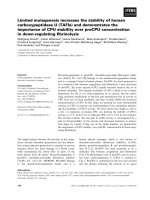
Summary Fish scale was decalcified and disaggregated and then collagen was prepared by limited pepsin digestion. The yields of collagens were very high on a dry weight basis; sardine 50.9%, red sea bream 37.5% and Japanese sea bass 41.0%, respectively. Th
Bạn đang xem bản rút gọn của tài liệu. Xem và tải ngay bản đầy đủ của tài liệu tại đây (209.19 KB, 6 trang )
International Journal of Food Science and Technology 2004, 39, 239–244
Fish scale collagen. Preparation and partial
characterization
Takeshi Nagai,1* Masami Izumi2 & Masahide Ishii3
1 Department of Food Science and Technology, National Fisheries University, Shimonoseki, Yamaguchi 7596595, Japan
2 Ribro Com, Inc., 1-5-10 Nishi-shinbashi, Minato-ku, Tokyo 1050003, Japan
3 Staff Labbi, 6-6-28 Akasaka Minato-ku, Tokyo 1070052, Japan
(Received 25 October 2002; Accepted in revised form 30 June 2003)
Summary
Fish scale was decalcified and disaggregated and then collagen was prepared by limited
pepsin digestion. The yields of collagens were very high on a dry weight basis; sardine
50.9%, red sea bream 37.5% and Japanese sea bass 41.0%, respectively. These scale
collagens were heterotrimers with a chain composition of (a1)2a2. Although the denaturation temperature of the collagen was lower than land animal collagen, fish scales will have
potential as an important collagen source for use in various industries.
Keywords
Alternative source of collagen from cattle skin, underutilized resources, yield.
Introduction
Collagen is the protein that is found in the highest
concentration, about 30%, in the living body. The
main sources of industrial collagen are limited to
those from bovine and pig skin and bones.
However, the existence of bovines infected with
Bovine Spongiform Encephalopathy (BSE) has
been reported in Japan (Yamauchi, 2002). It
becomes a matter of great important to solve the
problems created by BSE. One alternative is to
replace bovine collagen with another source. As
part of a study looking at the effective use of
underutilized resources, we have reported the
preparation and characterization of collagens
from aquatic organisms, mainly marine vertebrates and invertebrates (Nagai et al., 1999, 2000,
2001, 2002; Nagai & Suzuki, 2000a, b, c, 2002a, b).
Although there are many reports about collagen
from skin of marine organisms, there are few
studies of fish scales except for the studies of
Kimura’s group (Kimura et al., 1991) and those of
Shirai (Nomura et al., 1996). Kimura et al. (1991)
reported that collagen from carp scale could be
extracted with 0.5 m acetic acid and the yield was
*Correspondent: Fax: +81 832 33 1816;
e-mail: machin@fish-u.ac.jp
doi:10.1111/j.1365-2621.2004.00777.x
Ó 2004 Blackwell Publishing Ltd
about 7% on dry weight basis. On the contrary,
Nomura et al. (1996) reported the extraction of
collagen from sardine scale with different solvent
systems: 0.05 m Tris–HCl (pH 7.5) containing
0.5 m ethylenediaminetetraacetic acid (EDTA). Its
yield was very low, about 5%. It is possible for fish
scales to have potential as an important source of
collagen because they contain a large quantity of
collagen. This paper describes the preparation and
characterization of collagen from fish scales.
Materials and methods
Fish
Fish sardine Sardinops melanostictus (body weight
0.1–0.2 kg), red sea bream Pagrus major (1.0–
1.3 kg) and Japanese sea bass Lateolabrax japonicus (0.8–1.2 kg) were purchased from a fish
market in Shimonoseki City, Yamaguchi Prefecture, Japan. The scales were removed, washed with
distilled water and lyophilized.
Preparation of scale collagen
All the preparative procedures were at 4 °C. The
lyophilized scales (5.0 g) were treated with 0.1 n
NaOH to remove noncollagenous proteins and
239
240
Fish scale collagen T. Nagai et al.
pigments for 3 days by changing the solution once a
day, then washed with distilled water, dried, and
stored at )85 °C until used. The matter was
extracted with 0.5 m acetic acid for 3 days, and
the extract was centrifuged at 50 000 g for 1 h. The
supernatants were pooled and salted out by adding
NaCl to a final concentration of 0.9 m. Unfortunately, the collagen was not precipitated in this
solution. The resultant matter, obtained by centrifugation at 50 000 g for 1 h, was decalcified with
0.05 m Tris–HCl (pH 7.5) containing 0.5 m EDTA4 Na for 2 days and then disaggregated with 0.1 m
Tris–HCl (pH 8.0) containing 0.5 m NaCl, 0.05 m
EDTA-2 Na and 0.2 m 2-mercaptoethanol (2-ME)
for 3 days. After collecting the collagen fibrils with
cheesecloth, the residue was washed with distilled
water for 2 days by changing the water once a day.
The residue obtained was lyophilized. The lyophilized fibrils were suspended in 0.5 m acetic acid and
digested with 10% (w/w) pepsin (EC 3.4.23.1; 2·
crystallized, 3085 U mg)1 protein; Sigma, USA) at
4 °C for 24 h. The pepsin-solubilized collagen was
centrifuged at 50 000 g for 1 h and the supernatant
dialyzed against 0.02 m Na2HPO4 (pH 7.2) for
3 days, changing the solution once a day. The
resultant precipitate, obtained by centrifugation at
50 000 g for 1 h, was dissolved in 0.5 m acetic acid
and was salted out by adding NaCl to a final
concentration of 0.9 m, followed by precipitation of
the collagen by the addition of a final concentration
of 2.4 m NaCl at neutral pH. The resultant precipitate was obtained by centrifugation at 50 000 g
for 1 h, dissolved in 0.5 m acetic acid, and then
lyophilized.
Sodium dodecyl sulphate-polyacrylamide gel
electrophoresis
Sodium dodecyl sulphate-polyacrylamide gel
electrophoresis (SDS-PAGE) was performed as
described previously (Nagai et al., 2002). After
the electrophoresis, the gels were stained with
Coomassie Brilliant Blue R-250 (Fluka Fine
Chemical Co. Ltd., Tokyo, Japan) and destained
with 5% methanol and 7.5% acetic acid.
Peptide mapping
The collagen samples (0.5 mg) were dissolved in
0.1 m sodium phosphate buffer (pH 7.2) contain-
ing 0.5% SDS and heated at 100 °C for 5 min.
After cooling in ice, the digestion was done at
37 °C for 30 min using 5 lL of lysyl endopeptidase from Achromobacter lyticus (EC 3.4.21.50;
4.5 amidase activity mg)1 protein; Wako Pure
Chemicals, Osaka, Japan). After adding SDS to a
final concentration of 2%, the proteolysis was
stopped by boiling for 5 min. SDS-PAGE was
performed by the method of Laemmli (1970) using
15% gels.
Subunit composition
To separate the subunits of each collagen sample,
the sample was applied to a CM-Toyopearl 650M
(Tosoh Co., Tokyo, Japan) column chromatography. Fifteen milligrams of the collagen sample
were dissolved in 20 mm sodium acetate buffer
(pH 4.8) containing 6 m urea at 4 °C, denatured
at 45 °C for 30 min, and the solution was
centrifuged at 50 000 g at 20 °C for 1 h. The
supernatants were applied to a CM-Toyopearl
650M column (1.0 · 6.0 cm) previously equilibrated with the same buffer. Each subunit was
eluted with a linear gradient of 0–0.15 m NaCl in
the same buffer at a flow rate of 0.8 mL min)1.
The subunit quantity was detected by using
absorbance at 230 nm, and the fractions were
examined by SDS-PAGE.
Denaturation temperature
Denaturation temperature (Td) was measured by
the method of Nagai et al. (2002). Five millilitres
of a 0.03% collagen solution in 0.1 m acetic acid
was used for viscosity measurements. Td was the
temperature where the change in viscosity using a
Canon–Fenske type viscometer with an average
shear gradient of 400 s)1, was half completed.
Amino acid composition
Collagen samples were hydrolyzed under reduced
pressure in 6 m HCl at 110 °C for 24 h, and the
hydrolysates were analysed on a JASCO liquidchromatography system by on-line precolumn
derivatization with OPA. This system consisted
of a JASCO PU-2080 plus intelligent HPLCpump, a JASCO FP-2020 plus intelligent fluorescence detector, a JASCO CO-2060 plus intelligent
International Journal of Food Science and Technology 2004, 39, 239–244
Ó 2004 Blackwell Publishing Ltd
Fish scale collagen T. Nagai et al.
column thermostat, a JASCO DG-2083-53 3-line
degasser, a JASCO LG-2080-02 ternary gradient
unit, a JASCO AS-2057 plus intelligent sampler,
and a JASCO CrestPak C18S (/ 4.6 · 150 mm)
reversed-phase column. The excitation and emission wavelengths were set at 345 and 455 nm,
respectively. Eluents were filtered through Millipore membrane filters (pore size 0.45 lm).
Results and discussion
The scales were hardly solubilized with 0.5 m
acetic acid. The supernatants obtained by centrifugation were salted out by adding NaCl. Unfortunately the collagen was not precipitated in the
sample solution. As a result of decalcification and
disaggregation procedures, the collagen was easily
solubilized by limited pepsin proteolysis. Collagens solubilized by pepsin were effectively purified
by differential salt precipitation. The yields of the
collagens were very high and were in the range of
about 38–51% on a dry weight basis (sardine
50.9%, red sea bream 37.5% and Japanese sea
bass 41.0%, respectively). The results were similar
to previous reports (Nagai et al., 1999, 2000, 2001,
2002; Nagai & Suzuki, 2000a,b,c, 2002a,b),
suggesting that a great amount of collagen can
be obtained from aquatic animals. However,
Nomura et al. (1996) prepared collagen from sardine scale with different solvent systems: 0.05 m
Tris-HCl (pH 7.5) containing 0.5 m EDTA. Furthermore, they reported that the yield of the
collagen was only 5%, as acid solubilized collagen.
The preparative method reported here in was
superior to earlier reports and the collagen was
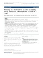
recovered in high yield from fish scale. The collagens obtained were examined by SDS-PAGE using
3.5% gel. It was found that the collagens from red
sea bream and Japanese sea bass comprised only
one a chain, a1, although red sea bream collagen
seemed to have a3 chain (Fig. 1). On the contrary,
sardine collagen had at least two different a chains,
a1 and a2 (Fig. 1). The a chains of these collagens
were different when compared with those from
porcine skin a chains. It suggests that these collagens are different to one another in primary
structure. In this electrophoretic separation the a3
chain was not separated from the corresponding a1
chain if other a chains, such as a3 and a4, were
present in these scale collagens.
Ó 2004 Blackwell Publishing Ltd
a
b
c
d
Figure 1 Sodium dodecyl sulphate-polyacrylamide gel electrophoresis of porcine skin type I collagen and fish scale
collagens on 3.5% gels containing 3.5 m urea. (a) Porcine,
(b) sardine, (c) red sea bream and (d) Japanese sea bass.
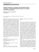
To compare the patterns of peptide fragments
with fish scale and porcine collagens, the digested
collagens were applied to SDS-PAGE using 15%
a
b
c
d
e
f
Figure 2 Peptide mapping of lysyl endopeptidase digests
from several fish scale collagens. (a) High molecular marker,
(b) porcine, (c) sardine, (d) Japanese sea bass, (e) red sea
bream and (f) low molecular marker.
International Journal of Food Science and Technology 2004, 39, 239–244
241
242
Fish scale collagen T. Nagai et al.
gel. The electrophoretic patterns of the three fish
scale collagens were similar to each other (Fig. 2).
In particular the protein bands with molecular
mass of 200, 120 or 30–40 kDa were nearly
identical in all these fish species. The pattern of
peptide fragments of porcine skin collagen was
quite different from those of other fish scale
collagens, although the pattern of porcine collagen
also shows some similarities in comparison with
those of fish scale collagens (Fig. 2).
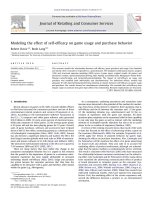
Figure 3 CM-Toyoperal 650M column chromatography of
denatured sardine scale collagen. A 1.0 · 5.0 column of
CM-Toyopearl 650M was equilibrated with 0.02 m sodium
acetate buffer (pH 4.8) containing 6 m urea, and maintained
at 37 °C. The collagen sample (15.0 mg) was dissolved in
5 mL of the same buffer, denatured for 30 min at 45 °C, and
then eluted from the column with a linear gradient of 0 to
0.15 m NaCl at a flow rate of 0.8 mL min)1. The fractions
indicated by the numbers were examined by sodium dodecyl
sulphate-polyacrylamide gel electrophoresis.
The denatured collagens were resolved by
CM-Toyopearl 650M column chromatography to
determine the subunit composition of fish scale
collagens. The chromatographic fractions were
identified by SDS-PAGE and sardine collagen
showed two a chains; a1 and a2 (Fig. 3). Similarly, red sea bream (Fig. 4) and Japanese sea bass
(Fig. 5) collagens comprised two a chains.
Although a band corresponding to a3 in Japanese
sea bass collagen was detected, it seemed to be
partially denatured. The scale collagens were
heterotrimers with a chain composition of
(a1)2a2. Kimura et al. (1991) prepared collagen
from carp scale and reported the properties. Carp
Figure 4 CM-Toyoperal 650M column chromatography of
denatured red sea bream scale collagen. The chromatographic conditions are shown in Fig. 3.
International Journal of Food Science and Technology 2004, 39, 239–244
Ó 2004 Blackwell Publishing Ltd
Fish scale collagen T. Nagai et al.
Figure 6 Thermal denaturation curve of fish scale collagen
solutions as measured by viscosity in 0.1 m acetic acid. The
incubation time at each temperature was 30 min. Collagen
concentration: 0.03%; (s) porcine skin collagen, (d)
sardine collagen, (h) red sea bream collagen, (+) Japanese
sea bass collagen.
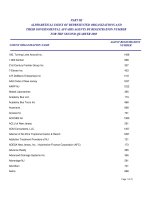
Table 1 Amino acid composition of scale collagens from fish
species, residues/1000
Amino acid
Figure 5 CM-Toyoperal 650M column chromatography of
denatured Japanese sea bass scale collagen. The chromatographic conditions are shown in Fig. 3.
scale collagen had three different a chains; a1, a2
and a3, giving a heterotrimer with a chain
composition of a1a2a3.
To determine the Td of the scale collagens
separated in three experiments, the changes in
viscosity and the Td were calculated from thermal
denaturation curves. It was calculated that the
Td s of fish scale collagens were as follows: sardine
28.5 °C, red sea bream 28.0 °C and Japanese sea
bass 28.0 °C (Fig. 6). On the contrary, the Td of
porcine skin collagen was measured at 37.0 °C,
this is about 9 °C higher than those of fish scale. It
was suggested that the tendency for the Td of
marine organism to be lower than that of land
animals is correlated with their environmental and
body temperature (Rigby, 1968).
Ó 2004 Blackwell Publishing Ltd
Hydroxyproline
Aspartic acid
Threonine
Serine
Glutamic acid
Proline
Glycine
Alanine
Half-cystine
Valine
Methionine
Isoleucine
Leucine
Tyrosine
Phenylalanine
Tryptophan
Lysine
Histidine
Arginine
Total
Sardine Red sea bream Japanese sea bass
86
47
24
41
71
111
340
115
2
18
13
11
22
3
12
0
25
7
52
87
46
26
39
72
109
340
116
2
19
12
10
22
2
13
0
23
7
55
85
48
25
42
75
108
341
114
2
18
12
10
23
2
13
0
24
6
52
1000
1000
1000
The amino acid composition in three fish scale
collagens is shown as residues per 1000 total
residues (Table 1). Glycine was the most abundant
amino acid in all of these collagens and the value
International Journal of Food Science and Technology 2004, 39, 239–244
243
244
Fish scale collagen T. Nagai et al.
was approximately 340/1000 residues. Alanine,
proline, hydroxyproline and glutamic acid had
relatively high contents in these collagens. On the
contrary, tryptophan was not detected in any
collagen samples.
It is known that the major components in fish
scale are as follows: water 70%, protein 27%, lipid
1% and ash 2%. Organic compounds comprise
40–90% in scales and most of them are collagen,
regardless of fish species. At present, great quantities of fish scales are produced in fish shops and
fish-processing factories. However, the effective
use of these scales is minimal. In this study,
collagen obtained from three types of fish scales
possessed properties typical of type I collagen.
Among them, surprisingly, sardine scale showed
the highest yield of collagen, about 51.0% on a dry
weight basis. From these results it is clear that fish
scales have the potential to be an alternative
source of collagen to porcine and cattle skin and
bone. Unless the problem of BSE infection in land
animals is resolved, fish scale as an alternative
source of collagen, will attract much attention in
the cosmetic and medical fields.
Acknowledgments
This work was supported in part by the grant from
the Kiei-Kai Research Foundation, Tokyo, Japan.
We would like to express our heartfelt gratitude to
the donor.
References
Kimura, S., Miyauchi, Y. & Uchida, N. (1991). Scale and
bone type I collagens of carp (Cyprinus carpio). Comparative Biochemistry and Physiology, 99B, 473–476.
Laemmli, U.K. (1970). Cleavage of structural proteins
during the assembly of the head of bacteriophage T4.
Nature, 227, 680–685.
Nagai, T. & Suzuki, N. (2002a). Collagen of the skin of
ocellate puffer fish (Takifugu rubripes). Food Chemistry,
78, 173–177.
Nagai, T., Nagamori, K., Yamashita, E. & Suzuki, N.
(2002). Collagen of octopus Callistoctopus arakawai arm.
International Journal of Food Science & Technology, 37,
285–289.
Nagai, T., Ogawa, T., Nakamura, T. et al. (1999). Collagen
of edible jellyfish exumbrella. Journal of the Science of
Food and Agriculture, 79, 855–858.
Nagai, T. & Suzuki, N. (2000a). Isolation of collagen from
fish waste material-skin, bone and fins. Food Chemistry,
68, 277–281.
Nagai, T. & Suzuki, N. (2000b). Partial characterization of
collagen from purple sea urchin (Anthocidaris crassispina)
test. International Journal of Food Science & Technology,
35, 497–501.
Nagai, T. & Suzuki, N. (2000c). Preparation and characterization of several fish bone collagens. Journal of Food
Biochemistry, 24, 427–436.
Nagai, T. & Suzuki, N. (2002b). Preparation and partial
characterization of collagen from paper nautilus (Argonauta argo, Linnaeus) outer skin. Food Chemistry, 76,
149–153.
Nagai, T., Worawattanamateekul, W., Suzuki, N. et al.
(2000). Isolation and characterization of colagen from
rhizostomous jellyfish (Rhopilema asamushi). Food Chemistry, 70, 205–208.
Nagai, T., Yamashita, E., Taniguchi, K., Kanamori, N. &
Suzuki, N. (2001). Isolation and characterisation of
collagen from the outer skin waste material of cuttlefish
(Sepia lycidas). Food Chemsitry, 72, 425–429.
Nomura, Y., Sakai, H., Ishii, Y. & Shirai, K. (1996).
Preparation and some properties of type I collagen from
fish scales. Bioscience, Biotechnology, and Biochemistry,
60, 2092–2094.
Rigby, B.J. (1968). Amino-acid composition and thermal
stability of the skin collagen of the Antarctic ice-fish.
Nature, 219, 166–167.
Yamauchi, K. (2002). Bovine Spongiform Encephalopathy
and People. Tokyo, Japan: Iwanami Press.
International Journal of Food Science and Technology 2004, 39, 239–244
Ó 2004 Blackwell Publishing Ltd