Nanoscale p hotodynamic ag ents for c olorectal
Bạn đang xem bản rút gọn của tài liệu. Xem và tải ngay bản đầy đủ của tài liệu tại đây (32.87 MB, 26 trang )
Review
Journal of
Biomedical Nanotechnology
Copyright © 2016 American Scientific Publishers
All rights reserved
Printed in the United States of America
Vol. 12, 1348–1373, 2016
www.aspbs.com/jbn
Nanoscale Photodynamic Agents for Colorectal
Cancer Treatment: A Review
Leping Yang1 , Jun He1 , Yu Wen1 , Wenjun Yi1 , Qinglong Li1 , Liangwu Lin2 ,
Xiongying Miao1 , Wei Chen1 3 ∗ , and Li Xiong1 ∗
1
Department of General Surgery, Second Xiangya Hospital, Central South University, Changsha 410011, Hunan, P. R. China
State Key Laboratory for Powder Metallurgy, Central South University, Changsha Hunan 410083, P. R. China
3
Department of Physics and the SAVANT Center, The University of Texas at Arlington, Arlington, Texas 76019-0059, USA
2
Colorectal cancer is the most common form of gastroenteric cancer worldwide. Photodynamic therapy is emerging as an
attractive method to treat cancers. Candidate targets of photodynamic therapy include epidermal growth factor receptors,
cholesterol and low-density lipoprotein, estrogen receptors, the nucleus and DNA, folic acid receptors, cholecystokinin A
receptors, lectin saccharide receptors, and tumor-specific antibodies. Specifically, in colorectal tumors, anti-DR5 antibody
and cancer-specific antibody moieties are involved. Cancer cells incorporate greater quantities of sugars, and glycoconjugated photosensitizer has remarkable internalization and cytotoxicity in colon/colorectal cancer cells. Simultaneously,
to circumvent the bio-distribution limitation, other molecules, including lectins, Hyaluronic acid, and peptides, have also
been considered for colorectal cancer. Other novel strategies indirectly targeting colorectal cancer include pH-responsive
PS, enzymatically activated photosensitization, and cancer-suppressing immune cells, mainly macrophages. Recently,
46
nanoparticles have gained attention as a versatile platform for multi-functional photodynamic therapy. In this review, we
summarize the targeting strategies investigated and highlight the potential of nanoparticles for target photodynamic therapy in colorectal cancer.
KEYWORDS: Photodynamic Therapy, Colorectal Cancer, Active Targeting Delivery, Nanoparticles.
CONTENTS
Introduction . . . . . . . . . . . . . . . . . . . . . . . . . .
Carbohydrates . . . . . . . . . . . . . . . . . . . . . . . .
Optimization and Characteristics . . . . . . . . . . .
New Advances . . . . . . . . . . . . . . . . . . . . . .
Hyaluronic acid (HA) . . . . . . . . . . . . . . . . . . . .
Antibody and Antibody Moiety in Targeting to CRC
Antibody: Death Receptor . . . . . . . . . . . . . . .
Antibody Moiety . . . . . . . . . . . . . . . . . . . . .
Peptides . . . . . . . . . . . . . . . . . . . . . . . . . . . .
Folic Acid . . . . . . . . . . . . . . . . . . . . . . . . . . .
Non-Cancer-Cell Targeting . . . . . . . . . . . . . . . .
pH Responsiveness Conjugate . . . . . . . . . . . .
Enzymate-Activated Photosensitizers . . . . . . . .
Macrophage . . . . . . . . . . . . . . . . . . . . . . . .
Prospective and Discussion . . . . . . . . . . . . . . . .
Acknowledgments . . . . . . . . . . . . . . . . . . . .
References and Notes . . . . . . . . . . . . . . . . . .
∗
INTRODUCTION
.
.
.
.
.
.
.
.
.
.
.
.
.
.
.
.
.
.
.
.
.
.
.
.
.
.
.
.
.
.
.
.
.
.
.
.
.
.
.
.
.
.
.
.
.
.
.
.
.
.
.
.
.
.
.
.
.
.
.
.
.
.
.
.
.
.
.
.
.
.
.
.
.
.
.
.
.
.
.
.
.
.
.
.
.
Authors to whom correspondence should be addressed.
Emails: ,
Received: 10 January 2016
Accepted: 28 March 2016
1348
J. Biomed. Nanotechnol. 2016, Vol. 12, No. 7
.
.
.
.
.
.
.
.
.
.
.
.
.
.
.
.
.
.
.
.
.
.
.
.
.
.
.
.
.
.
.
.
.
.
.
.
.
.
.
.
.
.
.
.
.
.
.
.
.
.
.
.
.
.
.
.
.
.
.
.
.
.
.
.
.
.
.
.
1348
1351
1351
1354
1355
1357
1357
1357
1358
1360
1361
1361
1362
1363
1365
1366
1366
Based on statistics published in 2012, colorectal cancer (CRC) is the third most prevalent cancer worldwide (1.4 million cases), with a high mortality rate at
694,000 deaths per year.1 In 2010, there were more than
270,000 new reported CRC cases and 130,000 related
deaths in China.
Photodynamic therapy and imaging involve the incorporation of photosensitizers (PS) by cancer cells or
other types of cells, followed by irradiation at certain wavelengths of light or non-irradiation instead of
fluorescence.2 3 Photodynamic therapy (PDT) mainly utilizes reactive oxygen species (ROSs) in situ, such as
singlet oxygen (1 O2 ) and free radicals, contributing to irreversible damage to cells4 through apoptosis, necrosis, and
autophagy.5 In the last decade, PDT has made remarkable
progress, with a variety of therapeutic regimens applied to
a range of clinical conditions, including cancers, infectious
diseases,6–9 and autoimmune disorders.10–12
Improvements are needed considering that the drawbacks of photodynamic therapy include poor selectivity,
1550-7033/2016/12/1348/026
doi:10.1166/jbn.2016.2284
Yang et al.
Nanoscale Photodynamic Agents for Colorectal Cancer Treatment: A Review
Leping Yang, graduated from Xiangya Medical University, is an assistant professor
in General Surgery Department of Second Xiangya Hospital, Central South University,
Changsha China and a visiting scientist in Stanford University. He is interested in photodynamic therapy including nanomaterials of drug delivery systems for photosensitizers
and combination of surgery and photodynamic therapy.
Jun He holds the assistant researcher in Research Laboratory for New Nanotechnology
and Photodynamic Therapy, General Surgery Department, Second Xiangya Hospital of
Central South University (Changsha, China). He graduated from the Xiangya Medical
School, Central South University, in 2015 and is performing his master study in Second
Xiangya Hospital of Central South University.
Yu Wen is an assistant professor in General Surgery Department of Second Xiangya
Hospital, Central South University, Changsha China and a visiting scientist in University
of Pittsburgh Medical Center. He conducts a broad research in photodynamic therapy
including nanomaterials of drug delivery systems for photosensitizers and combination
of surgery and photodynamic46
therapy and robot surgery as well. He recently initiates the
cancer stem cell targeting nano-drugs of photodynamic therapy involved in the earlier
stage of colorectal neoplasms.
Wenjun Yi is an assistant professor in General Surgery Department of Second Xiangya
Hospital, Central South University, Changsha China, and received degrees from the
Central South University, China (M.D. and Ph.D.). He majors in oncology surgery, and
focus on combination of photodynamic therapy and surgery. He is going to carry out the
laparoscopic assisted photodynamic therapy to increase the efficacy of radical resection.
Qinglong Li is professor of surgical oncology and the associated director of Surgery
Department, Second Xiangya Hospital, Central South University, and Secretary of
Surgery Committee of Hunan Province Medical Association. He is good at surgical
treatments to liver, bile duct and pancreatic diseases, especially combined photodynamic
therapy and nanomedicine research for cholangiocarcinoma. He is funded by Hunan
Provincial Science and Technology Department with several projects.
J. Biomed. Nanotechnol. 12, 1348–1373, 2016
1349
Nanoscale Photodynamic Agents for Colorectal Cancer Treatment: A Review
Yang et al.
Liangwu Lin holds the assistant researcher in State Key Laboratory for Powder Metallurgy, Central South University (Changsha, China). He received degrees from the Hunan
University, China (M.D. and Ph.D.). His research interests include synthesis, performance optimization, targeting mechanism and Photodynamic Therapy (PDT) efficiency
of targeting nano-photosensitizers, self-excited targeting nano-photosensitizers for the
treatment of cancer stem cells and deep tumors, and the application of luminescent
nanomaterials in the field of nanomedicine.
Xiongying Miao is the director of General Surgery Department, Second Xiangya Hospital, Central South University, and committee member of Chinese Professional Committee of General Surgery integrated with Traditional Chinese and Western Medicine,
committee member of Surgery Branch of China Medical Association. He is interested
at innovative multidisciplinary treatments to liver, bile duct and pancreatic diseases,
especially combined photodynamic therapy for liver cancer and cholangiocarcinoma.
He is funded by Famous Doctor Program of Xiangya Medical School and National
Natural Science Foundation of China, and Hunan Provincial Science and Technology
Department etc. with several research projects on nanomedicine.
Wei Chen is a professor in Physics Department and the director of the center for
Security Advances Via Applied Nanotechnology, the University of Texas at Arlington
(UTA). He earned his doctorate (Ph.D.) in Materials Chemistry from Peking University,
Beijing, China in 1992. From 1994 to 1998, he conducted research in nanoclusters and
quantum
devices at the Chinese Academy of Sciences Laboratory of Semiconductor
46
Materials Science, where he served as a deputy director and a senior research scientist. He received an outstanding young scientist award from the Chinese Academy of
Sciences, and has been honored for distinguished research by the Chinese Department
of Education and Peking University. From 1998 to 1999, Dr. Chen joined the Inorganic Chemistry and Nanometer Structure Consortium, University of Lund, Sweden as
a Senior Visiting Scientist. In 1999, he served in a similar role at the Centre for Chemical Physics, University of Western Ontario in Canada. In 2000, Dr. Chen joined Nomadics Inc., where he serves
as a senior and leading scientist in Nanotechnology. In 2006, he joined UTA as an assistant professor in Nano-Bio
Physics and he was promoted to associate professor in 2011 and full professor in 2013. Dr. Chen has more than 210
publications and 8 US patents. He serves as Editor-in-Chief for Reviews in Nanoscience and Nanotechnology, American editor for the Journal of Nanoscience and Nanotechnology and an associate editor for Journal of Biomedical
Nanotechnology published by American Scientific Publishers.
Li Xiong is a visiting scholar at Harvard University, and distinguished associate professor in General Surgery Department of Second Xiangya Hospital, Central South
University in Changsha China. Since 2009, he has performed research of photodynamic therapy. He likes invention and has some patents which focus on the surgery
and photodynamic therapy. Dr. Xiong pioneered the laparoscopy assisted photodynamic
therapy and intellectual photodynamic lancet for surgeons. He has initiated 2 projects
funded by National Natural Science Foundation of China with an established living
mice colonoscopy platform and ongoing developing on all in one machine of X-ray
activated nanophotosensitizers.
1350
J. Biomed. Nanotechnol. 12, 1348–1373, 2016
Yang et al.
Nanoscale Photodynamic Agents for Colorectal Cancer Treatment: A Review
prolonged skin photosensitization, scarring of healthy
tissue following irradiation, inter-patient fluctuations in
response, and intralesion heterogeneity. Ongoing problems
include difficulties in predicting dose-dependent responses
in cancer cells and normal tissues, and suboptimal targeting
strategies.
A great deal of research has focused on enhancing the targeting action of photosensitizers by coupling
PS with antibodies,13 peptide,14–18 carbohydrates,19–21 and
hyaluronic acid (HA),22 23 which binds selectively with
cancer cells. Cancer cells exhibit significant differences
compared with normal healthy cells, for example, overexpressing some types of antigens/antibodies and secreting biomolecules through paracrine or autocrine pathways.
These biomolecules might influence the cancer cells and
surrounding tissues to create a friendly environment for
cancers.24–27 The overexpressed receptors on the cancer
cell surface or in the cell membrane bind to antigens or
ligands to promote internalization or surface interactions.
Activation by a specific ligand containing PS might trigger responses involving cell-signal pathways. For example,
caspase 8 activity is triggered by the binding antibodies
against death receptor 5 (DR5) and decreases the viability of cancer cells.13 28 29 In contrast, a non-internalized
conjugate would remain outside the cell membrane, except
for the minimal amount of free PS that would diffuse
into cells. The PDT would thus have an insufficient effect,
though this technique is appropriate for selective fluorescence imaging. However, the 46
extracellular matrix and
immune cells in cancer tissue should also be evaluated.
A tumor-cell extracellular matrix is significantly different
compared to that of a normal cell, with characteristics such
as an acidic pH microenvironment30–33 and infiltration of
inflammatory cells.32 34–36 Methods leveraging these disparities might produce significant effects, as ECM provides
wide opportunities to observe agent activity.
The conjugate containing photosensitizer and a biomolecule required for targeting should neither dramatically compromise the binding specificity, nor influence the
photosensitization of PS.37 In some ways, the conjugate
might have improved binding compared to a free-targeted
biomolecule in vivo, considering the complexity and diversity of biological conditions.
Nanoparticles have attracted considerable interest for
use in PDT. One strategic advantage of NPs for biomedical applications is their potential for diverse modifications.
They can serve as platforms for the assembly of multifunctional structures.38–40 These structures retain high solubility
and colloidal properties for use in complex environments
(e.g., blood and tissues). They are ideal for targeting due to
their passive accumulation in tumor tissue, i.e., enhanced
permeability and retention (EPR) effect.41 42 Modified NPs
could be used as targeted vectors, significantly improving
the concentration in the targeted cell.
Here, we summarize targeting strategies for photodynamic therapy in one specific cancer, colon cancer and/or
J. Biomed. Nanotechnol. 12, 1348–1373, 2016
colorectal cancer, including targeting strategies and general applications for many types of cancers.
CARBOHYDRATES
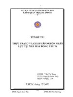
Tumors consume higher levels of glucose than normal cells
consume, a phenomenon known as the Warburg effect43
(illustration in Fig. 144 ). Moreover, the effect extends to
a variety of other carbohydrates, including glucose, mannose, and galactose. These carbohydrates link with specific proteins on the surface of cells. Lectin in particular
is known as a carbohydrate-binding protein.45 Targeting
has been established based on glycoconjugates, which couple photosensitizers with carbohydrates. Monsigny et al.46
showed that glycosylmoieties of glycoconjugates could be
recognized by membrane lectins that participate in internalizing the conjugate and releasing it in certain organelles
(endoplasmic reticulum and Golgi apparatus). Over the
past 15 years, a variety of photosensitizers have been coupled to saccharides, including glucose, lactose, mannose,
galactose, maltose, and glucosamine, to verify the feasibility and efficiency of using glycoconjugates for PDT.
The advantages of glycoconjugates for PDT are that they
increase the hydrophilicity of photosensitizers and have
potential for selective recognition and/or enhanced cell
uptake by cancer cells due to the added glyco-moieties.47
Optimization and Characteristics
Effect of Sugars and Linkage
Sugars. Many groups have demonstrated that
glycoconjugate derivatives consisting of photosensitizer
and saccharide show improved uptake by cancer cells
and phototoxicity in various cancer types. Zheng et al.48
studied the effect of lipophilicity on the photodynamic
effect of purpurin derivatives with alkyl chains of various lengths located in the 11 -O- and 132 -N-positions.
Along with other reports, the results suggest that the
lipophilicity of the glycoconjugate might be a dominant
parameter in controlling penetration through the cellular
membrane.49–51 Although glycoside residues binding with
the photosensitizer influence the ultimate lipophilicity of
the glycoconjugated compound and hence the variance in
cell penetration, the sugar serving as a ligand could also
serve an important role in cellular uptake of the conjugate.
In colorectal cancer, HT29 cells (human colorectal cancer) overexpress -glucose receptors.52 Laville et al.53
studied the photodynamic efficiency of diethylene glycolinked glycoconjugated porphyrins. Their data showed that
the relative drug uptake of the glycoconjugated porphyrins
was similar independent of the cell line studied (in HT29,
retinoblastoma Y79, and melanoma B16 cell lines). However, a partial saturation effect was observed when the cells
were incubated with glycosylated albumin before incubation with the corresponding glycosylated photosensitizers.
To obtain more information on this phenomenon, they used
a semi-quantitative method to verify the glyco-receptor on
1351
Nanoscale Photodynamic Agents for Colorectal Cancer Treatment: A Review
Yang et al.
the three cells, and demonstrated that the number of mannose and galactose receptors is similar for B16 and Y79
cells and that glucose receptors are overexpressed on HT29
cells. These studies suggest that the targeting of sugar
receptors may not be highly selective. Li et al.54 confirmed
that the binding affinity between a benzochlorin-galactose
conjugate and an isolated galectin was only two times
greater than that of a benzochlorin-glucose conjugate. Furthermore, Li also showed that the parent benzochlorin
unit exhibits fairly significant nonspecific galectin binding,
which is a general characteristic of the tetrapyrrolic ring
present in most photosensitizers, including porphyrins,
chlorins, and bacteriochlorins.53 These results indicate that
the choice of sugars to facilitate active internalization
should be made based on specific lectins expressed by specific cancer types.
Incorporation of glycoconjugates by cancer cells, followed by irradiation by a suitable wavelength of light, leads
to cell death, mainly via ROSs. While this phototoxicity has
been demonstrated in a sugar-dependent pathway,21 49 55
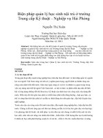
Makoto et al.49 demonstrated this phenomenon by comparing the photocytoxicity in HeLa cells of four glycoconjugated porphyrins containing different sugar moieties,
namely 5,10,15,20-tetrakis[4-( -D-glucopyranosyloxy)phenyl]
porphyrin, 5,10,15,20-tetrakis[4-( -D-galactopyranosyloxy)
phenyl]porphyrin, 5,10,15,20-tetrakis [4-( -D-xylopyranosyloxy)phenyl]porphyrin, and 5,10,15,20-tetrakis[4-( 46 D-arabinopyranosyloxy)phenyl]porphyrin (Fig. 2). The
results showed the phototoxicity of the samples varied up to five-fold, indicating that the type of sugar
makes a significant difference. This phenomenon has
been repeatedly verified by many reports using different
types of saccharides coupled to porphrins, chlorins, and
phthalocyanines.47 51 55 56
Clearly, differences in cell uptake of each conjugate
Figure 1. The metabolic switch towards aerobic glycolysis
are
involved in sugar-dependent photocytoxicity. Howcommonly observed in cancer cells—the Warburg effect—
occurs after upregulation of some enzymes (indicated in
ever, the relationship is not always as expected, suggestbold) that play an important role in glucose metabolism.
ing that agent internalization is not the dominant factor
The increased glucose utilization through the glycolytic pathcontrolling sugar-dependent photocytoxicity.49 Another
way generates metabolic intermediates (indicated in italic)
potential explanation is the heterogeneity of the subcelthat cancer cells need to sustain their rapid proliferation.
lular location of individual glycoconjugated tetrapyrrolic
One of these intermediates, glucose 6-phosphate, is used
for the synthesis of nucleic acid through the pentose phosderivatives.50 Mitochondrial fluorescence of glycoconjuphate pathway to allow rapid DNA replication. The abungated porphrins determined by absorption spectroscopy
dant production of pyruvate stimulates lipid synthesis that
represented 40–50% of the total intracellular dye conis necessary for the formation of membranes in dividing
centration, while the non-glycoconjugate only represented
tumor cells. Finally, secretion of lactate by the tumor cells
induces acidification of the tumor microenvironment, which
20–30% of the total concentration.50 These results were
creates a niche that favors further tumor progression and
similar to those of Laville and Tedesco.53 In contrast,
inhibits the action of some anticancer drugs. Aldo, aldolase;
the electronic absorption of glycoconjugate was influEno, enolase; GAPDH, glyceraldehyde 3-phosphate dehydroenced by the sugar moieties, thereby indicating that
genase; GLUT1, glucose transporter 1; HK, hex-okinase; LDH,
the interaction between glycoconjugate and endogenous
lactate dehydrogenase; PFK-I, phosphofructokinase 1; PGI,
phosphoglucose isomerase; PGK, phosphoglycerate kinase;
biomolecules such as albumin plays a considerable role in
PGM, phosphoglycerate mutase; PK, pyruvate kinase; and
sugar-dependent cytotoxicity.49
TPI, triose phosphate isomerase. Reprinted with permission
Linkage. Individual protein-saccharide interactions are
from [44], A. Annibaldi, et al., Glucose metabolism in cancer
typically weak56 and the number of saccharides conjugated
cells. Curr. Opin. Clin. Nutr. Metab. Care 13, 466 (2010). © 2010,
to the photosensitizer varies. Multivalent interactions in
Wolters Kluwer.
1352
J. Biomed. Nanotechnol. 12, 1348–1373, 2016
Yang et al.
Nanoscale Photodynamic Agents for Colorectal Cancer Treatment: A Review
Figure 2. The percentage of cell survival (%) as a function
of the concentration of the glycoconjugated porphyrins p-1a,
p-1b, p-1c and p-1d. p-1a, p-1b, p-1c and p-1d represent 5,10,
15,20-tetrakis[4-( -D-glucopyranosyloxy)phenyl]porphyrin, 5,
10,15,20-tetrakis[4-( -D-galactopyranosyloxy)phenyl]porphyrin,
5,10,15,20-tetrakis[4-( -D-xylopyranosyloxy)phenyl]
porphyrin, and 5,10,15,20-tetrakis[4-( -D-arabinopyranosyloxy)
phenyl]porphyrin, respectively. Reprinted with permission
from [49], M. Obata, et al., Sugar-dependent photodynamic
effect of glycoconjugated porphyrins: A study on photocytotoxicity, photophysical properties and binding behavior
to bovine serum albumin (BSA). Biochim. Biophys. Acta
1770, 1204 (2007) © 2007, Elsevier. 46
biological systems, known as the “cluster glycoside effect,”
have high affinity and high specificity. Momenteau57 first
systematically linked a series of neutral tri- and tetraglycosylated porphyrins to mono- or disaccharides at the
meso position of 5,10,15,20-tetraarylporphyrins via the
phenyl groups, demonstrating that compounds bearing
three mono-saccharide units are, in general, more phototoxic than symmetrical compounds. This finding has been
accepted widely and verified by a number of reports concerning the effect of linkage on PDT efficiency in vitro
and in vivo.50 58
Numerous binding strategies have been employed to
attach glycosyls to photosensitizers. These include a direct
linker, such as an ether when ortho- and meta-position substitution induces a constrained structure; an extended and
more flexible linker such as an diethylene glyco-linker; and
a more complicated linker such as triazole47 or a glycodendrimeric structure,19 which would decrease the photoactivity of PS compared to direct linkage in HT29 cells. The
geometry of the photosensitizer and the bulkiness of the
linker influence hydrophilicity and receptor binding.19 47
Maintaining Stability of the Glycoconjugate
Glycoconjugated photosensitizers have been used for
decades through explicitly synthesized processes. However, the metabolization of glycosylated photosensitizers
J. Biomed. Nanotechnol. 12, 1348–1373, 2016
has been less well studied both in vivo and in cell-based
assays. Metabolism in vivo is particularly important when
considering the use of glycoconjugated compounds in PDT
treatment, as deglycosylation will affect the amphiphilic
properties, biodistribution, blood clearance, and drug-cell
interactions.58
The stability of a glycoconjugate is largely determined
by the metabolization of glycoconjugated compounds,
especially the cleavage of the glycoside bond by glycosidases. In cancer photodynamic therapy, the level of glycosidase activity and expression in certain tumor types,
including colorectal cancer, is higher than in normal
tissues.59 Specifically, glucosidases are present in human
plasma and erythrocyte membranes,60 and the activity of
-gluosidases is approximately 20-fold lower than that
of -glucosidases.58 Therefore, metabolism of glycoconjugated photosensitizers can occur at a number of levels,
so elucidation of the nature of glycoconjugated PS under
physiological conditions is a vital issue in the use of the
compounds in animals and humans.58
Laville et al.58 studied the metabolism of
tri(glucosyloxyphenyl)chlorin in human colon tumor cells
in detail. As in previous reports,46 the resulting deglycosylation depends on the nature of the link between the
drug and the glycosylmoieties.
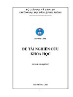
The three sugar motifs in the conjugate hydrolyze
sequentially, resulting in partially or totally deglucosylated compounds, and these processes are connected to the
oxidative metabolism of the corresponding porphyrins by
the chlorin system (Fig. 3). Analysis of the phototoxicity
of the final metabolite, with loss of three or less glucose
Figure 3. Possible metabolic pathway of glycoconjugated
chlorin 2a/3a. Reprinted with permission from [58], I. Laville,
et al., A study of the stability of tri(glucosyloxyphenyl) chlorin,
a sensitizer for photodynamic therapy, in human colon tumor
cells: A liquid chromatography and MALDI-TOF mass spectrometry analysis. Bioorg. Med. Chem. 12, 3673 (2004). © 2004,
Elsevier.
1353
Nanoscale Photodynamic Agents for Colorectal Cancer Treatment: A Review
units and oxidation of the porphyrin macrocycle, shows
biological activity lower than that of the initial compound.
Considering the difficulty of controls and measurement,
there have been no studies in vivo concerning whether the
physiological media play a role in the stability and phototoxicity of glycoconjugates. However, the susceptibility of
glyco-compounds to glycosidases has been verified, as has
binding to a variety of proteins in the body.49 Both points
highlight the importance of the stability of the glycoconjugate in maintaining its photoactivity and therapeutic efficiency. Further research is needed to evaluate the roles of
conjugated moieties and linkage strategies in developing
PDT for clinical applications.
New Advances
Two-photon absorption (2PA) processes had been applied
to coupling with saccharides, improving their water solubility and targeting to lectin receptors that are overexpressed in certain malignant cells. The advantages of 2PA
processes are greater penetration depth because two photons are absorbed simultaneously and the stronger signal,
which allows greater spatial precision than traditional onephoton excitation, preventing damage to adjacent healthy
tissue.51 55 61 The fundamental advantage of two-photon
excitation compared with single-photon and up-converting
excitation is shown in Figure 4.62 A group from Centre
Universitaire of France has synthesized a variety of glycoconjugated porphyrins for targeted
46 two-photon photodynamic therapy.51 Their results showed that two-photon
Yang et al.
compounds exhibited about twice the 1 O2 production of
the monomer. Unfortunately, no significant photodynamic
effect was measured for these two-photon compounds in
HT29 cells (human colorectal cancer cells). One possible
explanation is that the solubility of these compounds is
not optimal in a culture medium containing albumin to
simulate the physiological condition.
Importantly, glycoconjugated nanoparticle systems have
been used widely in recent years, including mesoporous
silica nanoparticles (MSN) and porous silicon nanoparticles (pSiNPs) functionalized with mannose or galactose
that exhibit enhancement of cytotoxicity in breast cancer, colon cancer, and retinoblastoma (RB) (summarized
in Table I). The significant advantages of nanoparticles
(monodispersity, high specific surface area, tunable pore
size and diameter, and versatile functionalization) allow
them to be employed as multifunctional conjugates for photodynamic therapy, imaging, and anti-cancer drug delivery.
Interestingly, Lu et al.56 synthesized self-assembled
nanoparticles containing a glycomoiety to form
glycopolymer-porphyrin, which then aggregated in aqueous solutions due to the specific structure (hydrophobic
porphyrins in the middle and hydrophilic glycopolymers
at both ends) to an average size of approximately 160 nm.
Their results revealed that the glucose in the conjugate
remained bioactive and further showed a stronger multivalent reaction with lectins due to the cargo of nanoparticles.
The results indicate a dual inhibition of cancer cells by
ROS-dependent pathway and ROS-independent pathways.
Figure 4. Comparison of the excitation profiles of (A) single-photon, (B) two-photon, and (C) upconverting excitation. From the
light-excitation pattern with 488- and 960-nm (0.16 NA) lasers in the cuvettes, it can clearly be seen that only in two-photon
excitation (B), the excitatory beam is focused in a spot in the focal plane. Conversely, in single-photon excitation (A), additional
light emanates from above and below the focal plane. Bottom figures: repetitive scanning in the focal plane (x-y plane) in a
´
fluorescein-stained formvar film shows that in two-photon excitation, only the focal plane photobleaches. The Jabłonski
diagrams
illustrate the differences in photon absorption between the various systems. In TPE, two photons of the same wavelength must
arrive simultaneously in time and space to excite the electron. Conversely, upconverting fluorochromes (C) contain metastable
states, as in this example for Europium (III) ions, which are sufficiently stable to allow sequential absorption of long-wavelength
photons. As a result, both TPE and photon upconversion show anti-Stokes shifts. Reprinted with permission from [62], H. C.
Ishikawa-Ankerhold, et al., Advanced fluorescence microscopy techniques–FRAP, FLIP, FLAP, FRET and FLIM. Molecules 17, 4047
(2012). © 2012, Multidisciplinary Digital Publishing Institute.
1354
J. Biomed. Nanotechnol. 12, 1348–1373, 2016
Yang et al.
Table I.
Nanoscale Photodynamic Agents for Colorectal Cancer Treatment: A Review
Carbohydrate-conjugated NP for PDT in various cancer types.
Cancer type
Colorectal cancer
Breast cancer
Retinoblastoma
(RB)
Prostate cancer
Carbohydrate
Nanoparticle
Photosensitizer
References
Galactose/mannose
Mannose
Mannose
Mannose
Mesoporous silica nanoparticles (MSN)
Mesoporous silica nanoparticles (MSN)
pSiNP
Mesoporous silica nanoparticles (MSN)
Porphyrin/biphotonic photosensitizer
Porphyrin
pSINP (biophotonic PS)
Biophotonic photosensitizer
[206, 207]
[208]
[209]
[207]
Mannose-6-phosphate
derivative
Mesoporous silica nanoparticles (MSN)
Biophotonic photosensitizer
[210]
Another strategy in CRC therapy involves targeting the
carbohydrate antigen on the surface of the cancer cells.
Guillaume Poiroux et al.63 first demonstrated the feasibility
of a covalent coupling of a PS with a plant lectin for photochemotherapy and dramatic cell death of T/Tn-positive
leukemic cells treated with TrMPyP-MorG conjugate was
observed. However, an investigation of the binding and
photophysical properties of PdTPPS [5,10,15,20-tetrakis
(4-sulfonatophenyl) porphyrin-Pd(II), a soluble PS] noncovalently bound to a lectin (Con A) indicated a much
lower rate constant for oxygen quenching by the PdTTPS
triplets bound to Con A than by the free PdTPPS, but
the cells remained good producers of O2 .64 A very recent
report65 showed that phthalocyanine-PEG gold nanoparticles conjugated to lectin to target the T antigen (the
Thosen-Friedenreich carbohydrate antigen, overexpressed
in more than 90% of primary tumors), with the structure
shown in Figure 5, exhibited comparable
cytotoxicity in
46
HT29 cells and SK-BR-3 cells. This result is supported
Figure 5. Schematic of the T-antigen-specific lectin, jacalin
(green) conjugated to the mixed monolayer of the phthalocyanine C11Pc photosensitizer (blue) and the thiol-functionalized
PEG (black) on the gold nanoparticle surface. Reprinted with
permission from [65], G. Obaid, et al., Cancer targeting with
biomolecules: A comparative study of photodynamic therapy
efficacy using antibody or lectin conjugated phthalocyaninePEG gold nanoparticles. Photochem. Photobiol. Sci. 14, 737
(2015). © 2015, The Royal Society of Chemistry and Owner
Societies.
J. Biomed. Nanotechnol. 12, 1348–1373, 2016
by an earlier report utilizing lectin jacalin conjugated to
C11Pc-PEG gold nanoparticles to target the T antigen on
the surface of HT29 cells.66 As there are significantly more
T antigens on HT29 cells than there are HER-2 receptors
on SK-BR-3 cells (ca. 4 4 × 10⁷ vs. ca. 1–2 × 10⁶),65 conjugating exogenous lectin to photosensitizers might be an
alternative targeted PDT treatment for colorectal cancer.
HYALURONIC ACID (HA)
Hyaluronic acid (HA), an anionic glycosoaminoglycan consisting of D-glucoronic acid and N-acetyl-Dglucosamine units, has been extensively investigated
for pharmaceutical applications owing to its excellent
biocompatibility, biodegradable, and non-immunogenic
characteristics.67–69 We consider this polysaccharide conjugate separately because it interacts with the CD44
receptor70–73 and RHAMM,74–76 which are overexpressed
on the surface of various tumor cells.23 77 Although expression of the HA receptor was up-regulated in cancer cells,
resulting in a high affinity of HA for tumors, normal cells
also express CD44 but not HA receptors.78
As a targeting moiety for photodynamic therapy
or imaging, HA also triggers intracellular signaling
Figure 6. Effect of PDT on HCT-116 cancer cells. HCT-116
cells were incubated or not (control) for 24 h with 20 g mL−1
of MSN functionalized with hyaluronic acid (MSN-HA) or not
(MSN). Cells were submitted to laser irradiation (14 J cm−2 )
and allowed to grow for two (A) or three (L) days. The bar
graph represents living cells, and the data are represented as
the mean ± SD of three independent experiments. Reprinted
with permission from [81], M. Gary-Bobo, et al., Hyaluronic
acid-functionalized mesoporous silica nanoparticles for efficient photodynamic therapy of cancer cells. Photodiagn. Photodyn. Ther. 9, 256 (2012). © 2012, Elsevier.
1355
Nanoscale Photodynamic Agents for Colorectal Cancer Treatment: A Review
Yang et al.
46
Figure 7. Cellular uptake of DOX@PHANs. (a) Flow cytometric quantification of HCT-116 cancer cells. The cells were treated
with DOX@ PHANs in the presence or absence of free HA (10 mg mL−1 ). (b) CLSM images of HCT-116 cancer cells and CV-1
normal cells. The cells were treated with DOX@PHANs in the presence or absence of free HA (10 mg mL−1 ). In each panel, the
cell nuclei stained with DAPI are blue; DOX is red; and Ce6 is cyan. Original magnification is 40×. The scale bars are 20 m.
(c) Mean fluorescence intensity of DOX and Ce6 in CLSM images (n = 3). Reprinted with permission from [83], C. S. Lee, et al.,
Photochemically triggered cytosolic drug delivery using pH-responsive hyaluronic acid nanoparticles for light-induced cancer
therapy. Biomacromolecules 15, 4228 (2014). © 2014, American Chemical Society.
influencing cellular proliferation, differentiation, and
migration.79–82 Recently, a variety of investigations have
examined its potential for targeting nanovectors and
non-nanovectors to cancer cells. Specially, Gary-Bobo
et al.81 synthesized hyaluronic acid-functionalized mesoporous silica nanoparticles and conducted a cytotoxicity
test in colon cancer cells (MTT results seen in Fig. 6).
The results showed that MSN-HA induced a pronounced
and significant effect on cancer cells, with 68% and
1356
100% cell lysis at days two and three after irradiation,
respectively, through an active mechanism involving CD44
receptors. The results were significant compared with
photo-activatable MSN.
More recently, a polyaniline nanoparticle designed for
photothermal therapy (PTT) combined with HA targeting moiety has shown excellent treatment efficacy in vivo
with HCT-116 cells (human colon cancer) at volumes in
each treatment group of 133.55 mm3 (PS-HA/AuNPS),
J. Biomed. Nanotechnol. 12, 1348–1373, 2016
Yang et al.
Nanoscale Photodynamic Agents for Colorectal Cancer Treatment: A Review
792 mm3 (saline), 699 mm3 (AuNPs), and 471 mm3 (free
PheoA).82 In addition, synergistic treatment with dualkilling gold nanoparticles (AuNPs) has been performed,
combining PDT with PTT and functionalizing the system
with HA. The results indicate in vitro and in vivo photoactivity of the hybrid targeted nanomaterial in aqueous solution and BALB/c nude mice, respectively, while in vivo
examination of the therapeutic efficacy is was established
in the A549 cell line (lung cancer cells).69
A group from Korea established a photochemically
triggered cytosolic drug-delivery system combined with
tumor-targeting pH-responsive hyaluronic-acid nanoparticles (PHANs) and anticancer therapeutics for therapeutic application in HCT116 cells. The main killing effect
was due to the anticancer drug doxiorubicin (DOX), while
the photosensitizer or PDT served as a switch for drug
release. The in vitro cellular uptake of DO@PHANs confirmed receptor-mediated cellular internalization. Furthermore, competitive inhibition was also observed using free
HA in HCT-116 cells by flow cytometry and CLSM, as
shown in Figure 7.83 The polysaccharide is modified by
acetylation and functionalized with a tertiary amine group
containing a polypeptidic pH-responsive moiety, which
might interfere with binding while still maintaining sufficient affinity to the receptor.
ANTIBODY AND ANTIBODY MOIETY IN
TARGETING TO CRC
46
The antibody or antibody moiety that binds to antigens
that are overexpressed on the surface of cancer cells can be
used to deliver agents into the cell84 85 or anchor to the surface area. Del Governatore et al.86 first applied 17.1A mAb
(which recognizes the epithelial membrane antigen found
on many cancers of the gastrointestinal tract87 ) coupled
with photosensitizer Ce6, to target the colorectal cancer.
The conjugate showed selective accumulation in colorectal
cancer cells compared with the control non-target ovarian
cancer cells. This result suggests that cell-specific accumulation of PS through antigen/antibody interaction could
result in remarkable enhancement of PDT to induce death
of gastrointestinal cancer cells in vitro and in vivo.
Antibody: Death Receptor
Death receptors (DRs), including TNF receptor 1 (TNFR1),
Fas, DR4 and DR5, are attractive targets for cancer
therapy88 and imaging.28 89 These DRs would induce apoptosis in cancer cells on binding with their trimeric cognate ligands; this mechanism is known as TRAIL-induced
apoptosis.90 It is encouraging that there are many reports
concerning chemotherapies engagement of DR5 leading to
therapeutic potential in a variety of cancer types.89 91 92
However, the emergence of various drug-resistance phenomena associated with DRs and weak DR5 activation
through insufficient receptor clustering in patients limits the
application of DR-agonistic antibodies.29 93 94
J. Biomed. Nanotechnol. 12, 1348–1373, 2016
PDT resolves this problem, and as demonstrated by
Daniela Schmid et al.,29 stage-II and stage-III colorectal
tumors express significantly higher levels of DR5 compared to normal colon tissue and other healthy tissue. Several studies were conducted focusing on the PS delivery
system and molecular imaging based on DR5. Abdelghany
et al.94 first evaluated the therapeutic efficiency of DR5
combined with PDT in colorectal cancer. They synthesized
a conjugate based on Chitosan/Alginate nanoparticles that
used TMP as a photosensitizer and functioned with DR5
antibody. To enhance the loading efficiency of TMP to
the nanoparticle, alginate was added to increase the likelihood of drug entrapment. Their results showed that the
conjugate trapped 9.1 g TMP per mg particle formulation. The release profile of the TMP from the nanoparticles indicated a biphasic release, with over 60% released
within two days and over 80% release after eight days, as
seen in detail in Figure 8. As expected, TMP-loaded DR5targeting nanoparticles showed significant improvement of
photocytoxicity to HCT 116 cells (human colorectal cell
line) compared with non-targeted nanoparticles after irradiation. Furthermore, they confirmed that anti-DR5 antibody displayed on the nanoparticles can also induce a
cytotoxic effect alone, independent of TMP. As described
above, this effect is a result of TRAIL-induced apoptosis.
Along with the reports from other researchers, it has
demonstrated that anti-DR5-NPs preserved the ability to
induce apoptosis more completely than free antibody
in vitro because the antibodies anchored on the NPs could
drive the clustering of receptors.95 This differences indicate advantages of NP for eliciting the intrinsic anticancer
mechanisms of biomolecules designed for targeting.13 29
Antibody Moiety
Many studies have demonstrated that the use of antibodies to target cancer cells through biological reactivity augments the efficiency of PDT, eliminating the cancer cells
specifically at relatively low concentrations of the PS.96–99
The potential for the application of antibody conjugates is
limited by the large size of mAbs, especially in vivo, which
inhibits the conjugate from penetrating solid, deep-seated
and poorly vascularized tumors.85 100 To address this limitation, antibody fragments such as Fab and scFv have
been proposed as an alternative to improve the potential
of immunoconjugates used in biological conditions.101–103
Staneloudi et al.104 designed conjugates via an isothiocyanate group, porphyrins linked with colorectal tumorspecific scFv (LAG3-scFv), for further characterization
and investigation in Caco-2 cells (human Caucasian colon
adenocarcinoma cell line). Previously, reports have shown
that scFv was more susceptible to interference with antigen binding than their monoclonal counterparts,105 as was
confirmed by the optimal loading ratios for affinity with
the antigen, at 5:1 and 20:1 (PS: scFv).106 After irradiation (red light 630 ± 15 nm), using scFv conjugates
without the final centrifugation clean up exhibited >90%
1357
Nanoscale Photodynamic Agents for Colorectal Cancer Treatment: A Review
Yang et al.
Figure 8. Characterization of TMP-loaded alginate/chitosan nanoparticles. (A) Size distribution of TMP-loaded nanoparticles
using dynamic light scattering. (B) SEM (left-hand panel) and TEM pictures (right-hand panel) of nanoparticles. (C) Controlled
drug release of TMP in PBS at 37 C under shaking, quantified by comparison to a calibration curve of TMP at fluorescence of
426/654 nm. (D) Stability of nanoparticles in 10% FBS supplemented media over time at 37 C. Reprinted with permission from
[94], S. M. Abdelghany, et al., Enhanced antitumor activity of the photosensitizer meso-tetra(N-methyl-4-pyridyl) porphine tetra
tosylate through encapsulation in antibody-targeted chitosan/alginate nanoparticles. Biomacromolecules 14, 302 (2013). © 2013,
American Chemical Society.
lysis of Caco-2 cells in the low micromolar range, while
LAG3 scFv (selectively attached to Caco-2 cells) conjugates caused 30% inhibition of cell growth.
The drawbacks of scFv use as a46
target moieties are instability (because they lack the stabilizing constant region
of the parent antibody)107 and the significantly reduced
immune reaction compared to the full antibody results in
low affinity to the corresponding antigen.97 The strategies
currently exploited to increase the stability of scFv fragments involve mutagenesis and co-expression of chaperone
proteins to facilitate the proper folding of the scFv108 109
and using scFv derived from phage display libraries,
which show good stability as well as appropriate affinities and internalizing characteristics.110 111 In addition, the
long hydrophilic PEG chains of the Mal-PEG2000 -DSPE
micelles were postulated to stabilize the scFv.107
Nanoparticles have the potential to resolve problems with the versatile photosensitizers that have been
explored.112 Carrier nanoparticles are commonly modulated by surface modifiers, and poly(ethylene glycol)
(PEG) is extensively used for its ability to reduce toxicity and extend circulation time.113–116 The construction
of PEGylated nanoparticles and the effect of the length
of PEG is illustrated in Figure 9.117 However, as mentioned above, PEG might also serve as a stabilizer of
scFv. As nanoparticles can load thousands of molecules
per nanoparticle, the presentation of multiple targeting
molecules on the surface of the NP offers optimal binding
for monovalent antibody fragments and hence improves
the binding affinity.97 118 Current investigations utilizing
NPs with antibodies or antibody moieties for PDT are
1358
summarized in Table II. Meanwhile, Stuchinskaya et al.99
showed that attachment of the anti-HER-2 antibody to gold
nanoparticles entrapping PS phthallocyanine did not influence the generation of singlet oxygen. These data indicate that NP coupled with antibody or antibody fragment
enhances the selectivity and efficiency of PDT.
PEPTIDES
Peptides, particularly small peptides, have attracted attention for application to targeted PDT, with significant advantages including easy synthesis, coupling to photosensitizers
and nanoparticles, low molecule size and, high binding
Figure 9. Ligand presentation on PEGylated nanoparticles.
(A) PEG masks surface charge. (B) Ligand presentation is
masked by 30 kDa PEG and by PEG folding. (C) A sufficiently short PEG modified with ligand on the termini can
result in ligand exposure. It is essential to find the shortest
PEG that masks nanoparticle charge to maximize ligand exposure. Reprinted with permission from [117], J. T. Duskey, et al.,
Nanoparticle ligand presentation for targeting solid tumors.
AAPS Pharmscitech. 15, 1345 (2014). © 2014, Springer.
J. Biomed. Nanotechnol. 12, 1348–1373, 2016
Yang et al.
Nanoscale Photodynamic Agents for Colorectal Cancer Treatment: A Review
Table II. Nanoparticles functionalized with antibodies or antibody moieties, current investigations in various cancers or bacteria.
PMBN∗ , poly[2-methacryloyloxyethyl phosphorylcholine-co-n-butyl methacrylate-co-p-nitrophenylcarbonyloxyethyl methacrylate].
Cell-line/bacteria
Antibody
13
Nanoparticle
Photosensitizer
Results
The viability of the non-targeted
group almost two-fold greater than
that of the targeted group.
Colorectal cancer
Anti-DR5 antibody
Chitosan/alginate
nanoparticles
Meso-tetra(N-mthyl4pyridyl)porphine
tetra tosylate (TMP)
Breast cancer65 99
Anti-HER-2 antibody
PEG gold
nanoparticles
Phthalocyanine
Anti-HER-2 antibody
PEG gold
nanoparticle
Head-neck squamous
cell-carcinoma211
Anti-EGFR ScFv
Iron oxide (IO) NP
Pc4
The targeted NPs demonstrated a
significantly stronger inhibitory
effect on tumor growth than
non-targeted NPs.
Cholesteatoma212
Anti-EGFR antibody
Nanocapsule
Indocyanine green
(ICG)
Epithelial ovarian-ca
ncinoma213
Cetuximab (C225)
Preformed liposome
Benzoporphyrin
derivative derivative
monoacid A(BPD)
The keratinocyte cell death rate was
70.12% ± 2 50%, whereas
negligible mucosal cell death was
observed.
The cell viability was 7 ± 4% and
56 ± 15% after treatment by
targeted and non-targeted PDT,
respectively, at a light dose of
10 J/cm2 .
Pancreatic ductal
adenocarci-noma214
Anti-VEGF mAb
Nanophotoactivatable
liposome
Benzoporphyrin
derivative monoacid
A(BPD)
NanoPAL-PDT achieved significantly
enhanced tumor reduction.
Skin squamous cell
carcinoma215 216
Anti-EGFR antibody
ICG-embedded
ormosil PEBBLE
nanoparticles
(ICG-PEBBLE)
PMBN∗
Indocyanine green
(ICG)
PDT using ICG-PEBBLE or
ICG-PEBBLE-Anti-EGFR
decreased skin tumor sizes.
Verteporfin
Tumor size was significantly
decreased within eight days in
mice treated with
verteporfin-PMBN-antibody
complex compared to controls.
PEG-iron-gold
nanoparticles
Methylene blue dye
Combined treatment (PDT and PTT)
can be highly effective for in vitro
killing of MDRB.
46
Anti-EGFR antibody
Multidrug-resistantbacteria217
Anti-DT104 antibody
affinity for the biological target.14 119–121 The biochemical, metabolic, and physiological alterations of tumor
cells include numerous overexpressed receptors that can
react with peptides.122 123 In addition, some of these peptides/receptors promote internalization of the conjugates.
Benson et al.14 synthesized a group of phthalocyaninepeptide conjugates that bind with EGFR to target colorectal
cancer cells. Based on docking studies (all their synthetic
conjugates showed lower docking energies than peptide
alone, with the lowest at −17 kcal/mol), conjugates bind
to EGFR with higher affinity than the peptides alone, as
EGF ligand binds to EGFR. In mouse studies, the fluorescence signal of cancer cells is significantly brighter than
the background of adjacent skin regions at 24 h (shown
in detail in Fig. 10). Unfortunately, the low cytotoxicity
makes it unsuitable for use in eradicating cancer cells but
indicates potential for an imaging application of Pc-peptide
conjugates. However, another group developed a series of
J. Biomed. Nanotechnol. 12, 1348–1373, 2016
The viability of the targeted group
was reduced to 5%. Over 95% of
the non-targeted group survived.
C11Pc phthalocyanine The cytotoxicity in cells
overexpressing HER-2 was twice
that seen in normal cells.
conjugates containing EGFR peptide (Erlotinib) and photosensitizer zinc(II) phthalocyanine and showed high photocytotoxicity toward HpeG2 cells with IC50 values (defined
as the dye concentration required to kill 50% of the cells)
of 9.61–91.77 nm at a relatively low dose (l = 670 nm,
80 mW cm−2 , 1.5 J cm−2 ).17 Furthermore, it has been established that the diversity of molecules contributes to the
heterogeneity of tumors;124–126 therefore, dual or multiple
biomarker targets with PDT might have stronger killing
effects on cancer cells than the common single-peptide
strategy, especially in vivo. A dual-targeted AuNP system
consisting of epidermal growth factor (EGFpep ) and transferrin (Tfpep ) peptides loaded with Pc 4 has been designed
and tested for cellular uptake and cytotoxic drug efficacy
in human glioblastoma and astrocytoma cell lines.127
To survey the peptides functionalized as target-moiety
conjugates for PDT, research has been conducted in various
cancer cells in the past year with EGF peptide,17 127 128
1359
Nanoscale Photodynamic Agents for Colorectal Cancer Treatment: A Review
Figure 10. Fluorescent images (exc 630 nm/em 700 nm) of
nude mice bearing s.c. tumor implants of A431 (top)(control)
or HT-29 (bottom) cancer cells at various times following intravenous administration of phthalocyanine-peptide conjugate.
The tumor positions are circled, and the left panel of HT-29
mouse shows the eGFP tumor fluorescence (exc 490 nm/em
535 nm). Reprinted with permission from [14], B. G. Ongarora,
et al., Phthalocyanine-peptide conjugates for epidermal growth
factor receptor targeting. J. Med. Chem. 55, 3725 (2012).
© 2012, American Chemical Society.
nuclear-localization signal peptide (NLS),129 RGD (glind
of integrin),18 130–133 Tf peptide,127 134 135 leuprorelin (binding to LHRH receptor),131 folic acid,131 and SST
(somatostatin).136 These investigations suggest that peptides could target PDT in vitro and in vivo and indicate
the need for more research to determine the possibility of
leveraging these techniques against CRC.
FOLIC ACID
Another biomolecule which has been examined for photodynamic therapy of CRC is46folic acid or folates,
which are low molecular weight pterin-based vitamins
required by eukaryotic cells for one-carbon metabolism
and de novo nucleotide synthesis. The folate receptor is a
glycosylphosphatidylinositol-anchored, high-affinity membrane folate-binding protein that is overexpressed in a wide
variety of human tumors, including more than 90% of
ovarian carcinomas.137 138 On the other hand, normal tissue distribution of the folate receptor is highly restricted,
making it a useful marker for targeted drug delivery to
tumors. This methodology is currently being used for the
selective delivery of imaging and therapeutic agents to
tumor tissues.139 Folic acid, a high-affinity ligand for the
folate receptor (Kd = ∼10−10 M), retains its receptor binding property when covalently derivatized by its gammacarboxyl group. Studies have shown that folate conjugates
are taken into receptor-bearing tumor cells via folate
receptor-mediated endocytosis.140 Folic acid is potentially
superior to antibodies as a targeting ligand because of its
small size, lack of immunogenicity, ready availability, and
simple, well-defined conjugation chemistry.138
Motivated by its significant advantages—including low
molecular weight; water solubility; stability to diverse
solvents, pHs, and heat; facile conjugation chemistry;
lack of immunogenicity; and high affinity for the
receptors141–143 —folic acid serving as a specific and selective recognition and internalization-media for cancer has
drawn wide attention recently. It has been established
1360
Yang et al.
that folate receptor (FR) overexpresses on a variety of
epithelial cancer cells, including cancers of ovary, lung,
kidney, breast, brain, and colon.144–147 Although normal
tissue cells also express restricted level of FR, the variance
in isoforms of FR between normal and malignant tissues
provides obvious accumulation of targeted agents in cancer sites.142 Besides, the unligated folate receptor unloading agents after vesicular trafficking to many organelles
might recycle to the cell surface,143 resulting in a potentially multifold effect of receptor mediated endocytosis.
Raphael and coworkers reported for the first time synthesis
of 4-carboxyphenylporphyrin (Por-COOH)-folic acid (FA)
conjugates with a linker of carboxyl group and evaluated
the photodynamic activity in KB cells that were stably
overexpressing the FR (279 × 103 receptors/cell).148 Following, another photosensitizer-FA conjugate employing
meta-tetrahydroxyphenylchlorin and a short poly(ethylene
glycol) (PEG) was developed and the preliminary in vitro
studies with KB cells was conducted.149
Consistent with the mainstream method of utilizing
nanoparticles as multi-functionalized vector or converting designed conjugate into nano-scale formation for photodynamic therapy, recent trials shed light on targeting
enhancement of FA with NPs applied in CRC.150 Chitosan nanoparticle is the polymer of 2-amino-2-deoxy- D-glucan integrated by glycosidic linkages. The primary
amino groups on the molecular chain of chitosan present
special properties that can link with photosensitizers such
as 5-aminolevulinic acid (5-ALA), a precursor in heme
group synthesis leading to final resultant of protoporphyrin
IX (PpIX). Additionally, chitosan shares enhanced internalization compared with other biological polymers due
to its more cationic property, allowing it to travel through
cell membranes more easily. Yang et al. synthesized the
folic acid-chitosan conjugate carrying 5-ALA and verified its targeting and uptake efficiency in different human
colorectal cancer cell lines (HT29 and Caco-2).150 Apparently, the expression level of folate receptor in various
cancer and cell lines would influence the engulfment of
folic acid-chitosan conjugate, as the difference of fluorescent intensity of PpIX was reflecting the expression level
of folate receptor in HT29 cells and Caco-2.150 In spite of
the restriction of occupation in CRC in vitro or in vivo,
remarkable evolvements have been achieved in applying
FA conjugated compounds for photodynamic therapy, parts
of them presented in Table III.
To investigate the efficiency of folic acid-CdTe nanoconjugates for tumor targeting, pure CdTe quantum dots and
folic acid coated CdTe quantum dots were incubated with
human nasopharyngeal epidermal carcinoma cell line with
positive folic acid receptors (KB cells) and lung cancer cells with negative folic acid receptors (A549 cells).
Figure 11 displays the results of the uptake of the CdTe
quantum dots (on the left) and the CdTe-folic acid nanoconjugates (one the right) by the KB cells after incubation for
2, 4, and 8 hours, respectively.151 Clearly, the uptake of
J. Biomed. Nanotechnol. 12, 1348–1373, 2016
Yang et al.
Table III.
Nanoscale Photodynamic Agents for Colorectal Cancer Treatment: A Review
Parts of investigations utilizing FA for targeting mission.
Cancer types
Ovarian cancer
Cervical cancer
Prostate cancer
Glioblastoma multiforme
Structure involvements
Characteristics
References
CCL21-FA-upconversion nanoparticles
CCL21 attracts CD4+ , CD8+ T cells and dendritic cells;
folic acid adheres to FR; UCNs@mesoporous silica is
served as upconversion fluorescence media.
[142]
MitoTPP-FA-nanographene oxide (NGO) Dual targeting nanosystem, containing cationic porphyrin
derivative (MitoTPP) and FA conjugated to NGO acts
as a nano-vector and quencher for PS
GO-FA-ZnO nanohybrid
Graphene oxide (GO)-ZnO hybrid possesses several
excellent attributes: extended light absorption range,
efficient charge transportation and separation, and
possible tumor targeting; functionalized by FA.
[218]
[219]
TiO−2@C-FA/MTX
TiO2 -doped mesoporous carbonaceous (TiO2 @C)
nanoparticle is employed as photosensitizer and
vector, added by FA and a chemotherapeutic agent
mitoxantrone (MTX).
[220]
Lf-FA-PLGA nanoparticles
Etoposide-encapsulated poly(lactide-co-glycolide)
(PLGA) NPs with surface Lf (Lactoferrin) and FA is
designed to facilitate permeation through BBB and
inhibit the GBM growth
[221]
the CdTe-folic acid nanoconjugates by the KB cells is very
high, while the uptake of the KB cells to the pure TGAcoated CdTe quantum dots is negligible. Figure 12 shows
that the uptake of the CdTe quantum dots (on the left) and
the CdTe-folic acid nanoconjugates (one the right) by the
A549 cells after incubation for 2, 4, and 8 hours, respectively. The results show that almost no CdTe quantum dots
or CdTe-folic acid conjugates were uptaken by the A549
46
cells.151 These observations demonstrate clearly the affinity
and selectivity of folic acid as a targeting ligand for tumor
cells with positive folate receptors.
NON-CANCER-CELL TARGETING
Tumor tissues exhibit a remarkable range of responsiveness to external stimuli, physical signals such as temperature, electric field, magnetic field, and ultrasound;
and chemical signals such as pH, ionic strength, redox
potential, and enzymatic activities.30 A variety of investigations have explored these abnormal environmental
signals, mainly pH,152–155 redox response,156 and enzymatic activities157–159 to accumulate more photosensitizers
in cancer tissues. It is important to highlight the conceptual differences between targeting cancer cells directly and
targeting traits of the tumorous matrix or stromal cells that
support the homeostasis and progression of cancer, such
as inflammatory cells. Thus, we propose these indirect targeting strategies be called “non-cancer-cell targeting.” As
shown in the sections below, non-cancer-cell targeting for
colorectal cancer with PDT has mainly been explored with
a pH-responsive photosensitizer, enzyme-activated photosensitizer, and macrophage targeting.
pH Responsiveness Conjugate
It has been established that tumor cells are immersed
in acidic extracellular media (pH ≈ 6 5), while the
J. Biomed. Nanotechnol. 12, 1348–1373, 2016
physiological environment maintains a balanced pH ≈
7 4.160–162 Thus, it is hypothesized that photosensitizers functionalized with such specific pH-responsive moieties ultimately result in enhancement of internalization,
see Table IV. The processes involved are as follows:
the pH-responsive conjugate possesses a pH-dependent
charge-switching property; the pH-responsive moiety is
protonated, giving it a positive charge; it thus strongly
attaches to the cellular membrane, which generally
exhibits a net negative charge due to the phosphate group
of phosphatidylserine, through elecrostatic interaction (the
whole processes is illustrated in Fig. 13).163
Kojima et al.164 employed hyperbranched poly(gylcidol)
modified by reaction with succinic anhydride to obtain pHsensitive polymers and adding temperature-sensitive polymers to synthesize a dual-stimulus-sensing complex. The
resulting nanocapsule could serve as a vector to harvest
the photosensitizer rose bengal (RB). Recently, cancerrecognizing polymeric photosensitizer (CRPP) has been
developed by three-step synthetic reactions in sequence,
resulting in a polymer mPEG-poly(Bz-L-Asp) containing photosensitizer Ce6 and pH-responsive imidazole
groups.163 The results of in vitro cellular internalization
with HCT-116 cells at various pH values demonstrated
that the interaction between the positively charged CRPP
and the negatively charged cellular membrane could be
strengthened, leading to enhanced cellular internalization.
Then, the authors conducted in vivo studies in Balb/c
nude mice bearing CT26 tumors and detected a significant enhanced and lasting fluorescence signal postinjection compared with mice treated with free Ce6.
More recently, another pH-responsive moiety, pHLIP
(Weerakkody et al.160 had demonstrated its fast pH-driven
ability for tumor targeting), was anchored to nanoparticles hollow gold nanospheres (HAuNS), which have
shown much stronger loading ability compared with gold
1361
Nanoscale Photodynamic Agents for Colorectal Cancer Treatment: A Review
KB cells with QDs
Yang et al.
KB cells with QD-FA
2 hours
4 hours
46
8 hours
Figure 11. Micrographs of KB cells incubated with QD/QDFA obtained through fluorescence microscopy as observed under the
20× objective. Green = Unlabeled human nasopharyngeal epidermal carcinoma cell line overexpressing surface receptors for
folic acid. Red = KB cells labeled with folic acid conjugated quantum dots. Reprinted with permission from [151], P. Suriamoorthy,
et al., Folic acid-CdTe quantum dot conjugates and their applications for cancer cell targeting. Cancer Nanotechnol. 1, 19 (2010).
© 2010, Springer-Verlag.
nanorods and gold vesicles.165 Its in vitro cytotoxicity to
Hela cells suggests that the introduction of pHLP contributed to greater cytotoxicity and confirmed the ability
of pHILP to target tumor cells at low pH value, some of
these investigations are listed in Table IV.166
Enzymate-Activated Photosensitizers
Enzymes are another novel activatable photosensitizing
system that has drawn attention in recent years. Although
this concept appears to be very new, it can be assigned
to a much larger, intensively investigated area, enzymeresponsive drug-delivery systems.167–172 Enzymes play a
central role in cell regulation and therefore are important
targets for drug development and therapeutics. When the
enzymatic activity is associated with a particular tissue or
1362
the enzyme is found at higher concentrations at the target
site, the agents anchored onto a carrier can be unloaded
via enzymatic conversion of the carrier.173 Although versatile carrier nanoparticles are responsible for a dramatic
improvement,168 174 the availability of the enzyme-activated
photosensitizer is still low. The most urgent issue is to
develop photosensitizers that show no photosensitization
without enzyme activity, while eliciting the robust production of ROSs given enzyme activity. To our best knowledge,
there are few investigations in the library concerning
enzyme-activated photosensitizers for PDT until
now, while the proof of concept has been validated.175 176
Work from Huaxia Shi and colleagues157 offers an
approach to enzyme-activated photosensitizers. They synthesized a dual-activity targeting complex, diiodostyryl
J. Biomed. Nanotechnol. 12, 1348–1373, 2016
Yang et al.
Nanoscale Photodynamic Agents for Colorectal Cancer Treatment: A Review
A-549 cells with QD
A-549 cells with QDFA
2 hours
4 hours
8 hours
46
Figure 12. Micrographs of A549 cells incubated with QD/QDFA obtained through fluorescence microscopy as observed under
the 20× objective. Green = Human lung carcinoma cell line lacking folic acid receptors. Reprinted with permission from
[151], P. Suriamoorthy, et al., Folic acid-CdTe quantum dot conjugates and their applications for cancer cell targeting. Cancer
Nanotechnol. 1, 19 (2010). © 2010, Springer-Verlag.
bodipy conjugated hyaluronic acid nanoparticles (DBHANPs) (diiodostyryl bodipy is designed for enzymeactivated functionality as well as photosensitization; HA
nanoparticles retain targeting of CD44 receptors) (Fig. 14).
Fluorescence images of DBHA-NPs incubated with HCT116 cells observed by confocal laser scanning microscopy
suggested that the conjugates are mainly located in the
lysosomes after entering the HCT-116 cells, which are full
of enzymes and hence induce disaggregation to produce
the desired photoactive agents. In addition, the evidence
from practical application of the DBHA-NPs in mice bearing HCT-116 cells is in accordance with results in vitro and
suggests the feasibility of the photosensitizing conformation. However, compared with common photosensitizers
that would directly produce ROSs after appropriate illumination without prerequisite enzymatic catalysis, the use
of this novel photosensitizer is considered to have several
J. Biomed. Nanotechnol. 12, 1348–1373, 2016
uncertainties based on its distinct and complicated reaction
mechanism, including difficulties in dose-control, administration and illumination interval control, and heterogeneity
in response of various cells.
Macrophage
Tumor-associated macrophages (TAMs) are often the most
abundant immune cells in tumor stroma, participating in a
variety of cancer processes, including tumor-cell growth,
angiogenesis, matrix remodeling, and metastases through
the production of a plethora of cytokines.35 177 178 More
information can be seen in Figure 15.179 Indeed, the
abundance of TAMs has been correlated with poor prognosis in various human cancers.180–182 Therefore, investigations considering TAMs, especially the M2 type,
which is the predominant pro-tumor effector, as an
alternative cancer-therapy target have been performed
1363
Nanoscale Photodynamic Agents for Colorectal Cancer Treatment: A Review
Yang et al.
Figure 13. Schematic representation of the cancer-recognizing polymeric photosensitizer (CRPP). (a) Chemical structure of
CRPP and (b) schematic representation
46 of the pH-dependent charge-switching behavior of CRPP and chemical structural representation of the protonation of the imidazole groups in CRPP at an acidic pH. Schematic representations of (c) the accumulation
of CRPP in tumors and (d) its enhanced cellular internalization via electrostatic interactions between the positively charged CRPP
and negatively charged cellular membrane; the internalized CRPP can generate singlet oxygen under laser irradiation, which
leads to the killing of tumor cells. Reprinted with permission from [163], S. Jeong, et al., A cancer-recognizing polymeric photosensitizer based on the tumor extracellular pH response of conjugated polymers for targeted cancer photodynamic therapy.
Macromol. Biosci. 14, 1688 (2014). © 2014, WILEY-VCH Verlag GmbH & Co. KGaA, Weinheim.
using various methods.183–185 These attempts resulted
in an immunocompromised state due to simultaneous
inhibition of M2 macrophages in normal tissues, where
they are involved in parasite containment, promotion of
Figure 14. Schematic illustration of DBHA-NPs as theranostic agents for PDT both in vitro and in vivo. Reprinted with
permission from [157], H. Shi, et al., Tumor-targeting, enzymeactivated nanoparticles for simultaneous cancer diagnosis
and photodynamic therapy. J. Mater. Chem. B 4, 113 (2016),
© 2016, Royal Society of Chemistry.
1364
tissue remodeling, and immune regulation.186 187 However,
efficient PDT-induced cytotoxicity elicited by local irradiation with a precise wavelength of light and active targeting
of TAMs might be a good candidate for inducing tumor
immune dysfunction.
Noriyuki et al.180 investigated glucose-chlorin for eliminating immune cells and showed that the conjugate
induced death of M2 macrophages more effectively than
PDT with chlorin alone. Further studies in vivo with allograft models established by subcutaneously implanting
mouse colon cancer cells suggested that glucose-chlorin
PDT significantly suppressed tumor growth compared
with control chlorin PDT, with an approximately sixfold difference in remaining tumor volume between the
two treatments. More recently, Li et al.188 validated
the feasibility of using targeted scavenger receptor-A
(SR-A), which can bind a wide range of polyanionic
ligands, including polyribonucleotides, polysaccharides,
and glycated proteins,189 conjugated with a novel photosensitizer zinc(II) phthalocyanine tetra-substituted with
6,8-disulphonate-2-naphthyloxy (ZnPcS8 ) to selectively
J. Biomed. Nanotechnol. 12, 1348–1373, 2016
Yang et al.
Table IV.
Nanoscale Photodynamic Agents for Colorectal Cancer Treatment: A Review
pH-responsive conjugate designed for targeted PDT or combination with other therapies.
pH-responsive motiety
Characteristics of response
Purposes
References
Cis-aconitic anhydride
(CA)
CA is cleaved at low pH, and the surface of
Zn-Por will be amino-positively charged to
facilitate internalization by cells
To develop a pH-sensitive MSN-based
drug-delivery system and combine it with
photodynamic therapy
[222]
Nanogel aggregate
Lower pH, lower level of nanogel aggregate
and less photo-interference between C60
molecules
At pH values lower than the pKb of amidazole,
the quenching of the chromophore is
precluded and production of 1 O2 is ensured
To fabricate and evaluate a pH-sensitive nanogel
aggregate targeting GC-g-DMA-g-C60
[223]
To develop and evaluate a pH-controlled
5,10,15,20-Tetrakis
(N-(2-(1H-imidazol-4-yl)ethyl)benzamide)
porphyrin (TIEBAP).
[224]
Expansile
nanoparticles (eNP)
eNP can enlarge their size in response to pH,
thus improving PDT efficacy.
To fabricate and evaluate a pH-, thermal- and
redox-potential triple-responsive nanogel
system (TRN).
[166]
Imidazole
The cancer-recognizing polymeric
To design and assess a CRPP in photodynamic
photosensitizer (CRPP) containing imidazole
therapy
exhibits pH-dependent charge switching.
Imidazole
eliminate TAMs. Practically, the optimal therapeutic
protocol should consider growth inhibition of cancer cells
and TAMs. Therefore, the preferential targets in photodynamic tumor therapy could enable the agents to accumulate in cancer cells and TAMs simultaneously.
PROSPECTIVE AND DISCUSSION
Photodynamic therapy is a topical area for cancer treat46
ment under extensive investigations and the new trend is
to enable PDT for deep cancer treatment.225–231 Targeting is always a challenge for effective treatment.232–236
Active targeting of PDT to CRC is widely researched,
and reports from the literature indicate its promise, but
there has been no demonstration of clinical applications.
The typical number of receptors per tumor cell and the
Figure 15. Role of tumor associated macrophages in tumour
progression. Reprinted with permission from [179], T. L.
Rogers, et al., Tumour macrophages as potential targets
of bisphosphonates. J. Transl. Med. 9, 177 (2011). © 2011,
Licensee BioMed Central Ltd.
J. Biomed. Nanotechnol. 12, 1348–1373, 2016
[163]
number of PS needed to kill a cancer cell are 105 and
107 , respectively, suggesting that each receptor might be
required to deliver as many as 100 PS.97 In addition, multimolecular reactions are often required for efficient binding
between ligands and receptors that initiate internalization.
Nanoparticles have potential as a platform to entrap thousands of biomolecules of varying size, charge, and function. Nonetheless, the optimal ratio of the components
having different functions is not known.
The receptors used for targeting, which are overexpressed on the surface of cancer cells, would be triggered
by binding of ligands, probably followed by metabolism
and activity. For example, anti-DR5 antibody binds to
DR5, inducing activation of caspase 8 and causing cancercell apoptosis, which in turn decreases the incorporation
of the conjugate containing PS. Although the anti-DR5
antibody/antigen reaction has a clear anticancer effect, the
decreasing amount of PS in cancer cells dramatically hinders the efficiency of PDT. Therefore, more investigations
should be undertaken to determine the interrelation of PDT
and impairment of internalization because the targeting
moiety binds to a receptor on the surface of cancer cell.
In recent years, a new concept has been explored
extensively: “theranostics,” defined as the combination
of diagnostic, imaging, and therapeutic agents on the
same platform.190–198 With nanoparticles evolving into a
preferential platform for this strategy, their biocompatibility, multifunctional properties, and stability in water
and organic environments are essential. For imaging or
diagnostics, agents on nanoparticles might be used in
fluorescence imaging (based on attached fluorophores),
photoacoustic imaging (based on intrinsic or attached
absorbers), SERS (based on intrinsic gold or silver),
MRI (based on attached paramagnetic relaxation agents),
positron emission tomography (PET, based on attached
PET isotopes), and single-photon emission computed
1365
Nanoscale Photodynamic Agents for Colorectal Cancer Treatment: A Review
tomography (based on attached -emitting isotopes).97
Anchoring an active targeting moiety on the theranostic
platform will increase the precision of targeting for both
imaging and therapeutic purposes. Sehgal et al.197 synthesized a conjugate containing the photosensitizer ZnPc
and anti-CEA antibody for targeted imaging of colorectal cancer. The results showed that the conjugate was
retained on the targeting cell surface through a noninternalizing receptor. One difficulty is that the photosensitizer would actually cause cell death without irradiation
with intense light because of the imaging process. There
is thus an intrinsic contradiction of “active targeting theranostics;” that internalization is needed for therapy, while
non-internalization is necessary for fluorescence imaging. Work is still needed to optimize targeted theranostic
regimens.
As benefits of nanoparticles for clinical application
become more widely recognized, there is emerging
concern regarding the safety of nano-materials.198–202
“Nanotoxicology” is based on the assumption that materials that are largely harmless on the normal macroscopic
scale can exhibit completely different biological properties and present hazards to human and the environment
on the nano scale.97 The factors contributing to the side
effects of nanoparticles include size, charge, concentration,
bioactivity of the outer coating (capping material, functional groups), and oxidative, photolytic and mechanical
stability.203 In this review, we focus
46 on strategies for targeting PDT to colorectal cancer, with some discussion of
work on other cancers. We also suggest that nanoparticles
have great potential as vectors for biomolecules. While
the number of PS needed to induce a sufficient anticancer
effect would decrease due to selective accumulation in
cancer cells, concerns over nanotoxicity have decreased.
However, surface functionalized moieties might directly
influence the toxicity of nanoparticles.204 The cytotoxicity of carbon nanotubes decreases with increasing numbers
of functional groups such as C6 H4 SO3 H groups, which
is consistent with the results seen from functionalized
fullerenes.205
Acknowledgments: This work was supported by the
National Natural Science Foundation of China (Grant No.
81372628), the Natural Science Foundation of Hunan
Province (Grant No. 12JJ5048), the Planned Science
and Technology Project of Hunan province (Grant Nos.
2014FJ6015, 2013WK3029, 2012FJ3129), the Changsha
Science and Technology Plan (K1205018-31), and the
Innovation Project of Ministry of Science and Technology (Grant No. X8C1Y0K9). Wei Chen would like
to acknowledge the financial support from the DHSDNDO-ARI program (2011-DN-077-ARI053-02,3,4&5)
and the U.S. Army Medical Research Acquisition Activity
(USAMRAA) under Contracts of W81XWH-10-1-0279
and W81XWH-10-1-0234.
1366
Yang et al.
REFERENCES
1. J. Ferlay, I. Soerjomataram, R. Dikshit, S. Eser, C. Mathers,
M. Rebelo, D. M. Parkin, D. Forman, and F. Bray, Cancer incidence
and mortality worldwide: Sources, methods and major patterns in
GLOBOCAN 2012. Int. J. Cancer 136, E359 (2015).
2. A. Juarranz, P. Jaen, F. Sanz-Rodriguez, J. Cuevas, and S. Gonzalez,
Photodynamic therapy of cancer. Basic principles and applications.
Clin. Transl. Oncol. 10, 148 (2008).
3. T. M. Liu, S. W. Chu, C. K. Sun, B. L. Lin, P. C. Cheng, and
I. Johnson, Multiphoton confocal microscopy using a femtosecond
Cr: Forsterite laser. Scanning 23, 249 (2001).
4. I. Roy, T. Y. Ohulchanskyy, H. E. Pudavar, E. J. Bergey, A. R.
Oseroff, J. Morgan, T. J. Dougherty, and P. N. Prasad, Ceramicbased nanoparticles entrapping water-insoluble photosensitizing
anticancer drugs: A novel drug-carrier system for photodynamic
therapy. J. Am. Chem. Soc. 125, 7860 (2003).
5. E. Buytaert, M. Dewaele, and P. Agostinis, Molecular effectors of
multiple cell death pathways initiated by photodynamic therapy.
Biochim. Biophys. Acta. 1776, 86 (2007).
6. M. R. Hamblin and T. Hasan, Photodynamic therapy: A new
antimicrobial approach to infectious disease? Photochem. Photobiol. Sci. 3, 436 (2004).
7. L. M. Baltazar, A. Ray, D. A. Santos, P. S. Cisalpino, A. J.
Friedman, and J. D. Nosanchuk, Antimicrobial photodynamic therapy: An effective alternative approach to control fungal infections.
Front. Microbiol. 6, 202 (2015).
8. G. Jori, C. Fabris, M. Soncin, S. Ferro, O. Coppellotti, D. Dei,
L. Fantetti, G. Chiti, and G. Roncucci, Photodynamic therapy in the
treatment of microbial infections: Basic principles and perspective
applications. Lasers Surg. Med. 38, 468 (2006).
9. E. Souza, A. C. Medeiros, B. C. Gurgel, and C. Sarmento, Antimicrobial photodynamic therapy in the treatment of aggressive periodontitis: A systematic review and meta-analysis. Lasers Med. Sci.
31, 187 (2016).
10. T. N. Demidova and M. R. Hamblin, Macrophage-targeted photodynamic therapy. Int. J. Immunopathol. Pharmacol. 17, 117 (2004).
11. T. Garg, G. Rath, and A. K. Goyal, Ancient and advanced
approaches for the treatment of an inflammatory autoimmune
disease-psoriasis. Crit. Rev. Ther. Drug Carrier Syst. 31, 331
(2014).
12. K. A. Muili, S. Gopalakrishnan, S. L. Meyer, J. T. Eells, and J. A.
Lyons, Amelioration of experimental autoimmune encephalomyelitis in C57BL/6 mice by photobiomodulation induced by 670 nm
light. PLoS One 7, e30655 (2012).
13. J. D. Twomey, S. R. Kim, L. Zhao, W. P Bozza, and B. Zhang,
Spatial dynamics of TRAIL death receptors in cancer cells. Drug
Resist. Updat. 19, 13 (2015).
14. B. G. Ongarora, K. R. Fontenot, X. Hu, I. Sehgal, S. D.
Satyanarayana-Jois, and M. G. Vicente, Phthalocyanine-peptide
conjugates for epidermal growth factor receptor targeting. J. Med.
Chem. 55, 3725 (2012).
15. V. Biju, Chemical modifications and bioconjugate reactions of
nanomaterials for sensing, imaging, drug delivery and therapy.
Chem. Soc. Rev. 43, 744 (2014).
16. E. E. Kamarulzaman, A. M. Gazzali, S. Acherar, C. Frochot,
M. Barberi-Heyob, C. Boura, P. Chaimbault, E. Sibille, H. A.
Wahab, and R. Vanderesse, New peptide-conjugated chlorin-type
photosensitizer targeting neuropilin-1 for anti-vascular targeted
photodynamic therapy. Int. J. Mol. Sci. 16, 24059 (2015).
17. F. L. Zhang, Q. Huang, J. Y. Liu, M. D. Huang, and J. P. Xue,
Molecular-target-based anticancer photosensitizer: Synthesis and
in vitro photodynamic activity of erlotinib-Zinc(II) phthalocyanine
conjugates. Chemmedchem. 10, 312 (2015).
18. D. Gao, L. Gao, C. Zhang, H. Liu, B. Jia, Z. Zhu, F. Wang, and
Z. Liu, A near-infrared phthalocyanine dye-labeled agent for integrin alphavbeta6-targeted theranostics of pancreatic cancer. Biomaterials 53, 229 (2015).
J. Biomed. Nanotechnol. 12, 1348–1373, 2016
Yang et al.
Nanoscale Photodynamic Agents for Colorectal Cancer Treatment: A Review
19. S. Ballut, A. Makky, B. Chauvin, J. P. Michel, A. Kasselouri,
P. Maillard, and V. Rosilio, Tumor targeting in photodynamic therapy. From glycoconjugated photosensitizers to glycodendrimeric
one. Concept, design and properties. Org. Biomol. Chem. 10, 4485
(2012).
20. M. Gary-Bobo, O. Hocine, D. Brevet, M. Maynadier, L. Raehm,
S. Richeter, V. Charasson, B. Loock, A. Morere, P. Maillard,
M. Garcia, and J. O. Durand, Cancer therapy improvement with
mesoporous silica nanoparticles combining targeting, drug delivery
and PDT. Int. J. Pharm. 423, 509 (2012).
21. M. Tanaka, H. Kataoka, M. Mabuchi, S. Sakuma, S. Takahashi,
R. Tujii, H. Akashi, H. Ohi, S.Yano, A. Morita, and T. Joh, Anticancer effects of novel photodynamic therapy with glycoconjugated chlorin for gastric and colon cancer. Anticancer Res. 31, 763
(2011).
22. K. Y. Choi, H. Chung, K. H. Min, H. Y. Yoon, K. Kim, J. H.
Park, I. C. Kwon, and S. Y. Jeong, Self-assembled hyaluronic
acid nanoparticles for active tumor targeting. Biomaterials 31, 106
(2010).
23. F. Li, B. C. Bae, and K. Na, Acetylated hyaluronic
acid/photosensitizer conjugate for the preparation of nanogels with
controllable phototoxicity: Synthesis, characterization, autophotoquenching properties, and in vitro phototoxicity against HeLa cells.
Bioconjug. Chem. 21, 1312 (2010).
24. F. Danhier, O. Feron, and V. Preat, To exploit the tumor microenvironment: Passive and active tumor targeting of nanocarriers for
anti-cancer drug delivery. J. Control. Release 148, 135 (2010).
25. H. Li, X. Fan, and J. Houghton, Tumor microenvironment: The role
of the tumor stroma in cancer. J. Cell. Biochem. 101, 805 (2007).
26. Y. S. Lee, I. Choi, Y. Ning, N. Y. Kim, V. Khatchadourian, D. Yang,
H. K. Chung, D. Choi, M. J. LaBonte, R. D. Ladner, K. C.
Nagulapalli Venkata, D. O. Rosenberg, N. A. Petasis, H. J. Lenz,
and Y. K. Hong, Interleukin-8 and its receptor CXCR2 in the
tumour microenvironment promote 46
colon cancer growth, progression and metastasis. Br. J. Cancer 106, 1833 (2012).
27. S. Chen and E. H. Huang, The colon cancer stem cell microenvironment holds keys to future cancer therapy. J. Gastrointest. Surg.
18, 1040 (2014).
28. S. R. Chavva, A. Pramanik, B. P. V. Nellore, S. S. Sinha, B. Yust,
R. Kanchanapally, Z. Fan, R. A. Crouch, A. K. Singh, B. Neyland,
K. Robinson, X. M. Dai, D. Sardar, Y. F. Lu, and P. C. Ray, Theranostic graphene oxide for prostate cancer detection and treatment.
Part. Part. Syst. Charact. 31, 1252 (2014).
29. D. Schmid, F. Fay, D. M. Small, J. Jaworski, J. S. Riley,
D. Tegazzini, C. Fenning, D. S. Jones, P. G. Johnston, D. B.
Longley, and C. J. Scott, Efficient drug delivery and induction of
apoptosis in colorectal tumors using a death receptor 5-targeted
nanomedicine. Mol. Ther. 22, 2083 (2014).
30. W. Gao, J. M. Chan, and O. C. Farokhzad, pH-responsive nanoparticles for drug delivery. Mol. Pharm. 7, 1913 (2010).
31. K. H. Min, J. H. Kim, S. M. Bae, H. Shin, M. S. Kim, S. Park,
H. Lee, R. W. Park, I. S. Kim, K. Kim, I. C. Kwon, S. Y. Jeong, and
D. S. Lee, Tumoral acidic pH-responsive MPEG-poly(beta-amino
ester) polymeric micelles for cancer targeting therapy. J. Control.
Release 144, 259 (2010).
32. J. L. Barnett and M. Robinson, Optimizing acid-suppression therapy. Manag. Care. 10, 17 (2001).
33. B. P. Mahoney, N. Raghunand, B. Baggett, and R. J. Gillies, Tumor
acidity, ion trapping and chemotherapeutics. I. Acid pH affects the
distribution of chemotherapeutic agents in vitro. Biochem. Pharmacol. 66, 1207 (2003).
34. S. I. Grivennikov, F. R. Greten, and M. Karin, Immunity, inflammation, and cancer. Cell 140, 883 (2010).
35. A. Mantovani, P. Allavena, A. Sica, and F. Balkwill, Cancer-related
inflammation. Nature 454, 436 (2008).
36. D. M. Mosser and J. P. Edwards, Exploring the full spectrum of
macrophage activation. Nat. Rev. Immunol. 8, 958 (2008).
J. Biomed. Nanotechnol. 12, 1348–1373, 2016
37. M. Mitsunaga, M. Ogawa, N. Kosaka, L. T. Rosenblum,
P. L. Choyke, and H. Kobayashi, Cancer cell-selective in vivo
near infrared photoimmunotherapy targeting specific membrane
molecules. Nat. Med. 17, 1685 (2011).
38. D. K. Chatterjee, L. S. Fong, and Y. Zhang, Nanoparticles in photodynamic therapy: An emerging paradigm. Adv. Drug Deliv. Rev.
60, 1627 (2008).
39. T. L. Doane and C. Burda, The unique role of nanoparticles in
nanomedicine: Imaging, drug delivery and therapy. Chem. Soc. Rev.
41, 2885 (2012).
40. L. Dykman and N. Khlebtsov, Gold nanoparticles in biomedical
applications: Recent advances and perspectives. Chem. Soc. Rev.
41, 2256 (2012).
41. B. Zhao, J. J. Yin, P. J. Bilski, C. F. Chignell, J. E. Roberts, and
Y. Y. He, Enhanced photodynamic efficacy towards melanoma cells
by encapsulation of Pc4 in silica nanoparticles. Toxicol. Appl. Pharmacol. 241, 163 (2009).
42. A. K. Iyer, K. Greish, T. Seki, S. Okazaki, J. Fang, K. Takeshita,
and H. Maeda, Polymeric micelles of zinc protoporphyrin for tumor
targeted delivery based on EPR effect and singlet oxygen generation. J. Drug Target 15, 496 (2007).
43. O. Warburg, Origin of cancer cells. Science 123, 309 (1956).
44. A. Annibaldi and C. Widmann, Glucose metabolism in cancer cells.
Curr. Opin. Clin. Nutr. Metab. Care 13, 466 (2010).
45. R. Lotan and A. Raz, Lectins in cancer cells. Ann. N. Y. Acad. Sci.
551, 385 (1988).
46. M. Monsigny, A. C. Roche, C. Kieda, P. Midoux, and
A. Obrenovitch, Characterization and biological implications of
membrane lectins in tumor, lymphoid and myeloid cells. Biochimie.
70, 1633 (1988).
47. D. Lafont, Y. Zorlu, H. Savoie, F. Albrieux, V. Ahsen, R. W. Boyle,
and F. Dumoulin, Monoglycoconjugated phthalocyanines: Effect of
sugar and linkage on photodynamic activity. Photodiagn. Photodyn.
Ther. 10, 252 (2013).
48. G. Zheng, W. R. Potter, S. H. Camacho, J. R. Missert, G. S.
Wang, D. A. Bellnier, B. W. Henderson, M. A. J. Rodgers, T. J.
Dougherty, and R. K. Pandey, Synthesis, photophysical properties,
tumor uptake, and preliminary in vivo photosensitizing efficacy of a
homologous series of 3-(1 -alkyloxy) ethyl-3-devinaylpurpurin-18N-alkylimides with variable lipophilicity. J. Med. Chem. 44, 1540
(2001).
49. M. Obata, S. Hirohara, K. Sharyo, H. Alitomo, K. Kajiwara,
S. Ogata, M. Tanihara, C. Ohtsuki, and S. Yano, Sugar-dependent
photodynamic effect of glycoconjugated porphyrins: A study on
photocytotoxicity, photophysical properties and binding behavior to
bovine serum albumin (BSA). Biochim. Biophys. Acta. 1770, 1204
(2007).
50. I. Laville, T. Figueiredo, B. Loock, S. Pigaglio, P. Maillard, D. S.
Grierson, D. Carrez, A. Croisy, and J. Blais, Synthesis, cellular internalization and photodynamic activity of glucoconjugated
derivatives of tri and tetra(meta-hydroxyphenyl)chlorins. Bioorg.
Med. Chem. 11, 1643 (2003).
51. S. Achelle, P. Couleaud, P. Baldeck, M. P. Teulade-Fichou, and
P. Maillard, Carbohydrate-porphyrin conjugates with two-photon
absorption properties as potential photosensitizing agents for photodynamic therapy. Eur. J. Org. Chem. 2011, 1271 (2011).
52. S. Griegel, M. F. Rajewsky, T. Ciesiolka, and H. J. Gabius,
Endogenous sugar recptor (lectin) profiles of human retinoblastoma and retinoblast cell-lines analyzed by cytological markers,
affinity-chromatography and neoglycoprotein-targeted photolysis.
Anticancer Res. 9, 723 (1989).
53. I. Laville, S. Pigaglio, J. C. Blais, F. Doz, B. Loock, P. Maillard,
D. S. Grierson, and J. Blais, Photodynamic efficiency of diethylene
glycol-linked glycoconjugated porphyrins in human retinoblastoma
cells. J. Mater. Chem. 49, 2558 (2006).
54. G. Li, S. K. Pandey, A. Graham, M. P. Dobhal, R. Mehta,
Y. Chen, A. Gryshuk, K. Rittenhouse-Olson, A. Oseroff, and R. K.
1367
Nanoscale Photodynamic Agents for Colorectal Cancer Treatment: A Review
55.
56.
57.
58.
59.
60.
61.
62.
63.
64.
65.
66.
67.
68.
69.
Pandey, Functionalization of OEP-based benzochlorins to develop
carbohydrate-conjugated photosensitizers. Attempt to target betagalactoside-recognized proteins. J. Org. Chem. 69, 158 (2004).
G. Garcia, F. Hammerer, F. Poyer, S. Achelle, M. P. TeuladeFichou, and P. Maillard, Carbohydrate-conjugated porphyrin
dimers: Synthesis and photobiological evaluation for a potential
application in one-photon and two-photon photodynamic therapy.
Bioorg. Med. Chem. 21, 153 (2013).
J. Lu, W. Zhang, L. Yuan, W. Ma, X. Li, W. Lu, Y. Zhao, and
G. Chen, One-pot synthesis of glycopolymer-porphyrin conjugate
as photosensitizer for targeted cancer imaging and photodynamic
therapy. Macromol. Biosci. 14, 340 (2014).
M. Momenteau, P. Maillard, M. A. De Be Linay, D. Carrez, and
A. Croisy, Tetrapyrrolic glycosylated macrocycles for an application in PDT. J. Biomed. Opt. 4, 298 (1999).
I. Laville, S. Pigaglio, J. C. Blais, B. Loock, P. Maillard,
D. S. Grierson, and J. Blais, A study of the stability of
tri(glucosyloxyphenyl)chlorin, a sensitizer for photodynamic therapy, in human colon tumoural cells: A liquid chromatography
and MALDI-TOF mass spectrometry analysis. Bioorg. Med. Chem.
12, 3673 (2004).
E. GilMartin, J. RodriguezBerrocal, M. P. DelaCadena, and
A. FernandezBriera, Alterations of glycosidases in human colonic
adenocarcinoma. Clin. Biochem. 30, 17 (1997).
G. Goi, C. Bairati, G. Segalini, A. B. Burlina, L. Massaccesi,
A. Lovagnini, and A. Lombardo, Alterations in the activity of several glycohydrolases in red blood cell membrane from type 2 diabetes mellitus patients. Metabolism. 48, 817 (1999).
E. Dahlstedt, H. A. Collins, M. Balaz, M. K. Kuimova, M. Khurana,
B. C. Wilson, D. Phillips, and H. L. Anderson, One- and twophoton activated phototoxicity of conjugated porphyrin dimers with
high two-photon absorption cross sections. Org. Biomol. Chem.
7, 897 (2009).
H. C. Ishikawa-Ankerhold, R. Ankerhold,
and G. P. Drummen,
46
Advanced fluorescence microscopy techniques–FRAP, FLIP, FLAP,
FRET and FLIM. Molecules 7, 4047 (2012).
G. Poiroux, M. Pitie, R. Culerrier, B. Segui, E. J. Van Damme,
W. J. Peumans, J. Bernadou, T. Levade, P. Rouge, A. Barre, and
H. Benoist, Morniga G: A plant lectin as an endocytic ligand for
photosensitizer molecule targeting toward tumor-associated T/Tn
antigens. Photochem. Photobiol. 87, 370 (2011).
V. Bogoeva, L. Petrova, and P. Kubat, Binding of palladium (II)
5, 10, 15, 20-tetrakis (4-sulfonatophenyl) porphyrin to a lectin
for photosensitizer targeted delivery. J. Photochem. Photobiol., B
153, 276 (2015).
G. Obaid, I. Chambrier, M. J. Cook, and D. A. Russell, Cancer targeting with biomolecules: A comparative study of photodynamic therapy efficacy using antibody or lectin conjugated
phthalocyanine-PEG gold nanoparticles. Photochem. Photobiol.
Sci. 14, 737 (2015).
G. Obaid, I. Chambrier, M. J. Cook, and D. A. Russell, Targeting the oncofetal Thomsen-Friedenreich disaccharide using jacalinPEG phthalocyanine gold nanoparticles for photodynamic cancer
therapy. Angew. Chem. Int. Ed. 51, 6158 (2012).
H. B. Fan, H. R. Tao, Y. N. Wu, Y. Y. Hu, Y. N. Yan, and
Z. J. Luo, TGF-beta 3 immobilized PLGA-gelatin/chondroitin
sulfate/hyaluronic acid hybrid scaffold for cartilage regeneration.
J. Biomed. Mater. Res. A 95, 982 (2010).
H. Fan, H. Tao, Y. Wu, Y. Hu, Y. Yan, and Z. Luo, TGF-beta
3 immobilized PLGA-gelatin/chondroitin sulfate/hyaluronic acid
hybrid scaffold for cartilage regeneration. J. Biomed. Mater. Res. A
95, 982 (2010).
J. Shi, R. Ma, L. Wang, J. Zhang, R. Liu, L. Li, Y. Liu, L. Hou,
X. Yu, J, Gao, and Z. Zhang, The application of hyaluronic
acid-derivatized carbon nanotubes in hematoporphyrin monomethyl
ether-based photodynamic therapy for in vivo and in vitro cancer
treatment. Int. J. Nanomed. 8, 2361 (2013).
1368
Yang et al.
70. C. Underhill, CD44-the hyaluronan receptor. J. Cell Sci. 103, 293
(1992).
71. D. Liu and M. S. Sy, A cysteine residue located in the transmembrane domain of CD44 is important in binding of CD44 to
hyaluronic acid. J. Exp. Med. 183, 1987 (1996).
72. J. Lesley, R. Hyman, and P. W. Kincade, CD44 and its interaction
with extracellular matrix. Adv. Immunol. 54, 271 (1992).
73. J. Li, Y. He, W. Sun, Y. Luo, H. Cai, Y. Pan, M. Shen, J. Xia, and
X. Shi, Hyaluronic acid-modified hydrothermally synthesized iron
oxide nanoparticles for targeted tumor MR imaging. Biomaterials
35, 3666 (2014).
74. J. Entwistle, C. L. Hall, and E. A. Turley, Receptors: Regulators of
signalling to the cytoskeleton. J. Cell. Biochem. 61, 569 (1996).
75. M. Slevin, J. Krupinski, J. Gaffney, S. Matou, D. West, H. Delisser,
R. C. Savani, and S. Kumar, Hyaluronan-mediated angiogenesis in
vascular disease: Uncovering RHAMM and CD44 receptor signaling pathways. Matrix. Biol. 26, 58 (2007).
76. B. Yang, B. L. Yang, R. Savani, and E. A. Turley, Identification
of a common hyaluronan binding motif in the hyaluronan binding proteins RHAMM, CD44 and link protein. EMBO J. 13, 286
(1994).
77. W. Park, K. S. Kim, B. C. Bae, Y. H. Kim, and K. Na, Cancer cell
specific targeting of nanogels from acetylated hyaluronic acid with
low molecular weight. Eur. J. Pharm. Sci. 40, 367 (2010).
78. T. J. Brown, The development of hyaluronan as a drug transporter
and excipient for chemotherapeutic drugs. Curr. Pharm. Biotechnol.
9, 253 (2008).
79. J. Entwistle, C. L. Hall, and E. A. Turley, HA receptors: Regulators
of signalling to the cytoskeleton. J. Cell. Biochem. 61, 569 (1996).
80. Y. Lee, H. Lee, Y. B. Kim, J. Kim, T. Hyeon, H. Park, P. B.
Messersmith, and T. G. Park, Bioinspired surface immobilization
of hyaluronic acid on monodisperse magnetite nanocrystals for targeted cancer imaging. Adv. Mater. 20, 4154 (2008).
81. M. Gary-Bobo, D. Brevet, N. Benkirane-Jessel, L. Raehm,
P. Maillard, M. Garcia, and J. O. Durand, Hyaluronic acidfunctionalized mesoporous silica nanoparticles for efficient photodynamic therapy of cancer cells. Photodiagn. Photodyn. Ther.
9, 256 (2012).
82. B. P. Jiang, L. Zhang, Y. Zhu, X. C. Shen, S. C. Ji, X. Y. Tan,
L. Cheng, and H. Liang, Water-soluble hyaluronic acid-hybridized
polyaniline nanoparticles for effectively targeted photothermal therapy. J. Mater. Chem. B 3, 3767 (2015).
83. C. S. Lee and K. Na, Photochemically triggered cytosolic drug
delivery using pH-responsive hyaluronic acid nanoparticles for
light-induced cancer therapy. Biomacromolecules 15, 4228 (2014).
84. A. J. Bullous, C. M. Alonso, and R. W. Boyle, Photosensitiserantibody conjugates for photodynamic therapy. Photochem. Photobiol. Sci. 10, 721 (2011).
85. G. A. van Dongen, G. W. Visser, and M. B. Vrouenraets,
Photosensitizer-antibody conjugates for detection and therapy of
cancer. Adv. Drug. Deliv. Rev. 56, 31 (2004).
86. M. Del Governatore, M. R. Hamblin, E. E. Piccinini, G. Ugolini,
and T. Hasan, Targeted photodestruction of human colon cancer
cells using charged 17.1A chlorin e6 immunoconjugates. Br. J.
Cancer. 82, 56 (2000).
87. M. Balzar, M. J. Winter, C. J. de Boer, and S. V. Litvinov, The biology of the 17-1A antigen (Ep-CAM). J .Mol. Med. (Berl) 77, 699
(1999).
88. M. W. den Hollander, J. A. Gietema, S. de Jong, A. M. E.
Walenkamp, A. K. L. Reyners, C. Oldenhuis, and E. G. E. de, Vries
Translating TRAIL-receptor targeting agents to the clinic. Cancer
Lett. 332, 194 (2013).
89. I. J. Burvenich, F. T. Lee, G. A. Cartwright, G. J. O’Keefe,
D. Makris, D. Cao, S. Gong, A. C. Chueh, J. M. Mariadason, M. W.
Brechbiel, R. A. Beckman, K. Fujiwara, R. von Roemeling, and
A. M. Scott, Molecular imaging of death receptor 5 occupancy and
J. Biomed. Nanotechnol. 12, 1348–1373, 2016
Yang et al.
90.
91.
92.
93.
94.
95.
96.
97.
98.
99.
100.
101.
102.
103.
104.
105.
106.
Nanoscale Photodynamic Agents for Colorectal Cancer Treatment: A Review
saturation kinetics in vivo by humanized monoclonal antibody CS1008. Clin. Cancer Res. 19, 5984 (2013).
N. Ozoren and W. S. El-Deiry, Cell surface death receptor signaling
in normal and cancer cells. Semin. Cancer Biol. 13, 135 (2003).
D. Daniel and N. S. Wilson, Tumor necrosis factor: Renaissance as
a cancer therapeutic? Curr. Cancer Drug Tar. 8, 124 (2008).
N. J. Roberts, S. B. Zhou, L. A. Diaz, and M. Holdhoff, Systemic
use of tumor necrosis factor alpha as an anticancer agent. Oncotarget 2, 739 (2011).
H. Kindler, D. Richards, L. Garbo, E. Garon, J. Stephenson,
C. Rocha-Lima, H. Safran, D. Chan, D. Kocs, and F. Galimi, A randomized, placebo-controlled phase 2 study of ganitumab (AMG
479) or conatumumab (AMG 655) in combination with gemcitabine
in patients with metastatic pancreatic cancer. Ann. oncol. 23, 2834
(2012).
S. M. Abdelghany, D. Schmid, J. Deacon, J. Jaworski, F. Fay, K. M.
McLaughlin, J. A. Gormley, J. F. Burrows, D. B. Longley, R. F.
Donnelly, and C. J. Scott, Enhanced antitumor activity of the photosensitizer meso-Tetra(N-methyl-4-pyridyl) porphine tetra tosylate through encapsulation in antibody-targeted chitosan/alginate
nanoparticles. Biomacromolecules 14, 302 (2013).
F. Fay, K. M. McLaughlin, D. M. Small, D. A. Fennell, P. G.
Johnston, D. B. Longley, and C. J. Scott, Conatumumab (AMG
655) coated nanoparticles for targeted pro-apoptotic drug delivery.
Biomaterials 32, 8645 (2011).
M. Bhatti, G. Yahioglu, L. R. Milgrom, M. Garcia-Maya, K. A.
Chester, and M. P. Deonarain, Targeted photodynamic therapy with
multiply-loaded recombinant antibody fragments. Int. J. Cancer
122, 1155 (2008).
Y. Y. Huang, S. K. Sharma, T. Dai, H. Chung, A. Yaroslavsky,
M. Garcia-Diaz, J. Chang, L. Y. Chiang, and M. R. Hamblin,
Can nanotechnology potentiate photodynamic therapy? Nanotechnol. Rev. 1, 111 (2012).
46 G. Yahioglu, J. A. Levitt,
M. K. Kuimova, M. Bhatti, M. Deonarain,
I. Stamati, K. Suhling, and D. Phillips, Fluorescence characterisation of multiply-loaded anti-HER2 single chain Fv-photosensitizer
conjugates suitable for photodynamic therapy. Photochem. Photobiol. Sci. 6, 933 (2007).
T. Stuchinskaya, M. Moreno, M. J. Cook, D. R. Edwards, and D. A.
Russell, Targeted photodynamic therapy of breast cancer cells using
antibody-phthalocyanine-gold nanoparticle conjugates. Photochem.
Photobiol. Sci. 10, 822 (2011).
P. A. Trail, H. D. King, and G. M. Dubowchik, Monoclonal antibody drug immunoconjugates for targeted treatment of cancer. Cancer Immunol. Immun. 52, 328 (2003).
K. A. Smith, P. N. Nelson, P. Warren, S. J. Astley, P. G. Murray, and
J. Greenman, Demystified...recombinant antibodies. J. Clin. Pathol.
57, 912 (2004).
K. E. Mironova, G. M. Proshkina, A. V. Ryabova, O. A.
Stremovskiy, S. A. Lukyanov, R. V. Petrov, and S. M. Deyev,
Genetically encoded immunophotosensitizer 4D5scFv-miniSOG is
a highly selective agent for targeted photokilling of tumor cells
in vitro. Theranostics 3, 831 (2013).
J. Hongrapipat, P. Kopecková, J. Liu, S. Prakongpan, and
J. Kopecek, Combination chemotherapy and photodynamic therapy
with fab’ fragment targeted HPMA copolymer conjugates in human
ovarian carcinoma cells. Mol. Pharm. 5, 696 (2008).
C. Staneloudi, K. A. Smith, R. Hudson, N. Malatesti, H. Savoie,
R. W. Boyle, and J. Greenman, Development and characterization
of novel photosensitizer : scFv conjugates for use in photodynamic
therapy of cancer. Immunology 120, 512 (2007).
W. W. Cheng and T. M. Allen, The use of single chain Fv as targeting agents for immunoliposomes: An update on immunoliposomal
drugs for cancer treatment. Expert Opin. Drug Deliv. 7, 461 (2010).
R. Hudson, M. Carcenac, K. Smith, L. Madden, O. Clarke,
A. Pelegrin, J. Greenman, and R. Boyle, The development and
J. Biomed. Nanotechnol. 12, 1348–1373, 2016
107.
108.
109.
110.
111.
112.
113.
114.
115.
116.
117.
118.
119.
120.
121.
122.
123.
124.
125.
characterisation of porphyrin isothiocyanate–monoclonal antibody
conjugates for photoimmunotherapy. Br. J. Cancer. 92, 1442 (2005).
W. W. Cheng and T. M. Allen, Targeted delivery of anti-CD19 liposomal doxorubicin in B-cell lymphoma: A comparison of whole
monoclonal antibody, Fab’ fragments and single chain Fv. J. Control. Release 126, 50 (2008).
P. Jurado, V. de Lorenzo, and L. A. Fernández, Thioredoxin fusions
increase folding of single chain Fv antibodies in the cytoplasm
of Escherichia coli: Evidence that chaperone activity is the prime
effect of thioredoxin. J. Mol. Biol. 357, 49 (2006).
X. Hu, L. O’Hara, S. White, E. Magner, M. Kane, and J. G. Wall,
Optimisation of production of a domoic acid-binding scFv antibody fragment in Escherichia coli using molecular chaperones and
functional immobilisation on a mesoporous silicate support. Protein
Expres. Purif. 52, 194 (2007).
D. Graus-Porta, R. R. Beerli, and N. E. Hynes, Single-chain
antibody-mediated intracellular retention of ErbB-2 impairs Neu
differentiation factor and epidermal growth factor signaling. Mol.
Cell. Biol. 15, 1182 (1995).
C. Marty, Z. Langer-Machova, S. Sigrist, H. Schott, R. A.
Schwendener, and K. Ballmer-Hofer, Isolation and characterization
of a scFv antibody specific for tumor endothelial marker 1 (TEM1),
a new reagent for targeted tumor therapy. Cancer Lett. 235, 298
(2006).
P. Zhang, W. Steelant, M. Kumar, and M. Scholfield, Versatile photosensitizers for photodynamic therapy at infrared excitation. J. Am.
Chem. Soc. 129, 4526 (2007).
J. M. Rabanel, P. Hildgen, and X. Banquy, Assessment of PEG on
polymeric particles surface, a key step in drug carrier translation.
J. Control. Release 185, 71 (2014).
A. Vonarbourg, C. Passirani, P. Saulnier, and J. P. Benoit, Parameters influencing the stealthiness of colloidal drug delivery systems.
Biomaterials 27, 4356 (2006).
Z. Amoozgar and Y. Yeo, Recent advances in stealth coating
of nanoparticle drug delivery systems. Wiley Interdiscip. Rev.
Nanomed Nanobiotechnol. 4, 219 (2012).
W. S. Cho, M. Cho, J. Jeong, M. Choi, H. Y. Cho, B. S. Han, S. H.
Kim, H. O. Kim, Y. T. Lim, and B. H. Chung, Acute toxicity and
pharmacokinetics of 13 nm-sized PEG-coated gold nanoparticles.
Toxicol. Appl. Pharmacol. 236, 16 (2009).
J. T. Duskey and K. G. Rice, Nanoparticle ligand presentation for
targeting solid tumors. AAPS Pharmscitech. 15, 1345 (2014).
E. Katz and I. Willner, Integrated nanoparticle-biomolecule hybrid
systems: Synthesis, properties, and applications. Angew. Chem. Int.
Ed. Engl. 43, 6042 (2004).
A. M. Bugaj, Targeted photodynamic therapy—A promising strategy of tumor treatment. Photochem. Photobiol. Sci. 10, 1097
(2011).
E. Ranyuk, N. Cauchon, K. Klarskov, B. Guerin, and J. E. van
Lier, Phthalocyanine-peptide conjugates: Receptor-targeting bifunctional agents for imaging and photodynamic therapy. J. Med. Chem.
56, 1520 (2013).
A. Srivatsan, M. Ethirajan, S. K. Pandey, S. Dubey, X. Zheng, T. H.
Liu, M. Shibata, J. Missert, J. Morgan, and R. K. Pandey, Conjugation of cRGD peptide to chlorophyll a based photosensitizer
(HPPH) alters its pharmacokinetics with enhanced tumor-imaging
and photosensitizing (PDT) efficacy. Mol. Pharm. 8, 1186 (2011).
M. Kudo, Signaling pathway and molecular-targeted therapy for
hepatocellular carcinoma. Dig. Dis. 29, 289 (2011).
W. M. Sharman, J. E. van Lier, and C. M. Allen, Targeted photodynamic therapy via receptor mediated delivery systems. Adv. Drug.
Deliv. Rev. 56, 53 (2004).
A. M. Broome, N. Bhavsar, G. Ramamurthy, G. Newton, and J. P.
Basilion, Expanding the utility of -galactosidase complementation: Piece by piece. Mol. Pharm. 7, 60 (2009).
M. Gerlinger, A. J. Rowan, S. Horswell, J. Larkin, D. Endesfelder,
E. Gronroos, P. Martinez, N. Matthews, A. Stewart, and P. Tarpey,
1369
Nanoscale Photodynamic Agents for Colorectal Cancer Treatment: A Review
126.
127.
128.
129.
130.
131.
132.
133.
134.
135.
136.
137.
138.
139.
140.
141.
142.
143.
144.
145.
Intratumor heterogeneity and branched evolution revealed by multiregion sequencing. N. Engl. J. Med. 366, 883 (2012).
A. Mitra, L. Mishra, and S. Li, EMT, CTCs and CSCs in tumor
relapse and drug-resistance. Oncotarget 6, 10697 (2015).
S. Dixit, K. Miller, Y. Zhu, E. McKinnon, T. Novak, M. E. Kenney,
and A. M. Broome, Dual Receptor-targeted theranostic nanoparticles for localized delivery and activation of photodynamic therapy
drug in glioblastomas. Mol. Pharm. 12, 3250 (2015).
J. D. Meyers, Y. Cheng, A. M. Broome, R. S. Agnes, M. D.
Schluchter, S. Margevicius, X. Wang, M. E. Kenney, C. Burda, and
J. P. Basilion, Peptide-targeted gold nanoparticles for photodynamic
therapy of brain cancer. Part. Part. Syst. Charact. 32, 448 (2015).
R. Vankayala, C. L. Kuo, K. Nuthalapati, C. S. Chiang, and K. C.
Hwang, Nucleus-targeting gold nanoclusters for simultaneous in
vivo fluorescence imaging, gene delivery, and NIR-light activated
photodynamic therapy. Adv. Funct. Mater. 25, 5934 (2015).
A. Yuan, B. Yang, J. Wu, Y. Hu, and X. Ming, Dendritic nanoconjugates of photosensitizer for targeted photodynamic therapy. Acta.
Biomater. 21, 63 (2015).
H. You, H. E. Yoon, P. H. Jeong, H. Ko, J. H. Yoon, and Y. C. Kim,
Pheophorbide-a conjugates with cancer-targeting moieties for targeted photodynamic cancer therapy. Bioorg. Med. Chem. 23, 1453
(2015).
K. Haedicke, D. Kozlova, S. Grafe, U. Teichgraber, M. Epple, and
I. Hilger, Multifunctional calcium phosphate nanoparticles for combining near-infrared fluorescence imaging and photodynamic therapy. Acta. Biomater. 14, 197 (2015).
Q. Chen, X. Wang, C. Wang, L. Feng, Y. Li, and Z. Liu, Druginduced self-assembly of modified albumins as nano-theranostics
for tumor-targeted combination therapy. ACS Nano 9, 5223 (2015).
H. Zhang, L. Hou, X. Jiao, Y. Ji, X. Zhu, and Z. Zhang, Transferrinmediated fullerenes nanoparticles as Fe 2+ -dependent drug vehicles
for synergistic anti-tumor efficacy. Biomaterials 37, 353 (2015).
S. Dixit, T. Novak, K. Miller, Y. Zhu,
46 M. E. Kenney, and A. M.
Broome, Transferrin receptor-targeted theranostic gold nanoparticles for photosensitizer delivery in brain tumors. Nanoscale 7, 1782
(2015).
T. Wang, N. Zabarska, Y. Wu, M. Lamla, S. Fischer, K. Monczak,
D. Y. Ng, S. Rau, and T. Weil, Receptor selective rutheniumsomatostatin photosensitizer for cancer targeted photodynamic
applications. Chem. Commun. (Camb) 51, 12552 (2015).
J. Sudimack and R. J. Lee, Targeted drug deliver via the folate
receptor. Adv. Drug Deliv. Rev. 41, 147 (2000).
S. Wang and P. S. Low, Folate-mediated targeting of antineoplastic
drugs, imaging agents, and nucleic acids to cancer cells. J. Control.
Release 53, 39 (1998).
C. L. Leamon and P. S. Low, Folate-mediated targeting: From diagnostics to drug and gene delivery. Drug Discov. Today 6, 44 (2001).
A. C. Antony, Folate receptors. Annu. Rev. Nutr. 16, 501 (1996).
A. C. Antony, The biological chemistry of folate receptors. Blood
79, 2807 (1992).
K. Y. Lee, E. Seow, Y. Zhang, and Y. C. Lim, Targeting CCL21folic acid-upconversion nanoparticles conjugates to folate receptoralpha expressing tumor cells in an endothelial-tumor cell bilayer
model. Biomaterials 34, 4860 (2013).
P. Chan, M. Kurisawa, J. E. Chung, and Y. Y. Yang, Synthesis
and characterization of chitosan-g-poly(ethylene glycol)-folate as
a non-viral carrier for tumor-targeted gene delivery. Biomaterials
28, 540 (2007).
S. Mansouri, Y. Cuie, F. Winnik, Q. Shi, P. Lavigne,
M. Benderdour, E. Beaumont, and J. C. Fernandes, Characterization
of folate-chitosan-DNA nanoparticles for gene therapy. Biomaterials 27, 2060 (2006).
S. Q. Liu, N. Wiradharma, S. J. Gao, Y. W. Tong, and Y. Y. Yang,
Bio-functional micelles self-assembled from a folate-conjugated
block copolymer for targeted intracellular delivery of anticancer
drugs. Biomaterials 28, 1423 (2007).
1370
Yang et al.
146. J. F. Ross, P. K. Chaudhuri, and M. Ratnam, Differential regulation
of folate receptor isoforms in normal and malignant tissues in vivo
and in established cell lines. Physiologic and clinical implications.
Cancer 73, 2432 (1994).
147. M. Garcia-Diaz, S. Nonell, A. Villanueva, J. C. Stockert,
M. Canete, A. Casado, M. Mora, and M. L. Sagrista, Do folatereceptor targeted liposomal photosensitizers enhance photodynamic
therapy selectivity? Biochim. Biophys. Acta 1808, 1063 (2011).
148. R. Schneider, F. Schmitt, C. Frochot, Y. Fort, N. Lourette,
F. Guillemin, J. F. Muller, and M. Barberi-Heyob, Design,
synthesis, and biological evaluation of folic acid targeted
tetraphenylporphyrin as novel photosensitizers for selective photodynamic therapy. Bioorg. Med. Chem. 13, 2799 (2005).
149. J. Gravier, R. Schneider, C. Frochot, T. Bastogne, F. Schmitt,
J. Didelon, F. Guillemin, and M. Barberi-Heyob, Improvement
of meta-tetra(hydroxyphenyl)chlorin-like photosensitizer selectivity
with folate-based targeted delivery. Synthesis and in vivo delivery
studies. J. Med. Chem. 51, 3867 (2008).
150. S. J. Yang, F. H. Lin, K. C. Tsai, M. F. Wei, H. M. Tsai, J. M.
Wong, and M. J. Shieh, Folic acid-conjugated chitosan nanoparticles enhanced protoporphyrin IX accumulation in colorectal cancer
cells. Bioconjug. Chem. 21, 679 (2010).
151. P. Suriamoorthy, X. Zhang, G. Hao, A. G. Joly, S. Singh, M. Hossu,
X. Sun, and W. Chen, Folic acid-CdTe quantum dot conjugates and
their applications for cancer cell targeting. Cancer Nanotechnol.
1, 19 (2010).
152. J. Murtagh, D. O. Frimannsson, and D. F. O’Shea, Azide conjugatable and pH responsive near-infrared fluorescent imaging probes.
Org. Lett. 11, 5386 (2009).
153. J. V. John, C. W. Chung, R. P. Johnson, Y. I. Jeong, K. D. Chung,
D. H. Kang, H. Suh, H. Chen, and I. Kim, Dual stimuli-responsive
vesicular nanospheres fabricated by lipopolymer hybrids for tumortargeted photodynamic therapy. Biomacromolecules 17, 20 (2016).
154. W. Hou, X. Zhao, X. Qian, F. Pan, C. Zhang, Y. Yang, J. M. de la
Fuente, and D. Cui, pH-sensitive self-assembling nanoparticles for
tumor near-infrared fluorescence imaging and chemo-photodynamic
combination therapy. Nanoscale 8, 104 (2015).
155. W. Fan, W. Bu, B. Shen, Q. He, Z. Cui, Y. Liu, X. Zheng, K. Zhao,
and J. Shi, Intelligent MnO2 nanosheets anchored with upconversion nanoprobes for concurrent pH-/H2 O2 -responsive UCL imaging and oxygen-elevated synergetic therapy. Adv. Mater. 27, 4155
(2015).
156. C. W. Chu, J. H. Ryu, Y. I. Jeong, T. W. Kwak, H. L. Lee,
H. Y. Kim, G. M. Son, H. W. Kim, and D. H. Kang, Redoxresponsive nanophotosensitizer composed of chlorin e6-conjugated
dextran for photodynamic treatment of colon cancer cells. J. Nanomater. 2016, 1 (2016).
157. H. Shi, W. Sun, C. Liu, G. Gu, B. Ma, W. Si, N. Fu,
Q. Zhang, W. Huang, and X. Dong, Tumor-targeting, enzymeactivated nanoparticles for simultaneous cancer diagnosis and photodynamic therapy. J. Mater. Chem. B 4, 113 (2016).
158. J. Chen, T. W. Liu, P. C. Lo, B. C. Wilson, and G. Zheng, “Zipper”
molecular beacons: A generalized strategy to optimize the performance of activatable protease probes. Bioconjug. Chem. 20, 1836
(2009).
159. T. M. Mawn, A. V. Popov, N. J. Beardsley, K. Stefflova,
M. Milkevitch, G. Zheng, and E. J. Delikatny, In vivo detection
of phospholipase C by enzyme-activated near-infrared probes. Bioconjug. Chem. 22, 2434 (2011).
160. D. Weerakkody, A. Moshnikova, M. S. Thakur, V. Moshnikova,
J. Daniels, D. M. Engelman, O. A. Andreev, and Y. K. Reshetnyak,
Family of pH (low) insertion peptides for tumor targeting. Proceedings of the National Academy of Sciences of the United States of
America 110, 5834 (2013).
161. P. Vaupel, Tumor microenvironmental physiology and its implications for radiation oncology. Semin. Radiat. Oncol. 14, 198 (2004).
J. Biomed. Nanotechnol. 12, 1348–1373, 2016
Yang et al.
Nanoscale Photodynamic Agents for Colorectal Cancer Treatment: A Review
162. I. F. Tannock and D. Rotin, Acid pH in tumors and its potential for
therapeutic exploitation. Cancer Res. 49, 4373 (1989).
163. S. Jeong, W. Park, C. S. Lee, and K. Na, A cancer-recognizing
polymeric photosensitizer based on the tumor extracellular pH
response of conjugated polymers for targeted cancer photodynamic
therapy. Macromol. Biosci. 14, 1688 (2014).
164. C. Kojima, K. Yoshimura, A. Harada, Y. Sakanishi, and K. Kono,
Synthesis and characterization of hyperbranched poly(glycidol)
modified with pH- and temperature-sensitive groups. Bioconjug.
Chem. 20, 1054 (2009).
165. J. Lin, S. Wang, P. Huang, Z. Wang, S. Chen, G. Niu, W. Li,
J. He, D. Cui, and G. Lu, Photosensitizer-loaded gold vesicles with
strong plasmonic coupling effect for imaging-guided photothermal/photodynamic therapy. ACS Nano 7, 5320 (2013).
166. M. Yu, F. Guo, J. Wang, F. Tan, and N. Li, Photosensitizer-loaded
pH-responsive hollow gold nanospheres for single light-induced
photothermal/photodynamic therapy. ACS Appl. Mater. Interfaces
7, 17592 (2015).
167. Y. L. Sun, Y. Zhou, Q. L. Li, and Y. W. Yang, Enzyme-responsive
supramolecular nanovalves crafted by mesoporous silica nanoparticles and choline-sulfonatocalix[4]arene[2] pseudorotaxanes for
controlled cargo release. Chem. Commun. (Camb) 49, 9033 (2013).
168. R. de la Rica, D. Aili, and M. M. Stevens, Enzyme-responsive
nanoparticles for drug release and diagnostics. Adv. Drug. Deliv.
Rev. 64, 967 (2012).
169. S. Kashyap, N. Singh, B. Surnar, and M. Jayakannan, Enzyme and
thermal dual responsive amphiphilic polymer core–shell nanoparticle for doxorubicin delivery to cancer cells. Biomacromolecules
17, 384 (2016).
170. M. C. Koetting, J. F. Guido, M. Gupta, A. Zhang, and N. A. Peppas,
pH-responsive and enzymatically-responsive hydrogel microparticles for the oral delivery of therapeutic proteins: Effects of protein size, crosslinking density, and hydrogel degradation on protein
delivery. J. Control. Release 221, 1846
(2016).
171. M. P. Christie, P. Simerska, F. E. Jen, W. M. Hussein, M. F. Rawi,
L. E. Hartley-Tassell, C. J. Day, M. P. Jennings, and I. Toth, A drug
delivery strategy: Binding enkephalin to asialoglycoprotein receptor
by enzymatic galactosylation. PLoS One 9, e95024 (2014).
172. U. Piotrowska and M. Sobczak, Enzymatic polymerization of cyclic
monomers in ionic liquids as a prospective synthesis method for
polyesters used in drug delivery systems. Molecules 20, 1 (2015).
173. T. L. Andresen, S. S. Jensen, and K. Jorgensen, Advanced strategies
in liposomal cancer therapy: Problems and prospects of active and
tumor specific drug release. Prog. Lipid. Res. 44, 68 (2005).
174. L. Sanchez-Sanchez, A. Tapia-Moreno, K. Juarez-Moreno, D. P.
Patterson, R. D. Cadena- Nava, T. Douglas, and R. Vazquez-Duhalt,
Design of a VLP-nanovehicle for CYP450 enzymatic activity delivery. J. Nanobiotechnology 13, 66 (2015).
175. D. Gabriel, M. A. Campo, R. Gurny, and N. Lange, Tailoring protease-sensitive photodynamic agents to specific diseaseassociated enzymes. Bioconjug. Chem. 18, 1070 (2007).
176. D. Gabriel, N Busso, A. So, H. van den Bergh, R. Gurny, and
N. Lange, Thrombin-sensitive photodynamic agents: A novel strategy for selective synovectomy in rheumatoid arthritis. J. Control.
Release 138, 225 (2009).
177. L. Bingle, N. J. Brown, and C. E. Lewis, The role of tumourassociated macrophages in tumour progression: Implications for
new anticancer therapies. J. Pathol. 196, 254 (2002).
178. B. Z. Qian and J. W. Pollard, Macrophage diversity enhances tumor
progression and metastasis. Cell 141, 39 (2010).
179. T. L. Rogers and I. Holen, Tumour macrophages as potential targets
of bisphosphonates, J. Transl. Med. 9, 177 (2011).
180. N. Hayashi, H. Kataoka, S. Yano, M. Tanaka, K. Moriwaki,
H. Akashi, S. Suzuki, Y. Mori, E. Kubota, S. Tanida, S. Takahashi,
and T. Joh, A novel photodynamic therapy targeting cancer cells
and tumor-associated macrophages. Mol. Cancer. Ther. 14, 452
(2015).
J. Biomed. Nanotechnol. 12, 1348–1373, 2016
181. H. Roca, Z. S. Varsos, S. Sud, M. J. Craig, C. Ying, and
K. J. Pienta, CCL2 and interleukin-6 promote survival of human
CD11b+ peripheral blood mononuclear cells and induce M2-type
macrophage polarization. J. Biol. Chem. 284, 34342 (2009).
182. J. J. Chen, Y. C. Lin, P. L. Yao, A. Yuan, H. Y. Chen, C. T.
Shun, M. F. Tsai, C. H. Chen, and P. C. Yang, Tumor-associated
macrophages: The double-edged sword in cancer progression.
J. Clin. Oncol. 23, 953 (2005).
183. G. Rao, H. Wang, B. Li, L. Huang, D. Xue, X. Wang, H. Jin,
J. Wang, Y. Zhu, and Y. Lu, Reciprocal interactions between tumorassociated macrophages and CD44-positive cancer cells via osteopontin/CD44 promote tumorigenicity in colorectal cancer. Clin.
Cancer Res. 19, 785 (2013).
184. M. Cieslewicz, J. Tang, L. Y. Jonathan, H. Cao, M. Zavaljevski,
K. Motoyama, A. Lieber, E. W. Raines, and S. H. Pun, Targeted
delivery of proapoptotic peptides to tumor-associated macrophages
improves survival. Proceedings of the National Academy of Sciences 110, 15919 (2013).
185. S. Zeisberger, B. Odermatt, C. Marty, A. Zehnder-Fjällman,
K. Ballmer-Hofer, and R. Schwendener, Clodronate-liposomemediated depletion of tumour-associated macrophages: A new and
highly effective antiangiogenic therapy approach. Br. J. Cancer.
95, 272 (2006).
186. C. Lamagna, M. Aurrand-Lions, and B. A. Imhof, Dual role of
macrophages in tumor growth and angiogenesis. J. Leukocyte Biol.
80, 705 (2006).
187. A. Sica and A. Mantovani, Macrophage plasticity and polarization:
In vivo veritas. J. Clin. Invest. 122, 787 (2012).
188. X. S. Li, M. R. Ke, M. F. Zhang, Q. Q. Tang, B. Y.
Zheng, and J. D. Huang, A non-aggregated and tumour-associated
macrophage-targeted photosensitiser for photodynamic therapy:
A novel zinc(II) phthalocyanine containing octa-sulphonates. Chem.
Commun. (Camb) 51, 4704 (2015).
189. C. Neyen, A. Pluddemann, S. Mukhopadhyay, E. Maniati,
M. Bossard, S. Gordon, and T. Hagemann, Macrophage scavenger
receptor a promotes tumor progression in murine models of ovarian
and pancreatic cancer. J. Immunol. 190, 3798 (2013).
190. D. Y. Lee and K. C. P. Li, Molecular theranostics: A primer for the
imaging professional. Am. J. Roentgenol. 197, 318 (2011).
191. S. M. Janib, A. S. Moses, and J. A. MacKay, Imaging and drug
delivery using theranostic nanoparticles. Adv. Drug Deliv. Rev.
62, 1052 (2010).
192. S. S. Kelkar and T. M. Reineke Theranostics: Combining imaging
and therapy. Bioconjug. Chem. 22, 1879 (2011).
193. L. B. Josefsen and R. W. Boyle, Unique diagnostic and therapeutic
roles of porphyrins and phthalocyanines in photodynamic therapy,
imaging and theranostics. Theranostics 2, 916 (2012).
194. F. U. Rehman, C. Zhao, H. Jiang, and X. Wang, Biomedical applications of nano-titania in theranostics and photodynamic therapy.
Biomater. Sci. 4, 40 (2015).
195. H. S. Nalwa and T. J. Webster (eds.), Cancer Nanotechnology:
Nanomaterials for Cancer Diagnosis and Therapy, American Scientific Publishers, Los Angeles, CA (2008).
196. H. S. Nalwa, A special issue on reviews in nanomedicine,
drug delivery and vaccine development, J. Biomed. Nanotechnol.
10, 1635 (2014).
197. I. Sehgal, H. Li, B. Ongarora, D. Devillier, and M. G. Vicente,
Synthesis and biological investigations of a ZnPc-antiCEA bioconjugate for imaging of colorectal cancer. J. Porphyr. Phthalocya.
17, 150 (2013).
198. G. Oberdorster, A. Maynard, K. Donaldson, V. Castranova,
J. Fitzpatrick, K. Ausman, J. Carter, B. Karn, W. Kreyling, D. Lai,
S. Olin, N. Monteiro-Riviere, D. Warheit, and H. Yang, A report
from the ILSI research foundation/risk science institute nanomaterial toxicity screening working group. Principles for characterizing
the potential human health effects from exposure to nanomaterials:
Elements of a screening strategy. Part. Fibre toxicol. 2, 8 (2005).
1371
Nanoscale Photodynamic Agents for Colorectal Cancer Treatment: A Review
199. G. Oberdorster, Safety assessment for nanotechnology and
nanomedicine: Concepts of nanotoxicology. J. Intern. Med. 267, 89
(2010).
200. B. Fubini, I. Fenoglio, M. Tomatis, and F. Turci, Effect of chemical
composition and state of the surface on the toxic response to high
aspect ratio nanomaterials. Nanomedicine 6, 899 (2011).
201. X. He, W. G. Aker, M. J. Huang, J. D Watts, and H. M. Hwang,
Metal oxide nanomaterials in nanomedicine: Applications in photodynamic therapy and potential toxicity. Curr. Top. Med. Chem.
15, 1887 (2015).
202. H. S. Nalwa, A special issue on reviews in biomedical applications
of nanomaterials, tissue engineering, stem cells, bioimaging, and
toxicity. J. Biomed. Nanotechnol. 10, 2421 (2014).
203. H. F. Krug and P. Wick, Nanotoxicology: An interdisciplinary challenge. Angew. Chem.-Int. Edit. 50, 1260 (2011).
204. C. M. Sayes, F. Liang, J. L. Hudson, J. Mendez, W. Guo, J. M.
Beach, V. C. Moore, C. D. Doyle, J. L. West, W. E. Billups, K. D.
Ausman, and V. L. Colvin, Functionalization density dependence of
single-walled carbon nanotubes cytotoxicity in vitro. Toxicol. Lett.
161, 135 (2006).
205. J. D. Fortner, D. Y. Lyon, C. M. Sayes, A. M. Boyd, J. C. Falkner,
E. M. Hotze, L. B. Alemany, Y. J. Tao, W. Guo, K. D. Ausman,
V. L. Colvin, and J. B. Hughes, C60 in water: Nanocrystal formation and microbial response. Environ. Sci. Technol. 39, 4307
(2005).
206. M. Gary-Bobo, Y. Mir, C. Rouxel, D. Brevet, I. Basile,
M. Maynadier, O. Vaillant, O. Mongin, M. Blanchard-Desce,
A. Morere, M. Garcia, J. O. Durand, and L. Raehm, Mannosefunctionalized mesoporous silica nanoparticles for efficient twophoton photodynamic therapy of solid tumors. Angew. Chem. Int.
Ed. Engl. 50, 11425 (2011).
207. M. Gary-Bobo, Y. Mir, C. Rouxel, D. Brevet, O. Hocine,
M. Maynadier, A. Gallud, A. Da Silva,
46 O. Mongin, M. BlanchardDesce, S. Richeter, B. Loock, P. Maillard, A. Morere, M. Garcia,
L. Raehm, and J. O. Durand, Multifunctionalized mesoporous silica nanoparticles for the in vitro treatment of retinoblastoma: Drug
delivery, one and two-photon photodynamic therapy. Int. J. Pharm.
432, 99 (2012).
208. D. Brevet, M. Gary-Bobo, L. Raehm, S. Richeter, O. Hocine,
K. Amro, B. Loock, P. Couleaud, C. Frochot, A. Morere,
P. Maillard, M. Garcia, and J. O. Durand, Mannose-targeted mesoporous silica nanoparticles for photodynamic therapy. Chem. Commun. (Camb) 12, 1475 (2009).
209. E. Secret, M. Maynadier, A. Gallud, A. Chaix, E. Bouffard,
M. Gary-Bobo, N. Marcotte, O. Mongin, K. El Cheikh,
V. Hugues, M. Auffan, C. Frochot, A. Morere, P. Maillard,
M. Blanchard-Desce, M. J. Sailor, M. Garcia, J. O. Durand,
and F. Cunin, Two-photon excitation of porphyrin-functionalized
porous silicon nanoparticles for photodynamic therapy. Adv. Mater.
26, 7643 (2014).
210. O. Vaillant, K. El Cheikh, D. Warther, D. Brevet, M. Maynadier,
E. Bouffard, F. Salgues, A. Jeanjean, P. Puche, C. Mazerolles,
P. Maillard, O. Mongin, M. Blanchard-Desce, L. Raehm,
X. Rebillard, J. O. Durand, M. Gary-Bobo, A. Morere, and
M. Garcia, Mannose-6-phosphate receptor: A target for theranostics
of prostate cancer. Angew. Chem. Int. Ed. Engl. 54, 5952 (2015).
211. D. Wang, X. Qin, G. Qian, L. Halig, B. Fei, Z. Chen, Z. G. Chen,
N. F. Saba, D. M. Shin, H. Xu, and A. Y. Wang, EGFR targeted
iron-oxide nanoparticles for photodynamic therapy in head and
neck cancer. Cancer Res. 74, 3223 (2014)
212. S. Tangutoori, B. Q. Spring, Z. Mai, A. Palanisami, L. Mensah, and
T. Hasan, Simultaneous delivery of cytotoxic and biologic therapeutics using nanophotoactivatable liposomes enhances treatment efficacy in a mouse model of pancreatic cancer. Nanomedicine 12, 223
(2015).
1372
Yang et al.
213. Y. Mir, S. A. Elrington, and T. Hasan, A new nanoconstruct for
epidermal growth factor receptor-targeted photo-immunotherapy of
ovarian cancer. Nanomedicine 9, 1114 (2013).
214. M. B. Gluth, Y. C. Kaufmann, J. L. Dornhoffer, and S. Ferguson,
Immunotargeted photodynamic therapy for cholesteatoma: In vitro
results with anti-EGFR-coated indocyanine green nanocapsules.
Otol. Neurotol. 36, 7 (2015).
215. A. M. Gamal-Eldeen, S. M. El-Daly, I. H. Borai, H. A. Wafay,
and A. R. Abdel-Ghaffar, Photodynamic therapeutic effect of indocyanine green entrapped in polymeric nanoparticles and their antiEGFR-conjugate in skin cancer in CD1 mice. Photodiagnosis
Photodyn. Ther. 10, 446 (2013).
216. N. Kameyama, S. Matsuda, O. Itano, A. Ito, T. Konno, T. Arai,
K. Ishihara, M. Ueda, and Y. Kitagawa, Photodynamic therapy
using an anti-EGF receptor antibody complexed with verteporfin
nanoparticles: A proof of concept study. Cancer Biother. Radiopharm. 26, 697 (2011).
217. X. Dai, Z. Fan, Y. Lu, and P. C. Ray, Multifunctional nanoplatforms for targeted multidrug-resistant-bacteria theranostic applications. ACS Appl. Mater. Interfaces 5, 11348 (2013).
218. J. Xu, F. Zeng, H. Wu, C. Yu, and S. Wu, Dual-targeting nanosystem for enhancing photodynamic therapy efficiency. ACS Appl.
Mater. Interfaces 7, 9287 (2015).
219. Z. Hu, J. Li, C. Li, S. Zhao, N. Li, Y. Wang, F. Wei, L. Chen,
and Y. Huang, Folic acid-conjugated graphene–ZnO nanohybrid
for targeting photodynamic therapy under visible light irradiation.
J. Mater. Chem. B 1, 5003 (2013).
220. Z. Li, Y. Ou-Yang, Y. Liu, Y. Q. Wang, X. L. Zhu, and
Z. Z. Zhang, Folic acid-conjugated TiO2 -doped mesoporous carbonaceous nanocomposites loaded with Mitoxantrone HCl for
chemo-photodynamic therapy. Photochem. Photobiol. Sci. 14, 1197
(2015).
221. Y. C. Kuo and Y. C. Chen, Targeting delivery of etoposide to inhibit
the growth of human glioblastoma multiforme using lactoferrinand folic acid-grafted poly(lactide-co-glycolide) nanoparticles. Int.
J. Pharm. 479, 138 (2015).
222. X. Yao, X. Chen, C. He, L. Chen, and X. Chen, Dual pHresponsive mesoporous silica nanoparticles for efficient combination of chemotherapy and photodynamic therapy. J. Mater. Chem.
B 3, 4707 (2015).
223. S. Kim, D. J. Lee, D. S. Kwag, U. Y. Lee, Y. S. Youn, and E. S. Lee,
Acid pH-activated glycol chitosan/fullerene nanogels for efficient
tumor therapy. Carbohydr. Polym. 101, 692 (2014).
224. X. Zhu, W. Lu, Y. Zhang, A. Reed, B. Newton, Z. Fan, H. Yu,
P. C. Ray, and R. Gao, Imidazole-modified porphyrin as a pHresponsive sensitizer for cancer photodynamic therapy. Chem. Commun. (Camb) 47, 10311 (2011).
225. W. Chen and J. Zhang, Using nanoparticles to enable simultaneous radiation and photodynamic therapies for cancer treatment.
J. Nanosci. Nanotechnol. 6, 1159 (2006).
226. W. Chen, Nanoparticle self-lighting photodynamic therapy for cancer treatment, J. Biomed. Nanotechnol. 4, 369 (2008).
227. L. Ma, X. J. Zou, and W. Chen, New X-ray induced nanoparticle photosensitizers for cancer treatment. J. Biomed. Nanotechnol.
10, 1501 (2014).
228. H. Homayonia, K. Jianga, X. J. Zou, M. Hossu, L. H. Rashidia, and
W. Chen, Enhancement of protoporphyrin IXperformance in aqueous solutions for photodynamic therapy, Photodiagnos and Photodynamic Therapy. 12, 258 (2015).
229. L. Ma, X. J. Zou, B. Bui, W. Chen, K. H. Song, and T. Solberg,
X-ray excited ZnS: Cu, Co afterglow nanoparticles for photodynamic activation. Appl. Phys, Lett. 105, 13702 (2014).
230. P. Juzenas, W. Chen, Y. P. Sun, M. Coelho, R. Generalov,
Na. Generalova, and I. L. Christensen, Quantum dots and nanoparticles for photodynamic and radiotherapy of cancer. Advanced Drug
Delivery Reviews 60, 1600 (2008).
J. Biomed. Nanotechnol. 12, 1348–1373, 2016