Tài liệu hay tiếng anh
Bạn đang xem bản rút gọn của tài liệu. Xem và tải ngay bản đầy đủ của tài liệu tại đây (456.63 KB, 18 trang )
Survey of Biomolecules Part III:
Amino Acids, Peptides, and Proteins
Lecture Supplement:
Take one handout from the stage
1
Why Bother With Protein Structure?
Structure controls function
•Enzyme selectivity
•Drug design
•Many others
Fundamental protein structure = amide polymer
R
H
H
O
R
H
H
N
N
N
H
H
R
H
O
H
N
N
O
R
O
H
R
H
O
n
Repeating unit
2
Amino Acids
Basic building block of protein structure = amino acid
•All have amine and carboxylic acid groups
•All are primary amines (-NH2) except proline
•Side chains attached to α-carbon vary
•Most have S configuration at α-carbon, except glycine (R = H)
•Amine + carboxylic acid = proton transfer possible
R
α-carbon
H
H
OH
R
H
O
N
N
H
O
Neutral (unionized) form
H
H
H
Keq > 1 at physiological pH
O
Zwitterionic (ionized) form
3
Amino Acids
The 21 natural amino acids categorized by side chain properties:
•Hydrophilic versus hydrophobic •Acidic versus basic versus neither (nonacidic)
Hydrophobic nonacidic side chains
H
H2N
H3C
H
COOH
Glycine (Gly)
H2N
H
H
H
COOH
H2N
Alanine (Ala)
H2N
COOH
Valine (Val)
H
COOH
Leucine (Leu)
H2N
COOH
Isoleucine (Ile)
Achiral
H
N
CH3S
COOH
H
Proline (Pro)
HN
H
H
H2N
COOH
Tryptophan (Try)
H2N
H
COOH
Phenylalanine (Phe)
H2N
COOH
Methionine (Met)
2o amine (HNR2)
4
Amino Acids
Hydrophobic acidic side chains
HS
HSe
H
H2N
COOH
Cysteine (Cys)
H
H2N
COOH
Selenocysteine (Sec)
Rare
Hydrophilic nonacidic side chains
O
HO
COOH
Serine (Ser)
H2 N
H
H
H
H
H2 N
H2N
H2N
O
OH
COOH
Threonine (Thr)
H2N
COOH
Asparagine (Asn)
H2N
COOH
Glutamine (Gln)
5
Amino Acids
Hydrophilic acidic side chains
O
HO
O
HO
H
H
H
HO
H2 N
H2N
COOH
Aspartic acid (Asp)
H2N
COOH
Glutamic acid (Glu)
COOH
Tyrosine (Tyr)
Hydrophilic basic side chains
H
N
H2N
H
H2N
Lysine (Lys)
N
HN
COOH
H
H
NH2
HN
H2 N
Arginine (Arg)
COOH
H2 N
COOH
Histidine (His)
6
Amino Acids Form Peptides
Amino acids link via peptide bond (an amide); form chains
Example:
OH
CH3
H
H
OH
Ala
H
O
Ser
OH
N
N
N
H
H
OH
H
O
O
Val
- 2 H2O
CH3
H
H
Serine rotation?
O
N
OH
N
H
N
O
H
O
OH
7
Amino Acids Form Peptides
Ala
Ser
CH3
H
H
N-terminus
Val
O
N
OH
N
H
C-terminus
N
O
H
O
OH
•A tripeptide (three amino acids)
•Naming: Val-Ser-Ala or Ala-Ser-Val? N-terminus → C-terminus
•Amino acid sequence = primary structure
•Like amino acids, peptides and proteins also have zwitterionic forms:
CH3
H
O
CH3
H
N
O
N
H2N
N
O
H
OH
COOH
H3N
N
O
CO2
H
OH
8
How Does Peptide Bond Influence Structure?
O
O
H
N
N
H
Trans
Cis
Amino acid chain
opposite sides C-N bond
Amino acid chain
same side of C-N bond
•Torsional strain: trans < cis; equilibrium favors trans isomer by ~ 2 kcal mol -1
•Amide is conjugated:
Conjugation effects:
Barrier to rotation around C-N bond ~16 kcal mol -1
O δ−
O
δ+
N
H
C
C
N
is planar
H
9
The Protein Conformation Problem
3 staggered
trans or cis
H
Consider major conformational isomers of a glycine peptide:
O
N
O
3 staggered
Each glycine has 2 x 3 x 3 = 18 major conformations Verify with models
A small protein consisting of 14 glycine has 1814 = 3.8 x 10¹⁷ major conformations!
Number of conformations ↑ significantly if more amino acids, or side chains present
Problem: Protein function requires well-organized and restricted structure
Solutions: •Local conformational restrictions: cis/trans isomers and planarity
•Intramolecular hydrogen bonds
Results: •Reduced protein flexibility
•Reduced structure randomness
10
Secondary Structure
•Structural randomness reduced by intramolecular hydrogen bonds
•Causes three basic motifs: the secondary structures of proteins
α-Helix
H
H
N
H
•Clockwise spiral down
R
N
O
R
•H-bonds parallel to axis
H
O
H
•Side chains point out from center
N
H
R
•Elastic coil: Thinkbook binding
O
H
N
R
H
N
O
H
H
H
N
R
O
O
axis of helix
11
Secondary Structure
β-Sheet: Two or more aligned, H-bonded amino acid chains
R
O
H
H
O
R
N
N-terminus
O
H
H
O
N
H
O
O
R
H
N
N
N
O
R
H
O
R
H
O
R
H
O
H
R
O
H
R
O
H
R
O
H
N
N
N
N
R
H
N
N
C-terminus
R
O
O
H
H
O
N
N
R
O
H
H
O
N-terminus
N
R
O
H
H
O
R
C-terminus
O
H
•Parallel (N-termini same end) or antiparallel (N-termini opposite ends)
R R
R
R R
R R
R
R R
•The illustrated β-sheet is antiparallel
• β-Sheet more rigid/less elastic than α-helix
•Significant component of keratin (hair, wool) and silk
•Make your own silk: Thinkbook page 100
R R
R
R R
R
R
R R
R
R R
12
Secondary Structure
(Random) Coil: not really random, just hard to describe
13
Tertiary Structure
•Tertiary structure: aspects of protein structure determined by side chain composition
Response to environment: side chain orientation depends on environment
Polar environment
(water)
Nonpolar environment
(core of cell membrane)
Hydrophilic side chains
point out
point in
Hydrophobic side chains
point in
point out
Disulfide bridges: form loop within one chain, or bond two separate chains
Cys
S
S
H
H
S
S
Cys
Found in:
•Insulin (3)
•Keratin (hair)
•Others
14
Quaternary Structure
Quaternary structure: association of two or more subunits by noncovalent bonds
•Subunits = polypeptides, carbohydrates, coenzymes, etc.
•Large surface areas → noncovalent forces can be significant magnitude
15
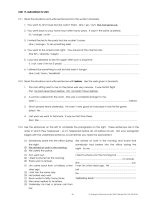
Protein Structure Representations
Myoglobin •stores O2 in muscle tissue via heme
•~70% α-helix
•a globular protein (~spherical shape)
Helix = fuchsia
Sheet = yellow
Coil = white
16
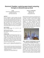
Protein Structure Representations
Retinol Binding Protein •Important for vision
Helix = fuchsia
Sheet = yellow
Coil = white
17
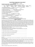
Protein Structure Representations
Lactate Dehydrogenase
•Released in bloodstream by damaged muscles
•Indicative of heart damage or failure
•Quaternary structure = four identical units
•Subject of Chem 153L experiments
Helix = fuchsia
Sheet = yellow
Coil = white
18