Early stages of host invasion by pseudomonas aeruginosa and effect of cyclic diguanylate signaling
Bạn đang xem bản rút gọn của tài liệu. Xem và tải ngay bản đầy đủ của tài liệu tại đây (7.97 MB, 189 trang )
EARLY STAGES OF HOST INVASION BY PSEUDOMONAS
AERUGINOSA AND EFFECT OF CYCLIC DIGUANYLATE
SIGNALING
AYSHWARYA RAVICHANDRAN
A THESIS SUBMITTED FOR THE DEGREE OF
DOCTOR OF PHILOSOPHY
DEPARTMENT OF BIOLOGICAL SCIENCES
NATIONAL UNIVERSITY OF SINGAPORE
2010
ACKNOWLEDGEMENTS
I express my heartfelt gratitude to my supervisor, A/P Sanjay Swarup for his constant
guidance and supervision throughout the period of this project.
I sincerely thank National University of Singapore for providing me with Research
Scholarship to complete this project. I would also like to thank Research Centre for
Excellence in Mechanobiology for funding part of this study and support.
I am extremely thankful to Dr. Yasushi Ishihama, Keio University, Japan for
performing phoshoproteome analysis on our samples without which my publication
would not have been possible. Helium-ion imaging was conducted under Dr. Daniel
Pickard and I am thankful for his guidance and facility. I extend my sincere gratitude to
Dr. Gerard Michel, Centre National de la Recherche Scientifique, France for his kind
gesture of sending antibodies and guidance in P. aeruginosa type II secretion systemrelated experiments. I would also like to thank Dr. Zhang Lian-Hui for providing
workspace in his laboratory during the initial stages of this project and Dr. Ganesh
Anand for his valuable scientific discussions time-to-time. I express my thanks to
Malarmathy Ramachandran and Karen Lam who have been very instrumental in
helping me with optimization of experimental methods used in this study.
I express my gratitude to Protein and Proteomics centre for their mass spectrometry
services, Electron microscopy and the confocal microscopy facilities at the Faculty of
Medicine, and the Electron microscopy facility at Department of Biological Sciences. In
this regard, I thank Ms.Michelle Mok, Ms. Wang Xianhui and Mdm. Loy Gek Luan.
My thanks are due to our lab officers Ms. LiewChye Fong, Dennis Heng andJiun Fu. I
extend my gratitude to all my lab mates especially Chui Ching, Weiling and Tanujaa
for their cooperation, help and constant support. I would also like to thank all theother
undergraduates and attachment students who have in one way or other helped this project.
I am lucky to have great friends at NUS especially Sheela, Gauri, Sravanthy, Karthik,
and Prasanna for their criticism, discussions and moral support.
I have been blessed with wonderful family that lives across the globe, a constant source
of encouragement and love; especially my parents Dr. Ravichandran and Dr.
Rajarajeswari, who inspired me to take up research. A special mention goes to Mrs.
Chandrika and Mr. Nagarajan, my guardians in Singapore. Last but not least, my
husband Mr. Vigneshwaran and parents-in-law have always been greatly supportive of
my career endeavors. I have no words to thank these people, without whom I could not
have endured this tough journey.
CONTENTS
ACKNOWLEDGEMENTS
i
SUMMARY
vii
ABBREVIATIONS
ix
LIST OF TABLES
xii
LIST OF FIGURES
xiii
PUBLICATIONS
xv
CHAPTER 1
INTRODUCTION
1.1
General Introduction
1
1.2
Objectives
3
CHAPTER 2
REVIEW OF LITERATURE
2.1
Bacterial invasion and infection mechanisms
5
2.2
Pseudomonas aeruginosa- an opportunistic pathogen
8
2.2.1 Chronic vs acute infection
9
2.3
Multifactorial nature of P. aeruginosa virulence mechanisms
10
2.4
Host surface-attachment, a key step in P. aeruginosa
invasion
Role of bacterial appendages in surface attachment
12
15
2.5.1. Flagellum- a primary adhesin
15
2.5.2. Type IV pili-mediated attachment
17
P. aeruginosa internalization by non-phagocytic cells
19
2.6.1.Host signaling pathways necessary for P. aeruginosa
invasion
20
Role of secretion systems in bacterial invasion
23
2.5
2.6
2.7
ii
2.7.1. Type II secretion system (T2SS) in P. aeruginosa
25
2.8
Co-ordinated regulation of P. aeruginosa virulence
mechanisms
29
2.9
C-di-GMP signaling
32
2.9.1.Role of c-di-GMP signaling in virulence regulation
34
2.9.2. MorA signaling
36
2.10
Bacterial Ser/Thr/Tyr phosphorylation system
38
CHAPTER 3
MATERIALS AND METHODS
3.1
Bacterial strains, plasmids and growth conditions
41
3.2
Gene expression studies
42
3.3
Cloning and genetic manipulation studies
43
3.4
Expression of recombinant proteins
45
3.5
Secretome analysis
46
3.5.1. Elastolytic activity assay
48
3.6
Intracellular protein extraction
49
3.7
Membrane protein preparation
49
3.8
Immunoblotting
50
3.9
Bacterial infection studies
51
3.9.1. Cell culture conditions
51
3.9.2. Infection assays
52
3.10
Extracellular matrix extraction
54
3.11
Sample preparation for Helium-ion microscopy
55
3.12
Phosphoproteome analysis
56
iii
3.12.1. 2-Dimensional Electrophoresis (2-DE) of P. putida
protein samples
3.12.2. Staining for phosphoproteins
CHAPTER 4
62
3.12.4. Analysis of LC-MS-MS data
64
CYCLIC DIGUANYLATE SIGNALING AFFECTS P.
AERUGINOSA ATTACHMENT AND ENTRY INTO LUNG
FIBROBLASTS
BACKGROUND
4.2
RESULTS AND DISCUSSION
CHAPTER 5
61
3.12.3.Sample preparation for phosphoproteome analysis by
Nano-LC-MS-MS
4.1
4.3
56
66
4.2.1. MorA affects bacterial attachment to host in P.
aeruginosa
67
4.2.2. Which appendage plays a major role in attachment
changes due to MorA- flagellum or pili?
72
4.2.3. Investigation of entry mechanism
74
CONCLUSIONS AND FUTURE DIRECTIONS
75
SECRETION OF EXTRACELLULAR PROTEASES IS
AFFECTED BY CYCLIC DIGUANYLATE SENSOR
REGULATOR MorAINP. AERUGINOSA
5.1
BACKGROUND
5.2
RESULTS AND DISCUSSION
79
5.2.1. C-di-GMP signaling affects T2SS secretome in P.
aeruginosa
81
5.2.2. Biological effects of increased extracellular protease
levels
87
iv
5.3
5.2.3. MorA affects invasion efficiency of P. aeruginosa
89
5.2.4. Mechanism of c-di-GMP regulation of P. aeruginosa
protease secretion
91
i) RNA levels of protease genes
91
ii) Protein levels of protease genes
93
iii) Levels of T2SS secreton assembly proteins
95
5.2.5. Does MorA affect invasion by degrading the
extracellular matrix?
96
CONCLUSIONS AND FUTURE DIRECTIONS
99
CHAPTER 6
Ser/Thr/Tyr PHOSPHOPROTEOMES OF P. PUTIDA AND
P. AERUGINOSA AND THEIR CROSSTALK WITH
CYCLIC DIGUANYLATE SIGNALING
6.1
BACKGROUND
6.2
RESULTS AND DISCUSSION
6.3
104
6.2.1. Gel-based approach for identification of
phosphoproteins
107
6.2.2.Phosphoproteome analysis of P. putida and P.
aeruginosa by Nano-LC-MS/MS method
110
6.2.3. Crosstalk of MorA-c-di-GMP signaling and protein
phosphorylation
124
CONCLUSIONS AND FUTURE DIRECTIONS
127
CHAPTER 7
REFERENCES
CONCLUDING REMARKS
131
132
v
APPENDICES
Supplementary information on gene cloning done in this
152
I
study
II
Methods for Ser/Thr/Tyr Phosphoproteome analysis
157
III
Supplementary information on host cell morphology
161
IV
Gene regulatory network of promoters affected by MorA
162
V
MALDI-ToF-ToF spectra of secreted proteins affected by
MorA
163
VI
MorA affects timing of flagellar biogenesis in P. aeruginosa
171
VII
Crosstalk of MorA and acetyl phosphate (AcP) signaling
172
vi
SUMMARY
Bacterial invasion plays a critical role in the establishment of P. aeruginosa infection,
which involves surface attachment of bacteria on the host cells followed by
internalization/ tissue penetration. Major virulence factors aiding bacterial invasion are
surface appendages and secreted proteases. The second messenger cyclic diguanylate (cdi-GMP) is well known to affect attachment of bacteria to surfaces, biofilm formation
and related virulence phenomena. MorA, a global regulator containing a GGDEF-EAL
domain has been previously shown to affect biofilm formation and timing of flagellar
biogenesis in P. aeruginosa PAO1 strain, and fimbriae expression in other clinical
strains. These domains are implicated in the turnover of c-di-GMP.
This study provides evidence that the global regulator MorA affects P. aeruginosa
attachment to host surface and levels of proteases secreted by the type II secretion system
(T2SS) hence regulating the invasion capacity of the pathogen. This is the first report on
control of c-di-GMP signaling on this secretion system. It was postulated that there may
be a common post-transcriptional signal acting between the regulatorMorA and the
effectors i.e. T2SS and pili/flagella since all the three are located at the bacterial poles.
Results confirm that the effect of MorA signaling on T2SS is post-transcriptional. Data
from this study suggest that the effect of MorA on host-surface attachment may be
mediated by pili, a key surface appendage.
Owing to growing importance of Ser/ Thr/ Tyr protein phosphorylation in bacteria, it was
hypothesized to be the common phenomenon bridging the altered c-di-GMP levels and
the observed effects on protease secretion and attachment to host surface. A
comprehensive phosphoproteome analysis was conducted on P. aeruginosa and P. putida
vii
that revealed several interesting leads suggesting many virulence and survival
mechanisms to be regulated by protein phosphorylation. This analysis uncovered a novel
crosstalk between two bacterial signaling paradigms namely- c-di-GMP second
messenger signaling and Ser/ Thr/ Tyr protein phosphorylation. Since not many
Ser/Thr/Tyr kinases have been characterized in bacteria, a direct correlation of c-di-GMP
levels and alteration in protein phosphorylation patterns need further investigation.
viii
ABBREVIATIONS
2-DE
2-Dimensional electrophoresis
ABC
ATP-binding cassette
AckA
acetate kinase
AcP
acetyl phosphate
Acyl-HSL
acyl homoserinelactone
aGM
asialoganglioside gangliotetrasylceramide
Amp
ampicillin
AP
alkaline phospahatase
BSA
bovine serum albumin
cAMP
cyclic adenosine monophosphate
c-di-GMP
cyclic di-guanylate monophosphate
CF
cystic fibrosis
CFTR
cystic fibrosis transmembrane conductance regulator
CHAPS
3-[(3-Cholamidopropyl)dimethylammonio]-1-propanesulfonate
CMR
Comprehensive Microbial Resource
Da
dalton
DGC
diguanylate cyclase
DMSO
dimethyl sulfoxide
DTT
dithiotreitol
ECL
enhanced chemiluminescence
ECM
extra-cellular matrix
ECP
extra-cellular protein
EDTA
ethylene-diamine-tetra-acetate
ETA
exotoxin A
GFP
green fluorescent protein
Gm
gentamycin
ix
GTP
Guanosine-5'-triphosphate
HAMMOC
hydroxy acid-modified metal oxide chromatography
HIM
Helium ion microscopy
HK
histidine kinase
HPA
β-hydroxypropanoic acid
IEF
isoelectric focusing
IPTG
isopropyl β-D-1-thiogalactopyranoside
KO
knockout
LA
lactic acid
LB
Luria-Bertani
LC-MS-MS
liquid chromatography followed by tandem mass spectrometry
LPS
lipopolysaccharide
m/z
mass/charge
MALDI
matrix-assisted laser desorption/ ionization
MOI
multiplicity of infection
OD
optical density
OE
overexpression
ORF
open reading frame
P. aeruginosa Pseudomonas aeruginosa
P. putida
Pseudomonas putida
PAGE
polyacrylamide gel electrophoresis
PBS
phosphate buffered saline
PDE
phosphodiesterase
PGPR
plant growth-promoting rhizobacterial
pI
isoelectric point
PMNs
polymorphonuclear leucocytes
PMT
photomultiplier tube
x
ppm
parts per million
Pta
phosphate acetyl transferase
PTM
post-transcriptional modification
qRT-PCR
quantitative Real-time Polymerase Chain Reaction
QS
quorum sensing
RR
response regulator
RT-PCR
reverse transcriptase polymerase chain reaction
S or Ser
serine
SE
standard error
SEM
scanning electron microscope
T or Thr
threonine
T2SS
type II secretion system
T3SS/TTSS
type III secretion system
T4P
type IV pili
T6SS
type VI secretion system
TCA
trichloroacetic acid
TEM
transmission electron microscopy
Tet
tetracycline
Ti
titania
ToF
time of flight
v/v
volume by volume
w/v
weight by volume
WT
wild type
Y or Tyr
tyrosine
Zr
zirconia
xi
LIST OF TABLES
Table 3.1
Bacterial strains and plasmids used in this study
39
Table 3.2
List of primers used for gene expression studies and cloning
experiments
42
Table 3.3
Immunoblot conditions for antibodies used in this study
48
Table 3.4
Optimization of parameters for 2-dimentional gel
electrophoresis of P. putida proteins
56
Table 5.1
MALDI-ToF-ToF identification of P. aeruginosa secreted
proteins affected by MorA
80
Table 6.1
List of identified phosphopeptides from P. putida PNL-MK25
107
Table 6.2
List of identified phosphopeptides from P. aeruginosa PAO1
111
Table 6.3
Specific roles of identified phosphoproteins
116
Table 6.4
Effect of MorA-c-di-GMP signaling on protein
phosphorylation
121
Table 6.5
Phosphopeptides of interest for validation functional
significance of phosphorylation
124
xii
LIST OF FIGURES
Figure 2.1
Bacterial infection strategies
7
Figure 2.2
P. aeruginosa virulence factors affecting different stages of
infection
11
Figure 2.3
Bacterial secretion systems
24
Figure 2.4
Type II secretion system in P. aeruginosa
26
Figure 2.5
Phenotypes regulated by c-di-GMP and binding sites/domains
33
Figure 2.6
Regulation of flagellum-based motility by c-di-GMP
signaling
36
Figure 2.7
Domain structure of MorA in P. putida and P. aeruginosa
Figure 2.8
Verification of transcriptional level effect of MorA on T3SS
genes
Figure 3.1
Strategy for insertion of peptide tag to LasB
43
Figure 3.2
Optimization of P. aeruginosa secreted protein extraction
45
Figure 3.3
Layout of bacterial infection assays
50
Figure 3.4
Optimization of antibiotic concentration and incubation time
for efficient clearance of external host-attached bacteria in
invasion assay
51
Figure 3.5
Workflow of phosphoproteome analysis
55
Figure 3.6
Optimization of 2-dimentional gel electrophoresis
58
Figure 3.7
Optimization of visualization of phosphoproteins
60
Figure 3.8
Workflow of sample preparation for phosphoproteome
analysis
61
Figure 4.1
P. aeruginosa attachment to host cells is affected by MorA
66
Figure 4.2
Host morphological changes correspond to effect of MorA on
bacterial attachment
67
xiii
Figure 4.3
P. aeruginosa cells actively divide during infection
69
Figure 4.4
Polar and lateral appendages mediate P. aeruginosa host
attachment
70
Figure 4.5
Entry mechanisms of P. aeruginosa WT
72
Figure 5.1
Type III effector secretion levels are not affected by MorA
77
Figure 5.2
Levels of secreted proteases are affected by MorA in P.
aeruginosa
79
Figure 5.3
Elastase activity in extracellular fraction of P. aeruginosa
PAO1 WT and morA KO strains
85
Figure 5.4
Invasion efficiency corresponds to altered elastolytic activity
87
Figure 5.5
RNA levels of major secreted proteases show no change due
to MorA
89
Figure 5.6
Elastolytic activity assay for LasB-FLAG construct
91
Figure 5.7
MorA does not affect intracellular levels of LasB
Figure 5.8
Levels of T2SS machinery proteins are unaltered by MorA
93
Figure 5.9
Optimization of extracellular matrix analysis
95
Figure 5.10
Mechanism of regulation of proteases secreted via T2SS
97
Figure 6.1
Effect of MorA on protein phosphorylation in P. putida
104
Figure 6.2
Total protein profiles of P. putida WT and morA KO are
consistent
105
Figure 6.3
Cellular localization and biological function of identified
phosphoproteins
115
Figure 6.4
Phosphorylation sites on T6SS-related proteins
119
Figure 6.5
Effect of c-di-GMP on protein phosphorylation in P. putida
and P. aeruginosa
120
xiv
LIST OF PUBLICATIONS/CONFERENCES FROM THIS STUDY
Publications
Ravichandran, A., Sugiyama, N., Tomita, M., Ishihama, Y., Swarup, S. Ser/Thr/Tyr
Phosphoproteome Analysis of Pathogenic and Non-Pathogenic Pseudomonas
Species.Proteomics 2009, 9, 1-12.
Ravichandran, A., Suriyanarayanan, T., Swarup, S. Global regulator MorA controls
bacterial invasion via protease secretion in Pseudomonas aeruginosa. Manuscript in
preparation.
Conferences
AyshwaryaRavichandran (Invited speaker), Understanding the cell surface-associated
events during bacterial infection processes. In Program, First Asian Helium Ion
Microscopy Workshop, National University of Singapore, September 10, 2009.
Ishihama, Y., Sugiyama, N., Ohnuma, S.,Tomita, M., Ravichandran, A., Swarup, S.
Phosphoproteome Analysis of Pathogenic and Non-Pathogenic Pseudomonas Species. In
Program and Abstracts, 57th ASMS Conference on Mass Spectrometry, Philadelphia, USA,
May 31-June 4, 2009.
Ravichandran, A., Sugiyama, N., Tomita, M., Ishihama, Y., Swarup, S. Phosphoproteome
Analysis of Pathogenic and Non-Pathogenic Pseudomonas Species.In Program (abstract
accepted), HUPO 8th Annual World Congress, Toronto, Canada, September 26-30, 2009,
Pg 68.
Ravichandran, A., Ramachandran, M., Pickard, D.S., Swarup, S. Mechanics of initial
stages of P. aeruginosa infection process. In Program and Abstracts, The
3rdMechanobiology Workshop, National University of Singapore, November 3-5, 2009,
Pg 72.
Ravichandran, A., Sugiyama, N., Tomita, M., Ishihama, Y., Swarup, S. Ser/Thr/Tyr
Phosphoproteome Analysis of Pathogenic and Non-Pathogenic Pseudomonas Species.In
Program and Abstracts, Joint 5th Structural Biology and Functional Genomics and 1st
Biological Physics International Conference, National University of Singapore, December
9-11, 2008, Pg 152.
Ravichandran, A., Lam Mok Sing, K.M., Ramachandran, M., Lim, C.T., Low, B.C., Jin,
S., Swarup, S. Mechanics of initial attachment of P. aeruginosa PAO1 to human host cells.
In Program and Abstracts, 2ndMechanobiology Workshop, National University of
Singapore, November 3-5, 2008.
Ravichandran, A., Sugiyama, N., Tomita, M., Ishihama, Y., Swarup, S. Ser/Thr/Tyr
Phosphoproteome Analysis of Pathogenic and Non-Pathogenic Pseudomonas Species.In
xv
Program and Abstracts,13thBiological Sciences Graduate Congress, National University
of Singapore, December 15-17, 2008, Pg 36.
Ravichandran, A., Heng, M.W., Swarup, S. Regulatory effect of c-di-GMP signalling on
metabolic and other pathways in Pseudomonas aeruginosa. Presented at the BactPath9
program, Monash University, Melbourne, Australia, September, 2007.
Ravichandran, A., Heng, M.W., Sugiyama, N., Ishihama, Y., Swarup, S. Effects of
cyclic-di-GMP signaling on protein phosphorylation and secretion in Pseudomonas sp. In
Program and Abstracts, 12th Biological Sciences Graduate Congress, University of
Malaya, Kuala Lumpur, Malaysia, December 17-19, 2007.
Ravichandran, A., Heng, M.W., Choy, W.K., Swarup, S. MorA, the regulator of biofilm
formation in P. aeruginosa also affects the levels of virulence-associated extra-cellular
proteases. In Program and Abstracts, Joint Third AOHUPO and Fourth Structural Biology
and Functional Genomics Conference, National University of Singapore, December 4-7,
2006, Pg 231.
Ravichandran, A., Heng, M.W., Choy, W.K., Swarup, S. MorA, the regulator of biofilm
formation in P. aeruginosa also affects the levels of virulence-associated extra-cellular
proteases. In Program and Abstracts, 11th Biological Sciences Graduate Congress,
Chulalongkorn University, Bangkok, Thailand, December 14-17, 2006, Pg 101.
xvi
CHAPTER 1
INTRODUCTION
1.1 General Introduction
P. aeruginosa is a well established opportunistic and nosocomial pathogen with an ability
to adapt to eclectic environments and grow utilizing a wide range of substrates. It
possesses an array of virulence determinants which aid in colonization on biotic and
abiotic surfaces as well as in dissemination. Extensive studies have been performed on P.
aeruginosa virulence mechanisms and their regulation (Ramos, 2004). With numerous
crosstalks and complex overlaps discovered in different strains under various conditions,
this area of study is thriving as novel regulation mechanisms are being unraveled
constantly. The reason is that the pathogen is highly adaptable to changes in its
environment and devises new methods of survival/infection by manipulating its
multifactorial virulence mechanisms (Ramos & Filloux, 2007). Hence, there are unclear
or unknown regulatory mechanisms to be explored.
Initial stages of P. aeruginosa infection include attachment to host surface followed by
internalization into host cells eventually leading to invasion of tissue. Known key
adhesins include the surface appendages- flagellum and pili. Their interaction with host
causes changes at the host-pathogen interface leading to internalization of P. aeruginosa.
Both pili and flagella are nanomachines known to be regulated by complex mechanisms
at various levels such as transcriptional, post-transcriptional, assembly and function
(Jarrell 2009). To penetrate tissues, P. aeruginosa secretes various proteins that cleave
the host connective tissue and/or gains access to more host cells. Though P. aeruginosa
possess almost all of the many Gram-negative secretion machineries discovered to date,
1
significance of type II and type III secretion systems have been well-established and
widely studied (Wooldridge 2009). The type II secretion system of P. aeruginosa secretes
many proteases and lipases which cleave the extracellular matrix components such as
fibronectin, elastin and collagens while type III secretion system (T3SS) injects proteins
directly into host cytoplasm leading to cytoplasmic rearrangements and morphological
changes aiding in invasion of host tissue. These virulence factors are known to be
affected by quorum sensing mechanism involving small molecule trafficking and/or
levels of nucleotide second messengers namely cyclic-AMP and cyclic diguanylate
monophosphate (c-di-GMP) in P. aeruginosa and other Gram-negative pathogens.
In recent years, the significance of c-di-GMP second messenger signaling is becoming
apparent in the regulation of a multitude of cellular process and virulence mechanisms in
all classes of bacteria (Tamayo et al., 2007). In particular, c-di-GMP levels have been
reported to impart significant effects flagellar motility and attachment to surfaces (Wolfe
& Visick, 2008). Though such common phenotypes are known to be affected by this
molecule, its mechanism of action and the level of regulation have been known to be
distinctive across the different bacterial species studied. Since several proteins involved
in the turnover of c-di-GMP in each species have been found, it is believed that each
protein may respond to unique environmental cues and alter c-di-GMP levels in a
temporal/ spatial manner to bring about phenotypic changes. Hence, each case showing a
phenotypic difference poses a challenge in understanding the underlying mechanism.
Our laboratory studies one such protein MorA, a membrane-localized global sensor
regulator with domains involved in c-di-GMP turnover (Choy et al., 2004). We have
previously reported that MorA affects timing of flagellar biogenesis, flagellar number and
2
surface attachment in P. putida, and biofilm formation in P. aeruginosa. We have further
evidence that MorA also affects timing of flagellar biogenesis and swimming speeds in P.
aeruginosa. More details can be found in secion 2.9.2. Others have also shown that MorA
regulates colony morphology and twitching motility via another appendage, namely
fimbriae in a P. aeruginosa strain isolated from cystic fibrosis lung (Meissner et al.,
2007). This species being pathogenic, effect on its surface appendage may have an
impact on its ability to attach and infect host cells. Though MorA has not been shown to
affect T3SS, similar proteins P. aeruginosa and other species are well-known to regulate
this secretion system (Kulasekara et al., 2006).
1.2. Objectives
The overall aim of this study was to investigate the role of MorA-c-di-GMP signaling in
P. aeruginosa virulence mechanisms. As previous studies have proven that function of
bacterial surface appendages and protein secretion are critical in early stages of bacterial
invasion, this study aimed to focus on the effect of MorA signaling on these factors.
The specific aims of this study were
i) To understand the role of MorA in P. aeruginosa attachment to host surface via
surface appendages and subsequent entry into host cell
ii) To study the effect of MorA-c-di-GMP signaling on P. aeruginosa secretion that aid
in invasion of host
iii) To investigate the mechanism(s) by which MorA may control host invasion of P.
aeruginosa
3
In this thesis, the second chapter (Chapter 2) provides review of bacterial invasion
mechanisms, relevant virulence properties of P. aeruginosa and their regulation, known
c-di-GMP signaling mechanisms and Ser/Thr/Tyr phosphorylation in bacteria. Chapter 3
gives the details of all the materials and methods that were used during the entire study.
Chapter 4 discusses effect of MorA on P. aeruginosa-host attachment, bacterial surface
structures aiding interaction, and changes at the host-pathogen interface leading to
internalization. In the next chapter (Chapter 5), the focus is on secreted proteases that are
shown to be affected by MorA signaling and their biological significance. Experiments to
investigate the mechanism of MorA regulation on protease secretion are also illustrated.
Lastly, Chapter 6 provides evidence that protein phosphorylation could be a likely
mechanism for large-scale post-transcriptional affects of c-di-GMP signaling. A
comparative interspecies analysis of Pseudomonas phosphoproteomes indicates that the
key survival and virulence pathways of Pseudomonas sp. may involve Ser/Thr/Tyr
phosphorylation.
4
CHAPTER 2
REVIEW OF LITERATURE
2.1. Bacterial invasion and infection mechanisms
Invasive bacteria actively induce their own uptake by phagocytosis in normally
nonphagocytic cells and then either establish a protected niche within which they survive
and replicate in the cytosol or vacuole, or disseminate from cell to cell by means of an
actin-based motility process. Through their interactions, pathogens can modify
epithelium function to enhance their penetration across the epithelial barrier and to
exploit mucosal host defenses for their own benefit. Apoptosis and antiapoptosis, as well
as cell cycle– and inflammation-related signaling pathways, are reprogrammed after
infection to help the cell to survive the stress induced by the infection. The success of an
infection depends on the messages that the two players -the bacterium and the host cellsend to each other. The mechanisms underlying bacterial attachment, entry, phagosome
maturation, and dissemination reveal common strategies as well as unique tactics evolved
by individual species to establish infection.
To enter nonphagocytic cells such as intestinal epithelial cells, some microbial pathogens
express a surface protein which can bind eukaryotic surface receptors often involved in
cell-matrix or cell-cell adherence. These interactions trigger a cascade of signals,
including protein phosphorylations and/or recruitment of adaptors and effectors, and
activation of cytoskeleton components. These events lead to the formation of a vacuole
that engulfs the bacterium through a “zippering” process in which relatively modest
cytoskeletal rearrangements and membrane extensions occur (Cossart & Sansonetti
5
2004). The Yersinia outer-membrane protein invasin strongly binds to integrin receptors
that are normally implicated in adherence of cells to the extracellular matrix (Isberg &
Barnes 2001). Similarly in L. monocytogenes, internalins A and B contribute to bacterial
entry, and both processes are dependent on the presence of raft microdomains, suggesting
that for entry, Listeria take advantage of raft microdomains, which are known to be
enriched in receptors and signaling molecules.
Pathogens can also bypass the first step of adhesion and interact directly with the cellular
machinery that regulates the actin cytoskeleton dynamics by injecting effectors through a
dedicated secretory system. The effector molecules cause massive cytoskeletal changes
that trigger the formation of a macropinocytic pocket, loosely bound to the bacterial
body. Both Shigella and Salmonella use this mechanism to enter the cell. Contact
between bacteria and a cell is mediated by the type III secretory system (TTSS). The
protein components of the translocon are associated with membrane rafts enriched in
signaling molecules. Following this, a macropinocytic pocket is formed involving
localized but massive rearrangements of the cell surface, characterized by the formation
of intricate filopodial and lamellipodial structures.
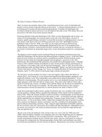
Figures 2.1A and 2.1B show the overall internalization and dissemination process of
Salmonella typhimurium and Shigella flexneri. Once in close contact with the epithelium,
Salmonellae induce degeneration of the enterocyte's microvilli, followed by profound
membrane "ruffling" localized to the area of bacteria–host cell attachment. This is
accompanied by extensive endocytosis and internalization of the bacteria into host cells
as described above. The bacterial adhesins leading to bacterial internalization are not only
6
A Salmonella
B Shigella
Figure 2.1. Bacterial infection strategies. A. Salmonella enterica typhimurium crossing
the epithelial barrier by entering via either M cells or enterocytes. B. Shigella entry into
rectal and colonic mucosa via M cells. Both A and B show changes in membrane
structure (membrane ruffling) due to binding of bacterial protein translocon with
signaling molecules in lipid raft-rich areas of host membrane. Subsequent events include
M cell destruction and subepithelial invasion by bacteria of macrophages. A & B adapted
from (Sansonetti & Phalipon 1999).
limited to invasin or TTSS; other bacterial surface structures including appendages and
surface polysaccharides in other pathogens are also capable of inducing host cellular
changes to gain entry into host. These have been discussed in detailed in the context of P.
aeruginosa later in this chapter.
7
2.2. Pseudomonas aeruginosa- an opportunistic pathogen
Pseudomonas aeruginosa is a ubiquitous bacterial species in the environment commonly
inhabiting soil and water. It possesses a large genome encoding eclectic arrays of
metabolic, catabolic, and virulence-related proteins and regulatory systems that define its
infinite ability to adapt to a wide range of environments and hosts (Stover et al., 2000).
Healthy individuals are generally not susceptible to P. aeruginosa infection; nevertheless,
several underlying conditions such as extensive burns, eye trauma, mechanical
ventilation, human immunodeficiency virus infection and malignancy increase the risk of
an acute spell (Fleiszig & Evans 2003); (Sadikot et al., 2005). It can cause urinary tract
infections, respiratory system infections, dermatitis, corneal infections, soft tissue
infections, bacteremia, bone and joint infections, gastrointestinal infections and a variety
of systemic infections. The main reason for chronic P. aeruginosa infections in hospital
environment and in cystic fibrosis (CF) patients are attributed to its ability to establish
biofilms in lungs, on implanted medical device or damaged tissue. A very typical
microbiological diagnostic finding is the recovery of various P. aeruginosa phenotypes
from chronically infected respiratory tract specimens of CF patients. Apart from the beststudied mucoid P. aeruginosa phenotype (Govan and Deretic, 1996), it is known that
dwarf colonies can be isolated from the chronically infected CF lung (Zierdt & Schmidt
1964). These ‘small colony variants’ (SCV) show increased antibiotic resistance to a
broad range of antimicrobial agents and their recovery in CF patients could be correlated
with parameters revealing poor lung function and inhaled antibiotic therapy (Haussler et
al., 1999). Treatment becomes problematic by the significant intrinsic resistance of P.
aeruginosa and the emergence of multidrug- resistant strains (Zaborina et al., 2006);
8