Mathematical modeling of circular dorsal ruffles and lamellipodial dynamics in single and collective cell migration
Bạn đang xem bản rút gọn của tài liệu. Xem và tải ngay bản đầy đủ của tài liệu tại đây (17.86 MB, 206 trang )
MATHEMATICAL MODELING OF
CIRCULAR DORSAL RUFFLES AND
LAMELLIPODIAL DYNAMICS IN SINGLE
AND COLLECTIVE CELL MIGRATION
LAI TAN LEI
B.Eng.(Hons.),NUS
A THESIS SUBMITTED
FOR THE DEGREE OF DOCTOR OF PHILOSOPHY
NUS GRADUATE SCHOOL FOR INTEGRATIVE SCIENCES AND
ENGINEERING
NATIONAL UNIVERSITY OF SINGAPORE
2012
Acknowledgements
Many thanks to the Biophysics Team at the Institute of High Performance
Computing for their valuable insights and criticisms of this work, especially
my co-supervisor Dr Chiam Keng Hwee, who has been extremely patient
and whose guidance has been invaluable. I would also like to thank my
collaborators, Mr Zeng Yukai, Mr Leong Man Chun, Dr Vedula Sri Ram
Krishna, Asst Prof Koh Cheng Gee, Prof Philip R. LeDuc and Prof Benoit
Ladoux who provided the experimental expertise cited in this thesis, and my
main supervisor Prof Lim Chwee Teck for his support of my work.
Thank you to my beloved family for their continual support these years.
Last but not least, my husband who has been very encouraging through
these difficult times.
i
Contents
Acknowledgements i
Summary vii
List of Tables x
List of Figures xi
List of Abbreviations xiii
1 Introduction and Literature Review 1
1.1 The impact of cell migration: why study it? . . . . . . . . . . 1
1.2 Structural ingredients for cell motility . . . . . . . . . . . . . . 2
1.2.1 Actin, its polymer and associated proteins . . . . . . . 4
1.2.2 Myosin: powering motility . . . . . . . . . . . . . . . . 7
1.2.3 Integrins provide the foothold . . . . . . . . . . . . . . 7
1.3 Achieving single cell motility . . . . . . . . . . . . . . . . . . . 10
1.3.1 Beginning with protrusion: lamellipodium, filopodium,
circular dorsal ruffles and blebbing . . . . . . . . . . . 10
1.3.2 Stabilising protrusions with adhesions . . . . . . . . . . 12
ii
1.3.3 Deadhering the rear . . . . . . . . . . . . . . . . . . . . 14
1.3.4 Experimental models used for the study of single cell
migration - keratocytes and fibroblasts . . . . . . . . . 15
1.3.5 Theoretical models developed for single cell motility . . 17
1.4 Collective cell migration . . . . . . . . . . . . . . . . . . . . . 23
1.4.1 Migration in three dimensions (3D) . . . . . . . . . . . 23
1.4.2 Migration of sheets . . . . . . . . . . . . . . . . . . . . 25
1.5 Thesis overview . . . . . . . . . . . . . . . . . . . . . . . . . . 27
1.5.1 Part I: Investigating actin dynamics in circular dorsal
ruffles . . . . . . . . . . . . . . . . . . . . . . . . . . . 28
1.5.2 Part II: A mechano-chemical study of lamellipodial dy-
namics . . . . . . . . . . . . . . . . . . . . . . . . . . . 29
1.5.3 Part III: Collective migration on a contrained substrate 31
1.5.4 What have we learnt? . . . . . . . . . . . . . . . . . . 32
1.5.5 Publications . . . . . . . . . . . . . . . . . . . . . . . . 33
2 Part I: Investigating the effect of substrate stiffness on cir-
cular dorsal ruffles through mathematical modeling 35
2.1 Circular dorsal ruffles: overview and biological impact . . . . . 35
iii
2.1.1 Motivation and objectives . . . . . . . . . . . . . . . . 37
2.2 Experimental methods . . . . . . . . . . . . . . . . . . . . . . 37
2.2.1 Preparation and characterization of elastic substrates . 38
2.2.2 Cell culture . . . . . . . . . . . . . . . . . . . . . . . . 38
2.2.3 Fluorescent staining and visualization . . . . . . . . . . 39
2.2.4 Data analysis . . . . . . . . . . . . . . . . . . . . . . . 40
2.2.5 Results from experiments: CDR size is independent
of substrate stiffness but CDR lifetime increases with
substrate stiffness . . . . . . . . . . . . . . . . . . . . . 41
2.3 Development and results of mathematical model . . . . . . . . 42
2.3.1 Development of mathematical model . . . . . . . . . . 42
2.3.2 Rac-Rho antagonism tunes the level of actin available
for stress fibers and CDRs . . . . . . . . . . . . . . . . 58
2.3.3 Negative feedback by WGAP results in actin ring in-
stead of actin patch formation . . . . . . . . . . . . . . 61
2.3.4 Multiple CDRs spread and merge into a single CDR . . 62
2.3.5 CDR actin propagates as an excitable wave . . . . . . 64
2.4 Conclusion . . . . . . . . . . . . . . . . . . . . . . . . . . . . . 72
iv
3 Part II: Mechanochemical model of lamellipodial dynamics
during cell migration 75
3.1 The lamellipodium: experiments and models . . . . . . . . . . 75
3.1.1 Objective of model . . . . . . . . . . . . . . . . . . . . 78
3.2 Model to describe lamellipodial fluctuations . . . . . . . . . . 79
3.3 Results and discussion . . . . . . . . . . . . . . . . . . . . . . 89
3.3.1 Periodic protrusion-retraction cycles observed in sim-
ulations . . . . . . . . . . . . . . . . . . . . . . . . . . 89
3.3.2 Periodic protrusion-retraction requires sufficiently stiff
substrate . . . . . . . . . . . . . . . . . . . . . . . . . . 90
3.3.3 Periodic protrusion-retraction requires sufficient acti-
vation of integrins . . . . . . . . . . . . . . . . . . . . . 92
3.3.4 Excessive activation of focal adhesions, coupled with
stiff substrates, leads to continuous protrusion . . . . . 94
3.3.5 Phase diagram and relation to experimental observations 95
3.3.6 Period of protrusion-retraction cycle is only affected by
the time delay in signal propagation . . . . . . . . . . . 98
3.4 Conclusion . . . . . . . . . . . . . . . . . . . . . . . . . . . . . 98
4 Part III: Collective migration of epithelial cells in constrained
v
environment 100
4.1 Collective migration of 2D sheets: an introduction . . . . . . . 100
4.1.1 Objective of study . . . . . . . . . . . . . . . . . . . . 105
4.2 Methods and analysis . . . . . . . . . . . . . . . . . . . . . . . 106
4.2.1 Development of Cellular Potts Model . . . . . . . . . . 106
4.2.2 Analysis of results: calculating correlation . . . . . . . 115
4.3 Results and discussion . . . . . . . . . . . . . . . . . . . . . . 118
4.3.1 The migration of the cell sheet is stalled by low cell-
substrate adhesion coupled with the absence of cell po-
larization . . . . . . . . . . . . . . . . . . . . . . . . . 118
4.3.2 Migration velocity and correlated movement are con-
trolled by extent of polarization and geometrical con-
straints . . . . . . . . . . . . . . . . . . . . . . . . . . 124
4.4 Conclusion . . . . . . . . . . . . . . . . . . . . . . . . . . . . . 128
5 Conclusion 131
5.1 Future work: where can we go next? . . . . . . . . . . . . . . 139
References 143
vi
Summary
Cell motility is a phenomenon that has intrigued scientists for many years.
Increasingly, researchers realize the need for quantitative analysis of both
the mechanical as well as the biochemical aspects at multiple scales. The
objective of this thesis is therefore to use mathematical and computational
modeling to quantitatively study several specific processes in cell motility.
The reorganization of actin, being the building block of the cell cytoskeleton,
is crucial in driving cell movement. A good appreciation of the biochemical
nature of actin dynamics is essential in the understanding of cell migration.
This was achieved by studying the dynamics of circular dorsal ruffles (CDR),
an actin-based structure often seen in growth-factor stimulated migrating
cells. The presence of CDRs has been shown to be the precursor to lamel-
lipodia generation and cell motility. Experimentalists have found that the
appearance of CDRs is often accompanied by the disappearance of actin-rich
stress fibers. While the generation of CDRs can been attributed to the acti-
vation of the Rac, stress fibers have been shown to be stabilized by the pres-
ence of active Rho. I therefore represented the formation of CDRs, starting
from growth factor induced Rac activation interacting with pre-existing Rho
and the associated stress fibers, using a system of partial differential equa-
tions. The numerical simulation results showed that increasing the substrate
stiffness, which led to increased stress fiber formation prior to stimulation, in-
creased the lifetime of the CDR without altering the size of these structures.
A simplified model, which involved Rac and a Rac inactivator, showed that
vii
the dynamics of CDRs can be likened to wave propagation in an excitable
medium.
The study of CDRs showed that the actin cytoskeleton is highly dynamic,
with many proteins regulating its activity. Yet, cell migration cannot be
reenacted without considering the interaction of forces that drive motion.
An important part of a migrating cell is the lamellipodium, a thin protrusive
portion at the front of the migrating cell. I developed a model of lamellipo-
dial dynamics that incorporated actin polymerization and forces exerted on
the actin cytoskeleton. Through the use of a stretch-sensitive protein that
responded to substrate stiffness, the model showed that the lamellipodium
can exhibit periodic protrusion-retraction cycles, continuous protrusion and
unstable retraction, depending on the substrate stiffness and the relative
amounts of integrin and myosin activation. In particular, periodic behavior
similar to that seen in recent experiments can be achieved when the substrate
is sufficiently stiff.
Studying cell migration is incomplete without looking at how cells move when
interacting with one another, which is usually the case in vivo. Therefore, I
investigated the collective migration of cells on constrained substrates. Using
a lattice-based computational method known as the Cellular Potts Model, I
studied the collective migration of cells as a function of the substrate channel
width and found that the collective migration velocity decreased with increas-
ing channel width. Analysis of the velocity field showed that the component
of the cell velocities perpendicular to the channel’s long axis demonstrated
increasing correlation length with channel width whereas the parallel com-
viii
ponent was unaffected. The decrease in velocity as the adhesive substrate
channel width was increased was found to be a consequence of the ability
of the cell to polarize during motion. This study showed that the study of
collective cell migration can reveal long range migratory behaviour within
tissues which single cell migration would not elucidate.
While many aspects of cell migration still elude us, through these three
projects, I have shown that the actin cytoskeleton is a highly dynamic struc-
ture regulated by a plethora of proteins, such as the antagonistic Rac and
Rho. This, with the help of stretch-sensitive proteins, can enable the lamel-
lipodium of the cell to exhibit different behaviour depending on the substrate
stiffness. Finally, the collective migration of cells showed a dependence of mi-
gration velocity and velocity correlation distance on the size of the substrate.
ix
List of Tables
1 Summary of models in current literature . . . . . . . . . . . . 20
2 Reaction terms used in mathematical model describing circu-
lar dorsal ruffles . . . . . . . . . . . . . . . . . . . . . . . . . . 52
3 Parameters used in mathematical model describing circular
dorsal ruffles . . . . . . . . . . . . . . . . . . . . . . . . . . . . 56
4 Values of parameters used in simulation of lamellipodium . . . 85
5 Values of parameters used in Cellular Potts Model . . . . . . . 115
x
List of Figures
1 Structure of a eukaryotic cell . . . . . . . . . . . . . . . . . . . 3
2 Actin dynamics at the front . . . . . . . . . . . . . . . . . . . 8
3 Cell motility requires the right mix of proteins at the right places 13
4 Collective cell migration in vivo . . . . . . . . . . . . . . . . . 24
5 Cells exhibiting circular dorsal ruffles . . . . . . . . . . . . . . 28
6 NIH 3T3 fibroblasts stained for actin before and after PDGF
stimulation . . . . . . . . . . . . . . . . . . . . . . . . . . . . 43
7 NIH 3T3 fibroblasts stained for actin after PDGF stimulation
for two different substrates . . . . . . . . . . . . . . . . . . . . 44
8 Quantification of the size of CDRs observed in cells . . . . . . 45
9 Summary of events leading up to CDR formation from PDGF
stimulation . . . . . . . . . . . . . . . . . . . . . . . . . . . . 48
10 F-actin ratio for varying substrate stiffnesses . . . . . . . . . . 49
11 Simulations results for the effect of FAK concentration on CDRs 60
12 Simulation results for the effect of WGAP and multiple PDGF
receptor aggregates on CDRs . . . . . . . . . . . . . . . . . . 63
13 Phase diagram and time plots for Rac and WGAP . . . . . . . 67
xi
14 Schematic of the major components of the lamellipodium. . . 80
15 Different types of lamellipodial dynamics observed . . . . . . . 91
16 Phase diagram depicting the three types of lamellipodial dy-
namics . . . . . . . . . . . . . . . . . . . . . . . . . . . . . . . 93
17 Variation of total period in lamellipodium simulation . . . . . 97
18 Schematic of the Cellular Potts Model setup . . . . . . . . . . 108
19 Initial setup of simulations of cell sheet migration . . . . . . . 114
20 Experimental setup of MDCK cell sheet migration . . . . . . . 117
21 MDCK cell sheet migration when PDMS slab was removed . . 119
22 Time lapse of CPM simulation of migrating cell sheet . . . . . 120
23 CPM simulation of collective cell migration . . . . . . . . . . . 121
24 Cell migration stalls when the cell-substrate adhesion and ex-
tent of polarization is low. . . . . . . . . . . . . . . . . . . . . 122
25 v component of cell migration velocity decreases with increas-
ing adhesive substrate channel width. . . . . . . . . . . . . . . 125
26 Variation of correlation length of the cell velocity with adhe-
sive substrate channel width. . . . . . . . . . . . . . . . . . . . 127
27 Summary of thesis contribution to cell migration. . . . . . . . 138
xii
List of Abbreviations
Abbreviation Definition
2D Two dimensional
3D Three dimensional
3T3 fibroblasts 3-day transfer, inoculum 3 x 10
5
fibroblasts
Abl Abelson tyrosine-protein kinase
ADF Actin depolymerization factor
ADP Adenosine diphsophate
ATP Adenosine triphosphate
Cof Cofilin
CDR Circular dorsal ruffle
CPM Cellular Potts Model
D-actin Circular dorsal ruffle actin
DAPI 4’,6-diamidino-2-phenylindole
DNA Deoxyribonucleic acid
EGF Epidermal growth factor
Ena Drosophila Enabled
ER Endoplasmic reticulum
F-actin Filamentous actin
FAK Focal adhesion kinase
FilGAP Filamin A-associated RhoGAP
G-actin Monomeric actin
xiii
GAP GTPase activating protein
GEF Guanine nucleotide exchange factor
GTP Guanosine triphosphate
JNK c-Jun N-terminal kinase
LIMK LIM domain kinase
MCS Monte Carlo step
MDCK Madin Darby canine kidney
mDia Mammalian diaphanous (Diaphanous-related formin)
MLC Myosin light chain
MLCP mMosin light chain phosphatase
MLCP-P Phosphorylated (inactive) myosin light chain phos-
phatase
MTOC Microtubule organizing center
NIH National Institutes of Health
NPF Nucleation promoting factor
p130Cas CRK-associated substrate protein
PDGF Platelet-derived growth factor
PDMS Polydimethylsiloxane
PIP
2
Phosphatidylinositol (4,5)-bisphosphate
PIP
3
Phosphatidylinositol (3,4,5)-trisphosphate
PI-3 kinase Phosphoinositide 3-kinase
PIV Particle image velocimetry
PTEN Phosphatase and tensin homolog, a phosphatidylinositol
(3,4,5)-trisphosphate phosphatase
xiv
RNA Ribonucleic acid
ROCK Rho-associated protein kinase
RTK Receptor tyrosine kinase
Src Sarcoma
VASP Vasodilator-stimulated phosphoprotein
WASP WiskottAldrich syndrome protein
WAVE WASP-family verprolin-homologous protein
WGAP WAVE-binding RacGAP
WRP WAVE-associated RacGAP protein
xv
1 Introduction and Literature Review
1.1 The impact of cell migration: why study it?
The migration of cells has been a biological phenomenon that has intrigued
scientists, biologists and non-biologists alike, for centuries. With the inven-
tion of the microscope, cell migration was documented in the sixteen hun-
dreds by Leeuwenhoek where he observed microscopic organisms moving in
rainwater via ’little horns’ that extended and contracted [41]. The study of
organisms moving towards chemical targets in their environment quickly be-
came an exciting area of research in the late eighteen hundreds. The careful
study of bacteria response to light and oxygen by Engelmann [64] as well as
the characterization of phagocytosis by Mechnikov [245] were some of the im-
portant works marking the first forays into the complete understanding of cell
motility. Today, with the development of powerful microscopes, experimen-
talists are able to study cell motion in much greater detail. Cell migration
has been found to be important in numerous physiological events. For in-
stance, during embryonic development, cells move in response to chemical
cues to specific regions of the embryo, subsequently generating the appropri-
ate organs in the right locations which are essential for survival [138, 263].
Cells can also migrate towards growth factors which are released by platelets
at the site of trauma to facilitate wound-healing [248, 200]. In the immune
system, phagocytes have been seen to follow fast-moving bacteria through
the modification of their morphology, culminating in the engulfing of the
pathogen and therefore the elimination of the possible threat to the host
1
body [185]. In a less beneficial context, cancer cells are known to peel off
from the primary tumour sites and enter the blood stream, only to exit the
vascular system at other locations and give rise to secondary tumours, in a
process known as metastasis [132, 36]. Studies have shown that metastatic
cancers are often life-threatening, with a survival rates dipping to less than
20% in many cancers [28]. Given the numerous applications, it is clear that
an understanding of cell motility is crucial, not just for the development of
strategies to combat conditions arising from incomplete cell migration which
can lead to mental retardation and organ malfunction in infants [49, 111],
but also to provide possible treatments for cancer patients who, on the other
hand, face the problem of migratory cancer cells.
1.2 Structural ingredients for cell motility
Understanding cell motility begins with an appreciation of the components
of a cell. Briefly, the eukaryotic cell is mainly made up of a fluid known as
the cytoplasm, enclosed within a plasma membrane typically composed of
lipids. Genetic material which contains information for cell replication and
cellular function is found in the cell nucleus, another membrane enclosed
compartment in the cell, in deoxyribonucleic acid (DNA)-containing struc-
tures known as chromosomes. The cell transcripts this information into short
ribonucleic acid (RNA) sequences which are transported out of the nucleus
to be interpreted by other organelles in the cytoplasm. One such organelle
is the ribosome, a machine which reads RNA sequences and creates proteins
to be used by the cell. Another important component of the cell is the en-
2
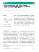
mitochondria
rough
endoplasmic
reticulum
smooth
endoplasmic
reticulum
Golgi
apparatus
nuclear
envelope
chromatin
free
ribosome
nucleus
plasma membrane
Figure 1: Main components of a typical eukaryotic cell. Illustration adapted
from />doplasmic reticulum (ER), a complex network of membrane continuous with
the nuclear envelope. The ER can be divided into two portions: the rough
ER which has ribosomes attached to its surface and therefore takes part in
the synthesis of proteins, and the smooth ER which does not have attached
ribosomes and instead, is involved in the synthesis of lipids as well as the
metabolism of carbohydrates. The packaging of proteins for transport is
typically carried out by the Golgi apparatus. To perform cellular functions,
energy is needed and this is provided by the mitochondria which generates
adenosine triphosphate (ATP), the source of energy in the cell.
3
1.2.1 Actin, its polymer and associated proteins
Apart from the organelles mentioned above, the cytoplasm of the cell contains
a vast array of other proteins and structures which maintain the everyday
activities of the cell. In cell migration, the skeleton of the cell, known as
the cytoskeleton, is arguably the structure in the center of activity. While
the cytoskeleton is a complex meshwork of actin filaments, microtubules and
intermediate filaments, the actin cytoskeleton has been identified as the main
player in cell migration. The actin cytoskeleton is generated from the actin
monomer, which is a 42 kDa globular protein (G-actin) that binds ATP and
is highly conserved in the eukaryotic kingdom [193]. The polymerization of
actin into filamentous structures (F-actin) form the actin cytoskeleton which
changes dynamically and therefore generates motility in cells. The process of
polymerization is preceded by nucleation which requires the formation of the
actin dimer. This first step, however, has been shown to be extremely un-
favourable energetically, with actin dimer dissociation equilibrium constants
as high as 5 M [223]. The cell overcomes this obstacle through the use of
actin-nucleating proteins, such as the Arp2/3 complex and its nucleation
promoting factors (NPF). The Arp2/3 complex is made up of seven sub-
units which activate upon binding to NPFs and the sides of existing actin
filaments at an angle of 70
◦
[85]. This forms a branching network of actin
filaments usually seen at the front of migrating cells [1, 16, 15]. On the other
hand, formins, a separate class of actin-nucleating proteins, do not require
pre-existing actin filaments for activation. Experiments suggest that formins
can stabilize the actin dimer during nucleation [199] by direct binding. This
4
leads to the formation of unbranched actin networks which are often seen
in stress fibers and filopodia [102, 119, 194]. Upon stable actin dimeriza-
tion, the actin filament is elongated by addition of actin monomers at the
fast-growing barbed end of the actin filament [165, 239] where the ATP is
located. Actin elongation is a tightly regulated process which requires coor-
dination among a vast array of actin binding proteins. For instance, capping
proteins prevent the elongation of actin filaments by blocking the addition
of new monomers at the barbed end [264]. Gelsolin, on the other hand, can
sever actin filaments, therefore regulating the length of actin filaments but
at the same time increasing the rate of actin dynamics [74, 236]. Actin elon-
gation can also be reduced by increasing the rate of depolymerisation of the
adenosine diphosphate (ADP) loaded end, also known as the pointed end,
of the actin filament which can be achieved by the actin-depolymerization
factor (ADF) and cofilin [264, 194]. Apart from proteins which hinder actin
filament elongation, other proteins promote actin network growth by stabi-
lizing the actin filament, for instance myosin [32], or increasing the pool
of ATP bound actin monomers, for instance profilin [61]. A third class
of actin-binding proteins keep the actin monomers in a sequestered form,
such as beta thymosins [264, 61, 214]. This facilitates rapid changes in the
actin cytoskeleton without the need for protein transcription, which is typi-
cally a much slower process. Aside from experiments, the dynamic nature of
the actin cytoskeleton has been intensively investigated using mathematical
models. Edelstein-Keshet and Ermentrout looked at the effect of polymer-
ization/depolymerization rates as well as filament fragmentation rate on the
length distribution of F-actin [67], with an extension into biological context
5
in an accompanying study [74]. They found that the combination of different
effects could lead to intermediate peaks in the length distribution which were
not observed when the factors were studied individually. In another paper,
Civelekoglu and Edelstein-Keshet [46] studied the dynamics of the actin cy-
toskeleton in a constrained space (for instance the cell) and found that in
order to see the results observed experimentally, the branching and filament
orientation cannot be random, which was also shown in Atilgan’s model [13].
Mogilner and colleagues, on the other hand, studied the effect that the actin
filament on the cell membrane and proposed the elastic Brownian ratchet
model for the interaction of the filament with the membrane [163, 166, 167]
in which an explicit relation between the velocity of the membrane and the
force exerted by the actin filament was derived. This model has been sub-
sequently used to by other researchers to represent the interaction between
the cell membrane and the barbed ends of the actin network [267, 273, 72].
In another approach, Gov and colleagues study the fluctuations of the cell
membrane which contains proteins that promote actin polymerization and
diffuse along the membrane in a curvature-dependent manner [226, 103, 104],
and are able to predict wavelike motion of the membrane which have been
seen in experiments. In a continuum approach, Prost’s group modelled the
actin network in the lamellipodium as an incompressible gel and were able
to generate the retrograde flow of actin observed by experimentalists, as well
as predict the force distribution from the leading edge to the rear of the
lamellipodium [137]. Other works, however, represented the actin network
as an interconnected system of cylinders to represent actin filaments and
crosslinking proteins [153, 130]. Kim et al. [130] found that using such a
6
structure, the actin network behaved as a viscoelastic material much like
what has been observed by experimentalists [133, 275]. They also found that
the actin cross-linking proteins were responsible for the elastic nature of the
actin cytoskeleton, which can be made even more elastic by prestressing the
filaments and therefore, pre-orientating the filaments along the direction of
stress.
1.2.2 Myosin: powering motility
While the actin cytoskeleton forms the foundation upon which motility can
be achieved, migration is very much a mechanical process that requires force
generation. This can be achieved by motor proteins and of particular interest
is the ubiquitous non-muscle myosin II. The non-muscle myosin II molecule
consists of two heads which bind to actin and enable movement by ”walking”
on the actin cytoskeleton through ATP hydrolysis [256]. The non-muscle
myosin II is especially prevalent along bundled actin filaments, which run
across the cell, known as stress fibers. Studies have shown that non-muscle
myosin II is responsible for the contractility of the rear end of a migrating
cell [256] and more recent work suggest that myosin generated forces can
influence the rate of protrusion of the leading edge of the cell [98, 99].
1.2.3 Integrins provide the foothold
In the same manner that friction provides the anchor upon which humans
can pivot their bodies to propel themselves forward, the cell requires pro-
7
Figure 2: Actin polymerization begins with nucleation, aided by Arp2/3
(green discs) or formins (dark blue discs). Polymerization occurs by addition
of ATP loaded actin monomers (white circles) to the barbed ends of the actin
filaments. As the actin filament ages, the ATP is hydrolyzed to form ADP-
actin (red circles). Capping proteins (light blue circles) prevent the addition
of actin monomers to the barbed ends while ADF/cofilin (yellow triangles)
increase the rate of depolymerization at the pointed ends. The binding of
profilin (black circles) to ADP-actin monomers catalyzes the exchange of
ADP for ATP. Figure adapted from Ref. [194].
8
teins which bind them to its extracellular environment such that myosin
generated forces can lead to an overall shift of the cell centroid. An exam-
ple is the integrin dimer which is a transmembrane protein that binds to
the actin network, usually indirectly via a complex aggregation of other pro-
teins, and the extracellular matrix [117]. Studies have shown that integrins
assemble into focal contacts which mature into focal adhesions under suitable
conditions [42, 44], such as the presence of activated RhoA. A more recent
study by Alexandrova et al. [3] showed that focal adhesions are first initiated
in the lamellipodium, which agrees with the results presented by Sheetz’s
group [98, 99], and cause a reduction in the retrograde flow of actin. When
the flow of actin was inhibited, the adhesions did not mature but instead
dissociated, suggesting that the adhesion strengthening requires mechanical
feedback from the connected actin cytoskeleton. This is further investigated
in Wolfenson’s study [265], which showed that the kinetic constants of pro-
teins associated with focal adhesions were altered when actomyosin contrac-
tility was attenuated, leading to focal adhesion disassembly. Apart from
mechanical factors, the maturation of focal adhesions have also been shown
to be regulated by the focal adhesion kinase (FAK). The phosphorylation of
FAK at Tyr397 leads to the recruitment of other proteins to FAK to form
a complex which causes downstream signalling events that culminate in the
maturation of the focal adhesion [161, 247, 187].
9

![mathematical foundations of scientific visualization, computer graphics, and massive data exploration [electronic resource]](https://media.store123doc.com/images/document/14/y/up/medium_upb1401358803.jpg)