Variational methods for modeling and simulation of tool tissue interaction
Bạn đang xem bản rút gọn của tài liệu. Xem và tải ngay bản đầy đủ của tài liệu tại đây (5.21 MB, 147 trang )
VARIATIONAL METHODS FOR MODELING
AND SIMULATION OF TOOL-TISSUE
INTERACTION
XIONG LINFEI
(B.Eng. Huazhong University of Science and Technology,
China)
A THESIS SUBMITTED FOR THE DEGREE OF
DOCTOR OF PHILOSOPHY
NATIONAL UNIVERSITY OF SINGAPORE
2014
DECLARATION
I hereby declare that the thesis is my original work and it has been written by
me in its entirety.
I have duly acknowledged all the sources of information which have been used
in the thesis.
This thesis has not been submitted for any degree in any university previously.
_________ ________
Xiong Linfei
02 May 2014
ACKNOWLEDGEMENT
First and foremost, I sincerely thank Dr. Chui Chee Kong and Prof Teo Chee
Leong, my supervisors, for their enthusiastic and continuous support and
guidance. I would send special thanks to Dr. Chui Chee Kong for his
insightful suggestions and critical comments which are quite important to my
PhD studies. During my PhD studies, he provided me not only with the
technical guidance, but also strong encouragement and kind affection.
I am grateful to Mr. Chng Chin Boon, Mr. Yang Tao, Dr. Fu Yabo, Dr. Wen
Rong and many other friends for their invaluable friendship, advice and help
during my PhD studies. Without their help and encouragement, I would not
have carried out this study smoothly.
I also thank Mr. Sakthi, Mrs. Ooi, Ms. Tshin and Mdm. Hamidah in the
Control and Mechatronics Lab for their help.
I would especially thank my parents and wife. My hard-working parents have
sacrificed their lives for my life and provided unconditional love and care. I
love them so much, and I would not have made it this far without them. My
wife has always stood by my side and I love her dearly and thank her for all
her advice and support. Their love gives me the strength to move forward.
XIONG LINFEI
02 May 2014
Contents
Summary I
List of Tables III
List of Figures IV
List of Symbols VII
List of Abbreviations VIII
Chapter 1 Introduction 1
1.1 Background and motivation 1
1.2 Variational methods for soft tissue modeling 3
1.3 Organizations 4
1.4 Contributions 5
Chapter 2 Literature Review 7
2.1 Non-physical based computational methods 7
2.2 Physical based computational methods 10
2.2.1 Non-continuum discrete models 10
2.2.2 Continuum mechanics based computational methods 12
2.3 Variational modeling methods 17
Chapter 3 Mathematical Modeling of Soft Tissue Deformation 22
Chapter 4 Modeling Vascular Tissue Mechanical Properties 27
4.1 Characterization of human artery tissue 27
4.1.1 Elongation tests on artery samples 29
4.1.2 Probabilistic approach 34
4.1.3 Verification using Monte Carlo Simulation 40
4.1.4 Validation of the proposed approach 41
4.1.5 Discussions and conclusions 43
4.2 Vascular tissue division analysis 46
4.2.1Modeling of the surgical tool 48
4.2.2 Soft tissue modeling 49
4.2.3 Tool-tissue interaction modeling 51
4.2.4 Genetic algorithm design 54
4.2.5 Experiment design and results 56
4.2.6 Discussions and conclusions 59
Chapter 5 Haptic Rendering for Soft Tissue Deformation 61
5.1 Modeling and simulating of gallbladder tissue 61
5.1.1 Gallbladder modeling 63
5.1.2 Experiments 67
5.1.3 Parameters identification using the Genetic Algorithm 69
5.1.4 Gallbladder wall modeling 70
5.1.5 Gallbladder organ tissue modeling 72
5.1.6 Applications 75
5.1.7 Discussions and conclusions 77
5.2 Haptic guidance for medical simulation 81
5.2.1 Haptic guidance for tracheal reconstruction simulation 83
5.2.2 Potential field modeling of haptic guidance force 85
5.2.3 Haptic rendering algorithm 88
5.2.4 Haptic rendering results 89
5.2.5 Discussions and conclusions 92
Chapter 6 Modeling and Simulating Bioresorbable Material Degradation Process 95
6.1 Related work in biodegradable materials 97
6.2 Modeling of the degradation process 98
6.2.1 FE modeling of the tool-tissue interaction 100
6.2.2 Energy modeling 101
6.2.3 Energy minimization and stable energy state 103
6.2.4 Simulating clip degradation 105
6.3 Experiments set up 106
6.3.1 In-vivo experiments 106
6.3.2 In-vitro experiments 107
6.4 Model calibration and validation 108
6.5 Discussions and conclusions 112
Chapter 7 Conclusions and Future works 116
7.1 Conclusions 116
7.2 Future works 118
Reference 121
List of publication 134
I
Summary
Virtual reality based surgical simulators provide a safe and effective way for
medical training, pre-operative surgical planning and robot assisted surgeries.
One of the main constraints in the development of high-fidelity simulators is
realistic modeling of medical procedures involving tool-tissue interaction. The
soft tissue constitutive laws, organ geometry, and the shape of the surgical tool
interacting with the organ are factors that affect the modeling realism of
medical simulation. Nonlinear mechanical property is an important attribute of
the soft tissue that needs to be considered in realistic deformation simulation.
Using variational principles, this dissertation investigates nonlinear soft tissue
deformation modeling and tool-tissue interaction simulation.
Since mechanical response of biological soft tissue always exhibits a large
variance due to its complex microstructure and different loading conditions, a
probabilistic approach was proposed to model the uncertainties in human
artery tissue deformation. Material parameters of the artery tissue were
represented by a statistical function with normal distribution. Mean and
standard deviation of the material parameters were determined using Genetic
Algorithm (GA) and inverse mean-value first-order second-moment
(IMVFOSM) method respectively. This approach was verified using computer
simulation with Monte-Carlo (MC) method and by comparisons between
predicted results and experimental data. The resultant biomechanical model
increases the accuracy of medical simulation as they explicitly takes into
account the heterogeneity of the mechanical soft biological tissues.
Mechanical properties of vascular tissue during division were studied. An
optimization method was introduced to estimate the spring and damper
parameters of the viscoelastic model. Experiments were performed on human
iliac arteries with laparoscopic scissors, similar to the surgical task of cutting a
blood vessel. The experimental data are modeled using linear viscoelastic
constitutive equations.
Nonlinear mechanical behaviors of gallbladder tissue were investigated with
GA based variational approach. Mechanical experiments on porcine
II
gallbladder tissue were performed to study tissue deformation. An exponential
strain energy function with a new volumetric function was proposed to model
the mechanical properties of gallbladder tissue. Comparisons between
predicted deformation and that of the experimental data on gallbladder tissues
demonstrate good applicability of this reality based variational approach. A
surgical simulation system based on the variational approach was also
developed with haptic guidance. Both the reaction force and guidance force
are modeled with different priorities in the simulation system. The user is
physically guided through the ideal motion path with a haptic device, giving
the user a kinesthetic understanding of the task. The simulation system was
applied in tracheal reconstruction surgery as well as an edutainment
manipulation task on rubber duck.
Finally, a variational based computational approach was proposed to model
degradation process of biodegradable clips. Biodegradable material is widely
applied in wound closure surgeries as it can help to maintain wound closure
until the wound is healed. The degradation process which considers both
material and geometry of the device as well as its deployment was modeled as
an energy minimization problem that was iteratively solved using active
contour and incremental finite element methods. Strain energy of the micro-
clip during degradation was modeled using active contour formulation.
Degradation rate is calculated from strain energy using the proposed
transformation. By relating strain energy to material degradation, the
degradation process was simulated with a degradation map. The simulating
results agreed with that of the in-vivo and in-vitro experimental results, which
validated our work.
This dissertation presents an advanced study of biomechanical modeling of
soft tissue using variational methods. The biomechanical models were
successfully implemented in medical simulation for surgical training planning
as well as medical device design.
III
List of Tables
Table.4.1.1 Estimated mean values of material parameters. 36
Table.4.1.2 Numerical values of
/
i
f
C
and standard deviation at different strain
stages in circumferential direction 38
Table.4.1.3 Numerical values of
/
i
f
C
and standard deviation at different strain
stages in longitudinal direction 39
Table.4.1.4 Standard deviation of artery material parameters in circumferential
direction 39
Table.4.1.5 Standard deviation of artery material parameters in longitudinal direction
39
Table.4.2.1 Average thickness of specimen, and number of cuts per specimen 57
Table.4.2.2 Fitting results of model parameters with experimental data 58
Table.5.1.1 Modeling results of the elongation test on the gallbladder wall tissue 71
Table.5.1.2 Modeling results of the indentation test on the gallbladder organ 74
Table.6.1 Value of time characteristic parameter 109
IV
List of Figures
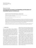
Figure.1.1 Time accuracy requirement of soft tissue modeling 3
Figure.2.1 Deformations of linear classic cylinder. (a) and (b) side view; (c) and (d)
top view 15
Figure.2.2 Deformations of nonlinear cylinder. (a) and (b) side view; (c) top view.
Comparisons between linear (wireframe) and nonlinear model (solid rendering) are
indicated in (b) and (c) [73] 15
Figure.2.3 Model fits of Franceschini et al[89]. one-cycle compression-tension (a)
and tension-compression (b) tests on specimens of white matter. The X axis denotes
the stretch ratio for the experimental data while the Y axis indicates the nominal
stress 20
Figure.2.4 Visual comparisons between the graph-cut method (outer line) and the
active contour segmentation (inner line) 21
Figure.4.1.1 The mechanical testing system; (1) power source (2) Strain gauge
amplified for load cell and pressure transducer(not shown), (3) Stepper motor control,
(4) Distance laser sensor, (5) Load cell, (6) translational stage with stepper motor. (7)
clamping feature and fixture, (8) base. 30
Figure.4.1.2 Stress and strain distribution of artery tissue. (a) Circumferential; (b)
Longitudinal directions. Blue solid line (—) denotes the random selected
experimental curves; red short dash line ( ) is the mean value curve of the
experimental curves 32
Figure.4.1.3 Stress and strain relationship of artery tissue. (a) Circumferential
direction; (b) Longitudinal direction. Green (-*) mean Black ( ) maximum and
minimum values of stress. Normal distribution of stress values is illustrated along
horizontal bars using red solid line 33
Figure.4.1.4 Comparison of simulated result and experimental mean value. (a)
Circumferential direction; (b) Longitudinal direction 37
Figure.4.1.5 CDFs of Engineering stress for artery tissue at seven strain values of
1.25, 1.30, 1.35, 1.40, 1.45, 1.50 and 1.55 from left to right. Red dash line is the
experimental CDFs; green heavy line is the CDFs from 10000 evaluations with direct
calculated material parameters; blue thin line is the CDFs from 10000 evaluations
with material parameters calculated from IMVFOSM method. (a) Circumferential
direction; (b) Longitudinal direction 41
Figure.4.1.6 Stress and strain relationship of artery tissue. (a) Circumferential
direction; (b) Longitudinal direction. Green (-*) mean values of stress, black ( )
maximum and minimum values of stress, blue (
) experimental data from
Yamada’s study, blue solid line is the experimental data from Sommer’s work.
Normal distribution of stress values is illustrated along horizontal bars 42
Figure.4.2.1 Laparoscopic scissors used in this section. (a) Aesculap laparoscopic
scissors, Model :PO004R; (b) Schematic view of the linage mechanism of
laparoscopic surgical instrument 48
Figure.4.2.2 Mass spring models used in medical simulation. (a) Maxwell model; (b)
Voigt model; (c) Kelvin model 50
Figure.4.2.3 Modified model with variables 51
V
Figure.4.2.5 Three pieces of human iliac artery were cut with five cuts. The cutting
process is divided in to three regions. (1) Contact region. (2) Cutting region. (3)
Completion region 58
Figure.4.2.6 Fitting result of experimental force using curve fitting and GA 58
Figure.5.1.1 Work flow of the study 63
Figure.5.1.2 Geometrical shape of the gallbladder organ in polar coordinates. The
major axis length is
1
D
, the minor axes lengths are
2
D
, and
3
D
(
123
D
DD
),
the gallbladder is subjected to a uniform internal pressure. The stress due to this
pressure at a surface point P has three components:
r
(radial),
(circumferential),
and
z
(axial) 64
Figure.5.1.3 Images of the experiments. (a) Indentation tests on gallbladder organ; (b)
Elongation tests on gallbladder wall tissue 68
Figure.5.1.4 Experimental results of uniaxial elongation tests on gallbladder wall
tissue in longitudinal and circumferential directions. Solid line shows the mean stress
of 5 specimens, vertical bar shows the standard deviation of stress 70
Figure.5.1.5 Mean experimental data (marked by *) and predicted result (solid line).
(a) Longitudinal; (b) Circumferential directions 72
Figure.5.1.6 Experimental results of uniaxial indentation tests on gallbladder organ in
longitudinal and circumferential directions. Solid line shows the mean stress of 5
specimens, vertical bar shows the standard deviation of stress 73
Figure.5.1.7 Mean experimental data (marked by purple point) and predicted result
(red solid line). (a) Longitudinal direction; (b) Circumferential direction 75
Figure.5.1.8 Segmented contour of gallbladder 76
Figure.5.1.9 Constructed 3D gallbladder model 76
Figure.5.1.10 Interactive manipulation of gallbladder model using haptic interface
device 77
Figure.5.2.1 Overview of the haptic guidance and visual simulation system 83
Figure.5.2.2 Three stages of potential energy (J) distribution around the predefined
path: (a)
=3; (b)
=6; (c)
=9 87
Figure.5.2.3 Potential field map at a fixed Z value around the path 88
Figure.5.2.4 Flow chart of the algorithm 89
Figure.5.2.5 3D tracheal model from CT scans; 3D tracheal model reconstructed from
CT scans, a physical based model is generated from the model for virtual interaction
90
Figure.5.2.6 Haptic simulation of tracheal reconstruction. (a) Image of the simulation
system;(b) and (c) Simulation images 91
Figure.5.2.7 Haptic guidance application of “rubber duck”: (a) Overview of the
application; (b) Manipulation point on the predefined path; (c) and (d) Manipulation
point is out of the predefined path 94
Figure.6.1 Work flow of the study 99
Figure.6.2 Computer simulation of clip-tissue interaction using ABAQUS: (a) Image
before deformation; (b) Image after deployment of clip into tissue 100
Figure.6.3 Energy distribution on clip at initial deployment before degradation,
energy is indicated from highest (red) to lowest (blue) 103
VI
Figure.6.4 In-vivo application of micro-clips on porcine vocal cord. Four micro-clips
of thickness 0.25mm are applied to appose the edges of the created epithelial flaps in
order to promote primary intention 106
Figure.6.5 Excised vocal folds with embedded micro-clips 2 weeks after deployment.
Micro-clips surface show various levels of degradation 107
Figure.6.6 Images of the in-vitro experiment: (a) Unloaded clips used in the
experiments; (b) Clips suspended and placed in tension using thread; (c) Clips
immersed in HBSS during the study 108
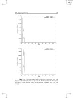
Figure.6.7 Plot of percentage mass remaining over different time intervals based on
the results of in-vitro immersion test (dash line).The degradation model mass
remaining prediction is also included (red line). (a) First group; (b) Second group . 110
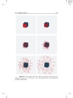
Figure.6.8 Degradation stages of the clip: five stages of degradation are simulated
from (a) to (l) in pairs with a certain time period:(a)-(c) 0.5 week; (d)-(f) 1 week; (g)-
(i) 1.5 weeks; (j)-(l) 2 weeks; (m)-(o) 2.5 weeks. The Green line indicates the original
shape of the clip; red line illustrates the degradation shape of previous stage; blue line
shows the degradation shape of current stage 111
Figure.7.1 Image shows the working condition of voice prosthesis. 1. Wound on the
tissue; 2.Biodegradable material layer; 3.Foundation layer 119
VII
List of Symbols
:
Inner product of two second-order tensors
Partial differential
Summation
Gradient
Square root
Vector norm
Integration
e
Euler number
min( )
Minimum
ln( )
Natural logarithm
exp( )
Exponential function
sin( )
Sine function
cos( )
Cosine function
VIII
List of Abbreviations
ALE Arbitrary-Lagrangian-Eulerian
BEM boundary element method
CDF cumulative distribution function
CT computerized tomography
DC direct calculating
EFFD extended free form deformation
FE finite element
FFD free form deformation
GA genetic algorithm
GVF gradient vector flow
HTK Histidine Tryptophan Ketoglutarate
IMVFOSM inverse mean-value first-order second-moment
L-H Legendre-Hadamard
MC Monte Carol
MIS minimally invasive surgery
MRA magnetic resonance angiography
MRI magnetic resonance imaging
PDE partial differential equation
TL total laryngectomy
VP voice prosthesis
1
Chapter 1 Introduction
1.1 Background and motivation
For minimally invasive surgeries, surgeons are required to be highly skilled to
perform the surgical operations [1]. Mastering and assessing operation skills
for the doctors can be difficult. Medical simulation is particularly attractive in
the field of surgical training because it avoids the participation of patients for
skills practice and enables the trainees to be trained before treating humans [2].
Virtual reality based surgical simulators present a safe, realistic, and efficient
way for surgical training, practice, and pre-operative planning. These
simulators simulate human anatomy environment and generate realistic
mechanical responses of human organs. Using medical simulators, new
surgeons can improve their surgical skills after exercising on a variety of
complex cases and receive feedback on their performance. Surgical simulation
systems are also useful for pre- and intra-operative planning of medical
procedures. Surgical and interventional radiology procedures often require a
patient-specific plan prior to performing an operation. Thus, simulation
systems which account for patient-specific anatomical details and tissue
properties can benefit the surgeons as well as increase the accuracy of the
surgical procedures [3, 4].
The key requirements in surgical simulation is establishing realistic human
anatomical environment and presenting accurate biomechanical responses of
organs during surgical procedures for the purposes of training, planning, and
assessing patient outcomes in a risk-free environment [4]. Developing realistic
virtual reality based surgical simulation system demands the acquisition of
specific biomechanical tissue information, development of efficient
computation strategies, employment of acceptable validation protocols, and
integration of advanced haptic rendering technologies [5]. A high-fidelity
surgical simulation system requires appropriately presentation of soft tissue
deformation during interactions similar to that of actual surgical manipulations.
The boundary conditions of soft tissues must be physically well defined and
2
their interactions with tools should be updated in real-time in order to create a
realistic visual and haptic interface.
The nonlinear mechanical response is an important attribute of soft tissue
properties which relates to simulation accuracy, and needs to be considered for
deformation simulation and haptic rendering in surgical simulation.
Experimental procedures such as inflation tests [6, 7], biaxial tests[8], as well
as tension and indentation tests [9-12] have been performed to study the
mechanical properties of soft biological tissue. These experiments showed that
the mechanical behavior of soft biological tissue was elastic, highly nonlinear
and anisotropic under finite strains, which is usually modeled within the
framework of hyperelasticity.
However, for realistic surgical simulation, there exists a trade-off between
computational speed and biomechanical simulation accuracy. Feedback from
surgeons reveals that a bad simulator is worse than no simulation, they also
insist that simulators must be realistic enough so that the errors are resulted
from incorrect manipulation of surgeons but not from the virtual environment
[13]. Relationship between computational speed and simulation accuracy for
different applications are summarized in Figure 1.1. Scientific analysis is
aiming at validating physical hypothesis of soft tissue for the design of new
procedures or implants. In this case, the accuracy of deformation is far more
important than computation time. On the other hand, surgery planning for
predicting the outcome of surgery or rehearsing complex operations, requiring
less computation time (from 30s to one hour) since several trials may be
necessary. For surgical procedure training, computation time of the level of
0.1s is required in order to achieve smooth user interaction whereas the
accuracy of deformation is not of primary importance [5]. In this dissertation,
we put our efforts to investigate the nonlinear mechanical properties of
biological soft tissue using computation approaches. The objective is to
provide an effective approach for realistic modeling and simulation of tool
tissue interaction. The findings of this work are utilized to build high-fidelity
medical simulation system.
3
Figure.1.1 Time accuracy requirement of soft tissue modeling
1.2 Variational methods for soft tissue modeling
Many studies have been conducted to investigate the biomechanical models of
soft tissue. Deformable models for soft tissue deformation can be classified
into two categories: physics based and non-physics based. Physics based
methods are based on continuum mechanical principles, and could obtain
accurate simulation results by directly solving the partial differential equations
(PDEs) using numerical or computational methods. Some of the prevailing
methods include the Finite element (FE) method [14], boundary element
method (BEM) [15], point-based method [16], and reduced model [17]. Non-
physics based models use intuitive methods instead of solving PDE. For
example, the mass-spring model [18] uses point masses connected by a
network of springs to represent continuous material, and meshless shape
matching model [19] computes deformations based on geometry shapes.
Numerical or computational models based on mechanical engineering
principles are employed to model the deformation of soft tissue realistically
[20]. They aim to provide accurate soft tissue modeling results while reducing
the computation cost. However, the balance between computational cost and
accuracy remains a research problem.
Variational principles for biomechanical systems, such as elasto-viscoelastic
behavior, have been known for a while, but have received renewed attention in
4
recent years. These principles can be written in a continuous or in an
incremental framework. In particular, a variational formulation of constitutive
models for standard generalized materials, including irreversible, dissipative,
and possibly rate-dependent behaviors, was proposed [21, 22] initially in an
isothermal context, and later extended to a fully coupled thermo-mechanical
context in [23]. These variational approaches could provide appropriate
mathematical basis for developing models of non-cohesive granular media
[24], porous plasticity [25], and nonlinear finite viscoelasticity [26].
The variational models can serve as an appropriate compensatory method to
model the nonlinear mechanical properties of soft tissue. Unlike the traditional
finite element method that always needs to consider the boundary condition in
interaction process, under the assumption of incompressible nonlinear body
[27], variational methods can be used for modeling of nonlinear biological soft
tissue deformation in the finite deformation regime. By defining the modeling
problem as an energy minimization process, the material parameters of
nonlinear model can be characterized within the variational framework. The
approach is qualified as variational since the constitutive updates consist of a
minimization problem within each load increment [26]. It displays great
advantages when dealing with nonlinear materials in an inexpensive
computationally way.
1.3 Organizations
The overall structure of the study takes the form of seven chapters, including
this introductory chapter. Chapter 2 begins by reviewing the literature on
surgical simulation in the context of nonphysical based and physical based
models and variational modeling of soft tissue deformation. Chapter 3
describes the variational principles of this dissertation study. Chapter 4
presents an investigation on statistical modeling of the uncertainties of human
artery tissue using probabilistic approach, and characterization of material
parameters in human vascular soft tissue during division. Chapter 5 presents
the study of constitutive laws for hyperelastic tissue and implementation for
surgical simulation with haptic rendering, as well as surgical simulation in
combination with haptic guidance. Chapter 6 discusses effects of strain energy
5
from tool-tissue interaction process on degradation mechanism of
biodegradable materials. Finally, the thesis concludes in Chapter 7 with a
discussion on future research in the area of realistic modeling of tool-tissue
interactions.
1.4 Contributions
The major contributions of this dissertation are:
Quantitative study the uncertainties in mechanical properties of
human arterial tissue using probabilistic approach
With the variational principles, a new probabilistic approach was proposed to
model the uncertainties of human arterial tissue deformation by assuming that
the instantaneous stress at a specific strain varies according to normal
distribution. Material parameters of the artery tissue were modeled with a
combined logarithmic and polynomial energy equation and characterized with
the experimental results obtained from human arteries. The statistical model is
able to present the soft tissue properties accurately. The interaction between
the uncertainty on the observations and the uncertainty on the estimated
parameters is a major phenomenon to consider when using biomechanical
models for medical simulation. By taking into account the inhomogeneous
mechanical properties of human biological tissue, the study can contribute to
realistic virtual simulation as well as an acceptable computational approach for
medical device validation.
Variationally modeling the nonlinear mechanical properties of
gallbladder organ and haptic implementation of the modeling results
We investigated the variational principles for biomechanical modeling of
gallbladder tissue. Mechanical experiments on porcine gallbladder tissue are
carried out to investigate soft tissue deformation properties. An exponential
strain energy function was proposed to describe the mechanical behavior of
the gallbladder tissue while the material parameters were calibrated with a
genetic algorithm based variational approach. The gallbladder tissue model is
assigned with hyperelastic properties and implemented in a medical simulation
6
system with haptic feedback. The nonlinear tissue model provides a realistic
material model for advanced surgical simulation.
Computation modeling of tool-tissue interaction process and their
effects on degradation process of biodegradable materials
Strain energy function is always accounting for the soft tissue deformation
modeling. The degradation process of the biodegradable clips is assumed to be
highly related to the strain energy on the clips resulted from tool-tissue
interaction process. The tool-tissue interaction process between biodegradable
clips and porcine vocal fold tissue was first modeled using FE analysis while
the FE results were used to calculate the strain energy of the clips using active
contour. Degradation process was defined as an energy minimization process
and solved within the variational framework. The degradation rate and
geometries of the clip during degradation was computed based on the physical
energy, and calibrated by experimental results. This work presents a
comprehensive study on the tool-tissue interaction and their effect on the
degradation process of biodegradable materials.
7
Chapter 2 Literature Review
Surgical simulation creates an efficient and safe platform for new surgeons to
gain necessary medical skills while reducing the needs for animals, cadavers,
and patients [28]. A goal of surgical simulation is the generation of realistic
human anatomical and physiological responses to surgical manipulations for
the purposes of training, planning, and assessing patient outcomes in a risk-
free environment [4]. It aims to assist medical practitioners by allowing them
to visualize, feel, and be fully immersed in a realistic environment. The
simulator should accurately represent the anatomical details and deformation
of the organ as well as provide realistic haptic feedback of tool-tissue
interaction.
Advanced modeling algorithms are important for accurate soft tissue
deformation modeling and haptic force feedback. During the past decades,
there has been growing interest in the medical and computer science field
around the simulation of medical procedures [5]. Computational modeling
and numerical methods have demonstrated their abilities in solving complex
boundary value problems for soft tissue modeling [29]. Different algorithms
have been proposed for computational modeling of soft tissue deformation.
These algorithms can be divided into two categories: Non-physical based
models, such as free form deformation [30] and deformable splines [31] which
are based on pure mathematical representation of the object’s surface and do
not generally provide a realistic simulation of its mechanical behavior.
Another category is physical based models, which can be classified into two
types: Non-continuum mechanics based methods, e.g., mass-spring models [32]
and continuum mechanics based methods, e.g., finite element methods [33].
This chapter will review the related works in soft tissue modeling.
2.1 Non-physical based computational methods
The non-physical computational methods for tool-tissue interaction modeling
include free-form deformation methods [34] and deformable splines [35].
These algorithms are based on pure mathematical representation of the
8
object’s surface, which fail to provide a realistic simulation of its mechanical
behavior. In such cases, physical accuracy is sacrificed for computational
efficiency and the system has no knowledge about the material properties of
the object being deformed [36]. The mostly used non-physical based model is
free form deformation model.
Free form deformation (FFD) is a space-warping technology that plays an
important role in computer-assisted geometric design and soft tissue
deformation animation [36]. Some useful deformation operations, which were
independent of control points, were developed by Barr in 1984 [37]. Complex
deformations, once achieved only by skilled and laborious manipulation of
numerous control points, could now be presented by applying these operators
to an object in a hierarchical fashion. However, the actions of Barr’s model
were constrained to against a single axis which reduces the potential of the
model for complex structure modeling. The restrictions of the model made it
only suitable for modeling of lattice shape.
To conquer the shape constraints of FFD, Extended Free Form Deformation
(EFFD) was proposed by Coquillart [38]. It allows the user to define the shape
of a lattice, which in turn induces the shape of the deformation. Animated
Free-Form Deformation[39], in which the deformation tool differentiates itself
from the object instead of interpolating the metamorphosis of the 3D lattice
which lies around the deformable object, was also proposed by Coquillart for
animating deformations. This technique allows reusing of deformations for
other objects and provides better control over the deformation.
A hierarchical transformation model of the motion of the breast was developed
by Rueckert [40] for non-rigid registration of contrast-enhanced breast MRI.
The local breast motion was described by a FFD based B-splines while the
global motion of the breast was modeled by an affine transformation. This
FFD based non-rigid registration algorithm shows better performance to
recover the motion and deformation of the breast than rigid or affine
registration algorithms. Liver motion during respiratory cycle was studied by
Rohlfing using an intensity-based FFD registration algorithm [41]. The
intensity based non-rigid image registration approach can achieve a
9
satisfactory level in abdominal organ motion modeling. The intensity-based
nonrigid registration algorithm was extended by using a novel regularization
term to constrain the deformation for breast images registration [42]. The
novel regularization term is a local volume-preservation (incompressibility)
constraint, which is motivated by the assumption that soft tissue is
incompressible for small deformations and short time periods. The intensity-
based free-form non-rigid registration algorithm was improved by
incorporation of the incompressible feature as it greatly reduces the problem
of shrinkage of contrast-enhanced structures while allowing motion artifacts to
be substantially reduced.
FFD enables smooth deformations of arbitrary structures, provides local
control over deformations, and serves as a computationally efficient algorithm
that is easy to implement. It can be extended in complex modeling work which
is usually carried out with physical based models[43, 44].
B-spline solids are employed to model skeletal muscle for the purpose of
building a data library of reusable, deformable muscles that are reconstructed
from actual muscle data[45]. Techniques are developed to construct
continuous representations of volume from discrete data. B-spline solids are
represented as mathematical three-dimensional vector functions in order to
obtain muscle fibre bundle orientations. As B-spline solids can be defined
completely with its control points and knot vectors, they can require
significantly less storage than a dense set of polygons.
Interphase correlation of the images during the respiratory process are studied
with B-spline registration models, intermediate phases are interpolated by
starting from two or three sets of 3D CT images acquired at different phase
points[46]. It demonstrates that the organ deformation during the breathing
process can be well modelled with a B-Spline deformable algorithm.
Deformable splines are also utilized in motion tracking for medical
applications. By formulating model parameters as tensor products of B-splines,
algorithms are proposed to quickly reconstruct left ventricle geometry/motion
from extracted boundary contours and tracked planar tags in MR images [47].
10
Furthermore, a thin plate spline model is developed for representing the heart
surface deformations[48]. The thin plate spline was extended to warp to the
stereo scenario, enabling efficient 3D tracking of the beating heart using stereo
endoscopic images. However, deformable splines are still quite complex and
computationally costlier than spring-mass type models which will be
introduced in next section, without actually offering better realism.
2.2 Physical based computational methods
This section discusses the physical based computational methods that are
employed in medical simulation.
2.2.1 Non-continuum discrete models
Among the physical based models, the discrete models, such as the mass-
spring systems[49] and Chain-mail representational models [50], are widely
used in soft tissue deformation modeling due to their low computational cost
and easily implementation [50-52]. Mass-spring models are usually utilized in
soft tissue deformation for solving linear elastic problems. For elastic
materials, Hooke's law represents the material behavior and relates the
unknown stresses and strains in following constitutive equation.
:C
(2.2.1)
where
is the Cauchy stress tensor, C is the fourth-order stiffness tensor,
is
the infinitesimal strain tensor, and
:
ij ij
A
BAB
is the inner product of two
second-order tensors (summation over repeated indices is implied).
Many works have been done under the framework of linear elasticity using
mass-spring models. Mass-spring models were first proposed to model facial
deformation [53, 54]. These early works solve static problems of Hooke’s law.
After that, dynamic models were introduced to model skin, fat and muscle
tissues [49, 55, 56]. Some studies have employed mass-spring-damper models
to simulate tissue deformation, but they fail to provide detail information on
the tissue properties required for the deformation simulation [54, 57, 58]. On
the other hand, a sophisticated apparatus was used for data acquisition to
11
enable virtual ultrasound display of the human thigh as well as force feedback
to the user [59]. The human thigh model was represented by a mass-spring
system which was characterized in an earlier study conducted by the same
author [60]. The two layer model was made up of a mesh of masses and linear
springs, and a set of nonlinear springs orthogonal to the surface mesh to model
volumetric effects. Realistic haptic force feedback was enabled by
incorporating a buffer model between the physical model and haptic device.
The buffer model was defined by a set of parameters and was continuously
adapted in order to fit the values provided by the physical model. This
computationally simple model can estimate the interaction force according to
the physical model at haptic update rates.
Although the mass-spring model can provide a fast computation and easy
implementation, they are not appropriate for the modeling of complex soft
tissue deformation in surgery. Primarily, most mass-spring systems are not
convergent [61]. As the mesh is refined, the simulation does not converge on
the true solution. Instead, the behavior of the model is dependent on the mesh
resolution and topology. In practice, spring constants are often chosen
arbitrarily, and one can present little quantitatively about the material being
modeled. In addition, there is often coupling between the various spring types.
For medical applications, as well as virtual garment simulation in the textile
industry, greater accuracy is required.
In order to overcome the accuracy problem in modeling of nonlinear
biological soft tissues, many researchers have explored new approaches to
implement the mass-spring methods. Basafa [62], in his study on realistic and
efficient simulation of liver surgery, proposed an extension of the mass-spring
modeling approach for more realistic force formation behavior while
maintaining the capability of real-time response. Schwartz [63] introduced an
extension of the linear elastic tensor–mass method for fast computation of
nonlinear viscoelastic mechanical forces and deformations for the simulation
of biological soft tissues with the aim of developing a simulation tool for the
planning of cryogenic surgical treatment of liver cancer. The Voigt model was
initially considered to approximate the properties of liver tissues. However, it
12
was later discovered, from experiments, that a linear model is not suitable for
modeling this application under various needle penetration loads [63].
Mass-spring models may be combined with other models to achieve a balance
in computational efficiency and modeling accuracy. A combined mass spring
and tensional integrity method is proposed and applied to simulate the
diaphragm motion [64]. A hybrid model which may allow real time
deformations and cuttings of anatomical structures was proposed [65]. The
quasi-static pre-compute elastic FE model introduced by the authors was
computationally efficient but did not allow topology change. Meanwhile, the
mass-spring model is well suited for the simulation of tearing and cutting, but
a limited number of elements are allowed for real-time simulation. So the
authors combined the above models in order to optimize the trade-off between
computation time and visual realism of the simulation. Similar study which
combined mass-spring models and Boundary Element Method (BEM) was
also proposed recently [66]. In this study, a BEM model is used to compute
the global deformation while a mass-spring model is employed to interactively
model the dynamic behaviours of organs. The hybrid model is suitable for
interactive surgical training applications, and provides visually accurate results
in simulating the deformation of biological soft tissues with experimental
inputs.
Problems still exist in relating mass-spring parameters with real material
parameters. The parameters of mass-spring models are typically determined in
an ad hoc fashion through trial-and-error which is not directly based on
continuum mechanics of deformable objects [67]. Algorithms have been
proposed to find alternative ways in determining the model parameters, in
which the parameters are determined using a finite element model as a
reference model by minimizing the error the stiffness matrices of the finite
element and mass-spring models through an optimization algorithm.
2.2.2 Continuum mechanics based computational methods
The computational methods which are based on continuum mechanics are
discussed in this section. The most computationally demanding soft tissue