Cell cycle lipidomics
Bạn đang xem bản rút gọn của tài liệu. Xem và tải ngay bản đầy đủ của tài liệu tại đây (2.23 MB, 159 trang )
CELL CYCLE LIPIDOMICS
LIM JING YAN
BSc (Hons.), NUS
A THESIS SUBMITTED
FOR THE DEGREE OF DOCTOR OF PHILOSOPHY
NUS GRADUATE SCHOOL FOR INTEGRATIVE
SCIENCES AND ENGINEERING
NATIONAL UNIVERSITY OF SINGAPORE
2013
2
Declaration
I hereby declare that the thesis is my original work and it has been written by me
in its entirety. I have duly acknowledged all the sources of information which
have been used in the thesis.
This thesis has also not been submitted for any degree in any university
previously.
Lim Jing Yan
11 Jan 2013
i
Acknowledgements
I would like to express my heartfelt gratitude to my supervisor, A/Prof. Markus R
Wenk, and co-supervisor, Prof Ong Choon Nam, for their encouragement,
guidance and support throughout the whole of my PhD. I appreciated the freedom
they gave me to explore in my study. I would also like to offer my sincere
gratitude to A/Prof Philipp Kaldis, who has given me timely advice and
invaluable suggestions throughout my PhD. I also thank Dr Aaron Z Fernandis for
showing me the ropes when I first started out in the lab. I am extremely grateful to
Dr Shui Guanghou, whose advice, encouragement, guidance and scientific
excellence have made this thesis possible.
I thank my dear lab mates, especially Husna, Sudar, Jacklyn, Charmaine and
Lissya, who have kept me sane and made this journey an interesting one. I am
also grateful to Dr Federico Torta and Madhu who had given me useful
suggestions and inputs in my thesis. I am thankful to Huimin and Dorothy for
helping me out with administrative work.
Thank you, NUS Graduate School for Integrative Sciences and Engineering
(NGS), for the generous funding through a research scholarship and wonderful
student support.
Finally, I would like to express my special appreciation to my mother, father and
brother for their unconditional care and love. And special thanks to my husband,
Jiunn Siong, who has been my constant source of love, strength and faith.
ii
Table of Contents
Acknowledgements i
Summary v
List of Tables vii
List of Figures viii
List of Abbreviations xii
1. INTRODUCTION 1
1.1 Lipidomics 1
1.1.1 Lipids 1
1.1.2 Mass spectrometry 3
1.2 Cell cycle and lipids 7
1.3 Rationale and objectives of this study 20
2. MATERIALS AND METHODS 21
2.1 Materials 21
2.2 Cell culture 21
2.3 Cell synchronisation 22
2.4 Flow cytometric analysis 23
2.5 Immunoblot analysis 23
2.6 Lipid analysis 24
2.6.1 Lipid extraction 24
2.6.2 HPLC-MS profiling of diverse lipids 25
2.7 Data analysis 26
3. CELL CYCLE SYNCHRONISATION AND LIPID PROFILE 28
3.1 Cell cycle synchronisation 28
3.2 Platform for cellular lipidomics setup and analysis 32
3.2.1 Cell number versus total lipid 32
3.2.2 FBS effect on general lipid profile 35
3.3 Materials and Methods 37
3.4 Results 38
3.4.1 Phospholipid profile of the cell cycle 38
3.4.2 Neutral lipid profile 61
iii
3.5 Discussion 70
4. CHOLESTEROL ESTERS’ ROLE IN CELL CYCLE 75
4.1 Introduction 75
4.2 Materials and Methods 79
4.2.1 Cell culture 79
4.2.2 RNAi transfection of HeLa cells 79
4.2.3 Fluorescence Imaging of lipid droplets 79
4.2.4 AlamarBlue assay for cell viability 80
4.2.5 Analysis of kinetics of cell cycle progression 80
4.2.6 Analysis of ACAT1 and cyclin levels using western blot 81
4.2.6 MS analysis of phospholipids, sphingolipids and cholesterol in ACAT1
and negative control cells 82
4.2.7 Time-lapse Imaging of ACAT1 KD and negative control cells 82
4.3 Results 83
4.3.1 Fluorescent imaging of lipid droplets at different cell cycle phases and
MS analysis of individual cholesterol ester species 83
4.3.2 Determination of ACAT1 protein expression, cell viability and lipid
profile of ACAT1 KD cells 86
4.3.3 Cell cycle progression of ACAT1 KD cells upon release from
aphidicolin synchronisation 89
4.3.4 Cell cycle progression of ACAT1 KD cells upon release from
hydroxyurea synchronisation 93
4.3.5 Cell cycle progression of ACAT1 KD cells upon release from
nocodazole synchronisation 95
4.3.6 Time lapse observation of cell division in ACAT1 KD and negative
control cells 97
4.4 Discussion 102
5. BREAST CANCER LIPIDOMICS 108
5.1 Introduction 108
5.2 Materials and Methods 113
5.2.1 Breast cancer samples 113
5.2.2 Lipid analysis 113
5.2.3 Statistical analysis 113
iv
5.3 Results 114
5.3.1 Phospholipid and sphingolipid profiles of breast tumor vs control 114
5.3.2 Sterol profile of breast tumor versus control 119
5.4 Discussion 120
6. CONCLUSION 125
7. FUTURE WORK 127
8. BIBLIOGRPAHY 130
v
Summary
Regulation of cell cycle is crucial for cell survival and function. Dysregulated cell
division can result in multiple disorders, including cancer, neurological, renal and
vascular proliferative diseases. Changes in cellular lipidome during the cell cycle
has become an increasingly important research area. Here, we investigated the
lipid composition of cells in different stages of the cell cycle using mass
spectrometry-based lipidomics approaches. We used two complementary
synchronisation methods (G1/S by aphidicolin and G2/M by nocodazole) with
two different cell lines (HeLa- human cervical cancer cell line and MCF7- human
breast cancer cell line). Among the lipid classes analysed, which included
phosphatidylcholine (PC), phosphatidylethanolamine (PE), phosphatidylinositol
(PI), phosphatidylglycerol (PG), sphingomyelin (SM), ceramide (Cer),
glucosylceramide (GluCer), triacylglycerol (TAG), diacylglycerol (DAG) and
sterols, cholesterol esters showed a significant increase in G2/M phase. This
highlights that cholesterol esters may be a fundamental lipid class for cell division,
as they could act as a store for cholesterol and fatty acids, which are essential
components of phospholipids and are critical for membrane biogenesis in cell
division.
Acyl-coA:cholesterol acyltransferase 1 (ACAT1) is the main intracellular enzyme
involved in cholesterol esterification. We confirmed the importance of cholesterol
esters in cell cycle using ACAT1 knocked-down (KD) cells which have lower
levels of cholesterol esters. Cell cycle kinetics of ACAT1 KD and negative
control cells were compared. A prolonged G2/M phase in ACAT1 KD cells was
vi
observed, indicating that cholesterol esters metabolism is crucial especially for
G2/M progression.
One of the consequences of cell cycle dysregulation is the development of cancer.
The connection between cell cycle and cancer is critical. The cell cycle machinery
controls cell proliferation and cancer is a condition of uncontrolled cell division.
Lipids have been reported to play a role in cancer. Hence, using breast cancer as a
model, we conducted a pilot study to profile and compare lipids in human breast
tumor and control tissues. Among other findings, we observed an increase in
cholesterol esters in the tumor samples when compared to control. This further
supports our previous results, where cholesterol esters were found to be important
for active cell division.
vii
List of Tables
Table 3.1 A summary of four common methods used in cell synchronisation, and
the advantages and problems faced while trying each method during this
experimental work. 31
Table 3.2 In depth analysis of the fold change of lipids in both cell lines for both
synchronisation methods 58
viii
List of Figures
Figure 1.1 Categories of lipids with examples 2
Figure 1.2 Schematic diagram of MRM in a triple quadrupole 6
Figure 1.3 The stages of the cell cycle 8
Figure 1.4 Summary of genes of lipid related proteins that are found to be
regulated in the cell cycle in two published cell cycle genomics study 10
Figure 1.5 Main functions of lipids in the cell cycle. 19
Figure 3.1 Graph representing the relationship between cell number and log value
of total normalised lipid signal intensities 34
Figure 3.2 Heatmap of lipid fold changes in HeLa cells cultured in media with
different FBS percentage for 24, 48 and 72 hours, as compared to the starting
point (FBS 10% 0h). 36
Figure 3.3 Schematic diagram of the work flow of cell cycle synchronisation and
sample collection 37
Figure 3.4 Cell cycle analysis of MCF7 cells synchronised at G1/S by aphidicolin
and then released into G2/M 39
Figure 3.5 Heatmap representation of individual polar lipid species changes in
MCF7 cells released from aphidicolin synchronisation 41
Figure 3.6 Bar graph of fold change in lipids that were significantly (p<0.05)
changed as MCF7 cells progressed from G1/S to G2/M after being released from
aphidicolin synchronisation. 42
Figure 3.7 Cell cycle analysis of HeLa cells synchronised and released from
aphidicolin 44
Figure 3.8 Heatmap representation of individual polar lipid species changes in
HeLa cells released from aphidicolin synchronisation. 46
Figure 3.9 Bar graph of fold change in phospholipids that were significantly
(p<0.05) changed as HeLa cells progressed from G1/S to G2/M after being
synchronised with aphidicolin. 47
ix
Figure 3.10 Cell cycle analysis of MCF7 cells synchronised by nocodazole and
released. 48
Figure 3.11 Heatmap representation of individual polar lipid species changes in
MCF7 cells released from nocodazole synchronisation. 50
Figure 3.12 Bar graph of fold change in lipids that were significantly (p<0.05)
changed as MCF7 cells progressed from G2/M to G1 after being synchronised
with nocodazole. 51
Figure 3.13 Cell cycle analysis of HeLa cells synchronised and released from
nocodazole 52
Figure 3.14 Heatmap representation of individual polar lipid species changes in
HeLa cells released from nocodazole synchronisation. 54
Figure 3.15 Bar graph of fold change in lipids that were significantly (p<0.05)
changed as HeLa cells progressed from G2/M to G1 after being synchronised with
nocodazole. 55
Figure 3.16 Venn diagrams summarising the overall similar lipids that display the
same trends in both cell lines 56
Figure 3.17 Bar charts showing the sum of each lipid class. Both aphidicolin (A)
and nocodazole 59
Figure 3.18 Bar charts showing trends in the total degree of unsaturation in fatty
acyl chains in cell cycle for aphidicolin (A) and nocodazole (B) synchronised
HeLa cells. 60
Figure 3.19 Changes in DAG and TAG as MCF7 progressed from G1/S to G2/M.
DAG and TAG levels at G2/M were normalised against those at G1/S. 62
Figure 3.20 Changes in DAG and TAG as HeLa progressed from G1/S to G2/M.
DAG and TAG levels at G2/M were normalised against those at G1/S 63
Figure 3.21 Changes in DAG and TAG as MCF7 progressed from G2/M to G1. 64
Figure 3.22 Changes in DAG and TAG as HeLa cells progressed from G2/M to
G1. 65
Figure 3.23 Fold changes in cholesterol and its derivatives in G2/M cells
compared to G1/S in (A) MCF7 and (B) HeLa, as the cells progressed from G1/S
to G2/M after aphidicolin synchronisation. 67
x
Figure 3.24 Fold changes in cholesterol and its derivatives in G1 cells compared
to G2/M in (A) MCF7 and (B) HeLa, as the cells progressed from G2/M to G1
after nocodazole synchronisation 69
Figure 4.1 Timeline to summarise the process from ACAT1 knockdown to cell
cycle synchronisation and cell collection. 81
Figure 4.2 Fluorescent microscope images of cells in different stages of the cell
cycle 84
Figure 4.3 Fold change of each cholesterol ester species in G2/M as compared to
G1/S. 85
Figure 4.4 Effects of ACAT1 KD by RNAi transfection in HeLa cells 86
Figure 4.5 Fold change in the percentage reduction of alamarBlue at 24h and 48h
against the 0h start point. 87
Figure 4.6 Lipid profile of ACAT1 KD and negative control cells. Error bars
represent + SD. 88
Figure 4.7 Cell cycle kinetics of ACAT1 KD and negative control cells which
were released from G1/S synchronisation by aphidicolin. 90
Figure 4.8 Western blots for ACAT1 KD and negative control cells released from
aphidicolin synchronisation 92
Figure 4.9 Cell cycle kinetics of ACAT1 KD and negative control cells which
were released from G1/S synchronisation by hydroxyurea. 94
Figure 4.10 Cell cycle kinetics of ACAT1 KD and negative control cells which
were released from G2/M synchronisation by nocodazole. 96
Figure 4.11 Time lapse observation of cell division after release from aphidicolin
synchronisation. 98
Figure 4.12 Analysis of cell division in unsynchronised ACAT1 and negative
control cells. 100
Figure 4.13 Time lapse images of ACAT1 KD cells arrested in mitosis 101
Figure 5.1 Overall phospholipid and sphingolipid changes in control versus tumor
breast tissue samples 115
xi
Figure 5.2 Bar charts showing lipid species that were significantly different in
tumor samples as compared to control breast tissue samples. 116
Figure 5.3 Bar charts representing the fold change in the total carbon chain length
(A) and degree of fatty acid unsaturation (B) 118
Figure 5.4 Fold change of sterol and its derivatives in control and tumor breast
tissues against control average. 119
xii
List of Abbreviations
Lipid class
Phosphatidic acid
PA
Phosphatidylcholine
PC
Phosphatidylethanolamine
PE
Phosphatidylinositol
PI
Phosphatidylglycerol
PG
Lysobisphosphatidic acid
Sphingomyelin
LBPA
SM
Ceramide
Cer
Glucosylceramide
GluCer
Triacylglycerol
TAG
Diacylglycerol
DAG
Cholesterol esters
Monosialodihexosylganglioside
CE
GM3
Enzyme
Acyl-CoA:cholesterol acyltransferase
ACAT
Glyceraldehyde-3-phosphate dehydrogenase
GAPDH
Reagents
Phosphate-buffered saline
PBS
Fetal bovine serum
FBS
Dulbecco's Modified Eagle Medium
Nocodazole
Aphidicolin
DMEM
NOCO
APH
Methods
Electrospray ionisation
ESI
Liquid chromatography
LC
Multiple Reaction Monitoring
Mass Spectrometry
MRM
MS
Fluorescence-activated cell sorting
FACS
Molecular Weight
Knocked-down
MW
KD
1
1. INTRODUCTION
1.1 Lipidomics
1.1.1 Lipids
Lipids are a group of biomolecules that are generally insoluble in water but
soluble in organic solvents, but with exceptions. In mammalian cells, lipids are
the second largest component in cellular mass after water. They are generally
categorised based on their common chemical properties, such as their headgroups,
or polarity (Cui and Thomas, 2009). Some of the main categories of lipids are
illustrated in Figure 1.1, particularly neutral lipids (triacylglycerol, diacylglycerol
and sterols) and polar lipids (phospholipids and sphingolipids). Within each group,
there are species with different fatty acyl chain lengths or varying degrees of
unsaturation. Many studies on fatty acid composition indicate that the number of
different fatty acid species found in lipids in a typical mammalian tissue ranges
from 30 to 60. Based on random permutations and combinations of various
positions of the fatty acyl backbone at sn-1 (stereospecific numbering) or sn-2 and
functional headgroups in mammalian lipids, there would be more than 1000
different lipid species (Wenk, 2005).
2
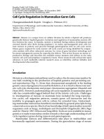
Figure 1.1 Categories of lipids with examples. (A) Fatty acyl chains which are
the components of lipids' hydrophobic fatty acid chains. (B) Neutral lipids which
consist of (i) glycerolipids, (ii) sterols and (iii) waxes. (C) Polar lipids which
consist of (i) glycerophospholipids (phospholipids for short) with two fatty acyl
chains and a phosphate headgroup which defines the lipid class; (ii) sphingolipids
which consist mainly of ceramides and sphingomyelin. Chemical structures are
obtained from LipidMaps.
A. Fatty acyls
Fatty acid (R- carbon chain of any length or
unsaturated bonds
(i) Glycerolipids
Triacylglyceroldiacylglycerol
(ii) Sterol lipids
Cholesterol
24-hydroxy Cholesterol
B. Neutral lipids
(iii) Waxes
Fatty ester
Fatty alcohol
Ethanolamine
(PE)
Serine
(PS)
Glycerol
(PG)
Inositol
(PI)
Headgroup:
choline (PC)
Types of headgroups:
C. Polar lipids
(ii) Sphingolipids
Ceramide
Sphingomyelin
(i) Glycerophospholipids (phospholipids)
3
Phospholipids are well known for their amphiphilic nature. They are involved in a
diverse array of functions such as signal transduction and execution of both
cellular proliferation and death programs, and are major structural components of
cellular membranes. The plasma membrane defines the outer most boundary for
the chemistry of life, while the inner membrane systems or organelles provide an
organisational framework to compartmentalise chemical reactions, ie. cellular
metabolism. Alterations in membrane lipid composition have an impact on a
broad range of cellular functions, like membrane permeability, transport systems,
activity of membrane-bound enzymes, cell growth, proliferation and viability
(Spector and Yorek, 1985). It is the combination of these activities, membrane
biogenesis and energy metabolism, that enable cells to grow and multiply. Hence,
it is logical to assume that cells need to coordinate membrane biogenesis with
basic metabolism to ensure successful replication with transfer of their genes
(Loewen, 2012).
1.1.2 Mass spectrometry
The end of the 20
th
century was marked by the genomics revolution, where the
human genome was finally unraveled. Proteomics was the hottest topic in the
beginning of the 21
st
century, with a lot of efforts trying to link genomics and
proteomics, thereby creating the field of transcriptomics. Genomic and proteomic
advances have shown the necessity to explore metabolic processes at the system
level (Ivanova et al., 2009). However, genes and transcripts do not always predict
the levels of active proteins or enzymes (Dennis, 2009), and hence, are not good
indicators of metabolite levels. It has also been understood that metabolite
4
concentrations represent sensitive markers of both genetic and phenotypic
changes (Cuperlovic-Culf et al., 2010). Hence, metabolomics came into the
picture, with lipidomics being a part of it.
Lipidomics is the systematic identification and analysis of the lipid molecular
species of a cell, living tissue or whole organism with emphasis on quantitating
compositional changes in response to perturbations, like cancer growth or drug-
induced alteration in metabolism (Brown, 2012). It is deemed as a logical
outcome of the history and traditions of lipid biochemistry, where mass
spectrometry technical advances play critical roles in providing a deeper
understanding of the cellular functions of lipids (Ivanova et al., 2009).
Mass spectrometry, with high sensitivity, specificity, selectivity and speed, is an
ideal tool to analyse lipids (Milne et al., 2006). The development of “soft”
ionization methods like matrix-assisted laser desorption/ionization (MALDI) and
electrospray ionization (ESI), and tandem mass spectrometry (MS-MS) made
lipid detection and measurement more accurate and comprehensive (Milne et al.,
2006; Wenk, 2005). Measurements of individual lipid species in a complex lipid
mixture is now possible, with minimal sample processing needed. Lipid analytes
and the matrix have good solubility in organic solvents, resulting in excellent
signal-to-noise ratios and reproducibility (Wenk, 2005). MALDI-TOF (time-of-
flight) has been successfully used to image the phospholipid distribution in tissues
(Malmberg et al., 2007; Puolitaival et al., 2008; Richter et al., 2007). However,
ESI is now more commonly chosen to profile complex lipid mixtures, as it offers
high sensitivity and specificity for a wide range of lipids (Wenk, 2005), including
5
phospholipids (Brügger et al., 1997), sphingolipids (Sullards and Merrill, 2001)
and even non-polar lipids like triacylglycerides, diacylglycerides (Han and Gross,
2001) and sterols. ESI involves the production of lipid ions by evaporating off
solvents in which the lipids are dissolved. The lipid ions can be generated by
adding a proton [M+H]
+
, by adducting a cation like Na
+
to form [M+Na]
+
, or by
removing proton to form [M-H]
-
. These ions are then channelled to mass
analysers such as a quadrupole or TOF. There, the lipid ions are selectively
filtered based on their mass-to-charge ratio (m/z), and will exit the analyser at
given voltages to reach the detector (Cui and Thomas, 2009).
Analysis of known substances can be done with limited range scanning and
selected ion monitoring. Tandem (MS-MS) method provides higher sensitivity for
comprehensive lipid analysis. It involves multiple steps of ion selection with
fragmentation between stages. The most commonly used triple quadrupole (QqQ)
consists of two mass analysers, Q1 and Q3, separated by a collision-induced
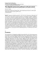
dissociation (CID) chamber (Cui and Thomas, 2009). Figure 1.2 shows a
schematic diagram of the process. Multiple reaction monitoring (MRM), which
monitors both Q1 and Q3 mass characteristic of each molecule, is often used for
quantification of already-known molecules. Product or daughter ions arising from
both positive and negative ion mode fragmentation processes yield a lot of
information on fatty acid, lysolipid and head-group related fragments which are
specific for each lipid type (Ivanova et al., 2009). This method offers a more
sensitive and accurate measurement of specified molecules than other MS
methods like the precursor ion (Q1) scan (Nakanishi et al., 2009). For instance,
6
PC 34:0 (phosphatidylcholine with 34 carbon atoms in its two saturated fatty acyl
chains) and PS 34:1 (phosphatidylserine with a total of 34 carbon atoms in its two
fatty acyl chains with 1 unsaturated bond) are isobaric in positive mode and not
distinguished by MS1 (Q1) alone. MS-MS is therefore needed to separate the two
species, based on their characteristic daughter ions in MS2 (Q3). In addition,
MRM can be quantitative when used with relevant internal standards (Wenk,
2005).
Figure 1.2 Schematic diagram of MRM in a triple quadrupole. The parent ion
with the intended m/z is selected in Q1 and sent for fragmentation in q2 where
collision-induced dissociation (CID) takes place. The parent ion is fragmented
into many smaller ions. The daughter or fragmented ion with a specific m/z
characteristic of the desired molecule is then allowed to enter Q3 and reach the
detector.
Apart from the presence of isobaric species, some of the limitations in MS
analysis include ion suppression and exact lipid identification. These would
require additional steps to resolve. For instance, separation of the complex lipid
mixture is enhanced by the addition of chromatography prior to the analyte's entry
into the mass spectrometer. This results in less ion suppression, high ionisation
yield and better sensitivity for minor species (Ivanova et al., 2009). Apart from
the traditional thin layer chromatography and gas chromatography (GC), high
Parent
mass
CID
Fragment
mass
Q1 Q3q2
detector
7
performance liquid chromatography (HPLC) including normal phase and reverse
phase chromatography has introduced more possibilities into the science of
separating lipids based on their chemical properties more efficiently and
selectively. The former separates lipids based on their headgroups, while the latter
separates lipids by the fatty acid composition (Pulfer and Murphy, 2003). More
recently, chromatographic columns have evolved to be more efficient by having
new column chemistries and reduced particle size (Wenk, 2005).
1.2 Cell cycle and lipids
The eukaryotic cell division cycle is one of the most rigorously studied biological
processes. It is divided into the sequential G1 (Gap 1), S (Synthesis) and G2 (Gap
2), collectively known as interphase, and M (mitotic) phases. In appropriate cues,
cells may exit from the cell cycle and enter quiescent state (G0) or proceed into
the next cycle. The two main events in the cell cycle are DNA synthesis in S
phase, and the division of the parent cell into two daughter cells in the M phase.
G1 and G2 are periods in the cell cycle where the cell prepares to replicate its
genomic material and then to divide respectively. The transition from one phase
of the cell cycle to another occurs in a unidirectional fashion and is traditionally
known to be regulated mainly by cyclins and cyclin-dependent kinases (Cdk) in a
spatial and temporal manner (Bollen et al., 2009; Malumbres, 2011). Different
phases of the cell cycle are controlled mainly by a unique pair of Cdk-cyclin.
While Cdk protein levels remain consistent throughout the cell cycle, protein
levels of their activating partners cyclins fluctuate during the cell cycle. This
8
results in the periodic activation of Cdk, which leads to the progression of cell
cycle (Figure 1.3) (Vermeulen et al., 2003).
Figure 1.3 The stages of the cell cycle. The respective Cdk/cyclin complexes
that are essential for cell cycle progression are indicated (Vermeulen et al., 2003).
Building of a new cell is an extraordinarily energy-consuming task. It is
dependent on various metabolic and biosynthetic processes, most of which are
crucial for biomass accumulation (Cai and Tu, 2012). In order to progress through
the cell cycle smoothly and eventually divide into two, the cell should ideally
duplicate its contents, which would then be distributed to the daughter cells. Two
main biosynthetic activities required by proliferating cells are ribose-5-phosphate
synthesis for nucleotides, and fatty acid synthesis for lipids, both of which are
G1
S
G2
M
Cdk
4/6
Cyclin
D
Cdk
2
Cyclin
E
Cdk
2
Cyclin
A
Cdk
1
Cyclin
A
Cdk
1
Cyclin
B
G0
Cell
Cycle
9
linked to glucose and glutamine metabolism (DeBerardinis et al., 2008). More and
more links have been made between cell metabolism and cell cycle control
(DeBerardinis et al., 2008; Vander Heiden et al., 2009). For instance, cyclins have
been linked to cell metabolism. Gene expression from cyclin D1- knockout mice
showed cyclin D1's inhibitory effects on numerous target genes in glycolysis,
lipogenesis and mitochondrial activity. Cyclin D1 knockdown in breast cancer
cells caused an increase in pyruvate kinase levels, thereby promoting glycolysis.
Levels of fatty acid synthase (FAS) and acetyl-coA carboxylase (ACC), which are
essential for fatty acid biosynthesis, were also increased (Sakamaki et al., 2006).
How metabolic changes affect the cell cycle and vice versa has become an
increasingly important question, with lipids being one of the major metabolites.
Cell cycle plays essential roles in various critical biological events, ranging from
multicellular development, wound healing and regeneration to gametogenesis
(Cho et al., 2001). Functional genomics studies have been carried out on various
biological models like bacteria (Laub et al., 2000), yeasts (Cho et al., 1998;
Spellman et al., 1998), primary cells (Bar-Joseph et al., 2008; Cho et al., 2001)
and cancer cell lines (Whitfield et al., 2002). These studies shed light into almost
all possible aspects of cell cycle control, including DNA replication in S phase,
sister chromatids segregation in mitosis, extracellular matrix remodelling in
cytokinesis. Efforts to plough through these vast amount of genomics data for
lipid related genes in human cell cycle have been unfulfilling, with only a subset
of genes of lipid related proteins being found to be regulated cyclically along with
human cell cycle (Figure 1.4).
10
Figure 1.4 Summary of genes of lipid related proteins that are found to be
regulated in the cell cycle in two published cell cycle genomics study.
Since lipids are the major constituent of cellular membranes, one would expect
cells to double their phospholipid mass during cell cycle in order for membranes
to be distributed evenly between the two daughter cells. Several groups have
attempted to understand how membrane phospholipid metabolism is regulated
within the cell cycle. Findings are so far contradictory, and seem to be cell type
specific. PCs are the main component of cellular membrane phospholipids
(Jackowski, 1996). Jackowski (1994) observed periodic membrane PC
degradation and synthesis during the cell cycle in a human macrophage cell line.
G1 cells rapidly synthesise and degrade PC, while maintaining a constant total
membrane phospholipid mass. PC degradation ceases in S phase to allow cells to
double their membrane phospholipid content for cell division. The activity of the
11
rate-limiting PC synthesis enzyme CTP:phosphocholine cytidylyltransferase is the
lowest in G2/M and it correlates with the cessation of phospholipid synthesis in
G2/M (Jackowski, 1994). However, it was later reported that radiolabeled
phospholipid precursors were rapidly incorporated during G2/M in breast cancer
cell line MCF7 and Chinese Hamster Ovary (CHO) cells, suggesting that lipid
synthesis occurs at G2/M as well. Key PC and PE biosynthetic enzymes,
including CTP:phosphocholine cytidylyltransferase, were also found to be highly
active at G2/M (Lin and Arthur, 2007).
A scanning electron microscopy (SEM) examination of the cell surface changes in
HeLa cells in different cell cycle stages reveals that the cell surface morphologies
are characteristic in each cell cycle phase (Lundgren and Roos, 1976). For
instance, mitotic cells were spherical and covered with microvilli structures of
varying length. As the cells entered G1, they appeared to be flatter, with shorter
microvilli and more blebs. All these morphological changes may be closely
associated with changes in the lipid profiles of the membranes at different stages
of the cell cycle. This is because each phospholipid has a different molecular
shape and they each form a different polymorphic phase depending on the overall
geometry when put together (Dowhan et al., 2008). For example, PC,
sphingomyelin (SM), PS, PI, phosphatidylglycerol (PG) are cylindrical in shape,
hence, they have the tendency to form bilayer phases. Phosphatidylethanolamine
(PE), on the other hand, is cone shaped. In the presence of PE, membranes tend to
form hexagonal phases. Lysophospholipids are inverted cone shaped, therefore
they form micelles readily.