Tổng quan về điện tâm đồ
Bạn đang xem bản rút gọn của tài liệu. Xem và tải ngay bản đầy đủ của tài liệu tại đây (1.29 MB, 24 trang )
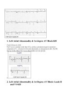
1. Left Atrial Abnormality & 1st degree AV Block-KH
Frank G.Yanowitz, M.D.
The P-wave is notched, wider than 0.12s, and has a prominent negative (posterior)
component in V1 - all criter for left atrial abnormality or enlargement (LAE). The PR
interval >0.20s. Minor ST-T wave abnormalities are also present.
2.
2. Left Atrial Abnormality & 1st Degree AV Block: Leads II
and V1-KH
Frank G.Yanowitz, M.D.
3
3. Left Atrial Enlargement & Nonspecific ST-T Wave
Abnormalities-KHFrank G.Yanowitz, M.D.
LAE is best seen in V1 with a prominent negative (posterior) component measuring 1mm
wide and 1mm deep. There are also diffuse nonspecific ST-T wave abnormalities which
must be correlated with the patient's clinical status. Poor R wave progression in leads V1-
V3, another nonspecific finding, is also present.
Left Atrial Enlargement: Leads II and V1-KHFrank
G.Yanowitz, M.D.
4
4. LVH and Many PVCs-KHFrank G.Yanowitz, M.D.
The combination of voltage criteria (SV2 + RV6 >35mm) and ST-T abnormalities in V5-6
are definitive for LVH. There may also be LAE as evidenced by the prominent negative P
terminal force in lead V1. Isolated PVCs and a PVC couplet are also present.
5. Severe RVHFrank G. Yanowitz, M.D. Copyright 1998
RVH features include the marked right axis deviation (+150 degrees), qR complex in lead
V1, R:S ratio in V6 <1, and right precordial lead ST depression.
Left Atrial Enlargement-KHFrank Yanowitz Copyright 1996
Left atrial enlargement is illustrated by increased P wave duration in lead II, top ECG, and
by the prominent negative P terminal force in lead V1, bottom tracing.
6. LVH - Best seen in the frontal plane leads!-KH
Frank G. Yanowitz, M.D. copyright 1997
7. LVH: Strain pattern + Left Atrial Enlargement-KH
Frank G. Yanowitz, M.D. copyright 1997
8. RVH with Right Axis Deviation
Frank G. Yanowitz, M.D. copyright 1997
Note the qR pattern in right precordial leads. This suggests right ventricular pressures greater
than left ventricular pressures. The persistent S waves in lateral precordial leads and the
RAD are other finding in RVH.
9.
9. Right Ventricular Hypertrophy (RVH) & Right Atrial
Enlargement (RAE)-KHFrank G.Yanowitz, M.D.
In this case of severe pulmonary hypertension, RVH is recognized by the prominent
anterior forces (tall R waves in V1-2), right axis deviation (+110 degrees), and "P
pulmonale" (i.e., right atrial enlargement). RAE is best seen in the frontal plane leads; the
P waves in lead II are >2.5mm in amplitude.
Right Axis Deviation & RAE (P Pulmonale): Leads I, II, III-
KH
10.
10. Right Atrial Enlargement (RAE) & Right Ventricular
Hypertrophy (RVH)-KHFrank G.Yanowitz, M.D.
RAE is recognized by the tall (>2.5mm) P waves in leads II, III, aVF. RVH is likely
because of right axis deviation (+100 degrees) and the Qr (or rSR') complexes in V1-2.
RAE & RVH-KH
11.
11. LVH with "Strain"-KHFrank G. Yanowitz, M.D.,
copyright 1997
12.
12. LVH & PVCs: Precordial Leads-KH .Frank G.Yanowitz,
M.D.
13.
13. LVH: Limb Lead Criteria-KH Frank G.Yanowitz, M.D.
In this example of LVH, the precordial leads don't meet the usual voltage criteria or
exhibit significant ST segment abnormalities. The frontal plane leads, however, show
voltage criteria for LVH and significant ST segment depression in leads with tall R waves.
The voltage criteria include 1) R in aVL >11 mm; 2) R in I + S in III >25mm; and 3)
(RI+SIII) - (RIII+SI) >17mm (Lewis Index).
LVH: Limb Lead Criteria-KH
In this example of LVH, the precordial leads don't meet the
usual voltage criteria or exhibit significant ST segment
abnormalities. The frontal plane leads, however, show
voltage criteria for LVH and significant ST segment
depression in leads with tall R waves. The voltage
criteria include 1) R in aVL >11 mm; 2) R in I + S in III
>25mm; and 3) (RI+SIII) - (RIII+SI) >17mm (Lewis
Index).
1. Right Atrial Enlargement (RAE)
P wave amplitude >2.5 mm in II and/or >1.5 mm in V1 (these
criteria are not very specific or sensitive)
Better criteria can be derived from the QRS complex; these
QRS changes are due to both the high incidence of RVH when
RAE is present, and the RV displacement by an enlarged right
atrium.
QR, Qr, qR, or qRs morphology in lead V1
(in absence of coronary heart disease)
QRS voltage in V1 is <5 mm and V2/V1
voltage ratio is >6 (Sensitivity = 50%;
Specificity = 90%)
In the above ECG, note the tall P waves in Lead II, and the Qr
wave in Lead V1.
2. Left Atrial Enlargement (LAE)
P wave duration > 0.12s in frontal plane (usually lead II)
Notched P wave in limb leads with the inter-peak duration >
0.04s
Terminal P negativity in lead V1 (i.e., "P-terminal force")
duration >0.04s, depth >1 mm.
Sensitivity = 50%; Specificity = 90%
3. Bi-Atrial Enlargement (BAE)
Features of both RAE and LAE in same ECG
P wave in lead II >2.5 mm tall and >0.12s in duration
Initial positive component of P wave in V1 >1.5 mm tall and prominent P-
terminal force
1. Introductory Information:
The ECG criteria for diagnosing right or left ventricular
hypertrophy are very insensitive (i.e., sensitivity ~50%, which
means that ~50% of patients with ventricular hypertrophy
cannot be recognized by ECG criteria). However, the criteria
are very specific (i.e., specificity >90%, which means if the
criteria are met, it is very likely that ventricular hypertrophy is
present).
2. Left Ventricular Hypertrophy (LVH)
General ECG features include:
> QRS amplitude (voltage criteria; i.e., tall R-waves in LV leads,
deep S-waves in RV leads)
Delayed intrinsicoid deflection in V6 (i.e., time from QRS onset to
peak R is >0.05 sec)
Widened QRS/T angle (i.e., left ventricular strain pattern, or ST-T
oriented opposite to QRS direction)
Leftward shift in frontal plane QRS axis
Evidence for left atrial enlargement (LAE) (lessonVII)
ESTES Criteria for LVH ("diagnostic", >5 points; "probable", 4 points)
CORNELL Voltage Criteria for LVH (sensitivity = 22%,
specificity = 95%)
S in V3 + R in aVL > 24 mm (men)
S in V3 + R in aVL > 20 mm (women)
Other Voltage Criteria for LVH
Limb-lead voltage criteria:
+ECG Criteria Points
Voltage Criteria (any of):
a. R or S in
limb leads
>20 mm
b. S in V1 or
V2 > 30
mm
c. R in V5 or
V6 >30
mm
3 points
ST-T Abnormalities:
Without digitalis
With digitalis
3 points
1 point
Left Atrial Enlargement in V1 3 points
Left axis deviation 2 points
QRS duration 0.09 sec 1 point
Delayed intrinsicoid deflection in
V5 or V6 (>0.05 sec)
1 point
R in aVL >11 mm or, if left axis deviation, R in aVL >13 mm
plus S in III >15 mm
R in I + S in III >25 mm
Chest-lead voltage criteria:
S in V1 + R in V5 or V6 >
35 mm
Example 1: (Limb-lead Voltage Criteria; e.g., R in aVL >11 mm; note
wide QRS/T angle)
clic
k here to view
Example 2: (ESTES Criteria: 3 points for voltage in V5, 3 points for ST-T
changes)
(Note also the left axis deviation of -40 degrees, and left atrial
enlargement)
3. Right Ventricular Hypertrophy
General ECG features include:
Right axis deviation (>90 degrees)
Tall R-waves in RV leads; deep S-waves in LV leads
Slight increase in QRS duration
ST-T changes directed opposite to QRS direction (i.e., wide QRS/T
angle)
May see incomplete RBBB pattern or qR pattern in V1
Evidence of right atrial enlargement (RAE) (lessonVII)
Specific ECG features (assumes normal calibration of 1 mV = 10 mm):
Any one or more of the following (if QRS duration <0.12 sec):
Right axis deviation (>90 degrees) in presence of disease
capable of causing RVH
R in aVR > 5 mm, or
R in aVR > Q in aVR
Any one of the following in lead V1:
R/S ratio > 1 and negative T wave
qR pattern
R > 6 mm, or S < 2mm, or rSR' with R' >10 mm
Other chest lead criteria:
R in V1 + S in V5 (or V6) 10 mm
R/S ratio in V5 or V6 < 1
R in V5 or V6 < 5 mm
S in V5 or V6 > 7 mm
ST segment depression and T wave inversion in right precordial leads is
usually seen in severe RVH such as in pulmonary stenosis and pulmonary
hypertension.
Example #1: (note RAD +105 degrees; RAE; R in V1 > 6 mm; R in aVR > 5 mm)
Example #2: (more subtle RVH: note RAD +100 degrees; RAE; Qr
complex in V1 rather than qR is atypical)
Example #3: (note: RAD +120 degrees, qR in V1; R/S ratio in V6 <1)
4. Biventricular Hypertrophy (difficult ECG diagnosis to make)
In the presence of LAE any one of the following suggests this diagnosis:
R/S ratio in V5 or V6 < 1
S in V5 or V6 > 6 mm
RAD (>90 degrees)
Other suggestive ECG findings:
Criteri
a for LVH and RVH both met
LVH criteria met and RAD or RAE present
It’s a PAC with RBBB aberration
F’ is for “fusion beat”; i.e. the fusion of a left ventricular PVC with the sinus initiated QRS
complexThe subsequent ventricular ectopics are
upgoing (anterior oriented) QRSs, suggestion
origin from the LV
This is a ventricular tachycardia with intermittent 2:1 exit block.The longer RR
intervals are twice the short intervals suggesting
that not every impulse form the ventricular focus
makes it out to the rest of the ventricles.
The first FLB is a late onset PVC, and the other three are fusion beats.Late PVCs
often occur coincidentally with sinus activation of
the ventricles. The degree of fusion may vary as
seen in this example.
2nd degree AV blockSome P waves conduct, and some do not
The ‘e’ represents a junctional escape beat; the ‘c’ represents a sinus capture.
Sometimes this goes by the name of “escape-capture
bigeminy”. Any pause in the rhythm may result in
an escape beat if the pause is too long
Sinus rhythm with 1st degree AV block; occasional PVCThanks to the PVC and
resulting pause, the sinus P wave becomes
separated form the preceding T wave. The 1st
degree AV block is quite marked.
Nonconducted PACsThis is the most common cause of an
unexpected pause in the rhythm. The P-waves of
the PACs are early relative to the sinus PP
intervals.
A junctional escape complexActually the sinus P wave is seen
partially superimposed on the junctional escape
beat thereby distorting the onset of the QRS.
2nd degree AV block type II (Mobitz)The PR intervals for two
consecutive beats are constant, followed by a
blocked sinus P wave. The QRS is wide
suggesting a bundle branch block