MECHANISMS OF BINDING DIVERSITY IN PROTEIN DISORDER: MOLECULAR RECOGNITION FEATURES MEDIATING PROTEIN INTERACTION NETWORKS
Bạn đang xem bản rút gọn của tài liệu. Xem và tải ngay bản đầy đủ của tài liệu tại đây (3.02 MB, 118 trang )
MECHANISMS OF BINDING DIVERSITY IN PROTEIN DISORDER:
MOLECULAR RECOGNITION FEATURES MEDIATING
PROTEIN INTERACTION NETWORKS
Wei-Lun Hsu
Submitted to the faculty of the University Graduate School
in partial fulfillment of the requirements
for the degree
Doctor of Philosophy
in the Department of Biochemistry and Molecular Biology,
Indiana University
July 2013
ii
Accepted by the Faculty of Indiana University, in partial
fulfillment of the requirements for the degree of Doctor of Philosophy.
A. Keith Dunker, Ph.D., Chair
Yaoqi Zhou, Ph.D.
Doctoral Committee
Thomas D. Hurley, Ph.D.
April 23, 2013
Vladimir N. Uversky, Ph.D.
iii
© 2013
Wei-Lun Hsu
ALL RIGHTS RESERVED
iv
ACKNOWLEDGEMENTS
I would like to take the opportunity to thank all the people who provided me with
their help and support. I fully appreciated what they have done for me.
I would like to give my sincere gratitude to my adviser, Dr. A. Keith Dunker for
his unreserved support and patient instruction during the past few years. His passion in
research and outstanding accomplishment in science inspire me in many aspects. The
great enthusiasm to the academic society he has especially makes me ways. Under
Keith’s guidance, I learned and was trained to combine bioinformatics analysis and
laboratory experimentation to do intrinsically disordered protein research, which gives
me a broad view to evaluate complicated biological questions in a systematic way. I
really appreciate all the help Keith offered while I was in the most difficult time in my
life. Without his support, I could not accomplish my dream to study in the U.S. In the
meanwhile, Keith is also a good instructor to train and encourage students to develop
their own innovative ideas and figure out solutions independently. He helped a lot to
shape me and show me how to approach problems. I am so lucky to have Keith as my
mentor that I could have the chance to explore my research interests, broaden my skill set
and figure out my future career plan upon completion of my Ph.D. study.
I also want to thank my research committee, Dr. Vladimir N. Uversky, Dr. Yaoqi
Zhou, Dr. Thomas D. Hurley and Dr. Pedro Romero for their valuable suggestions and
comments to help develop my thesis work. I would also like to show my thankfulness to
the Biochemistry and Molecular Biology department for continuing supporting in
students’ research and career development. I appreciated all the assistance from other
v
faculty members in our department as well, including Dr. Georgiadis, Dr. DePaoli-Roach,
Dr. Goebl, Dr. Meroueh, Dr. Zhang, Dr. Wek, Dr. Hoang and Dr. Takagi.
In addition, I want to say thanks to all the members in Dr. Dunker’s laboratory.
Without their support, I can’t accomplish what I have done. Thank you, Chris, Jingwei,
Bin, Eshel, Caron, Fei, Maya and Bo for always being my technical and mental support. I
also appreciated the chance to collaborate with other researchers outside of Indiana
University. I thank Dr. Sarah Bondos and Hao-Ching Hsiao at Texas A&M University
for sharing their fantastic work regarding to partner selection of Ubx protein, Dr. Lukasz
Kurgan and Fatemeh Miri Disfani at the University of Alberta for their development of
the MoRFpred disordered binding site predictor, Dr. Gil Alterovitz and Jonah Kallenbach
in Harvard Medical School for working together to construct the MoRF-partner binary
predictor.
Finally, I want to thank Yayue, Yunlong, Fucheng, Baohua, Hongying, Wenyan,
Sue, Shelly, Yan, Yanlu, my family and friends for their endless support. Thank you all!
vi
PREFACE
To innocence, and curiosity…
vii
ABSTRACT
Wei-Lun Hsu
Mechanisms of Binding Diversity in Protein Disorder: Molecular Recognition Features
Mediating Protein Interaction Networks
Intrinsically disordered proteins are proteins characterized by lack of stable
tertiary structures under physiological conditions. Evidence shows that disordered
proteins are not only highly involved in protein interactions, but also have the capability
to associate with more than one partner. Short disordered protein fragments, called
“molecular recognition features” (MoRFs), were hypothesized to facilitate the binding
diversity of highly-connected proteins termed “hubs”. MoRFs often couple folding with
binding while forming interaction complexes. Two protein disorder mechanisms were
proposed to facilitate multiple partner binding and enable hub proteins to bind to multiple
partners: 1. One region of disorder could bind to many different partners (one-to-many
binding), so the hub protein itself uses disorder for multiple partner binding; and 2. Many
different regions of disorder could bind to a single partner (many-to-one binding), so the
hub protein is structured but binds to many disordered partners via interaction with
disorder. Thousands of MoRF-partner protein complexes were collected from Protein
Data Bank in this study, including 321 one-to-many binding examples and 514 many-to-
one binding examples. The conformational flexibility of MoRFs was observed at atomic
resolution to help the MoRFs to adapt themselves to various binding surfaces of partners
or to enable different MoRFs with non-identical sequences to associate with one specific
viii
binding pocket. Strikingly, in one-to-many binding, post-translational modification,
alternative splicing and partner topology were revealed to play key roles for partner
selection of these fuzzy complexes. On the other hand, three distinct binding profiles
were identified in the collected many-to-one dataset: similar, intersecting and
independent. For the similar binding profile, the distinct MoRFs interact with almost
identical binding sites on the same partner. The MoRFs can also interact with a partially
the same but partially different binding site, giving the intersecting binding profile.
Finally, the MoRFs can interact with completely different binding sites, thus giving the
independent binding profile. In conclusion, we suggest that protein disorder with post-
translational modifications and alternative splicing are all working together to rewire the
protein interaction networks.
A. Keith Dunker, Ph.D., Committee Chair
ix
TABLE OF CONTENTS
List of Tables xi
List of Figures xii
List of Abbreviations xiv
Chapter 1: Introduction
1.1. Intrinsic Protein Disorder and Protein Functions 1
1.2. Intrinsic Protein Disorder in Protein-Protein Interactions 4
1.3. Characterization of Molecular Recognition Features (MoRFs) and their Binding
Partners 5
1.4. MoRFs in PDB: Their Length, delta ASA and Secondary Structures 6
1.5. Validation on MoRFs (Gunasekaran-Tsai-Nussinov Graph) 9
1.6. Two MoRF Mechanisms in Hub Proteins 10
1.7. Importance of Understanding the MoRF Mechanisms in Hub Proteins 13
Chapter 2: Materials and Methods
2.1. MoRF Datasets Preparation 17
2.2. Characterization of MoRF Clusters that Perform One-to-Many and Many-to-One
Binding 17
2.3. Removal of Redundant MoRFs in MoRF Clusters 20
2.4. Removal of Atypical MoRFs in MoRF Clusters 20
2.5. Secondary Structure Assignment on MoRFs 20
2.6. Sequence and Structure Similarity Analyses 20
2.7. Peptide-Protein Interaction Annotation 21
x
2.8. SCOP Classification of MoRF Partners 22
2.9. Network Analysis of MoRF Dataset 22
Chapter 3: Binding Diversity of Intrinsic Protein Disorder
3.1. One-to-Many Binding 24
3.1.1. Fifteen MoRF Sets with Similarly-Folded Partners 31
3.1.2. Eight MoRF Sets with Differently-Folded Partners 45
3.1.3. Alternative Splicing and Posttranslational Modifications in One-to-Many
Binding 56
3.2. Many-to-One Binding 59
3.2.1. Peptide-Protein Interactions and Protein-Protein Interactions 61
3.2.2. Binding Profiles: Independent and Overlapping (Similar vs. Intersecting) 64
3.2.3. Structurally Conserved MoRFs with Diverse Sequences 70
3.2.4. Selected Many-to-One Case Studies 73
3.2.5. Examples of Retro-MoRF and PP1-like MoRF 76
3.3. Many-to-Many Binding 78
Chapter 4: SCOP Folds of MoRF Partners
4.1. Partner Folds Selection in each MoRF Types 80
Chapter 5: Conclusion 84
References 91
Curriculum Vitae
xi
LIST OF TABLES
Table 1 7
Table 2 25
Table 3 26
Table 4 28
Table 5 31
Table 6 59
Table 7 60
Table 8 63
Table 9 67
Table 10 74
Table 11 76
Table 12 76
Table 13 77
Table 14 78
xii
LIST OF FIGURES
Figure 1 2
Figure 2 7
Figure 3 8
Figure 4 8
Figure 5 9
Figure 6 19
Figure 7 27
Figure 8 38
Figure 9 40
Figure 10 43
Figure 11 44
Figure 12 46
Figure 13 48
Figure 14 50
Figure 15 54
Figure 16 63
Figure 17 63
Figure 18 65
Figure 19 68
Figure 20 69
Figure 21 72
xiii
Figure 22 74
Figure 23 75
Figure 24 77
Figure 25 77
Figure 26 82
xiv
LIST OF ABBREVIATIONS
MoRF Molecular Recognition Feature
IDP Intrinsically Disordered Protein
NMR Nuclear magnetic resonance
ANS 1-Anilino-8-naphthalene-sulfonate
PTM Post Translational Modification
IDR Intrinsically Disordered Region
ASE Alternative Splicing Event
ELM Eukaryotic Linear Motif
LM Linear Motif
SLiM Short Linear Motif
RISP Regions of Increased Structural Propensity
PDB Protein Data Bank
RMSD Root Mean Square Deviation
OR Overlap Ratio
CI Confidence Interval
SCOP Structural Classification of Proteins
NR Nuclear Receptor
PPI Protein-Protein Interaction
UniProt Universal Protein Resource
iMoRF Immune-Related MoRF
1
CHAPTER 1
Introduction
1.1. Intrinsic Protein Disorder and Protein Functions
Intrinsically disordered proteins (IDPs) are a group of proteins that lack stable
tertiary structures either partially or in their entirety. Their structural conformations are
too dynamic to be described by a single conformation under physiological conditions.
IDPs still can be identified by more than 40 experimental methods, such as x-ray
crystallography (missing density), Nuclear magnetic resonance (NMR) (lack of chemical
dispersion in 1H-15N NOEs), far-UV (170-250nm) circular dichroism (lack of secondary
structure), protease sensitivity (readily cleaved by proteases), 1-Anilino-8-naphthalene-
sulfonate (ANS) binding (lack of hydrophobic cores) and so on. Protein disorder has
been found to exist in nature as disordered tails, linkers, domains, or entirely unfolded as
collapsed or extended forms (Figure 1) [1]. The existence of IDPs challenge the
traditional biochemistry view of sequence-structure-function paradigm since these
proteins still carry out important biological functions without well-defined structures. In
other words, the structure of a protein may not always define its function or a single
unique structure cannot describe their function. However, in some cases, these disordered
regions can adopt specific three dimensional structures after binding to another molecule.
There are some possible reasons why IDPs lack stable structures. Some researchers
believe IDPs are unstructured only when lacking a ligand/partner or other factors that
promote their folding, but others, including our laboratory, believe IDPs’ lack of structure
is encoded by their amino acid sequences just like structured proteins.
2
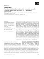
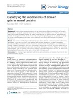
Figure 1. Various forms of protein structures: (A) structured domain, (B) disordered
domain, (C) disordered tails, (D) disordered linker, (E) collapsed disorder and (F)
extended disorder. Red parts of structures imply disordered regions. The diagram is
adapted from DisProt Database [1].
A
B
C
D
E
F
3
IDPs are often referred to using alternative names, such as naturally unfolded
proteins, intrinsically unstructured proteins, flexible/dynamic proteins, conformational
disorder, extended polypeptide, mobile domains, molten globule, random coils or
disordered proteins. Genomics and proteomics studies have revealed protein disorder is
highly abundant in various organisms, such us in humans and viruses. Eukaryotes
generally have higher intrinsically disordered contents than prokaryotes. A quantitative
and qualitative measurement of the extent of protein disorder in 3484 species with known
genomes was performed by Xue et al. [2]. Viruses were found to have the widest spread
of disorder content (from 7.3% in human coronavirus NL63 to 77.3% in avian carcinoma
virus) in their study.
Several studies have revealed the possibility of the hypothesis: protein disorder is
used for signaling because of its unique structural properties. Many bioinformatics
studies claim that disordered proteins involve more in signaling pathway, gene
regulation, molecular recognition and cell control particularly while structured proteins
often involve in catalysis, membrane transport and small molecules binding [3-7].
Many biological events in which disordered proteins participate are found to be
regulated by post translational modifications (PTMs) and alternative splicing events
(ASEs) [8,9]. Fukuchi et al. explored a variety of protein modification events in different
subcellular localizations and found protein disorder are highly enriched in nuclear
proteins (47%) compared to mitochondria proteins (13%) [8]. Also, phosphorylation and
O-linked glycosylation sites were frequently observed to localize in intrinsically
disordered regions (IDRs). They suspected the O-linked glycans are attached to IDRs in
order to protect the protein from proteolytic cleavage in the extracellular environment.
4
Besides PTMs, alternative splicing events (ASEs) have been associated with IDRs by
various laboratories [8,9].
1.2. Intrinsic Protein Disorder in Protein-Protein Interactions
Many proteins execute their biological functions through protein-protein
interactions. By binding to interacting partners, proteins can deliver signals to other
molecules. For example, hormone neurotransmitters and their receptors trigger various
signal transduction pathways following their mutual interaction, antibody recognition of
peptide antigens leads to B-cell activation, and the interaction between G-protein coupled
receptors and G-proteins leads to the transduction of many biological signals.
Protein-protein interaction networks underlie a wide variety of biological
functions, ranging from regulating cell division to responding to external signals. High
throughput methods have enabled researchers to map out sets of protein-protein
interactions over entire proteomes. Mapping protein-protein interactions leads to
networks that are far from random. While most proteins have only a few interacting
partners, the studies reveal complex networks in which a small number of proteins, called
hubs, are observed, to have multiple interacting partners. Indeed, in some cases hubs
bind to 15, 20, 50 or even more partner proteins. As expected for such network
architecture, deletion of a protein with only a few partners is typically less deleterious
than the deletion of a hub protein [10,11].
How do such networks arise from simpler precursors? Other networks of a
similar architecture arise because “the rich get richer”; units with more connections have
a higher probability of adding even more connections over time as compared to the units
with fewer connections. This suggests that highly connected proteins have special
5
features that facilitate their binding to multiple partners and that facilitate binding to new
partners that arise through mutation [12]. What are these special features?
Theoretical arguments [13,14] and experimental data [15,16] suggest that
unfolded or disordered protein can very readily change shape and thereby easily adapt to
multiple, distinct partners. The common involvement of disorder in hub proteins’
interactions has been supported by several subsequent studies [17-19]. Intrinsically
disordered proteins often bind to more than one partner. Thus, we proposed that the
special feature of hub proteins enabling their binding to multiple partners is likely to be
intrinsic disorder. In support of IDPs as being important for binding to multiple partners,
both hub proteins and their binding partners are observed to be enriched in disorder [19-
21], and many additional studies support these concepts [17,22-31].
1.3. Characterization of Molecular Recognition Features (MoRFs) and their Binding
Partners
With regard to IDP regions involved in binding, various descriptors have been
used, such as eukaryotic linear motif (ELMs) [32,33], linear motifs (LMs) [34], short
linear motif (SLiMs) [35,36], regions of increased structural propensity (RISPs) [37], and
molecular recognition features (MoRFs) [38]. All of these describe similar phenomena,
despite different approaches used by the various researchers for identification of binding
segments. The identification of ELMs, LMs, or SLiMs start from sequence pattern or
motif-based approaches, whereas the identification of RISPs and MoRFs start from short
regions with binding indicators located within longer regions of predicted disorder. The
motif-based and algorithmic approaches show significant overlap in their identification of
their binding sites [34], suggesting that the different approaches associated with the
6
different names are merely emphasizing different aspects of the same types of binding
interactions.
Because ELMs, LMs, and SLiMs all involve sequence motifs, these binding
regions can be identified by simple pattern recognition methods, albeit with a high error
rate due to their typically short length involving just a few key residues. Predicting
protein-protein interaction sites in proteins can be used to supplement experimental
approaches [39,40]. Predicting binding sites by sequence matches to the motifs of ELMs
[32,33], LMs [34], SLiMs [35,36], or other collections of sequence patterns [41-43]
provides one strategy for identifying potential binding sites located within IDPs or IDP
regions. Using sequence characteristics that indicate short binding regions within longer
regions of disorder offers a second strategy that does not depend on specific motifs, and
several predictors have been developed that use this second strategy [44-48]. Such
predictors have been used by experimentalists to help with the identification of binding
regions within longer regions of disorder [37,49].
1.4. MoRFs in PDB: Their Length, delta ASA and Secondary Structures
Table 1 lists the number of MoRFs we collected in each filtering step in our 2008
and 2012 datasets. The criteria we used for screening MoRFs are slightly different in two
aspects: the length of MoRF partners and the exact sequence we use for sequence
alignment. Basically, the MoRF dataset grew about 2.7 folds over the past 4 years.
7
Table 1. Description of MoRF datasets built in 2008 and 2012.
Data set
March 2008
June 2012
Initial MoRF dataset (5-25)
4289
8084
MoRF dataset with biological interaction (>400Å
2
)
3837
7064
MoRF dataset with globular partner
(>70 vs. > 40)
3148
6171
MoRFs mapped to UniProt
(ATOM vs. SEQRES)
1805
4839
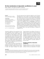
The following Figures (2-4) give us a general overview of our 2008 MoRF dataset
(4289 complexes) on MoRF length, surface area change upon binding (∆ASA) and
secondary structure.
Figure 2. A histogram of MoRF length of the 2008 MoRF dataset.
0
100
200
300
400
500
600
5 6 7 8 9 10 11 12 13 14 15 16 17 18 19 20 21 22 23 24 25
Count
MoRF Length
8
Figure 3. A scatter plot reveals a positive but not significant correlation between MoRF
length and surface area change (∆ASA) upon binding.
Figure 4. A pie chart of different MoRF types based on their secondary structures.
0
500
1000
1500
2000
2500
3000
3500
4000
5 7 9 11 13 15 17 19 21 23 25
∆ASA
MORF Length
17%
5%
73%
4%
1%
helix
sheet
coil
complex
N/A
9
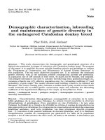
1.5. Validation on MoRFs (Gunasekaran-Tsai-Nussinov Graph)
Gunasekaran et al. developed a protocol [50] that we modified [38] to indicate
whether a MoRF is likely to be disordered when unbound. The Gunasekaran-Tsai-
Nussinov graph provides a scale that measures confidence with which one can say
whether a protein is ordered or disordered. The farther the point, which corresponds to a
given chain, is from the dividing black line (boundary), the greater the confidence with
which a protein can be classified into either of the classes. Points above the line
correspond to disordered chains like Figure 5 shows below. All the 842 MoRFs selected
form our 2008 MoRF dataset (a non-redundant set) are validated as likely to be
disordered before the binding events.
Figure 5. A Gunasekaran-Tsai-Nussinov graph example (adapted from Bioinformatics
28, i75-83).
Disordered
Ordered
10
1.6. Two MoRF Mechanisms in Hub Proteins
We further suggested two ways that disorder could be used by hub proteins for
binding to multiple partners: 1. One region of disorder could bind to many different
partners (one-to-many binding), so the hub protein itself uses disorder for multiple
partner binding; and 2. Many different regions of disorder could bind to a single partner
(many-to-one binding), so the hub protein is structured but binds to many disordered
partners via interaction with disorder [51]. Since this initial proposal, we [19,22,23] and
many others [20,21,24-31,52] have provided additional evidence that hubs and/or their
binding partners are especially enriched in intrinsic disorder, with both the many-to-one
and one-to-many processes involving the use of intrinsic disorder.
The C-terminal region of p53 uses disorder to bind to more than 45 different
proteins and to form a tetramer, but only six of these complexes and the tetramer have
had their structures deposited in the Protein Data Bank (PDB) [46]. One particular p53
segment “SHLKSKKGQSTSRHKKLMFKTE” (residues 367-388), which is both an
ELM and a MoRF and which is located at the C-terminus, morphs into an -helix when
binding with S100ββ, into a -sheet with sirtuin, into an irregular structure with CREB
binding protein (CBP) and into another irregular structure with cyclin A2 as a partner
[46].
Very different biological processes are transduced via these four different
interactions involving the same segment of p53: The CDK2/cyclin A2 complex regulates
progression of S phase of the eukaryote cell cycle by recognizing diverse but structurally
constrained target sequences (KXL/RXL motif) from various substrates, including p53
[53]; deacetylase enzymes like the Sir 2 protein, which is a homologue of Sirtuin, can
11
lead to down-regulation of p53-dependent transcription by binding to the acetylated p53
peptide on lysine 382 [54]; the recognition of acetylated lysine 382 in p53 by the
conserved bromo-domain of transcriptional coactivator CBP is very specific, leading to
the recruitment of p53 acetylation-dependent coactivator following DNA damage and to
the activation of cyclin-dependent kinase inhibitor p21 [55]; dimeric S100 calcium
binding protein B can sterically block the phosphorylation and acetylation sites of on p53
that are critical for the activation important transcription; finally, the peptide derived
from the region of p53 was found to undergo a disorder-to-order conformational change
while binding to Ca2+ loaded S100ββ [56]. Thus, this same intrinsically disordered
segment plays roles in a diverse set of signaling pathways.
The highly conserved 14-3-3 protein family has been reported to associate with
over 200 different but mostly phosphorylated proteins [57]. Phosphorylation plays a
central role in cellular regulation, either by altering a protein’s activity directly or by
inducing specific protein-protein interactions. Protein phosphorylation events are often
coupled with domain-binding motifs, highlighting a potential switch-like function of
phosphorylation. In part, the ability of 14-3-3 to associate with many different proteins is
the result of its specific phospho-serine/phospho-threonine binding activity. These
phosphorylation sites are often surrounded by disorder-promoting residues. From this
observation, a bioinformatics study suggested that over 90% of the 14-3-3 protein
partners do not adopt a defined three-dimensional structure in total or in part [58]. This
implies structural disorder in 14-3-3 partners is the key characteristic for promoting this
binding diversity. But how the 14-3-3 partners have diverged with respect to their
primary structure and yet still maintain binding to 14-3-3 as an unanswered question.