Báo cáo y học: "The dynamic pattern of end-tidal carbon dioxide during cardiopulmonary resuscitation: difference between asphyxial cardiac arrest and ventricular fibrillation/pulseless ventricular tachycardia cardiac arres" ppt
Bạn đang xem bản rút gọn của tài liệu. Xem và tải ngay bản đầy đủ của tài liệu tại đây (518.98 KB, 8 trang )
RESEARCH Open Access
The dynamic pattern of end-tidal carbon dioxide
during cardiopulmonary resuscitation: difference
between asphyxial cardiac arrest and ventricular
fibrillation/pulseless ventricular tachycardia
cardiac arrest
Katja Lah
1,2
, Miljenko Križmarić
2
, Štefek Grmec
1,2,3,4*
Abstract
Introduction: Partial pressure of end-tidal carbon dioxide (PetCO2) during cardiopulmonary resuscitation (CPR)
correlates with cardiac output and consequently has a prognostic value in CPR. In our previous study we
confirmed that initial PetCO2 value was significantly higher in asphyxial arrest than in ventricular fibrillation/
pulseless ventricular tachycardia (VF/VT) cardiac arrest. In this study we sought to evaluate the pattern of PetCO2
changes in cardiac arrest caused by VF/VT and asphyxial cardiac arrest in patients who were resuscitated accordi ng
to new 2005 guidelines.
Methods: The study included two cohorts of patients: cardiac arrest due to asphyxia with initial rhythm asystole or
pulseless electrical activity (PEA), and cardiac arrest due to arrhythmia with initial rhythm VF or pulseless VT. PetCO2
was measured for both groups immediately after intubation and repeatedly every minute, both for patients with or
without return of spontaneous circulation (ROSC). We compared the dynamic pattern of PetCO2 between groups.
Results: Between June 2006 and June 2009 resuscitation was attempted in 325 patients and in this study we
included 51 patients with asphyxial cardiac arrest and 63 patients with VF/VT cardiac arrest. The initial values of
PetCO2 were significantly higher in the group with asphyxial cardiac arrest (6.74 ± 4.22 kilopascals (kPa) versus 4.51
± 2.47 kPa; P = 0.004). In the group with asphyxial cardiac arrest, the initial values of PetCO2 did not show a
significant difference when we compared patients with and without ROSC (6.96 ± 3.63 kPa versus 5.77 ± 4.64 kPa;
P = 0.313). We confirmed significantly higher initial PetCO2 values for those with ROSC in the group with primary
cardiac arrest (4.62 ± 2.46 kPa versus 3.29 ± 1.76 kPa; P = 0.041). A significant difference in PetCO2 values for those
with and without ROSC was achieved after five minutes of CPR in both groups. In all patients with ROSC the initial
PetCO2 was again higher than 1.33 kPa.
Conclusions: The dynamic pattern of PetCO2 values during out-of-hospital CPR showed higher values of PetCO2
in the first two minutes of CPR in asphyxia, and a prognostic value of initial PetCO2 only in primary VF/VT cardiac
arrest. A prognostic value of PetCO2 for ROSC was achieved after the fifth minute of CPR in both groups and
remained present until final values. This difference seems to be a useful criterion in pre-hospital diagnostic
procedures and attendance of cardiac arrest.
* Correspondence:
1
Center for Emergency Medicine Maribor, Cesta proletarskih brigad 21, 2000
Maribor, Slovenia
Full list of author information is available at the end of the article
Lah et al. Critical Care 2011, 15:R13
/>© 2011 Lah et al.; licensee BioMe d Central Ltd. This is an open access article distributed under the terms of the Creative Commons
Attribution License ( which permits unrestricted use, distribution, and reproduction in
any medium, pro vided the original work is properly cited.
Introduction
Capnomet ry and capnography have gained a crucial role
in monitoring critically ill patients in the pre-hospital
setting [1-5]. They can be u sed as a detector for correct
endotracheal tube placement, to monitor the adequacy
of ventilation, ensure a proper nasogastric tube place-
ment, recognize changes in alveolar dead space, help
describe a proper emptying pattern of alveoli, help esti-
mate the deepness of sedation and relaxation in criti-
cally ill, help in diagnostics of severe pulmonary
embolism, and can be used in cardiac arrest patients as
a pr ognostic determinant of outcome and in monitoring
the effectiveness of cardiopulmonary resuscitation (CPR)
[6-11]. In our previous study [12], we found that initial
values of partial pressure of end-tidal carbon dioxide
(PetCO2) in asphyxial arrest were significantly higher
than in ventricular f ibrillation/pulseless ventricular
tachycardia (VF/VT) arrest. In asphyxial arrest there was
also no signi fican t difference in initial values of PetCO2
in patients with and without return of spontaneous cir-
culation (ROSC). In asphy xial arrest the initial values of
PetCO2 cannot be used as a prognostic factor of out-
come of CPR, as they can be in VF/VT arrest [13-16].
This difference, together with other criteria, can t here-
fore be useful for differentiating between the causes of
cardiac arrest in the pre-hospital setting [17]. In this
studywesoughttoevaluatethepatternofPetCO2
changes in cardiac arrest caused by VF/VT and a sphyx-
ial cardiac arrest in patients who were resuscitated
according to new 2005 guidelines [18-20].
Materials and methods
This prospective observational study was conducted at
the Center for Emergency Medicine, Maribor, Slovenia.
To facilitate a true comparison, the design of this study
was identical to our first one. Patients constitute two
cohorts. The study was approved by the Ethical Board
of the Ministry of Health, which granted waiver of
informed consent ( victims of cardiac arrest). Patients
who regained consciousness or their relatives were
informed after enrollment.
The first cohort included patients who suffered from
cardiac arrest due to asphyxia. The causes of asphyxia
were: asthma, severe acute respiratory failure, tumor of
the airway, suicide by hanging, acute intoxication, pneu-
monia and a foreign body in the airway. The definitive
cause of c ardiac arrest was confirmed in the h ospital by
further diagnostic and/or pat hology reports (post
mortem). The initial rhythm seen on the monitor for all
the patients in this group was either asystole or pulseless
electrical activity. We excluded patients in severe
hypothermia (core temperature <30°C) and patients with
incomplete measurements of PetCO2 in the first
10 minutes of CPR.
The second group include d patients who suffered
from primary ca rdiac arrest (acute myocardial infarction
or malignant arrhythmias). The definitive cause of car-
diac arrest was confirmed in the hospital by further
diagnostic and/or patholo gy reports (post mortem). The
initial rhythm seen on the monitor for all the patients in
this group was either VF or pulseless VT. We excluded
patients in severe hypothermia (core temperature <30°C)
and patients with incomplete measurements of PetCO2
in the first 10 minutes of CPR.
The inclusion/exclusion criteria for both groups are
presented in Table 1.
Resuscitation procedures were performed by an emer-
gency medical team (emergency medical physician and
two emergency medical technicians or registered nurses)
in accordance with 2005 ERC Guidelines. For manage-
ment of VF and pulseless VT, direct-current counter-
shocks were delivered by means of standard techniques.
PetCO2 measurements were made by infrared sidestream
capnometer (BCI Capnocheck Model 20600A1; BCI Inter-
national, Waukesha, WI, USA). Measurements for both
groups were made immediately after endotracheal intuba-
tion (first measurement) and then repeatedly every minute
continuously. Endotracheal i ntu bation was perf ormed at t he
beginning of CPR. Ventilation was performed by mechanical
ventilator (6 ml/kg, 10 breaths/minute; Medumat Standard
Weinmann, Hamburg, Germany). The carbon dioxide
(CO2) cuvette was located in a connector between the
Table 1 Inclusion/exclusion criteria for both groups
Inclusion
criteria:
VF/VT group Asphyxia group
Initial rhythm VF/VT Asystole or PEA
Age >18 years >18 years
Core
temperature
>30°C >30°C
Measurement
of PetCO2
values
Every minute in the first
10 minutes after
intubation
Every minute in the first 10
minutes after intubation
Aetiology Confirmed acute
myocardial infarction and/
or primary VF/VT
(electrocardiogram,
enzymes,
electrophysiological
studies)
Confirmed asphyxial cause
(acute asthma attack,
severe acute respiratory
failure, tumor of the
airway, suicide by hanging,
acute intoxication,
aspiration, foreign body in
the airway)
Exclusion
criteria:
CPR
procedures
Successful defibrillation in
the first cycle
VF or pulseless VT as the
initial rhythm on the
monitor
Aetiology Acute myocardial
infarction with asystole or
PEA as the initial rhythm
(autopsy or additional
investigations in the
hospital)
Acute myocardial infarction
as a cause of arrest
(autopsy or additional
investigations in the
hospital)
Lah et al. Critical Care 2011, 15:R13
/>Page 2 of 8
mechanical ventilator and the endotracheal tube; it was
applied to the endotracheal tube before the intubation.
We obtained the initial (first measurement after intu-
bation), average after one minute of CPR, and final
(measurement at admission to the hospital or discontin-
ued CPR) value of PetCO2 for both groups. We also
decided to obtain values of PetCO2 after two, three, five
and ten minutes of CPR. We performed the same proce-
dures for the patients with and without ROSC.
ROSC is defined as a return of spontaneous circula-
tion or as palpable peripheral arterial pulse and measur-
able systolic arterial pressure. In the present study
ROSC represented hospitalized patients.
All the data were co llecte d in Microsof t Excel t able s.
The paired Student t-test was used to compare initial
and subsequent PetCO2 v alues for each subject. For
other parameters, both groups were compared by Stu-
dent t-test and c
2
test. Continuou s variables are
described as the mean ± standard deviation. P <0.05
was considered significant.
Results
Between June 2006 and June 2009 resuscitation was
attempted in 325 patients (ROSC was 55%, admission to
hospital 40% and discharge rate was 23%). The study
environment, the pre-hospital environment and charac-
teristics of cardiac arrest are presented in Figure 1 as an
Utsteinstylereport.OfthosewhoreceivedCPR,211
were excluded; 8 patients had cardiac arrest of unknown
aetiology, 11 patients had cardiac arrest precipita ted by
trauma and 192 failed inclusion criteria or met exclusion
criteria, hence, leaving 51 patients with asphyxial cardiac
arrest and 63 patients with primary cardiac arrest.
Demographic and clinical characteristics for both groups
are presented in Table 2.
The causes of asphyxial cardiac arrest were acute
asthma attack (15 cases), severe acute respiratory failure
(15 cases), tumor of the airway (3 cases), suicide by
hanging (3 cases), pneumonia (4 cases), acute intoxica-
tion (8 cases), and foreign body in the airway (3 cases).
The values of PetCO2 for all patients are presente d in
Figure 2. The i nitial values of PetCO2 were significantly
higher in the group with asphyxial cardiac arrest (6.74 ±
4.22 kilopascals (kPa) versus 4.51 ± 2.47 kPa; P = 0.004).
The values of PetCO2 remained significantly higher
until the third minute of CPR, by then there was no
remaining significant difference betwee n the groups
(5.63 ± 3.11 kPa versus 5.36 ± 2.17 kPa; P = 0.654).
There is also no significant difference between the
groups at the final values of PetCO2 (5.96 ± 2.18 kPa
versus 5.12 ± 1.57 kPa; P = 0.105).
We also compared patients with and without ROSC
within both groups. The values of PetCO2 for both
groups according to ROSC are presented in Figure 3.
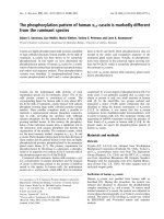
In the group with asphyxial cardiac arrest the initial
values of PetCO2 did not show significant difference when
we compared patients with and without ROSC (6.96 ±
3.63 kPa versus 5.77 ± 4.64 kPa; P = 0.313). We confirmed
significantly higher initial PetCO2 values for those with
ROSC in the group with primary cardiac arrest (4. 62 ±
2.46 kPa versus 3.29 ± 1.76 kPa; P = 0.041). The significant
difference in PetCO2 values for those with and without
ROSC was achieved after the fifth minute of CPR in
both groups (asphyxial arrest: 6.09 ± 2.63 kPa versus
4.47 ± 3.35 kPa; P = 0.006; primary arrest: 5.63 ± 2.01
kPa versus 4.26 ± 1.86; P = 0.015) and remained present
until final values of PetCO2 (asphyxial arrest: 5.87 ± 2.14
kPa versus 0.55 ± 0.49 kPa; P < 0.001, primary arrest:
4.99 ± 1.59 kPa versus 0.96 ± 0.39 kPa; P < 0.001). In all
patients with ROSC the initial PetCO2 was again higher
than 1.33 kPa.
After one minute of CPR we observed no significant dif-
ference in those with and without ROSC in both groups
(asphyxial arrest: 6.26 ± 3.03 kPa versus 7.31 ± 4.69 kPa;
P = 0.345, primary arrest: 5.35 ± 2.18 kPa versus 4.42 ±
2.09 kPa; P = 0.134). After two minutes (asphyxial arrest:
6.07 ± 2.66 kPa versus 6.96 ± 3.54; P = 0.316, primary
arrest: 5.48 ± 2.10 kPa versus 4.56 ± 2.31 kPa; P =0.351)
and three minutes (asphyxial arrest: 6.08 ± 2.29 kPa versus
4.82 ± 3.64 kPa; P = 0.143, primary arrest: 5.56 ± 2.14 kPa
versus 4.49 ± 1.86 kPa; P = 0.070) of CPR there still
was no significant difference among those with and
without ROSC.
We also observed a significant improvement i n inten-
sive care unit (ICU) survival rates for both groups. When
we compar ed the first and this study, a significant differ-
ence was achieved for patients who suffered from asphyx-
ial cardiac arrest (7/37 (16%) versus 20/31 (39.2%);
P = 0.02) and for those who suffered from VF/VT cardiac
arrest (38/103 (27%) versus 40/23 (63.5%); P < 0.01).
Discussion
In this study, which was conducted accordin g to ERC
2005 Guidelines, we confirmed higher values of initial
PetCO2 in asphyxial cardiac arrest than in primary
cardiac arrest. The high initial values of PetCO2 in
asphyxial cardiac arrest did not have a pro gnostic value
for ROSC.
The 2005 ERC Guidelines differ from the 2000 ERC
Guidelines mainly in a shift from primary rhythm-based
management of cardiac arrest to a focus on neurological
outcomes. The guidelines in the second study period are
intensely focused on cardiac massage; the compressions:
ventilation ratio is 30:2, the hands-off time is mitigated
and if the access time is longer than three minutes,
there are first two minutes of CPR befo re the first defi-
brillation. Only a single shock is administrated instead
of a three-shock sequence [21-26].
Lah et al. Critical Care 2011, 15:R13
/>Page 3 of 8
Nevertheless, the gen eral pattern of PetCO 2 changes
remains the same. In asphyxial cardiac arrest the initial
values are high, and do not have prognosti c value for
ROSC, then decreas e later in CPR and increase again in
patients with ROSC [27,28]. In primary cardiac arrest
the initial values are signi ficantly higher in patien ts with
ROSC. The difference fr om the first study [12] is shown
in the first and the second minute of CPR. In this s tudy
the significant difference between the two groups remains
until the third minute of CPR. This may be a result of a
Figure 1 All cardiac arrests placed in the Utstein template. CPC, cerebral performance categories; DNAR, do not attempt resuscitation; EMS,
emergency medical service; PEA, pulseless electrical activity; ROSC, return of spontaneous circulation; VF, ventricular fibrillation; VT, ventricular
tachycardia.
Lah et al. Critical Care 2011, 15:R13
/>Page 4 of 8
Table 2 Demographic and clinical characteristics for both groups of patients
Primary cardiac arrest - VF/VT (n = 63) Asphyxial cardiac arrest (n = 50) P-value
Age (years) 62.6 + 11.6 59.45 + 19.04 0.497¹
Gender(Male/Female) 50/13 27/23 0.003²
Response time (minute)ª 7.12 + 4.5 6.64 + 4.47 0.858¹
Witnessed arrest (Yes/no) 60/3 43/7 0.050²
Resuscitation by medical team (min) 29.7 + 17.2 28.2 + 21.3 0.58¹
ROSC (yes/no) 45/18 = 71% 27/24 = 53% 0.175²
Discharged from ICU (yes/no) 40/23 = 63% 20/30 = 39.2% 0.011²
Discharged alive (yes/no) 25/38 = 39.6% 9/41 = 17.6% 0.009²
CPC 1 to 2 (yes/no) 17/46 = 26.9% 5/45 = 9.8% 0.04²
Average number ob PetCO2 observations 9 (between 3 in 19) 9 (between 2 in 22) 0.312²
CPC cerebral performance category; ICU, intensive care unit; PetCO2 partial pressure of end-tidal carbon dioxide; ROSC return of spontaneous circulation.
ª Time elapsed between the 112 call and the arrival of emerg ency medical team to the patient.
¹ Student t-test.
² c
2
test.
Figure 2 End-tidal pCO2 during cardiopulmonary resuscitation in all patients included in study. All patients: asphyxial cardiac arrest (black
bar), primary cardiac arrest (dotted bar). CPR, cardiopulmonary resuscitation; PetCO2, partial pressure of end-tidal carbon dioxide.
Lah et al. Critical Care 2011, 15:R13
/>Page 5 of 8
higher emphasis on cardiac massage, which causes more
CO2 to be shifted from a peripheral compartment. Both
studies were conducted in out-of -hospital environ-
ments, which meant longer access times and different
first approaches. In the first study we started with
rhythm recognition in order to defibrillate as soon as
possible, whereas in this studywestartedwithcardiac
massage immediately after cardiac arrest was recog-
nized. This probably leads to more intens e shipment of
CO2 from the peripheral compartment, which then
causes values of PetCO2 to r emain higher for a longer
time. The pattern is restored after the third minute of
CPR, when the values decrease and later increase again
only in patients with ROSC. The significant difference
in PetCO2 values (and restart of a prognostic value of
PetCO2) for those with and without ROSC was
achieved after five minutes of CP R in both groups and
remained present until final values of PetCO2. In both
studies the initial PetCO2 values for all patients with
ROSC were higher than 1.33 kPa.
In the second study, where resuscitation was conducted
according to the 2005 ERC Guidelines, we also observed a
significant increase in ICU survival rates in both groups.
Assisted ventilation can be postponed in VF/VT
cardiac arrest [29,30]. On the other hand, quick interven-
tion with assisted ventilation in the field can be life saving
in asphyxial cardiac arrest [31-33]; therefor e, it is impor-
tant to be able to recognize the cause of cardiac arrest.
Limitations
This study has some limitations. First, our sample size is
reasonable (rigorous inclusion and exclusion criteria),
but a larger cohort may h ave afforded the opportunity
for complete subgroup analysis. Second, PetCO2 is only
an indirect measurement of cardiac out-put and a two-
compartment model of CO2 [12]. In the next study w e
should include point-of-care bedside blood gas analysis
and point-of-care ultrasound in the field. Third, better
results in the second study are the results of the
improvement of skills, methods of CPR (new guide lines)
and bystander CPR.
Conclusions
The dynamic pattern of PetCO2 values during out-of-
hospital CPR shows higher values of PetCO2 in the fi rst
two minutes of CPR in asphyxial and prognostic value
Figure 3 End-tidal pCO2 during cardiopulmonary resuscitation regarding aetiology of cardiac arrest and outcome.PetCO2during
cardiopulmonary resuscitation. Asphyxial with ROSC (black bar), asphyxial without ROSC (white bar), VF/VT with ROSC (dotted bar) and VF/VT
without ROSC (gray bar). Data are presented as mean values with one standard deviation. P-values were calculated by unpaired t-test for each
time period and show above bars. CPR, cardiopulmonary resuscitation; PetCO2, partial pressure of end-tidal carbon dioxide; ROSC, return of
spontaneous circulation.
Lah et al. Critical Care 2011, 15:R13
/>Page 6 of 8
of initial PetCO2 only in primary VF/VT cardiac arrest.
The prognostic value of PetCO2 for ROSC was achieved
after the fifth minute of CPR in both groups and
remained present until the final values.
The values of PetCO2 seem to be useful in differen-
tiating causes of cardiac arrest in the pre-hospital
setting.
Key messages
• Initial values of PetCO2 are higher in asphyxial
cardiac arrest than in primary cardiac arrest.
• Initial values of PetCO2 in as phyxial cardiac arrest
do not have a prognostic value for resuscitation
outcome.
• TheprognosticvalueofPetCO2forROSCwas
achieved after the fifth minute of CPR in both
groups and remained present until the final values.
• The values of PetCO2 seem to be useful in differ-
entiating the causes of cardiac arrest in a pre-hospi-
tal setting.
Abbreviations
ARDS: acute respiratory distress syndrome; CO2: carbon dioxide; CPC:
cerebral performance categories; CPR: cardiopulmonary resuscitation; EMS:
emergency medical service; ERC: European Resuscitation Council; ICU:
intensive care unit; kPa: kilopascals; PEA: pulseless electrical activity; PetCO2:
partial pressure of end-tidal carbon dioxide; ROSC: return of spontaneo us
circulation; VT/VF: ventricular fibrillation/pulseless ventricular tachycardia.
Acknowledgements
We thank Petra Klemen MD, MSc for checking the English language.
Author details
1
Center for Emergency Medicine Maribor, Cesta proletarskih brigad 21, 2000
Maribor, Slovenia.
2
Department of Emergency Medicine, Faculty of Medicine
University of Maribor, Slomškov trg 15, 2000 Maribor, Slovenia.
3
Faculty for
Health Sciences University of Maribor, Žitna ulica 15, 2000 Maribor, Slovenia.
4
Department of Family Medicine, Poljanski nasip 58, Faculty of Medicine
University of Ljubljana, 1000 Ljubljana, Slovenia.
Authors’ contributions
LK was involved in the writing of the study protocol, collected the data,
analysed and interpreted the data and wrote the draft of the manuscript.
MK was involved in designing the study protocol and statistical analysis and
interpreted the data. SG was involved in designing and writing the study
protocol, analysed and interpreted the data and made comments on the
draft of the manuscript.
Competing interests
The authors declare that they have no competing interests.
Received: 18 June 2010 Revised: 24 October 2010
Accepted: 11 January 2011 Published: 11 January 2011
References
1. Grmec S, Krizmaric M, Mally S, Kozelj A, Spindler M, Lesnik B: Utstein style
analysis of out-of-hospital cardiac arrest–bystander CPR and end expired
carbon dioxide. Resuscitation 2007, 72:404-414.
2. Axelsson C, Karlsson T, Axelsson AB, Herlitz J: Mechanical active
compression-decompression cardiopulmonary resuscitation (ACD-CPR)
versus manual CPR according to pressure of end-tidal carbon dioxide (P
(ET)CO2) during CPR in out-of-hospital cardiac arrest (OHCA).
Resuscitation 2009, 80:1099-1103.
3. Tachibana K, Imanaka H, Takeuchi M, Takauchi Y, Miyano H, Nishimura M:
Noninvasive cardiac output measurement using partial carbon dioxide
rebreathing is less accurate at settings of reduced minute ventilation
and when spontaneous breathing is present. Anesthesiology 2003,
98:830-837.
4. Rivers EP, Martin GB, Smithline H, Rady MY, Schultz CH, Goetting MG,
Appleton TJ, Nowak RM: The clinical implications of continuous central
venous oxygen saturation during human CPR. Ann Emerg Med 1992,
21:1094-1101.
5. Pytte M, Dorph E, Sunde K, Kramer-Johansen J, Wik L, Steen PA: Arterial
blood gases during basic life support of human cardiac arrest victims.
Resuscitation 2008, 77:35-38.
6. Kolar M, Krizmaric M, Klemen P, Grmec S: Partial pressure of end-tidal
carbon dioxide successful predicts cardiopulmonary resuscitation in the
field: a prospective observational study. Crit Care 2008, 12:R115.
7. Weil MH: Partial pressure of end-tidal carbon dioxide predicts successful
cardiopulmonary resuscitation in the field. Crit Care 2008, 12:90.
8. Krizmaric M, Verlic M, Stiglic G, Grmec S, Kokol P: Intelligent analysis in
predicting outcome of out-of-hospital cardiac arrest. Comput Methods
Programs Biomed 2009, 95:S22-S32.
9. Berek K, Schinnerl A, Traweger C, Lechleitner P, Baubin M, Aichner F: The
prognostic significance of coma-rating, duration of anoxia and
cardiopulmonary resuscitation in out-of-hospital cardiac arrest. J Neurol
1997, 244:556-561.
10. Vivien B, Amour J, Nicolas-Robin A, Vesque M, Langeron O, Coriat P, Riou B:
An evaluation of capnography monitoring during the apnoea test in
brain-dead patients. Eur J Anaesthesiol 2007, 24:868-875.
11. Fries M, Weil MH, Chang YT, Castillo C, Tang W: Microcirculation during
cardiac arrest and resuscitation. Crit Care Med 2006, 34:S454-S457.
12. Grmec Š, Lah K, Tušek Bunc K: Difference in end-tidal CO2 between
asphyxia cardiac arrest and ventricular fibrillation/pulseless ventricular
tachycardia cardiac arrest in the prehospital setting. Crit Care 2003, 7:
R139-R144.
13. Grmec S, Strnad M, Podgorsek D: Comparison of the characteristics and
outcome among patients suffering from out-of-hospital primary cardiac
arrest and drowning victims in cardiac arrest. Int J Emerg Med 2009,
2:7-12.
14. Vaagenes P, Safar P, Moossy J, Rao G, Diven W, Ravi C, Arfors K:
Asphyxiation versus ventricular fibrillation cardiac arrest in dogs.
Differences
in cerebral resuscitation effects–a preliminary study.
Resuscitation 1997, 35:41-52.
15. Idris AH, Wenzel V, Becker LB, Banner MJ, Orban DJ: Does hypoxia or
hypercarbia independently affect resuscitation from cardiac arrest? Chest
1995, 108:522-528.
16. Kamohara T, Weil MH, Tang W, Sun S, Yamaguchi H, Klouche K, Bisera J: A
comparison of myocardial function after primary cardiac and primary
asphyxial cardiac arrest. Am J Respir Crit Care Med 2001, 164:1221-1224.
17. Mithoefer JC, Mead G, Hughes JM, Iliff LD, Campbell EJ: A method of
distinguishing death due to cardiac arrest from asphyxia. Lancet 1967,
2:654-656.
18. International Liasion Committee on Resuscitation: 2005 International
Consensus on Cardiopulmonary Resuscitation and Emergency
Cardiovascular Care Science with Treatment Recommendations.
Resuscitation 2005, 67:181-314.
19. ECC Committee, Subcommittees and Task Forces of the American Heart
Association: 2005 American Heart Association Guidelines for
Cardiopulmonary Resuscitation and Emergency Cardiovascular Care.
Circulation 2005, 112:IV1-IV203.
20. European Resuscitation Council: European Resuscitation Council
Guidelines for Resuscitation 2005. Resuscitation 2005, 67:181-341.
21. Hallstrom A, Rea TD, Mosesso VN Jr, Cobb LA, Anton AR, Van Ottingham L,
Sayre MR, Christenson J: The relationship between shocks and survival in
out-of-hospital cardiac arrest patients initially found in PEA or asystole.
Resuscitation 2007, 74:418-426.
22. Hallstrom A, Herlitz J, Kajino K, Olasveengen TM: Treatment of asystole and
PEA. Resuscitation 2009, 80:975-976.
23. Geddes LA, Roeder RA, Rundell AE, Otlewski MP, Kemeny AE, Lottes AE: The
natural biochemical changes during ventricular fibrillation with
cardiopulmonary resuscitation and the onset of postdefibrillation
pulseless electrical activity. Am J Emerg Med 2006, 24:577-581.
Lah et al. Critical Care 2011, 15:R13
/>Page 7 of 8
24. Campbell RL, Hess EP, Atkinson EJ, White RD: Assessment of a three-phase
model of out-of-hospital cardiac arrest in patients with ventricular
fibrillation. Resuscitation 2007, 73:229-235.
25. Rittenberger JC, Menegazzi JJ, Callaway CW: Association of delay to first
intervention with return of spontaneous circulation in a swine model of
cardiac arrest. Resuscitation 2007, 73:154-160.
26. Baker PW, Conway J, Cotton C, Ashby DT, Smyth J, Woodman RJ,
Grantham H: Clinical investigators. Defibrillation or cardiopulmonary
resuscitation first for patients with out-of-hospital cardiac arrests found
by paramedics to be in ventricular fibrillation? A randomized control
trial. Resuscitation 2008, 79:424-423.
27. DeBehnke DJ, Hilander SJ, Dobler DW, Wickman LL, Swart GL: The
hemodynamic and arterial blood gas response to asphyxiation: a canine
model of pulseless electrical activity. Resuscitation 1995, 30:169-175.
28. Hendrickx HH, Safar P, Miller A: Asphyxia, cardiac arrest and resuscitation
in rats. II. Long term behavioral changes. Resuscitation 1984, 12:117-128.
29. Noc M, Weil MH, Tang W, Turner T, Fukui M: Mechanical ventilation may
not be essential for initial cardiopulmonary resuscitation. Chest 1995,
108:821-827.
30. Herff H, Bowden K, Paal P, Mitterlechner T, von Goedecke A, Lindner KH,
Wenzel V: Effect of decreased inspiratory times on tidal volume. Bench
model simulating cardiopulmonary resuscitation. Anaesthesist 2009,
58:686-690.
31. Yeh ST, Cawley RJ, Aune SE, Angelos MG: Oxygen requirement during
cardiopulmonary resuscitation (CPR) to effect return of spontaneous
circulation. Resuscitation 2009, 80:951-955.
32. Linner R, Werner O, Perez-de-Sa V, Cunha-Goncalves D: Circulatory
recovery is as fast with air ventilation as with 100% oxygen after
asphyxia-induced cardiac arrest in piglets. Pediatr Res 2009, 66:391-394.
33. Idris AH, Wenzel V, Becker LB, Banner MJ, Orban DJ: Does hypoxia or
hypercapnia independently affect resuscitation from cardiac arrest?
Chest 1995, 108:522-528.
doi:10.1186/cc9417
Cite this article as: Lah et al.: The dynamic pattern of end-tidal carbon
dioxide during cardiopulmonary resuscitation: difference between
asphyxial cardiac arrest and ventricular fibrillation/pulseless ventricular
tachycardia cardiac arrest. Critical Care 2011 15:R13.
Submit your next manuscript to BioMed Central
and take full advantage of:
• Convenient online submission
• Thorough peer review
• No space constraints or color figure charges
• Immediate publication on acceptance
• Inclusion in PubMed, CAS, Scopus and Google Scholar
• Research which is freely available for redistribution
Submit your manuscript at
www.biomedcentral.com/submit
Lah et al. Critical Care 2011, 15:R13
/>Page 8 of 8