Heart Disease in Pregnancy - part 8 pptx
Bạn đang xem bản rút gọn của tài liệu. Xem và tải ngay bản đầy đủ của tài liệu tại đây (374.37 KB, 37 trang )
Physical signs are often deceptively few. Clinical signs of acute right ventricu-
lar failure can be subtle. Tachypnea is the most frequent followed by tachycar-
dia. The jugular venous pressure may be raised and there may be increased left
parasternal pulsation, a third heart sound gallop and widely split second sound.
The lungs are usually clear but there may be focal crackles as surfactant is lost
from non-perfused segments of lung. At this stage the arrival of further emboli
will very probably be fatal, but otherwise the evidence of acute cor pulmonale
has usually resolved in just a day or two.
Massive PE
The patient, who may have seemed quite fit up to then, has suddenly collapsed
and is in shock, pale, cold, clammy and shut down or in actual circulatory arrest.
In patients with maintained consciousness, tachypnea and hyperpnea are strik-
ing with poor peripheral perfusion. Substernal chest pain may be confusing.
The lungs are usually clear with good air entry. Rarely, PE may trigger bron-
chospasm in people with asthma. The pulse is rapid and ill-sustained and blood
pressure maintained only with the patient supine. There may be pulsus para-
doxus as filling of the left ventricle and stroke volume fall on inspiration (see
Figure 17.1). The venous pressure will be raised but this cannot be observed
clinically because the patient is lying flat and also because of her heightened res-
piratory efforts. A third heart sound gallop is prominent but pulmonary valve
closure is soft (not accentuated as generally stated) and may be absent if the
right ventricular diastolic pressure has risen to equal the diastolic pressure in the
pulmonary artery. In less severe cases the second heart sound is widely split. A
systolic murmur of tricuspid regurgitation may be audible but it is often silent
because right ventricular pressure and flow may be insufficient to produce au-
dible turbulence, and indeed all the heart sounds become soft as the circulation
fails. When circulatory arrest occurs sinus rhythm is commonly maintained
(persistent electrical activity).
A low P
CO
2
may be coupled with a low PO
2
sometimes contributed to by cen-
tral right-to-left shunting if the foramen ovale is patent. This can lead to con-
comitant stroke, which may dominate the clinical scene. The association of
hypoxemia with hypocapnia and a respiratory alkalosis is always highly sugges-
tive of pulmonary embolism but hypoxemia is not invariable.
24,25
Recurrent PE
Patients with shortness of breath and features of pulmonary hypertension may
have had recurrent episodes of PE or give no such history but show widespread
perfusion defects on scanning.
An underlying thrombophilia is likely but sometimes tests reveal au-
toimmune disease with lupus or Behçet syndrome, and the pulmonary
hypertension has resulted from pulmonary arteritis with thrombosis in situ
rather than embolism. In rare cases pulmonary angiography or cardiac mag-
netic resonance imaging (CMRI) may show multiple pulmonary artery branch
stenoses.
Pulmonary embolism 249
Paradoxical embolism
Elevation of right atrial pressure favors paradoxical passage of emboli if the
foramen ovale is patent. A devastating stroke or unexplained systemic em-
bolism should lead to a search for a cardiac source and suspicion of paradoxical
embolism, so concomitant PE and occult DVT should also be sought.
26,27
Echocardiography with injection of a sonicated indicator while the patient
performs a Valsalva maneuver will force a right-to-left shunt, with passage of
bubbles through the defect and their appearance in the left atrium. The tech-
nique is more sensitive if imaging is performed from the transesophageal ap-
proach. If patency of the foramen ovale is revealed after systemic embolism,
device closure should be performed.
Non-thrombotic PE
Amniotic fluid, fat, tumor or air may embolize to the lungs.
28,29
Fat embolism
may occur after major fractures. Progressive pulmonary hypertension may re-
sult from multiple microtumor embolism in chorioncarcinoma. This sometimes
develops years after a normal pregnancy or abortion. A pregnancy test should
be performed if there is clinical suspicion. Air embolism is a complication of cen-
tral venous lines and special care is necessary when the right atrial pressure is
raised, in case of patency of the foramen ovale. A much smaller amount of air
than is tolerated on the right side can have devastating consequences when re-
leased into the systemic circulation.
Embolism of amniotic fluid is usually asymptomatic and is common peripar-
tum but, rarely, it causes sudden collapse during or after delivery, particularly
after surgical delivery in multiparous patients but is distinguished by the dis-
seminated vascular coagulopathy that usually follows.
Diagnostic strategy
The diagnostic strategy depends on the initial hemodynamic presentation.
30–32
Suspected PE always requires urgent confirmation or exclusion.
In patients whose general condition is good and who are hemodynamically
stable there is time for diagnostic imaging. Suspicion rests on clinical prob-
ability (see Figure 17.2) and diagnosis will follow the results of the baseline tests
and scans (Figure 17.3).
Diagnostic delay must be minimized in patients needing urgent reperfusion
(Figure 17.4). There is no time for imaging tests apart from immediate on-site
echocardiography. Patients who are in cardiogenic shock need reperfusion
treatment right away. Echocardiography also plays a central role in identifying
those patients without shock but whose hemodynamic instability and poorer
outlook are shown by right ventricular dilatation.
Baseline tests
Blood gases, ECG and chest radiograph are basic. They may be uninformative
diagnostically if they are all normal but they say much about the general condi-
tion of the patient and are useful in exclusion of other conditions.
250 Chapter 17
Arterial blood gases
These are helpful but not specific and may be normal. A normal alveolar–
arterial oxygen gradient does not exclude PE but a reduced P
O
2
in an apparently
fit patient is highly significant, especially when combined with a low P
CO
2
. Arte-
rial samples should be taken with the patient sitting up if possible.
25,33
The electrocardiogram
The ECG (Table 17.4) may reveal evidence of right ventricular overload with
clockwise rotation and right-sided T-wave inversion, low voltage, right axis and
rSr in V1 or occasionally right bundle-branch block.
24
The chest radiograph
This is usually normal but a near-normal film in the setting of severe respira-
tory and circulatory compromise is highly suggestive of massive PE. The chest
radiograph is useful in ruling out other lung pathology such as pneumonia or
pneumothorax. It may show non-specific abnormality such as patchy basal at-
electasis or pleural effusion or, rarely, one of the classic signs, a wedge-shaped
Pulmonary embolism 251
D-dimer a
Normal
In first trimester
In second trimester
In third trimester
Clinical probability
Low
a
Spiral CT
Negative
Perfusion scan,
CUS or
arteriography
Abnormal
PE unlikely
Abnormal
>700 ng/mL
>1000 ng/mL
>1420 ng/mL
<700 ng/mL
<1000 ng/mL
<1420 ng/mL
Moderate or high
Positive
Negative Positive
Normal Abnormal
? Infection
TREAT TREAT
Perfusion scan
or
Chest radiograph
Normal
Figure 17.3 Diagnostic strategy for pulmonary embolism (PE) in stable patients. CT,
computed tomography; CUS, compression ultrasonography.
a
Reliability in pregnancy
needs further confirmation.
opacity caused by segmental infarction or focal oligemia (Westermark’s sign),
indicating massive central embolic occlusion.
24
D-dimers
These are breakdown products of fibrin clot. They indicate on-going fibrino-
lysis. A normal level is a rapid test, currently much used to rule out throm-
boembolism but pregnancy itself increases the plasma D-dimer concentration
above the normal upper limit of 500 ng/mL.
Normal ranges at different stages of pregnancy were recently established
from quantitative assays in 50 normal pregnant women using a US Food and
Drug Agency (FDA)-approved ELISA (enzyme-linked immunosorbent assay)
method. D-dimer levels increased through pregnancy and exceeded 500 ng/mL
in 50%, 75% and 100% of women in the three trimesters. The study indicated
that levels above 700, 1000 and 1420 have >50% likelihood of being abnormal
252 Chapter 17
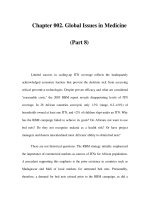
Short axis projections
DiastoleSystole
Figure 17.4 Transthoracic echocardiogram, short-axis projection, systole on the left,
diastole on the right, showing diastolic bowing of the ventricular septum toward the left
ventricle and reduced left ventricular volume in acute pulmonary embolism.
Table 17.4 The ECG in pulmonary embolism
T-wave inversion in leads III, aVF and right-sided chest leads
Right axis and clockwise rotation, dominant S–V5
rSr in V1; complete right bundle-branch block (rare)
Low voltage in limb leads
Qs in leads III and aVF
for each trimester (see Figure 17.3) but more studies are still needed before
these figures can be relied on.
Raised D-dimer levels are not specific but normal levels can be used to back up
clinical assessment of the low probability to rule out PE and remove the need for
imaging. Levels raised above the recently established norms in otherwise
healthy pregnant women are highly suggestive of PE,
34,35
but more studies are
still needed.
Diagnostic imaging (Table 17.5)
Echocardiography
Echocardiography is under-used as the most rapid diagnostic measure in emer-
gency circumstances.
36
It is also non-invasive and does not involve radiation.
Right ventricular dysfunction is found in about a third of all patients with acute
PE (see Figure 17.4). The degree of dilatation and severity of systolic dysfunc-
tion give both therapeutic and prognostic guidance and are the single most im-
portant prognostic factor for in-hospital death.
37
They are usually immediately
available in the accident and emergency department (A&E) to A&E staff, cardi-
ologists or obstetricians faced with a patient in shock or with recent onset of
Pulmonary embolism 253
Table 17.5 Diagnostic imaging in suspected pulmonary embolism
No lung scan needed if leg scan positive
Chest radiograph Usually normal or non-specific
Echocardiography Immediate availability
Shows RV (TTE), main PA branches (TOE)
Perfusion scan Positive scan with normal chest radiograph; start heparin
Useful if SCT negative and clinical probability high
Ventilation scan Useful if both radiograph and perfusion scan are abnormal
If abnormal consider antibiotics
If normal start heparin or both
Spiral CT scan Positive scan with normal chest radiograph; start heparin
Useful if perfusion is equivocal and chest radiograph or ventilation
are normal
May miss subsegmental PE
CMRI Becoming more generally available; shows RV too
Pulmonary angiography Essential for fragmentation or embolectomy
Gold standard but invasive
Involves radiation
CMRI, cardiac magnetic resonance imaging; PA, pulmonary artery; PE, pulmonary embolism; RV,
right ventricle; SCT, spiral computed tomography; TOE, transesophageal echocardiography;
TTE, transthoracic echocardiography.
puzzling symptoms, and their usefulness will increase further as hand-held
machines come into more general use.
Although detection of right ventricular dysfunction lacks specificity, this is of
much less importance in the largely healthy pregnant population than in the
older suspect population with a higher incidence of co-morbidity. Rarely,
echocardiography will reveal a clinically unsuspected cardiomyopathy, parti-
cularly peripartum cardiomyopathy with its high incidence of intraventricular
thrombi that may present with pulmonary (or systemic) embolism.
Otherwise unexplained right ventricular dilatation, poor function and tricus-
pid regurgitation are frequently a surprise in patients with negative clinical
findings who may have complained only of some shortness of breath, transient
dizziness or faintness, and who do not appear to be in distress. Bowing of the
ventricular septum towards the left ventricle in diastole indicates right ventric-
ular volume overload.
38,39
Rarely, worm-like emboli swim in the right atrium to
poke in and out of the tricuspid valve or extend into the ventricle or pulmonary
artery.
40
The central pulmonary arteries are not seen in transthoracic views for
which transesophageal imaging is needed.
Transesophageal echocardiography does not have the brilliant immediacy of
transthoracic echocardiography but needs no preparation or cooperation from
radiological colleagues to delay it. It shows the main pulmonary artery, the right
and the proximal left pulmonary artery, and any thrombi or filling defects.
41
Compression venous Doppler ultrasonography
Loss of venous compressibility indicates thrombosis. Augmentation of flow is
absent or reduced during compression. This is the primary diagnostic test for
DVT because it is non-invasive and totally safe for the fetus. The test is highly
sensitive and specific for proximal DVT with thrombosis of femoral veins, but
is not reliable for isolated iliac thrombosis (more prevalent during preg-
nancy) and ultrasound diagnosis of isolated calf vein thrombosis needs special
expertise.
13,42
About half of all patients with PE have no imaging evidence of DVT. Although
a normal ultrasound examination therefore does not rule it out, its identifica-
tion indirectly establishes the diagnosis of PE but false-positive results may be
obtained in the third trimester as a result of compression of the iliofemoral veins
by the uterus.
Real-time ultrasonography
The common femoral vein and popliteal vein can be visualized and intraluminal
clots detected, although their echogenicity varies according to their age. Real-
time imaging uses standard equipment, is easy, and can be repeated and com-
bined with compression. It cannot detect isolated iliac vein thrombosis.
Contrast phlebography
This is reserved for investigation of equivocal results of ultrasound examination
in patients with high clinical probability of DVT but with no evidence of PE. It is
254 Chapter 17
rarely indicated in pregnancy but the alternative may possibly be unnecessary
heparin treatment.
Ventilation–perfusion scans
Perfusion lung scans
These are indicated as the primary test for PE. They are performed by injecting
technetium-99m (
99m
Tc) coupled to microaggregates of human albumin and
scanning the distribution of radioactivity with a gamma camera. The radiation
dose to the fetus is minimal. A normal scan rules out PE. Unfortunately an ab-
normal scan cannot confirm the diagnosis, although non-specific abnormalities
are less frequent in pregnant patients than in an older age group. Large perfu-
sion defects with a normal chest radiograph are likely to be the result of PE and
make a ventilation scan unnecessary. The original classification stemming from
the PIOPED trial has been revised
44
and was followed by the attempt in the
PISAPED trial with the aim of eliminating equivocal results.
45
Ventilation scans
These employ inhaled xenon-133 (
133
Xe) or krypton-81m (
81m
Kr). An abnor-
mal perfusion scan followed by a normal ventilation scan is diagnostic of PE and
reported as ‘high probability of PE’. Matched abnormalities in perfusion and
ventilation scans with an abnormal chest radiograph are likely to be caused by
infection. One reason for abnormalities on the ventilation scan, especially
when a scan is delayed, is the patchy atelectasis of embolized segments of lung
that often follows in the next few days. The radiation dose is similar to that with
a perfusion scan.
Doubt has been expressed as to whether the ventilation scan is any more use-
ful than a chest radiograph in interpreting the perfusion scan.
Spiral computed tomography
With the development of more accurate scanners, spiral CT has increased in
popularity as the primary imaging test for PE.
46,47
This preference is because
ventilation–perfusion scans still produce so many equivocal results in older pa-
tients with co-morbidity, among whom reports of ‘intermediate risk of PE’ are
frequent and frustrating. They are especially likely when the chest radiograph is
abnormal. These limitations of ventilation–perfusion scans are much less of a
problem in the younger and otherwise healthy pregnant population.
Spiral CT produces a definite positive or negative result but is less accurate in
revealing segmental PE than central or lobar emboli. A normal study therefore
cannot rule out isolated peripheral subsegmental PE or be the basis for with-
holding anticoagulant treatment.
The technique has the disadvantages of both exposure to radiation and a fetal
dose of iodinated contrast, although the fetal radiation dose with spiral CT is
lower than with ventilation–perfusion scanning and neonatal hypothyroidism
has not been reported.
Pulmonary embolism 255
Magnetic resonance imaging
MRI with gadolinium enhancement now has similar accuracy to pulmonary
angiography and CMRI also allows assessment of ventricular function. It avoids
radiation and the use of radiographic contrast and imposes no risk, but is not
usually immediately, or as yet generally, available.
48
Both spiral CT and MRI can be extended to look for DVT but there is no point
if imaging for PE has been positive. Neither CT nor MRI are needed if leg vein
ultrasonography is positive.
Pulmonary angiography
Pulmonary angiography is safe during pregnancy with suitable abdominal
screening, but is rarely indicated except as part of the interventional treatment
of immediately life-threatening massive embolism. It is regarded as the gold
standard but carries a mortality rate of about 0.5%, is technically demanding
and often hard to interpret despite good image quality, for both of which the
skills of a radiologist may be needed especially for out-of-hours emergency
work. Safety and accuracy have been greatly increased by the use of selective
injections, digital subtraction and magnification.
48–50
Anticoagulants may be
withheld if pulmonary angiography is normal.
50,51
Management
Patients in cardiogenic shock or hemodynamically unstable
Massive and subacute massive PE
The management of a patient with a high clinical probability of PE and who is in
shock is aimed at restoring circulation and saving life (Table 17.6). The diagnosis
needs to be confirmed and action taken with no time lost (Figure 17.5). If the di-
agnosis is confirmed by right ventricular dilatation shown on transthoracic
echocardiography, percutaneous catheter fragmentation and thrombolysis
(Figure 17.6) should be carried out immediately and without delay for other
investigations.
18,31,52–54
It is usually successful if the embolism was truly acute. It
256 Chapter 17
Table 17.6 Massive pulmonary embolism (emergency treatment to save life)
Cardiopulmonary resuscitation (CPR) if circulatory arrest
Elevate legs
Oxygen
Central intravenous line
Start heparin
Consider dobutamine infusion
Consider inhaled nitric oxide
Thrombolytic drug
Per catheter clot fragmentation and/or extraction
will fail if the circulation has collapsed after apparent sudden onset, although all
or most of the material has been gradually accreted through recurrent episodes.
A pigtail catheter should be introduced by the brachial route or central vein
with the patient tilted head down. Fragmentation can be accomplished very
swiftly and, if it is successful, blood pressure and consciousness are restored
within minutes. The extreme emergency is over. The catheter is inserted via a
brachial or central route so as to avoid dislodging any thrombus in the pelvis or
vena cava, and to save the fetus from radiation if the patient is undelivered. Pro-
vided that the obstruction is caused by freshly arrived embolic material that is
still lying centrally, it can usually be moved on with dramatic improvement.
Formal angiography is not required but contrast is needed to guide the proce-
dure and should be used as sparingly as possible.
If attempts to fragment central emboli and move them on are unsuc-
cessful, per catheter embolectomy should be tried and, if all else fails, surgical
embolectomy.
Pulmonary embolism 257
Stable
Baseline tests
D-dimer*
Echocardiography
Normal RV
Normal RV
Perfusion
scan + positive
or
Peripheral
TREAT
PE EXCLUDED
Follow management
strategy for stable patients
Catheter fragmentation
Successful
Thrombolysis
Surgical embolectomy
Cardiogenic shock
Echocardiography
Dilated RV
Dilated RV
Central
Spiral CT
Unsuccessful
Figure 17.5 Management strategy in patients with pulmonary embolism (PE) again
stressing the key role played by echocardiography. RV, right ventricle.
a
Reliability in
pregnancy needs further confirmation.
Other measures are adjuvant. The legs should be elevated and oxygen given.
If consciousness has been lost chest compression will help to empty the right
ventricle and be directly therapeutic if it dislodges thrombi and assists in moving
them on. If the circulation is compromised and the right ventricle dilated, but
the patient is conscious and not in shock, there is time for perfusion or spiral CT
to assess the size and distribution of the clot burden.
A central line is inserted and unfractionated heparin is started. It is usual to
give inotropes and vasopressors but, unless an effective circulation returns rap-
idly, an attempt should be made to fragment the emboli per catheter. Dobuta-
mine is usually given even though endogenous neuroendocrine activation is
likely to be providing maximum stimulation already. Dobutamine provides
positive inotropic effect with pulmonary vasodilatation through its beta-
adrenergic action. Inhaled nitric oxide may release pulmonary vasoconstriction
and help to reduce right ventricular afterload.
Fluid loading is probably unhelpful and no more than 500 mL fluid should be
given, more only if it appears to have been beneficial (as ventricular interdepend-
ence may cause further compromise of left ventricular filling). This can most rap-
idly be appreciated by following the effect of the infusion echocardiographically.
Thrombolytic treatment with recombinant plasminogen activator (rtPA)
should be given only if the circulation remains compromised. This does not
258 Chapter 17
Figure 17.6 The right digital subtraction pulmonary arteriogram from a patient with
massive pulmonary embolism (a) before and (b) after restoration of flow to the lower
lobe by mechanical fragmentation. The pigtail catheter is clearly visible.
cross the placenta or directly injure the fetus but may cause bleeding and fetal
risk. It can be started immediately after completion of mechanical fragmenta-
tion and given directly into the pulmonary arteries (although there is no proof
of added efficacy by this route). Infusion of unfractionated heparin should
follow.
Major PE
If echocardiography reveals right ventricular dysfunction, septal bowing and
tricuspid regurgitation, but the circulation is not compromised, compensation
is marginal and the patient should be regarded as unstable. The fetus is at risk if
maternal output falls with exertional blood pressure dips. The patient is nursed
in an intensive care or high dependency unit with oxygen, heparin and trial of
dobutamine, but thrombolytic treatment is not indicated and recovery of right
ventricular function is often swift over hours rather than days. A slower or no
improvement suggests a subacute or acute on chronic pathogenesis with possi-
ble later need for elective surgical embolectomy if resolution fails.
Clinically stable patients
If the patient is hemodynamically stable with maintained blood pressure and
cardiac output, anticoagulant treatment with heparin may be all that is required
plus oxygen and pain relief. Absence of right ventricular dilatation on echocar-
diography adds reassurance but the risk of recurrent embolism continues until
existing venous thrombus has been autolysed or organized. Now is the time for
insertion of a prophylactic caval filter
18
if this is contemplated but the pro-
cedure, although improved, involves radiation, carries morbidity and is not
always successful in preventing recurrence.
Continuing management
Intravenous unfractionated heparin should be given, aiming for an adjusted
partial thromboplastin time (aPPT) between 1.5 and 2.5 times the control (anti-
Xa activity 0.3–0.6 IU).
18
Control should be very tight because the therapeutic
window is narrow. A changeover to low-molecular-weight heparin (LMWH)
can be made after a week or so if the patient is stable and echocardiography
shows that right ventricular function is restored. Follow-up ventilation–
perfusion scanning can be delayed until the puerperium. LMWH is probably
safe and effective but there are no data in pregnancy and it takes longer to re-
verse before delivery. After delivery a change can be made to warfarin
(coumadin) which should be continued until the ventilation–perfusion scan is
normal or there is no further improvement. Compression stockings should be
worn through the rest of the pregnancy and early puerperium.
Patients who have suffered PE are at risk from recurrence until any thrombus
in leg and pelvic veins has lysed, embolized or organized. Thrombolytic agents
do not work immediately or completely, but patients who are destined to sur-
vive sometimes improve before thrombolytic activity has started through on-
ward movement, compaction or shrinkage of embolized material. Heparin is
Pulmonary embolism 259
not lytic and resorption of non-embolized material depends on endogenous
lysis which is very active in the lungs. Patients who have suffered massive or
major embolism are probably less likely to harbor unstable peripheral thrombus
than patients suffering minor embolism, but are in a worse position to sustain
another onslaught until resolution of the first. Further embolism from legs or
pelvis remains a risk for the first few days and weeks.
Prevention of PE
Diagnosis of venous thrombosis
A full history is important because a positive history indicates the need to per-
form a full coagulation screen and to institute prophylactic measures in anyone
with a personal or family history of unexplained thrombosis. This must be done
before anticoagulants are started. Physical methods of prophylaxis include pos-
ture (sleeping semi-prone rather than supine in later pregnancy) and compres-
sion stockings. LMWH should be given to high-risk patients.
Prevention of PE relies on the prevention, rapid diagnosis and effective treat-
ment of venous thrombosis. Accurate objective diagnosis is important because
of the mortal risk from PE if DVT is untreated and also because long-term anti-
coagulant treatment in pregnancy carries risk to both mother and fetus, and a
positive diagnosis has implications for prophylaxis in future pregnancies. Accu-
rate diagnosis is mandatory (Table 17.7).
Clinical probability is assessed on medical history and clinical examination.
Thrombosis is most frequent in the left femoral vein and calf antenatally. Dis-
tinction has to be made from benign pregnancy associated swelling, which is
usually slowly progressive, painless and bilateral although occasionally rapid
and unilateral even in the absence of thrombosis.
Perspective
Although thromboembolism is frequently missed it is still rare compared with
the frequency with which it is suspected and the prevalence of high probability
ventilation–perfusion scans in pregnant women with suspected PE is very low.
260 Chapter 17
Table 17.7 Possible deep vein thrombosis (DVT)
High index of suspicion needed:
—
half of patients with a PE do not have any sign of a DVT
—
half of patients with an acutely swollen calf do not have a DVT
Investigation:
D-dimer: promising, needs more pregnancy data
—
real-time venous ultrasonography: available, easy, non-invasive
—
compression Doppler ultrasonography: needs expertise, non-invasive
—
phlebography: invasive, involves radiation, painful, may cause DVT
Withholding anticoagulation in pregnant women with a low clinical prob-
ability of PE, negative leg Doppler and normal or non-diagnostic ventilation–
perfusion scans is probably safe but simpler means of ruling out thromboem-
bolism are needed. The reliability of a negative pregnancy level D-dimer in
ruling out thromboembolism without the need for further investigation is
promising but more data are awaited.
References
1 Rutherford S, Montoro M, McGehee W, Strong T. Thromboembolic disease
associated with pregnancy: an 11 year review. Am J Obstet Gynecol 1991;164(suppl):
286.
2 Andersen BS, Steffensen FH, Sorensen HT, Nielsen G, Olsen J. The cumulative inci-
dence of venous thromboembolism during pregnancy and puerperium
—
an 11 year
Danish population based study of 63,300 pregnancies. Acta Obstet Gynaecol Scand
1998;77:170–3.
3 Report on Confidential Enquiries into Maternal Deaths in the United Kingdom 1994–96.
London: HMSO, 1998.
4 Danilenko-Dixon DR, Heit JA, Silverstein MD et al. Risk factors for deep vein throm-
bosis and pulmonary embolism during pregnancy or post partum: a population
based, case–control study. Am J Obstet Gynecol 2001;184:104–10.
5 Toglia MR, Weg JG. Venous thromboembolism during pregnancy. N Engl J Med
1996;335:108–13.
6 Hirsch DR, Mikkola KM, Marks PW et al. Pulmonary embolism and deep vein throm-
bosis during pregnancy or oral contraceptive use: prevalence of factor V Leiden. Am
Heart J 1996;131:1145–8.
7 Peek MJ, Nelson RA, de Swiet M, Letsky EA. Activated protein C resistance in normal
pregnancy. Br J Obstet Gynaecol 1997;104:1084–6.
8 Bauer KA. Hypercoagulability
—
a new co-factor in the protein C anticoagulant path-
way. Lancet 1994;330:566–7.
9 Preston FE, Rosendaal FR, Walker ID et al. Increased fetal loss in women with herita-
ble thrombophilia. Lancet 1996;348:913–16.
10 Carr MH, Towers VC, Eastenson AR, Pircon RA, Iriye BK, Adashek JA. Prolonged bed
rest during pregnancy: does the risk of deep vein thrombosis warrant the use of rou-
tine heparin prophylaxis? J Matern Fetal Med 1997;6:264–7.
11 Weiner CP, Kwaan AC, Xu C et al. Antithrombin III deficiency in women with hyper-
tension during pregnancy. Obstet Gynecol 1985;65:301–6.
12 Lindqvist P, Dahlback B, Marsal K. Thrombotic risk during pregnancy: a population
study. Obstet Gynecol 1999;94:595–9.
13 Kyrle PA, Eichinger S. Deep vein thrombosis. Lancet 2005;365:1111–202.
14 Khamashta MA, Mackworth-Young C. Antiphospholipid (Hughes’) syndrome
—
a
treatable cause of recurrent pregnancy loss. BMJ 1997;314:244.
15 Den Heyer M, Koster T, Blom HJ et al. Hyperhomocysteinaemias a risk factor for deep
vein thrombosis. N Engl J Med 1996;334:759–62.
16 Rosendaal FR. Risk factors for venous thrombosis disease. Thromb Haemost
1999;82:610–19.
17 Prandoni P, Lensing AW, Coigo A et al. The long term clinical course of acute deep
venous thrombosis. Arch Intern Med 1996;125:1–7.
Pulmonary embolism 261
18 Task Force on Pulmonary Embolism, European Society of Cardiology. Guidelines on
diagnosis and management of acute pulmonary embolism. Eur Heart J 2000;21:
1301–36.
19 The PIOPED Investigators. Value of the ventilation–perfusion scan in acute pul-
monary embolism. Results of the prospective investigation of pulmonary embolism
diagnosis. JAMA 1990;263:2753–9.
20 Sergysels R. Pulmonary gas exchange abnormalities in pulmonary embolism.
In Morpurgo M (ed.), Pulmonary Embolism. New York: Marcel Dekker, 1994:
pp 89–96.
21 Colman NC. Pathophysiology of pulmonary embolism. In Leclerk JR (ed.), Venous
Thromboembolic Disorders. Philadelphia: Lea Febiger, 1991: pp 65–73.
22 Coon WW, Coller FA. Clinicopathologic correlation in thromboembolism. Surg
Gynecol Obstet 1959;109:259–69.
23 Chan WS, Ray JG, Murray S et al. Suspected pulmonary embolism in pregnancy. Arch
Intern Med 2002;162:1170–5.
24 Stein PD, Terrin ML, Hales CA et al. Clinical, laboratory, roentgenographic and elec-
trocardiographic findings in patients with acute pulmonary embolism and no pre-
existing cardiac or pulmonary disease. Chest 1991;100:598–603.
25 Stein PD, Henry JW, Miller AC. Arterial blood gas analysis in the assessment of sus-
pected acute pulmonary embolism. Chest 1996;109:78–81.
26 Amarenco P. Patent foramen ovale and the risk of stroke: smoking gun guilty by asso-
ciation? Heart 2005;91:441–7.
27 Konstantinides S, Geibel A, Kasper W, Olschewski M, Blumel L, Just H. Patent fora-
men ovale is an important predictor of adverse outcome in patients with major pul-
monary embolism. Circulation 1998;97:1946–51.
28 Gei AF, Vadhera RB, Hankins GD. Embolism during pregnancy: thrombus, air and
amniotic fluid. Anesthesiol Clin North Am 2003;21:16.
29 Berkowitz KD, Goldstein DP. Gestational trophoblastic disease. Cancer 1995;76:
2079–85.
30 Kearon C. Diagnosis of pulmonary embolism. Can Med Assoc J 2003;168:1–22.
31 Kucher N, Luder CM, Dornhofer T, Windecker S, Meier B, Hess OM. Novel manage-
ment strategy for patients with suspected pulmonary embolism. Eur Heart J 2003;24:
366–76.
32 Nijkeuter M, Geleijns J, de Roos A, Meinders AE, Huisman MV. Diagnosing pul-
monary embolism in pregnancy: rationalising fetal radiation exposure in radiological
procedures. J Thromb Haemost 2004;2:1857–8.
33 Ang CK, Tan TH, Walters A, Wood C. Postural influence on maternal capillary oxygen
and carbon dioxide tension. BMJ 1969;4:201–3.
34 Hernandez J, Hambleton G, Kline JA. D-dimer concentrations in normal pregnancy.
Acad Emerg Med 2004;11:526–7.
35 Morse M. Establishing a normal range for D-dimer levels through pregnancy to aid in
the diagnosis of pulmonary embolism and deep vein thrombosis. J Thromb Haemost
2004;2:1202.
36 Cheriex EC, Sreeram N, Eussen Y, Pieters FA, Wellens HJ. Cross sectional Doppler
echocardiography as the initial technique for the diagnosis of acute pulmonary em-
bolism. Br Heart J 1994;72:52–7.
37 Wolfe MW, Lee RT, Feldstein ML, Parker JA, Come PC, Goldhaber SZ. Prognostic sig-
nificance of right ventricular hypokinesis and perfusion lung scan defects in pul-
monary embolism. Am Heart J 1994;127:1371–5.
262 Chapter 17
38 Mc Connell MV, Solomon SD, Rayan M, Come PC, Goldhaber RT. Regional right ven-
tricular dysfunction detected by echocardiography in acute pulmonary embolism.
Am J Cardiol 1996;78:469–73.
39 Kasper W, Geibel A, Tiede N et al. Distinguishing between acute and subacute massive
pulmonary embolism by conventional and Doppler echocardiography. Br Heart J
1993;70:352–6.
40 Chapoutot L, Nazerollas P, Metz D et al. Floating right heart thrombi and pulmonary
embolism: diagnosis, outcome and therapeutic management. Cardiology 1996;87:
169–74.
41 Pruszczyk P, Torbicki A, Pacho R et al. Non-invasive diagnosis of suspected severe
pulmonary embolism: transoesophageal echocardiography vs spiral CT. Chest 1997;
112:722–8.
42 Kearn C, Julian JA, Newman E, Ginsberg JS, for the McMaster Diagnostic Imaging
Practice Guidelines Initiative. Non-invasive diagnosis of deep vein thrombosis. Ann
Intern Med 1998;128:663–77.
43 Chan WS, Ray JG, Murray S, Coady GE, Coates G, Ginsberg JS. Suspected pulmonary
embolism in pregnancy: clinical presentation, result of lung scanning and subsequent
maternal and pediatric outcomes. Arch Intern Med 2002;162:1170–5.
44 Sostman HD, Colman RE, DeLong DM, Newman GE, Paine S. Evaluation of revised
PIOPED criteria for ventricular perfusion scintigraphy in patients with suspected pul-
monary embolism. Radiology 1994;193:103–7.
45 The PISA-PED Investigators. Value of perfusion lung scan in the diagnosis of pul-
monary embolism: results of the prospective study of acute pulmonary embolism
diagnosis (PISA-PED). Am J Respir Crit Care Med 1996;154:1387–93.
46 Doyle NM, Ramirez MM, Mastrobattista JM, Monga M, Wagner LK, Gardner MO.
Diagnosis of pulmonary embolism: a cost-effectiveness analysis. Am J Obstet Gynecol
2004;191:1019–23.
47 Mayo JR, Remy-Jardin M, Muller NL et al. Pulmonary embolism: prospective com-
parison of spiral CT with ventilation/perfusion scintigraphy. Radiology 1997;205:
447–52.
48 Goodman LR, Lipchik RJ, Kuzo RS et al. Subsequent pulmonary embolism after a
negative pulmonary angiogram
—
prospective comparison with scintigraphy. Radio-
logy 2000;215:535–42.
49 Johnson MS, Stine SB, Shah H, Harris VJ, Ambrosius WT, Trerotola SO. Possible pul-
monary embolus: evaluation with digital subtraction versus cut-film angiography
—
prospective study in 80 patients. Radiology 1998;207:131–8.
50 Oudkerk M, van Beek EJ, Wielopolski P et al. Comparison of contrast enhanced mag-
netic resonance angiography and conventional pulmonary angiography for the diag-
nosis of pulmonary embolism: a prospective study. Lancet 2002;359:1643–7.
51 de Swiet M. Management of pulmonary embolus in pregnancy. Eur Heart J
1999;20:1378–85.
52 Brady AJB, Crake T, Oakley CM. Percutaneous catheter fragmentation and distal dis-
persion of proximal pulmonary embolus. Lancet 1991;338:1186–9.
53 Mazeika PK, Oakley CM. Massive pulmonary embolism in pregnancy treated with
streptokinase and percutaneous catheter fragmentation. Eur Heart J 1994;15:
1281–3.
54 Sofocleous CT, Hinrichs C, Bahramipour P, Barone A, Abujudeh H, Contractor D. Per-
cutaneous management of life-threatening pulmonary embolism complicating early
pregnancy. J Vasc Interv Radiol 2001;12:1355–6.
Pulmonary embolism 263
CHAPTER 18
Hypertensive disorders
of pregnancy
Alexander Heazell, Philip N Baker
Hypertensive disorders of pregnancy are the most common medical problem
encountered in the second half of pregnancy, affecting about 6–10% of preg-
nancies.
1
Although there are many different causes of hypertension in preg-
nancy, the most clinically important condition is pre-eclampsia, affecting
between 1 and 3% of pregnancies.
2
Pre-eclampsia is associated with increased
maternal and fetal morbidity and mortality.
3
As there is no effective interven-
tion for pre-eclampsia, except delivery, pre-eclampsia is responsible for about a
half of induced pre-term deliveries, with the associated consequences of pre-
mature birth.
At present, the precise cause of pre-eclampsia is unknown, although it is un-
derstood to be a disorder of widespread endothelial dysfunction. It is hypothe-
sized that decreased invasion and subsequent remodeling of maternal spiral
arteries in the first trimester lead to reduced placental perfusion, and release
of factors into the maternal circulation, resulting in endothelial cell damage
(Figure 18.1).
4–6
As every organ has a blood supply, pre-eclampsia should be
regarded as a multi-system disease, which may affect each patient differently
(Tables 18.1 and 18.2). Therefore, the management of pre-eclampsia involves
far more than the treatment of hypertension alone.
In normal pregnancy the maternal blood pressure decreases in the first half of
pregnancy, rising to pre-pregnancy levels or higher from about 30 weeks’ gesta-
tion. Hypertension in pregnancy is defined as a blood pressure >140/90 mmHg
on two separate occasions, at least 4 hours apart, or a single diastolic reading
>110 mmHg.
7
If this occurs before 20 weeks’ gestation, it is presumed to
be chronic hypertension. If new hypertension occurs after 20 weeks’ gesta-
tion without proteinuria, it is termed ‘pregnancy-induced hypertension’.
7
Pre-eclampsia is defined as new onset hypertension with proteinuria
(>300 mg/24h or ++on urine dipstick) in the absence of a urinary tract infection
after 20 weeks’ gestation.
8
If a patient with pre-existing hypertension develops
proteinuria (>300 mg/24 h or ++), this is termed superimposed pre-eclampsia
(Table 18.3). Eclampsia is defined as a tonic–clonic seizure occurring in the pres-
ence of pre-eclampsia.
Much of the antenatal care in the UK is directed towards the identification of
pre-eclampsia. As there is currently no reliable screening test to identify women
264
Heart Disease in Pregnancy, Second Edition
Edited by Celia Oakley, Carole A Warnes
Copyright © 2007 by Blackwell Publishing
who will go on to develop pre-eclampsia, regular screening of blood pressure
and urine is carried out throughout pregnancy, because this has been shown to
reduce perinatal mortality sevenfold. In addition, women at risk of develop-
ing hypertension in pregnancy should be identified. The incidence of pre-
eclampsia is increased in primigravidae (or first pregnancy with that partner),
multiple pregnancies, women with a first-degree relative with hypertension in
pregnancy, and at extremes of reproductive age.
2,6,9,10
Important risk factors in
a patient’s past medical history include: chronic renal disease, chronic hyper-
tension (especially if poorly controlled), diabetes mellitus and thrombophil-
ia.
11–14
Patients with a positive past medical history should be classified as a
high-risk pregnancy, and managed in a specialist high-risk obstetric clinic, being
seen more frequently than ‘low-risk’ patients. Patients who have had previous
pre-eclampsia should also be managed as a high-risk pregnancy because the risk
of recurrence of some form of pre-eclampsia is between 20 and 40% depending
on the population studied.
15
It is important to note that pre-eclampsia is a heterogeneous condition, and
the diagnosis may be made from the presence of clinical features, such as those
above, in combination with the presence of hypertension and proteinuria (see
Table 18.1). Patients may also have biochemical and/or hematological abnor-
malities that may be found on further investigation (see Table 18.2).
Despite widespread abnormalities in the cardiovascular system, cardiologists
are rarely involved in the management of hypertensive disorders of pregnancy,
except in severe or resistant hypertension or in other unusual circumstances,
e.g. secondary hypertension resulting from coarctation of the aorta. Most
cardiologists therefore have little experience in the routine management of
Hypertensive disorders of pregnancy 265
Abnormal trophoblast invasion
and failure of conversion of
spiral arteries
Placental damage
Release of circulating factors
Normal utero
placental
artery
Endothelial dysfunction affecting
multiple organ systems
Brain
Kidney
Liver
Inappropriate
activation of
clotting cascade
Figure 18.1 Proposed pathophysiology of pre-eclampsia.
hypertension in pregnancy. This may be a particular problem in pre-pregnancy
counseling for women with hypertension, prophylaxis for pre-eclampsia, and
the use of pharmacological agents in pregnancy, or in the follow-up of patients
who have had pre-eclampsia. Therefore, this chapter addresses hypertension in
pregnancy with particular respect to these areas.
Blood pressure measurement in pregnancy
The measurement of blood pressure in pregnancy is subject to the same prob-
lems as in normal patients, such as: selection of appropriate cuff size, observer
error and bias, and blood pressure variability, all of which may affect the final
documented reading. However, in pregnancy there are specific concerns relat-
ing to the position of the patient. The blood pressure is lower in the second half
of pregnancy in patients lying supine. This is because the gravid uterus obstructs
266 Chapter 18
Table 18.1 Clinical features of pre-eclampsia
Central nervous system
Eclamptic convulsions
Cerebral hemorrhage, intraventricular or subarachnoid
Cerebral infarction: microinfarction or macroinfarction (e.g. cortical blindness caused by
infarction of occipital cortex)
Coagulation system
Thrombocytopenia
Microhemangiopathic hemolysis
HELLP syndrome (hemolysis, elevated liver enzymes, low platelets)
Disseminated intravascular coagulation
Eyes
Retinal detachment
Retinal edema
Kidney
Acute tubular necrosis
Acute cortical necrosis
Unspecified renal failure
Liver
Rupture of hepatic capsule
Infarction
Jaundice
Decreased synthesis of soluble clotting substances
HELLP syndrome
Respiratory system
Pulmonary edema
Laryngeal edema
Adult respiratory distress syndrome
the venous return from the lower limbs and reduces cardiac pre-load. Therefore
blood pressure should be measured in the left lateral or sitting position. As a re-
sult of difficulties in maintaining the sphygmomanometer cuff at the level of the
heart in the left lateral position, sitting is the preferred position.
In the UK (although not in the USA) the fourth Korotkoff sound (K4), rather
than the fifth (K5), was previously recommended for the measurement of dias-
tolic blood pressure. This was because of concern that K5 may be audible at zero
cuff pressure. However, a study of 250 patients found the diastolic blood pres-
sure was always >50 mmHg.
16
K5 is nearer to intra-arterial diastolic blood pres-
sure than K4, and can be recognized more consistently than K4.
17
K5 is now
recommended for the measurement of diastolic blood pressure in pregnancy in
the UK.
‘White coat hypertension’, in which the blood pressure is excessively high be-
cause of the clinical environment, is as much of a problem in pregnancy as in
non-pregnant women, if not more. Its impact is minimized by devices that allow
Hypertensive disorders of pregnancy 267
Table 18.3 Classification of hypertensive disorders in pregnancy
Chronic hypertension
Pregnancy-induced hypertension (gestational hypertension)
Pre-eclampsia
Pre-eclampsia superimposed on chronic hypertension
Table 18.2 Abnormalities that may be found on investigation indicating pre-eclampsia
Maternal
Elevated urea and creatinine
a, b
Elevated liver function tests
a
(aspartate transaminase, alanine transaminase)
Hemolysis
a
Hyperuricemia
a
Hypocalcemia
Increased packed cell volume
a
Proteinuria
a
Raised antithrombin III in plasma
Raised fibronectin in plasma
Raised von Willebrand’s factor in plasma
Thrombocytopenia
a
Fetal
Intrauterine growth restriction
Abnormal umbilical artery Doppler flow
Abnormal fetal cardiac trace (as measured by cardiotocography)
a
Tests commonly used in the UK.
b
Urea and creatinine both fall during pregnancy as a result of hemodilution; values should be
compared with ‘normal’ ranges for pregnancy.
blood pressure to be measured frequently and outside the hospital, such as 24-
hour ambulatory monitoring.
8
Automatic blood pressure devices, either ambulatory or static, are widely
used in antenatal clinics, delivery units and high-dependency areas, and need
to be calibrated specifically for pregnancy and specifically for pre-eclampsia.
Many of the machines in current use have not been validated. In pre-eclampsia,
in particular, the physical characteristics of blood vessels are altered. The pat-
tern of pressure changes between systole and diastole analyzed by oscillometric
machines is altered and the algorithm used to calculate diastolic pressure is no
longer accurate. In severe pre-eclampsia, machines that rely on oscillometric
measurement may underestimate diastolic blood pressure by 15 mmHg com-
pared with conventional sphygmomanometry; this may lead to inadequate
treatment of hypertension. Even machines using a microphone to detect
Korotkoff’s sounds are not immune to this problem. We recommend the
SpaceLabs 90207 and SpaceLabs Scout as the only automatic blood pres-
sure machines that have been shown to be accurate in severe pre-eclampsia
to date.
18
Prevention of pre-eclampsia
The possibility of prevention of pre-eclampsia is particularly important, because
delivery of the infant remains the only effective treatment for the condition.
There are two main groups of women at risk of pre-eclampsia: those who have
had hypertension in a previous pregnancy and those who have a medical
condition such as hypertension or renal disease that predisposes patients to
pre-eclampsia.
Early trials with antiplatelet agents, in particular low-dose aspirin 60–
150 mg/day, were encouraging, suggesting a 70% reduction in risk.
19,20
In con-
trast, larger trials such as CLASP (Collaborative Low-dose Aspirin Study in
Pregnancy) showed no overall reduction in risk; however, further analysis of
the CLASP data did suggest that early onset pre-eclampsia might be reduced by
as much as 50%, which is significant because it is early onset pre-eclampsia (be-
fore 34 weeks) that is associated with the highest perinatal morbidity.
21
Subse-
quent meta-analysis of 32 trials of anti-platelet therapy found a 15% reduction
in risk of developing pre-eclampsia; this benefit was independent of gestation
on entry to the trial or dose of aspirin administered.
22
The most common dose of
aspirin used in the UK is 75 mg/day, which should ideally be taken during the
first trimester, because this is when underlying pathological changes of pre-
eclampsia commence.
Several dietary measures have been used in an attempt to reduce the risk of
pre-eclampsia. There is no beneficial effect of a reduction in salt intake or mag-
nesium supplementation.
23,24
However, calcium supplementation of at least
1 g/day appears to result in a reduction of the risk of hypertension and pre-
eclampsia of 19% and 30%, respectively.
25
These effects seem to be most
marked in women with a high risk of pre-eclampsia. Supplementation of
268 Chapter 18
antioxidants, such as vitamins C and E, has been shown in a study of 283 high-
risk women to reduce incidence of pre-eclampsia from 26% to 8%.
26
A large
multicenter, placebo-controlled, randomized trial is being undertaken in the
UK and is expected to report results in 2006.
A Cochrane meta-analysis of antihypertensive treatment before conception
or during pregnancy concluded that there was a reduction in the number of pa-
tients presenting with severe hypertension, but there was no reduction in the
risk of developing pre-eclampsia, or in the fetal or neonatal outcomes.
27
There-
fore, patients who are known to be hypertensive before pregnancy should be
investigated in the usual way, and treatment should be commenced to achieve
optimal blood pressure before pregnancy. No antihypertensive drugs have been
shown conclusively to be teratogenic, although angiotensin-converting en-
zyme (ACE) inhibitors may be related to skull defects and renal dysfunction.
28
Therefore, the choice of drugs to be used before pregnancy may be made irre-
spective of any consideration of a future pregnancy. However, if the patient
wishes to conceive, it is better to control blood pressure with agents that are
suitable for use throughout pregnancy, rather than have to change drugs in the
first trimester, which may lead to suboptimal control of hypertension during
placental development. There are no levels of blood pressure above which preg-
nancy cannot be countenanced. Patients should be counseled about the risks of
pre-eclampsia and the possible prophylactic therapy.
Management of hypertension in pregnancy
There are two important differences between the management of hypertension
in pregnancy and the management of hypertension outside pregnancy. Most
cases of hypertension outside pregnancy are idiopathic or ‘essential’, i.e. follow
a chronic course, with the major purpose of treatment being to prevent long-
term complications such as stroke and myocardial infarction. A few patients
present outside pregnancy with acute severe hypertension, which must be
treated immediately because of the risk of hypertensive encephalopathy or
cerebral hemorrhage, but this is now uncommon. It is realized that acute low-
ering of blood pressure in the non-pregnant state has major risks to the individ-
ual. However, hypertension in pregnancy normally resolves post partum, so
there is no justification for treatment of hypertension to prevent long-term
complications. Importantly, pre-eclampsia carries acute risks of eclampsia and
cerebral hemorrhage; the latter complication is the cause of most of the mater-
nal mortality in the disease.
3
In contrast to the non-pregnant state, acute lower-
ing of blood pressure is often necessary in severe pre-eclampsia.
The most important difference between the management of hypertension
outside pregnancy and that in pregnancy is the multisystem nature of pre-
eclampsia, and the cardiovascular consequences of endothelial dysfunction, in-
cluding prothrombotic tendency, reduced intravascular volume and increased
endothelial permeability. Pre-eclampsia is extremely varied in its presentation,
and hypertension may even be mild or absent in patients who otherwise have
severe manifestations of the condition. It is essential that other features of the
Hypertensive disorders of pregnancy 269
syndrome must be looked for and managed appropriately (see Tables 18.1 and
18.2). The presence of these features is variable and different aspects of the pre-
eclamptic process may progress at different speeds. Progression is, however, re-
lentless and no intervention has been shown to halt the condition except
delivery.
As a result of this varied presentation, division of hypertensive disorders of
pregnancy and pre-eclampsia into mild, moderate and severe disease is compli-
cated and may not reflect clinical presentation. Therefore, the whole clinical
picture, including maternal symptoms, signs, investigations and fetal well-
being, must be assessed, before determining whether the patient has mild, mod-
erate or severe disease. As a result of the focus of this chapter, assessment of fetal
well-being and the timing of delivery are not discussed in depth.
Patients who have asymptomatic hypertension with no proteinuria, or no
abnormalities of renal, liver or clotting function, and whose blood pressure re-
mains <150mmHg systolic and <95mmHg diastolic, can be successfully man-
aged as outpatients, preferably when day-unit facilities are available, where a
clinician can commence and monitor response to antihypertensive medication.
Over 40% of patients who initially develop hypertension alone, will go on to de-
velop pre-eclampsia.
29
Therefore, these patients should be kept under regular
surveillance with regular blood pressure measurement, assessment of protein-
uria, full blood count, and renal, liver and clotting function.
Patients with blood pressure >160/100 mmHg without proteinuria should
immediately be assessed by an obstetrician or day-care unit. If proteinuria is
present admission will be required.
30
Patients should be managed in hospital,
not because hospital admission or bedrest affects the progression of pre-eclamp-
sia, but because admission allows intensive monitoring of mother and fetus,
namely 4-hourly blood pressure measurement, 24-hour urine collection, full
blood count, renal, liver and clotting function, and fluid balance monitoring. As
the only present ‘treatment’ to halt the development of pre-eclampsia is deliv-
ery, the purpose of this intensive monitoring is twofold: first, to assess the need
for, institute and monitor the response to antihypertensive medication for ma-
ternal safety, and so that the pregnancy may be continued to maximal fetal ma-
turity, and second, because close observation of blood pressure, organ function
and fetal well-being enables early recognition of the need for delivery.
The specific aims of antihypertensive therapy are to prevent maternal cere-
brovascular accident or eclampsia, and to prolong pregnancy to enable corticos-
teroids to be administered to aid fetal lung maturity if the fetus is <34 weeks.
Maternal blood pressure does not require to be lowered to ‘normal’ levels, be-
cause this may further compromise placental function. A target blood pressure
should be approximately 140/90 mmHg.
Management of mild-to-moderate hypertension
The Cochrane database meta-analysis of all trials of antihypertensive drugs for
mild-to-moderate hypertension suggested that treatment of lower degrees of
270 Chapter 18
blood pressure (where there is no acute maternal risk) does not improve the
fetal outcome, but reduces the incidence of severe hypertension.
27
However,
patients with chronic hypertension, who are at high risk of developing pre-
eclampsia, are likely to be receiving antihypertensives at the beginning of preg-
nancy and have been excluded from most of the trials in the Cochrane database.
There is equivocal evidence to suggest that pregnancy can be prolonged by ex-
pectant management of pre-eclampsia, with careful blood pressure control and
appropriate biochemical and hematological investigations, because the control
of blood pressure does not prevent deterioration of pre-eclampsia or the perina-
tal morbidity.
31,32
Therefore, an important factor about expectant management
is the gestation of the infant. Expectant management may be appropriate for a
woman presenting at 26 weeks, whereas at 38 weeks there is little to be gained
by continuation of the pregnancy.
Methyldopa
Despite limited efficacy as a hypotensive agent, methyldopa is still the most
commonly used drug for long-term control of blood pressure in pregnancy. It
has been shown to improve fetal outcome when compared with placebo and
there are long-term follow-up data at 7 years that show no detriment to the off-
spring in the methyldopa-treated group.
33,34
Recently, methyldopa has been re-
ported to be the only antihypertensive not to have effects on the fetoplacental
circulation,
35
although this may reflect its limited antihypertensive effect. The
usual dose range is 250 mg to 1 g three times a day. At high doses the sedative
and depressant effects of methyldopa are marked. Methyldopa should not be
used if there is a substantial risk of maternal depression when a beta-blocking
agent or calcium antagonist may be more suitable.
Beta-blocking drugs
Beta blockers such as labetalol (an alpha and beta blocker) are well established
in clinical practice in the UK. Administration of beta blockers has been associat-
ed with a reduction in the incidence of severe hypertension, probably via the re-
duction in maternal cardiac output and decreased peripheral resistance. Other
beta blockers have been tested, such as oxprenolol, which has been compared
with methyldopa, and atenolol compared with placebo. These have had varied
success in the reduction of the risk of the development of pre-eclampsia.
36,37
However, there are concerns that beta blockers, particularly atenolol, may lead
to the development of intrauterine growth restriction as a result of decreased
uteroplacental perfusion, because there is a reduction in fetal weight and pla-
cental weight after atenolol therapy.
38
It is not clear whether the risk of growth
retardation is specific to atenolol. As atenolol does not have intrinsic sympath-
omimetic activity (unlike oxprenolol) or mixed alpha and beta-antagonist ac-
tivity (labetalol), the pharmacological profile of each individual beta blocker
may be important. Despite the relatively mild beta-antagonist effects of la-
betalol, this drug should not be used in patients who are asthmatic.
Hypertensive disorders of pregnancy 271
Nifedipine
Nifedipine is the only calcium antagonist for which there is any extensive expe-
rience in pregnancy, and this is anecdotal rather than in the context of a robust
clinical trial. A retrospective study has shown that it is a useful antihypertensive
agent,
39
either alone or together with methyldopa or labetalol. Nifedipine has
been shown to be effective in lowering maternal blood pressure and reducing
cerebral artery vasospasm.
40
The use of long-acting nifedipine preparations is
particularly useful in enhancing compliance, and in women with labile blood
pressure.
Diuretics
Diuretics were formally used extensively for the ‘treatment’ or prevention of
pre-eclampsia. Meta-analysis has shown that they reduce edema but have no
impact on perinatal survival.
41
Diuretics are theoretically contraindicated be-
cause the circulating blood volume is already contracted in severe pre-eclamp-
sia and any further reduction might impair placental perfusion. Diuretics also
raise the concentration of serum urate that is used to monitor the progress of
pre-eclampsia (see Table 18.2). For these reasons, and because they are ineffec-
tive hypotensive agents, diuretics are not used to control blood pressure in
pregnancy.
ACE inhibitors
These drugs should not be used after the first trimester. They cause renal failure
in the fetus, which is shown before delivery as oligohydramnios and after deliv-
ery as oliguria and anuria.
42
The condition can be fatal for the fetus; both capto-
pril and enalapril have been implicated.
Management of severe hypertension
A blood pressure >170/110 mmHg results in direct endothelial damage, and is
just below the level of blood pressure (180–190/120–130 mmHg) at which cere-
bral autoregulation fails, leading to increased risk of cerebral hemorrhage.
43
There is also increased risk of placental abruption or asphyxia.
43
Therefore,
blood pressure of 170/110 mmHg requires urgent management. At present in-
travenous hydralazine or labetalol is the most common antihypertensive used
in this setting, although there is insufficient evidence at present to suggest
which is the more effective.
44
However, clinicians should use a drug with which
they are familiar, because the side-effect profiles of these agents may mimic
symptoms of pre-eclampsia (e.g. headache).
45
It is essential that the maternal
blood pressure be reduced to a safe level, without causing a precipitate decrease
in blood pressure, which may reduce uteroplacental perfusion, and in turn lead
to fetal hypoxia. Therefore, the fetal heart rate should be continuously moni-
tored during intravenous antihypertensive therapy. The management of severe
acute hypertension should be undertaken where one-to-one care can be pro-
vided, and there are facilities for regular blood pressure monitoring at least
every 15 minutes.
272 Chapter 18
Hydralazine
Hydralazine is the drug that has previously been used most commonly for blood
pressure control in acute severe hypertension. Intravenous boluses (10–20 mg
over 10–20 minutes) will lower the blood pressure to a safe level.
46
While the
intravenous bolus dose is being given blood pressure should be checked every
5 minutes. The drug may then be given by intravenous infusion between 1 and
5 mg/h. Side effects of hydralazine include headache, flushing, dizziness and
palpitations.
Labetalol
Labetalol is a combined alpha- and beta-adrenergic antagonist, and has become
the most frequently used antihypertensive for acute severe hypertension. Ini-
tially, 200 mg can be given orally. If there is no response to oral therapy, a bolus
dose of 50 mg can be given intravenously over 1 minute. This is followed by an
infusion of 20 mg/h, which may be increased in increments of 20 mg/h, up to a
maximum of 160 mg/h. Patients with a history of asthma or cardiac disease
should not be given labetalol because of its beta-blocking component.
Nifedipine
The calcium antagonist nifedipine is effective for oral control of acute severe hy-
pertension. The sublingual form of nifedipine should not be used in pregnancy,
because there may be a precipitate fall in blood pressure leading to fetal hy-
poxia.
47
In severe acute hypertension 10 mg of the standard preparation (not
long acting) may be given. Side effects include headache, dizziness and palpita-
tions. Recent studies have shown that co-administration of nifedipine and mag-
nesium sulfate does not potentiate either agent, calming initial concerns that
co-administration may lead to profound hypotension.
48,49
Anticonvulsant drugs
Patients who have severe pre-eclampsia are at increased risk of eclamptic
seizures, which are detrimental to both mother and fetus. In the USA parenter-
al magnesium sulfate has long been used to control seizures. Lucas et al. showed
that magnesium sulfate was considerably better than phenytoin for seizure pro-
phylaxis.
50
A randomized placebo-controlled trial of 10 110 women with hy-
pertension and proteinuria found a 58% lower risk of eclamptic seizures in
women given magnesium sulfate compared with placebo.
51
There was no effect
on fetal mortality, although there was a reduction in the incidence of placental
abruption. In a subsequent meta-analysis of this and other randomized studies
of magnesium sulfate in seizure prophylaxis, the relative risk of having an
eclamptic seizure was 0.33 for patients receiving magnesium sulfate compared
with placebo.
52
In addition magnesium sulfate was found to be more effective
than phenytoin in the prevention of eclampsia.
53
Magnesium sulfate (10%) is given as a 4 g loading dose intravenously over
10 minutes, followed by intravenous infusion of 1 g/h for 24 h; this should be
continued until after delivery of the baby. Provided that the respiratory rate is
Hypertensive disorders of pregnancy 273