Heart Disease in Pregnancy - part 6 ppt
Bạn đang xem bản rút gọn của tài liệu. Xem và tải ngay bản đầy đủ của tài liệu tại đây (606.58 KB, 37 trang )
arterial bed and decreased coronary flow reserve may contribute to these symp-
toms. Autonomic dysfunction may also occur, resulting in inappropriate
vasodilatation.
A subset of patients have dynamic left ventricular outflow tract obstruction,
which may be responsible for symptoms
3,14
(Figure 13.2). This is caused by se-
vere hypertrophy of the basal septum projecting into the left ventricular out-
flow tract and secondary systolic anterior motion of the mitral valve. The
abnormal mitral valve motion is produced either by a ‘Venturi effect’ from an
increase in velocity sucking the mitral valve into the outflow tract or by ‘drag
forces’ pushing redundant mitral valve leaflets into the outflow tract.
15
Sec-
ondary mitral regurgitation may be present from distortion of the mitral valve
apparatus and contribute further to symptoms. The mitral valve apparatus is
almost always abnormal in these patients, with anterior displacement of the
Hypertrophic cardiomyopathy and pregnancy 175
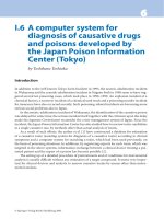
Figure 13.2 Schematic diagram of the left ventricle in hypertrophic cardiomyopathy
during systole. There is projection of the basal septum into the outflow tract with
systolic anterior motion of the mitral valve, which results in left ventricular outflow
tract obstruction. The obstruction is dynamic, dependent on the preload, afterload and
contractility of the heart. By permission of the Mayo Foundation for Medical Education
and Research.
papillary muscles, which position the mitral valve leaflets so as to contribute
further to this outflow tract obstruction.
16
It is the young patients with severe
outflow tract obstruction who are at highest risk for hemodynamic deteriora-
tion during pregnancy.
Diagnosis of hypertrophic cardiomyopathy in
patients of child-bearing age
As young patients with hypertrophic cardiomyopathy are symptom free, the
diagnosis of hypertrophic cardiomyopathy is primarily made through family
screening or after a routine medical examination when a murmur is heard on
auscultation or an abnormal ECG is found. Hypertrophic cardiomyopathy is
sometimes first recognized during pregnancy when systolic murmurs lead to
cardiologic referral. As the murmur of left ventricular outflow tract obstruction
is dynamic, maneuvers such as squat to stand should be performed. In patients
with hypertrophic cardiomyopathy, the length and intensity of a late systolic
ejection murmur should significantly increase upon assuming the standing
position.
The ECG is almost always abnormal in patients beyond the first decade of life
with hypertrophic cardiomyopathy (Figure 13.3).
17,18
It often shows features of
atrial and ventricular hypertrophy that are associated with marked ST- and
T-wave abnormalities. Echocardiography should always be performed in pa-
176 Chapter 13
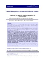
Figure 13.3 A 12-lead ECG from a patient with hypertrophic cardiomyopathy. There is
left ventricular hypertrophy present. There is high voltage as well as secondary ST–T-
wave abnormalities. There is left atrial enlargement.
Hypertrophic cardiomyopathy and pregnancy 177
tients with an abnormal ECG to look for the increase in left ventricular wall
thickening that is considered the ‘gold standard’ of diagnosis when present
(Figure 13.4). However, in some patients with an abnormal ECG, it may not
identify hypertrophy, especially if it is localized to an abnormal location such as
the apex. In a situation where there is significant unexplained abnormality of the
ECG, repeat echocardiography with contrast enhancement or magnetic reso-
nance imaging (MRI) should be performed.
The presence or absence of left ventricular outflow tract obstruction
should be documented both by examination and by echocardiography. Systolic
anterior motion of the mitral valve with a late peaking, high-velocity Doppler
signal in the left ventricular outflow tract is diagnostic of dynamic left ventricu-
lar outflow tract obstruction (Figure 13.5). The magnitude of the obstruction
is determined by the peak Doppler velocity (Figure 13.5 shows a velocity of
more than 6 m/sec) and should be measured at rest and during provocation
(after a premature ventricular contraction, during the strain phase of the Val-
salva maneuver or during inhalation of amyl nitrite). Secondary mitral
regurgitation, characterized by a posteriorly directed jet, should be identified
and semi-quantified.
Genetics and family screening
Hypertrophic cardiomyopathy is usually the result of a missense mutation in the
sarcomeric protein genes and transmitted in an autosomal dominant manner.
There are over 200 mutations that have been identified in 10 different sarcomeric
genes.
19
Thus, once the diagnosis of hypertrophic cardiomyopathy has been made,
all patients should undergo genetic counseling and family screening. This should be
performed even if the patient is completely asymptomatic. All first-degree relatives
of the proband should be screened with physical examination, an ECG and
echocardiography. In adolescence, screening should be performed every year
throughout their growth spurt. In adults, screening should be performed every
5 years because hypertrophy may appear late in patients with certain gene
mutations. In the future, genetic analysis should aid in helping screen the presence
or absence of hypertrophic disease in first-degree relatives of patients with
documented hypertrophic cardiomyopathy. Although initial data suggested that
certain specific mutations may predispose to sudden death or have a benign
course,
20
the accuracy of this prediction has been challenged with studies of unre-
lated hypertrophic cardiomyopathy patients that demonstrated that specific malig-
nant or benign mutations are rare and clinical outcomes cannot be successfully
predicted.
21,22
In women who wish to become pregnant, genetic counseling is essential.
There is a 50% chance that the child may have hypertrophic cardiomyopathy. If
hypertrophic cardiomyopathy becomes manifest in the very early childhood
years, severe disease occurs and the prognosis may be dismal.
23
The utility of
prenatal ultrasound screening is controversial. In the future prenatal molecular
diagnosis may be performed.
24
178 Chapter 13
(a)
(b)
Figure 13.4 A two-dimensional echocardiogram from a patient with hypertrophic
cardiomyopathy. There is an increase in left ventricular wall thickness, with a greater
increase in thickness of the ventricular septum. (a): parasternal long-axis view during
diastole. (b): parasternal long-axis view during systole.
Hypertrophic cardiomyopathy and pregnancy 179
(c)
(d)
Figure 13.4 Continued (c): short axis, diastole. (d): short axis, systole. There is systolic
anterior motion of the mitral valve causing left ventricular outflow tract obstruction.
Risks in pregnancy
The risks in pregnancy are related either to hemodynamic deterioration or ar-
rhythmias and sudden death. Most young woman with hypertrophic cardiomy-
opathy will go through pregnancy without difficulty.
25–29
The increased blood
volume and stroke volume of pregnancy are beneficial for the dynamic left ven-
tricular outflow tract obstruction. Most women who have no or mild symptoms
before the pregnancy will not develop more symptoms during the preg-
nancy course. There are some who become more dyspneic as a result of the larger
blood volume but this can be controlled with low-dose diuretics.
In patients who have moderate to severe symptoms before the pregnancy,
symptoms will worsen in 10–30% of patients, especially if there is pre-existing
left ventricular outflow tract obstruction. The higher the left ventricular outflow
tract gradient, the more likely that there will be progressive symptoms.
25
It is the
subset of patients with very severe obstruction (gradient >100 mmHg) who are
at highest risk of hemodynamic deterioration during pregnancy and delivery.
Sudden death or resuscitated ventricular fibrillation is unusual but has been re-
ported to occur in patients with hypertrophic cardiomyopathy during pregnan-
cy.
25,30–33
In an Italian series of 100 women with hypertrophic cardiomyopathy,
there were two pregnancy-related arrhythmic deaths.
25
The maternal mortality
rate was 10 per 1000 live births and was higher than the expected mortality rate
in the Italian population (relative risk 17.1, 95% CI 2.0–61.8). However, the ab-
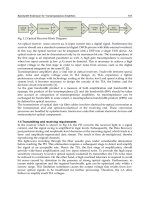
180 Chapter 13
200 mmHg
0 mmHg
6 m/s
Ao
Lv
Figure 13.5 Simultaneous left ventricular (LV) and aortic (Ao) pressure from
catheterization with continuous wave Doppler of the LV outflow tract. The peak
velocity of 6 m/sec by Doppler corresponds to the gradient of 150 mmHg by
catheterization.
solute risk of death was low and each of the two patients who died had significant
risk factors for sudden cardiac death: one had severe hypertrophy and severe out-
flow tract obstruction (>115mmHg) and the other a highly malignant family his-
tory of hypertrophic cardiomyopathy (five young relatives died suddenly).
Management in pregnancy
Even though the outcome is usually favorable, some patients will develop
symptoms for the first time during pregnancy, or pre-existing symptoms will get
worse. When symptoms are present, beta blockade should be started Table 13.1.
The dosage of beta blocker should be titrated to attain a resting heart rate less
than 70 beats/min (bpm). Beta blockers have the potential to cause growth re-
tardation, low Apgar scores or neonatal hypoglycemia in the infants but this is
rare.
21,34
Breast-feeding is not contraindicated but atenolol, acebutolol, nadolol
and sotalol are secreted in breast milk in larger amounts than the other beta
blockers. Verapamil is also safe for use in pregnancy if beta blockers are not tol-
erated, but can cause hemodynamic deterioration and sudden death if started in
a patient with a severe resting left ventricular outflow tract obstruction. When
verapamil is initiated in patients with severe left ventricular obstruction, this
should be done under monitored conditions in hospital.
Low-dose diuretics may be useful if patients develop pulmonary congestive
symptoms during the pregnancy as a result of the volume overload. However,
care must be taken not to cause too much of a reduction in preload because this
may exacerbate the left ventricular outflow tract obstruction. Periodic bedrest
in the left lateral decubitus position is recommended for all patients with hyper-
trophic cardiomyopathy and even mild symptoms.
In patients who have severe symptoms and severe outflow tract obstruction,
septal reduction therapy is recommended before proceeding with pregnancy.
Septal myectomy is considered the ‘gold standard’ for symptomatic patients
who have persistent symptoms despite optimal medical therapy.
35
In young
healthy women, the risk of the operation in experienced centers is less than 1%.
36
Septal myectomy relieves the gradient and produces an excellent reduction in
symptoms. Surgical myectomy has been performed during pregnancy in the
rare patient with a large outflow gradient who develops severe intractable
symptoms during pregnancy.
Hypertrophic cardiomyopathy and pregnancy 181
Table 13.1 General recommendations for management of patients with hypertrophic
cardiomyopathy during pregnancy
Document degree of left ventricular outflow tract (LVOT) obstruction and risk stratification
Risk stratification for sudden death
Beta blockade for symptoms
Avoid decrease in preload (dehydration, over diuresis)
Avoid inotropes (dopamine or dobutamine) and vasodilators (nifedipine)
In the hypotensive patient balance fluids and vasoconstrictor
Septal ablation has been considered an alternative to septal myectomy in pa-
tients with hypertrophic cardiomyopathy and left ventricular outflow tract ob-
struction.
37
However, the long-term effect of this procedure is unknown. It is
not known whether or not creating a large infarct in patients already at risk of
ventricular arrhythmias may enhance the propensity to develop dangerous ar-
rhythmias or cause adverse ventricular remodeling with time.
38
Therefore, in
women of child-bearing age, septal myectomy has been considered the proce-
dure of choice in our institution (Mayo Clinic)to which such rare patients are
referred.
Arrhythmias are uncommon in patients with hypertrophic cardiomyopathy
undergoing pregnancy.
25,26
Should atrial fibrillation or flutter occur, cardiover-
sion should be considered if there is hemodynamic compromise as a result of
the loss of atrial contraction and fast ventricular response. Beta blockers are usu-
ally the drug of choice in preventing further episodes, although low-dose amio-
darone can be used if recurrent episodes are present. Amiodarone has been used
safely in pregnancy. Fetal hypothyroidism may occur so that a neonatal assess-
ment after delivery is warranted but no congenital abnormalities have been
noted.
Aggressive risk stratification to determine those at risk for sudden death
should be performed on all patients with hypertrophic cardiomyopathy. The
major risk factors that predict sudden death include previous out-of-hospital
arrest or documented sustained ventricular tachycardia, and a strong family
history of hypertrophic cardiomyopathy with sudden death. Other ‘minor’ risk
factors for the occurrence of sudden death include severe hypertrophy (thick-
ness >3 cm), non-sustained ventricular tachycardia on Holter monitoring, a
drop in blood pressure with exercise and perfusion defects on MRI. If multiple
risk factors are present, implantation of an automatic defibrillator has been rec-
ommended.
6
Successful shocks with automated defibrillators have been re-
ported in pregnant patients with hypertrophic cardiomyopathy.
39
Amiodarone
may be a useful suppressive antiarrhythmic agent in the rare event of recurrent
intractable ventricular tachycardia during pregnancy.
Labour and delivery
Delivery should take place in high-risk centers experienced in the management
of this condition and where continuous ECG and blood pressure monitoring are
possible. Continued beta blockade and fluid replacement are necessary in the
presence of dynamic outflow tract obstruction. Normal vaginal delivery is safe
and cesarean section is indicated only for obstetric purposes. The use of
prostaglandins for induction of labour is inadvisable because of their vasodila-
tor effect, but oxytocic agents are well tolerated. Epidural anesthesia
should be avoided because of the production of hypotension, and blood
loss should be replaced promptly. After completion of the third stage, the
patient should sit up to avoid pulmonary congestion and may require intra-
venous frusemide. (Table 13.2)
182 Chapter 13
If there is evidence of left ventricular outflow tract obstruction with hemody-
namic deterioration after delivery, fluids and vasoconstriction with phenyl-
ephrine to increase afterload are recommended. Beta-adrenergic agents such as
dopamine or dobutamine should be avoided as a result of the increase in con-
tractility, outflow tract gradient and worsening hypotension that they cause.
Continuous monitoring by right heart catheterization may be needed in
selected cases and transesophageal echocardiography has been used to assess
the hemodynamics. Antibiotic prophylaxis is needed for dental procedures
during pregnancy and to cover surgical delivery.
Conclusion
With care in high risk patients the outcome of pregnancy in women with hy-
pertophic cardioimyopathy is usually good (Table 13.3).
References
1 Maron BJ, Gottdiener JS, Epstein SE. Patterns and significance of distribution of
left ventricular hypertrophy in hypertrophic cardiomyopathy. A wide angle, two
dimensional echocardiographic study of 125 patients. Am J Cardiol 1981;48:418–
28.
2 Davies MJ. The current status of myocardial disarray in hypertrophic cardiomyopa-
thy. Br Heart J 1984;51:361–3.
Hypertrophic cardiomyopathy and pregnancy 183
Table 13.2 Management of delivery in patients with hypertrophic cardiomyopathy
Delivery in hospital with continuous ECG and blood pressure monitoring
Normal vaginal delivery
Do not use prostaglandins
Prompt blood loss replacement
Sit the patient up after completion of the third stage to avoid pulmonary edema
Antibiotic prophylaxis
Table 13.3 Outcomes in pregnant patients with hypertrophic cardiomyopathy
Reference No. of No. of Cardiac Worsening of Intrauterine Observed
patients pregnancies symptoms symptoms deaths deaths
during during
pregnancy
a
pregnancy
Thaman et al.
26
127 271 36 (28%) 10 (<10%) 3 (2%) 0
Autore et al.
25
100 199 Not reported 6 of 40 patients Not reported 2
(15%)
Probst et al.
28
41 150 27% 0% 0 0
a
Numbers in parentheses are percentages.
3 Nishimura RA, Holmes DR, Jr. Hypertrophic obstructive cardiomyopathy. N Engl J
Med 2004;350:1320–7.
4 Teare D. Asymmetrical hypertrophy of the heart in young adults. Br Heart J
1958;20:1–8.
5 Brock R. Functional obstruction of the left ventricle; acquired aortic subvalvar steno-
sis. Guy’s Hospital Rep 1957;106:221–38.
6 Maron BJ, McKenna WJ, Danielson GK et al. American College of Cardiology/
European Society of Cardiology Clinical Expert Consensus Document on Hyper-
trophic Cardiomyopathy: a report of the ACC Foundation Task Force on Clinical Ex-
pert Consensus Documents and the European Society of Cardiology Committee for
Practice Guidelines. J Am Coll Cardiol 2003;42:1687–713.
7 Maron BJ. Hypertrophic cardiomyopathy: a systematic review. JAMA 2002;287:
1308–20.
8 Richardson P, McKenna RW, Bristow M et al. Report of the 1995 World Health Orga-
nization/International Society and Federation of Cardiology Task Force on the defi-
nition and classification of cardiomyopathies. Circulation 1996;93:841–2.
9 Maron BJ, Casey SA, Poliac LC, Gohman TE, Almquist AK, Aeppli DM. Clinical
course of hypertrophic cardiomyopathy in a regional United States cohort. JAMA
1999;281:650–5.
10 Niimura H, Bachinski LL, Sangwatanaroj S et al. Mutations in the gene for cardiac
myosin-binding protein C and late-onset familial hypertrophic cardiomyopathy.
N Engl J Med 1998;338:1248–57.
11 Nagueh SF, McFalls J, Meyer D et al. Tissue Doppler imaging predicts the develop-
ment of hypertrophic cardiomyopathy in subjects with subclinical disease. Circulation
2003;108:395–8.
12 Nagueh SF, Bachinski LL, Meyer D et al. Tissue Doppler imaging consistently detects
myocardial abnormalities in patients with hypertrophic cardiomyopathy and pro-
vides a novel means for an early diagnosis before and independently of hypertrophy.
Circulation 2001;104:128–30.
13 Ho CY, Sweitzer NK, McDonough B et al. Assessment of diastolic function with
Doppler tissue imaging to predict genotype in preclinical hypertrophic cardiomyopa-
thy. Circulation 2002;105:2992–7.
14 Maron MS, Olivotto I, Betocchi S et al. Effect of left ventricular outflow tract ob-
struction on clinical outcome in hypertrophic cardiomyopathy. N Engl J Med
2003;348:295–303.
15 Sherrid MV, Gunsburg DZ, Moldenhauer S, Pearle G. Systolic anterior motion begins
at low left ventricular outflow tract velocity in obstructive hypertrophic cardiomy-
opathy. J Am Coll Cardiol 2000;36:1344–54.
16 Klues HG, Maron BJ, Dollar AL, Roberts WC. Diversity of structural mitral valve al-
terations in hypertrophic cardiomyopathy. Circulation 1992;85:1651–60.
17 Frank S, Braunwald E. Idiopathic hypertrophic subaortic stenosis. Clinical analysis of
126 patients with emphasis on the natural history. Circulation 1968;37:759–88.
18 Ryan MP, Cleland JGF, French JA et al. The standard electrocardiogram as a screening
test for hypertrophic cardiomyopathy. Am J Cardiol 1995;76:689–94.
19 Van Driest SL, Ommen SR, Tajik AJ, Gersh BJ, Ackerman MJ. Sarcomeric genotyping
in hypertrophic cardiomyopathy. Mayo Clin Proc 2005;80:463–9.
20 Watkins H, Rosenzweig A, Hwang D et al. Characteristics and prognostic implications
of myosin missense mutations in familial hypertrophic cardiomyopathy. N Engl J Med
1992;326:1108–14.
184 Chapter 13
21 Van Driest SL, Ackerman MJ, Ommen SR et al. Prevalence and severity of ‘benign’
mutations in the beta-myosin heavy chain, cardiac troponin T, and alpha-
tropomyosin genes in hypertrophic cardiomyopathy. Circulation 2002;106:3085–90.
22 Ackerman MJ, VanDriest SL, Ommen SR, et al. Prevalence and age-dependence of
malignant mutations in the beta-myosin heavy chain and troponin t genes in hyper-
trophic cardiomyopathy: a comprehensive outpatient perspective. J Am Coll Cardiol
2002;39:2042–8.
23 Spirito P, Bellone P, Harris KM, Bernabo P, Bruzzi P, Maron BJ. Magnitude of left ven-
tricular hypertrophy and risk of sudden death in hypertrophic cardiomyopathy.
N Engl J Med 2000;342:1778–85.
24 Charron P, Heron D, Gargiulo M et al. Prenatal molecular diagnosis in hypertrophic
cardiomyopathy: report of the first case. Prenat Diagn 2004;24:701–3.
25 Autore C, Conte MR, Piccininno M et al. Risk associated with pregnancy in hyper-
trophic cardiomyopathy. J Am Coll Cardiol 2002;40:1864–9.
26 Thaman R, Varnava A, Hamid MS et al. Pregnancy related complications in women
with hypertrophic cardiomyopathy. Heart 2003;89:752–6.
27 Oakley GD, McGarry K, Limb DG, Oakley CM. Management of pregnancy in patients
with hypertrophic cardiomyopathy. BMJ 1979;i:1749–50.
28 Probst V, Langlard JM, Desnos M, Komajda M, Bouhour JB. [Familial hypertrophic
cardiomyopathy. French study of the duration and outcome of pregnancy.] Arch Mal
Coeur Vaiss 2002;95:81–6.
29 Turner GM, Oakley CM, Dixon HG. Management of pregnancy complicated by
hypertrophic obstructive cardiomyopathy. BMJ 1968;4:281–4.
30 Pelliccia F, Cianfrocca C, Gaudio C, Reale A. Sudden death during pregnancy in
hypertrophic cardiomyopathy. Eur Heart J 1992;13:421–3.
31 Shah DM, Sunderji SG. Hypertrophic cardiomyopathy and pregnancy: report of a
maternal mortality and review of literature. Obstet Gynecol Surv 1985;40:444–8.
32 Benitez RM. Hypertrophic cardiomyopathy and pregnancy: maternal and fetal out-
comes. J Maternal-Fetal Invest 1996;6:51–5.
33 Minnich ME, Quirk JG, Clark RB. Epidural anesthesia for vaginal delivery in a patient
with idiopathic hypertrophic subaortic stenosis. Anesthesiology 1987;67:590–2.
34 Oakley GD, McGarry K, Limb DG, Oakley CM. Management of pregnancy in patients
with hypertrophic cardiomyopathy. BMJ 1979;i:1749–50.
35 Maron BJ, Dearani JA, Ommen SR et al. The case for surgery in obstructive hyper-
trophic cardiomyopathy. J Am Coll Cardiol 2004;44:2044–53.
36 McCully RB, Nishimura RA, Tajik AJ, Schaff HV, Danielson GK. Extent of clinical
improvement after surgical treatment of hypertrophic obstructive cardiomyopathy.
Circulation 1996;94:467–71.
37 Sigwart U. Non-surgical myocardial reduction for hypertrophic obstructive car-
diomyopathy. Lancet 1995;
346:211–14.
38 Maron BJ. Surgery for hypertrophic obstructive cardiomyopathy: alive and quite
well. Circulation 2005;111:2016–18.
39 Piacenza JM, Kirkorian G, Audra PH, Mellier G. Hypertrophic cardiomyopathy and
pregnancy. Eur J Obstet Gynecol Reprod Biol 1998;80:17–23.
Hypertrophic cardiomyopathy and pregnancy 185
CHAPTER 14
Peripartum cardiomyopathy,
other heart muscle disorders
and pericardial diseases
Celia Oakley
Peripartum cardiomyopathy
Peripartum cardiomyopathy is a dilated cardiomyopathy that occurs in the peri-
partum period. Heart failure had been known of since the eighteenth century
but was first described in 1937 as ‘Idiopathic myocardial degeneration associ-
ated with pregnancy and especially the puerperium’.
1
With the emergence and
classification of the different forms of cardiomyopathy in the 1960s it became
known as a dilated cardiomyopathy with a temporal relationship to pregnan-
cy.
2
Biopsy shows myocarditis in a high proportion of cases
3,4
and further work
has pointed to a probable autoimmune mechanism, although this is still not
fully understood.
5
The sudden onset of heart failure in a previously healthy young woman who
had been looking forward to the birth brings bleak fear to the patient and her
family, and something similar to her doctors as the future is unknown to each.
Some patients rapidly deteriorate and will die without a device or transplanta-
tion whereas others make an astonishing almost complete recovery and it is im-
possible to know which way ‘the cat will jump’. More often the onset is less
dramatic and the failure less severe but improvement slow or absent. There is no
doubt that many mild cases are missed altogether. As the condition is rare (al-
though less rare than generally believed), personal experience is limited and the
literature tends to reflect a limited personal experience that may be likened to a
blind person describing an elephant, having felt only one part of it.
Definition
Peripartum cardiomyopathy (PPCM) is a dilated cardiomyopathy with a tem-
poral relationship to pregnancy. It was defined arbitrarily by Demakis et al.
6
in
1971 as unexplained left ventricular systolic dysfunction developing in the last
month of pregnancy or within 5 months of delivery. The definition required
that there be no other identifiable cause for the heart failure and excluded pa-
186
Heart Disease in Pregnancy, Second Edition
Edited by Celia Oakley, Carole A Warnes
Copyright © 2007 by Blackwell Publishing
tients with a previous history of possible myocardial disease.
6
At a workshop in
1997 the addition was made that the left ventricular dysfunction should be
demonstrated echocardiographically
7
(Table 14.1).
Although the definition aims to separate patients out with previously unsus-
pected dilated cardiomyopathy exacerbated by the pregnancy, it is not practica-
ble for all seemingly healthy women to have echocardiography studies on first
booking and, in practice, the differentiation is difficult and will largely depend
on when heart disease is first recognized. Any patient with a family history of
cardiomyopathy should have echocardiography performed even if she is appar-
ently fully fit.
Patients with pre-existing heart disease are not immune from developing a
PPCM and may be more likely to develop symptoms because of their reduced
cardiovascular reserve.
8,9
Epidemiology and prevalence
The prevalence of PPCM is not known because recognition and accurate diag-
nosis depend on the availability and application of echocardiography. This
would require its routine use in every parturient woman within sizeable de-
fined populations for insight to be gained about the true prevalence. M-mode
echocardiography was only just becoming available in the cardiology depart-
ments of major hospitals in the 1960s and so had not contributed to the diagno-
sis of the retrospective series of patients described in the key papers from New
Orleans in 1965
2
or from Chicago in 1971.
6
Even now cases go undiagnosed and
this is true of major centers as well as less highly developed ones.
The condition has been described from all around the world in both small per-
sonal series and reviews of accumulated cases from many sources, and over
different observation periods, so providing little idea of true prevalence.
10–16
It is no wonder that estimates are in truth just wild guesses. They vary
widely from 1 in 1485 to 1 in 15 000 live births even within the USA.
10–14
Much
higher figures come from South Africa (1 in 1000) and from Haiti (1 in
350–400) live births.
15
The high prevalence reported from Nigeria was caused
by heart failure induced by a local custom that decrees that parturient women
eat excessive local salt (kanwa) and lie on heated mud beds for 40 days post
partum.
17–18
Peripartum and other cardiomyopathic pericardial diseases 187
Table 14.1 Diagnostic criteria
• Cardiac failure developing in the last month of pregnancy or within 5 months of delivery
• Absence of other detectable cause
• Apparent absence of myocardial disease before the last month of pregnancy
• Left ventricular systolic dysfunction demonstrated by echocardiography:
—
ejection fraction <45%
—
fractional shortening <30% and/or
—
end-diastolic dimension >2.7 cm/m
2
A consensus opinion from the workshop in 1997 was of an incidence in be-
tween 1 in 3000 and 1 in 4000 in the USA
7,19
which suggests that there may be
750–1000 cases a year in the UK.
Etiology
Traditional predisposing causes of PPCM include multiple pregnancy, multipar-
ity, African race, older maternal age, pre-eclampsia and a history of previous
peripartum cardiomyopathy. Selenium deficiency,
20
infection, tocolytic
therapy and surgical delivery have also been suggested.
21,22
The hemodynamic burden is greater during pregnancy than in the puerperi-
um. Multiple pregnancy magnifies this. If twin pregnancy is a cause of PPCM
such patients should develop symptoms during the second trimester, as do pa-
tients with structural heart disease or a pre-existing dilated cardiomyopathy
when they exhaust a reduced cardiac reserve. With delivery and uterine con-
traction maternal blood volume is expanded by return of uteroplacental blood
and at the same time afterload increases resulting from loss of the low-resistance
placental bed. These volume shifts are exaggerated in multiple pregnancy.
Postpartum blood loss is needed to alleviate the hypervolemia but iatrogenic
overhydration may contribute to pulmonary edema after surgical delivery.
Over 90% of cases of PPCM present in the puerperium when the increased
hemodynamic load of pregnancy has diminished. This is inconsistent with the
condition being simply an exacerbation of a pre-existing dilated cardiomyopa-
thy, but would conform with an autoimmune origin developing as the dormant
immune system is reactivated after delivery.
24–26
The reported incidence of myocarditis ranges from 8.8% to 78%. The fre-
quent finding of myocarditis when endomyocardial biopsies are taken within a
month of onset of symptoms is in conformity with the autoimmune theory, as is
the marked capacity for hemodynamic recovery that is also shown by some pa-
tients with acute myocarditis seen outside pregnancy. The true prevalence of
myocarditis in PPCM may have been underestimated because many biopsies
may be taken after the changes have regressed and, even in fulminant cases, my-
ocarditis tends to be focal, bringing the possibility of sampling errors. The
diagnosis may also be missed if immunohistochemical staining is not performed.
Failure to include these in the so-called ‘Dallas criteria’ may have contributed to
the difficulty in recruiting patients to the trial of immunosuppression in acute
myocarditis.
27,28
It is probable that the most good would be done by immuno-
suppressive treatment if it is started as early as possible after onset. The difficulty
in recognizing heart failure in late pregnancy, when many women complain of
shortness of breath and develop swollen legs, means that insidious onset at that
time would be very likely to remain undiagnosed.
Viruses have long been suspected of playing a part in initiating both the in-
flammatory process in dilated cardiomyopathy and an autoimmune process
against exposed or damaged myocardial proteins, although no infective agent
has been found in cases of PPCM and the causative agent is probably not an in-
fective one. Fetal cells are known to enter the maternal circulation during preg-
188 Chapter 14
nancy and to remain there without rejection. If such foreign cells enter cardiac
tissue during the immunosuppressed state of pregnancy, they might well be
responsible for triggering a vigorous reaction after restoration of immune com-
petence and would explain cases of PPCM with a new partner after previous
healthy pregnancies. Persuasive support for abnormal immunological activa-
tion comes from the finding of high titers of autoantibodies both peripartum
and in dilated cardiomyopathy. In addition, several autoantibodies have been
found that were unique to PPCM and not present in patients with dilated car-
diomyopathy.
24–26
Some patients with PPCM give a family history of dilated cardiomyopathy
and autoantibodies are found in both conditions.
29–31
Dilated cardiomyopathy
is familial in about a quarter of cases but the heredity is far from straightforward.
Genetic studies may shed some light on PPCM in which a family history of either
dilated cardiomopathy or PPCM seems too frequent for chance. Both facilitative
and protective factors would account for the fewer than expected numbers in
cardiomyopathy families in which ‘latent cases’ with only mild echocardio-
graphic abnormality are a feature. As well as these and possible lurking infective
agents are the unique hormonal environment and possible maladaptive
responses to the hemodynamic changes of pregnancy, all of which have been
suggested as possible contributory causes.
32–34
Although the time of clinical onset does not necessarily mark the time of
onset of the cardiac dysfunction, fulminant cases usually present in the first few
days post partum but give no hint of a preceding cardiac problem. Milder cases
tend to present later in the puerperium with a much more insidious onset that
the women are unable to date. Only 9% of patients present in the last month of
pregnancy and it is likely that the problem almost universally starts early after
delivery, as would be in keeping with the immunological explanations for the
condition.
Pre-eclampsia has been cited as a possible contributory cause and is some-
times associated but pre-eclampsia does not cause systolic heart failure in
healthy young women, and cardiomyopathy is not found even in patients who
have been under close observation in hospital. Moreover PPCM usually devel-
ops post partum whereas delivery cures pre-eclampsia.
PPCM is no doubt polygenic with links to idiopathic dilated cardiomyopathy,
its expression determined by the interplay of many other endogenous and
environmental factors yet to be determined.
Diagnosis
Diagnosis rests on the recognition of left ventricular dysfunction around the
time of parturition that is believed to be new and for which other possible
causes have been excluded.
Heart failure
The recognition of heart failure or even that the patient is ill is not easy before
delivery when dyspnea, fatigue and edema are normally common in late
Peripartum and other cardiomyopathic pericardial diseases 189
pregnancy. Cough, orthopnea, paroxysmal nocturnal dyspnea, palpitation,
chest pain and abdominal pain may develop.
Most women are kept in hospital for only a few hours before being discharged
back into the care of their midwives, on whom much of the responsibility of
noticing something wrong will fall. Diagnosis should become obvious if symp-
toms worsen at a time when they should have improved, but the heart failure
may be missed until the new mother tries to resume her normal activities, to-
gether with the added work and loss of sleep involved in looking after a new baby.
The condition does not differ clinically from dilated cardiomyopathy. Exami-
nation will reveal a resting tachycardia, low blood pressure and pulse pressure,
raised cervical venous pressure, gallop rhythm, lung crackles and enlarged liver.
A mitral regurgitant murmur is sometimes heard. Fluid overload is a prominent
feature, particularly in patients who have just had a surgical delivery, and there
may be ascites.
Chest pain
As in myocarditis outside pregnancy, chest pain is frequently the herald symp-
tom that leads to diagnosis. It may suggest myocardial infarction and demand
urgent investigation.
35
Myocardial infarction is also a rare complication of preg-
nancy that usually occurs in the peripartum period. The differential diagnosis
may remain in doubt until coronary angiography, because cardiac markers may
be raised and echocardiography does not always show uniformly global hy-
pokinesia. Cocaine abuse can cause vasospasm with ischemic chest pain and
even cardiac infarction, which is often caused by a spontaneous coronary artery
dissection; if it involves the left anterior descending artery it may lead to exten-
sive anteroapical dysfunction and severe left ventricular failure. A few infarcts
being seen now are occurring in older women, smokers with the metabolic syn-
drome and atheroma. Either way urgent coronary angiography is needed for
diagnosis and prompt appropriate treatment.
Embolism
Endocardial thrombosis is frequent and either pulmonary or systemic em-
bolism may be the first clinical event that brings the cardiac problem to
light.
36–39
Echocardiography, a routine first investigation for a possible embolic
source, may show ventricular thrombus and severe biventricular dysfunction
that had previously escaped notice because of lack of formal cardiovascular
examination or pulmonary auscultation. This is because there may have been
little in the way of cardiac symptoms before the embolus despite the marked
compromise. Most peripartum women are healthy and pass from home to
hospital and back with little time for anything apart from the actual birth. Even
after such patients have been re-admitted to hospital on account of embolism,
the signs of cardiac dysfunction are sometimes missed. Although this empha-
sizes the huge cardiovascular reserve that exists, it also indicates the subtlety of
the clinical signs or their elusiveness unless clinicians’ thoughts turn to the
heart. Even florid failure with edema may be attributed to other causes.
190 Chapter 14
The higher prevalence of intracardiac thrombosis and embolism in peripar-
tum cardiomyopathy than in dilated cardiomyopathy outside pregnancy is at-
tributable to the hypercoagulable state existing in pregnancy, which increases
the risk of intracavitary thrombus caused by stasis in poorly contracting cham-
bers or endocardial inflammation.
Echocardiography reveals the problem but only if it is performed. This is more
likely if the patient has an embolus or is short of breath after delivery, but much
less likely if she complains only of fatigue or chest pain and is not examined. This
could mean an unknown but possibly large number of milder non-fatal cases
remaining unrecognized and either improving spontaneously or presenting
later with a dilated cardiomyopathy.
Arrhythmias
The increased tendency to cardiac arrhythmias seen in pregnancy is attributed
to an increased sympathetic drive that is intensified by the neuroendocrine
activation of cardiac failure.
Arrhythmias are frequent and palpitation is often the presenting feature of a
PPCM. Frequent ectopic beats, supraventricular and ventricular tachycardias
(Figure 14.1b, c), atrial flutter and fibrillation may all be seen in the same pa-
tient and contribute to hemodynamic instability.
40,41
A new mother, tired and
therefore harassed, may ignore the rapid heart beat attributing it to her fatigue.
Her ventricular dysfunction may then be worsened and it may be uncertain
whether the arrhythmia caused the ventricular dysfunction, ‘tachycardia fail-
ure,’ or whether it was caused by an underlying PPCM.
Peripartum and other cardiomyopathic pericardial diseases 191
Figure 14.1 (a) ECG from a patient with a
fulminant peripartum cardiomyopathy showing
low voltage and QS waves in leads V1–V3, poor
R-wave progression and T-wave inversion in left
ventricular leads, suggesting (old rather than
evolving) anteroapical infarction. (b,c) Rhythm
strips from the same patient showing
supraventricular tachycardia (b) and a burst of
ventricular tachycardia (c).
192 Chapter 14
Differential diagnosis
The clinical differential diagnosis is from other causes of heart failure, other
cardiomyopathies, pre-existing dilated cardiomyopathy, acute myocardial
infarction, ritodrine-induced pulmonary edema in patients given the drug by
infusion in saline, fluid overload after surgical delivery, tachycardia-induced
failure, massive pulmonary embolism, amniotic fluid embolism, and infective,
metabolic and toxic causes of heart failure (Table 14.2).
Investigations
Laboratory blood tests
Hematology and biochemical findings are usually normal, but atrial and brain
natriuretic peptides are elevated and the D-dimer will be raised above the usu-
ally raised upper limits for pregnancy and the puerperium in patients with in-
tracardiac thrombi or those who have had emboli. Cardiac markers, especially
troponin, may be above normal or enzymes may be within normal with a raised
troponin.
The laboratory work-up will include renal and liver function tests, thyroid
function, serological tests for viral and rickettsial infections, syphilis and HIV,
and tests for alcohol and cocaine, plus autoimmune studies to exclude collagen
vascular disease, sarcoidosis and pheochromocytoma as clinically indicated.
Electrocardiogram
Sinus tachycardia is often interrupted by multiple supraventricular and ventric-
ular ectopic beats, bursts of tachycardia, atrial flutter or fibrillation. Low voltage
is usual particularly in the standard leads. The QRS may be widened, reflecting
left ventricular dilatation. Left or right bundle-branch block may come and go
or a QS pattern in the chest leads may suggest myocardial infarction (see Figure
14.1). Occasionally the ECG is within normal limits or just shows non-specific
ST- and T-wave abnormalities or T-wave inversion.
The chest radiograph
Portable anteroposterior films are not very helpful and if possible a departmen-
tal posteroanterior film should be obtained. The cardiac diameter is usually in-
creased and the lungs show pulmonary congestion or frank pulmonary edema.
Small pleural effusions are commonly present.
Table 14.2 Differential diagnosis
• Pre-existing dilated cardiomyopathy
• Peripartum myocardial infarction (coronary artery dissection, thrombosis or embolism,
cocaine-induced spasm)
• Pulmonary embolism – thrombus or amniotic fluid
• Fluid overload
Echocardiography
All four cardiac chambers are usually dilated and there is marked left ventricu-
lar hypokinesia. This is typically global but may be more focal, suggesting possi-
ble infarction. Wall thicknesses are normal. All valves except the aortic may
show regurgitation. All the indices of contractility are reduced. Spontaneous
echo contrast reflects slowed flow (Figure 14.2) and thrombus may be seen in
either or both ventricles or in the atria if they are fibrillating. A small pericardial
effusion is commonly present.
Cardiac catheterization and angiocardiography
Hemodynamic measurement is not needed if the diagnosis is clear. When per-
formed, pulmonary artery wedge pressure and ventricular diastolic pressures
will be elevated, but pulmonary artery pressure is usually normal or barely
raised. If significant pulmonary hypertension is recorded this suggests a pre-
existing condition and is not seen in acute PPCM.
Coronary angiography shows normal coronary arteries and left ventricular
angiography is contraindicated if echocardiography has shown ventricular
thrombus and the added contrast load is better avoided because all the informa-
tion is available from echocardiography.
Endomyocardial biopsy
The indications for biopsy in dilated cardiomyopathy are controversial but
biopsy is needed when a specific cause is suspected. This should include PPCM
in which the prevalence of myocarditis remains uncertain. Biopsy should be
performed only if the operator is experienced in the technique and has access to
an experienced pathologist for interpretation. Biopsies should be taken from
the right ventricle if echocardiography has shown this to be free from thrombus.
If biopsy is to be performed it should be done as soon as practicable after onset
and after prior consultation with the cardiac histopathologist, who should be
fully informed of the clinical details. Biopsies should be obtained from as many
different sites in the ventricle as possible.
Treatment
Patients with PPCM should be made known to and discussed with cardiologists
in a specialist cardiac intensive care unit. If they do poorly and need transfer
they can then be moved without delay so that a ventricular assist device can be
installed as a bridge to recovery or transplantation performed. The condition of
these patients often changes quickly (for both better and worse) and decisions
must be made (and if necessary changed) equally rapidly.
The patient with acute-onset heart failure needs to be managed in a cardiac
unit so that her vital signs, heart rate and blood pressure, cardiac rhythm, oxy-
gen saturation and urinary output can be monitored, treatment adjusted
and appropriate action taken in case of sudden arrhythmia or cardiac arrest.
This can be difficult in severely ill patients with prenatal presentation, in whom
invasive monitoring during delivery will be advisable and when continuous
Peripartum and other cardiomyopathic pericardial diseases 193
194 Chapter 14
(a)
(b)
(c)
Figure 14.2 Echocardiograms from another patient. (a) Long-axis, (b) apical four-
chamber and (c) short-axis views at the level of the papillary muscles, showing a
moderately dilated and globally poorly contracting left ventricle (end-diastolic
dimension 66 mm, ejection fraction 25%). The left atrium is also dilated and the apical
four-chamber view shows spontaneous echo contrast in the left ventricle.
assessment of the fetus is also essential. Fortunately such cases are excep-
tionally rare.
The timing and mode of delivery need to be discussed among cardiologists,
obstetricians and anesthetists. In most cases this will be vaginal delivery waiting
for spontaneous labour if the cardiac condition has been stabilized and the fetus
is well. Labour should be induced if she fails to respond. If there is concern about
the baby surgical delivery may be necessary, but otherwise vaginal delivery
with good pain relief and accelerated second stage are to be preferred. Epidural
anesthesia is contraindicated if the patient is still heparinized. The patient and
her partner should be kept fully informed by the consultant throughout and
concerns, doubts and options discussed.
Heart failure is treated conventionally except that angiotensin-converting
enzyme (ACE) inhibitors and angiotensin receptor blockers (ARBs) are con-
traindicated if the patient is still undelivered. Reliance has to be placed on
hydralazine and nitrates. Carvedilol and amlodipine may also be used before
delivery when needed. Diuretics are needed for volume overload, but remem-
ber the risk of uteroplacental hypoperfusion in undelivered patients. Spirono-
lactone is helpful in severe failure. Low blood pressure need not be a deterrent
to pressing ahead cautiously with vasodilators, provided that urine output and
cerebration are unaffected. If the cardiac output is rising in response to unload-
ing, the low blood pressure will not fall further and a systolic pressure of
80 mmHg is well tolerated by a young patient. Beta blockers should be intro-
duced slowly because they cause transient deterioration before improvement,
but are particularly useful for deterring arrhythmias. Digoxin is indicated only if
needed for control of atrial fibrillation. The addition of pentoxifylline has been
claimed to improve the outcome.
42
Intravenous dobutamine should be used when inotropic support is needed. A
Swan–Ganz catheter is not needed if peripheral perfusion and oxygen satura-
tions are satisfactory, the lung bases clear and urine output is good. It is only in
the sickest patients on a balloon pump, and needing frequent adjustment of
drug infusion rates, that a Swan–Ganz catheter adds the information on which
fine adjustments are made. Continuous hemofiltration may be needed to re-
move excess fluid pending hemodynamic improvement.
Transplantation
43
is always a last resort in this condition, with the capacity for
such dramatic improvement. A left ventricular assist device may be needed as a
bridge to recovery or to transplantation, and buys time. Priority is given to such
young patients but assessment and preparation take time and organs are not al-
ways available, all of which underline the need for cardiologists in hospitals
without these facilities not to keep hold of these patients.
Anticoagulant treatment
Anticoagulant treatment is important in these patients with their high throm-
botic risk and should be started immediately whether or not ventricular throm-
bi have been shown or whether embolism has occurred or spontaneous echo
contrast is seen. They should be continued until all thrombi have disappeared,
Peripartum and other cardiomyopathic pericardial diseases 195
D-dimer has returned to normal and while ventricular function remains
impaired.
Unfractionated heparin should be used in undelivered patients because the
level of anticoagulation can be assessed from the activated partial thromboplas-
tin time and low-molecular-weight heparin cannot be as quickly reversed with
protamine. Warfarin is used after delivery. All the drugs mentioned are compat-
ible with breast-feeding.
Immunosuppressive treatment
Immunosuppressive treatment should probably be given to all patients in
whom biopsy has shown myocarditis, although benefit is not yet evidence
based. Many patients improve rapidly without it and others deteriorate despite
it. The myocarditis trial was unsatisfactory for many reasons
—
biopsy criteria,
delayed onset of treatment and protocol violations
—
as well as being unrelated
to the condition in pregnancy.
28
Immunoglobulins
A small study of intravenous immunoglobulin in women with PPCM suggested
that improvement in ejection fraction was greater in patients given this treat-
ment than in those who did not receive it.
44
Follow-up
It may be wise to continue left ventricular support with an ACE inhibitor and a
beta blocker for a year after recovery of ventricular function. Patients whose left
ventricular ejection fraction is still depressed should remain on treatment,
probably also including warfarin, and all patients should be followed up long
term with at least annual echocardiography irrespective of whether or not they
have apparently recovered fully.
Prognosis
Prognosis depends on recovery of left ventricular function.
45–50
As with the fig-
ures for prevalence the reported mortality rate from small series around the
world varies widely (from 7% to 56%). A realistic figure for the short-term
mortality rate is between 20 and 25%, and almost half of deaths occur in the first
3 months. The mortality rate was 28% in a series of black women from South
Africa. Cytokine levels were highest in the women who died.
45
About a third of
patients show a return to normal left ventricular function within 6 months and
more than half show significant improvement. The long-term prognosis de-
pends on whether left ventricular function eventually returns to normal or the
patient develops a dilated cardiomyopathy.
There is still very little information about the response to any subsequent
pregnancies. Women who have recovered from PPCM may have reduced con-
tractile reserve on dobutamine stress testing, and it has been suggested that such
women might not tolerate a subsequent pregnancy although there are as yet no
follow-up data for this suggestion.
51
196 Chapter 14
In a retrospective series from Brazil, 11 of 18 patients had persistent left ven-
tricular function. Deterioration was not seen in subsequent pregnancies in any
of those who had recovered.
46
Information on 44 patients was recovered by
questionnaire from members of the American College of Cardiology by
Elkayam.
47
The recurrence rate was 21%, but with no deaths among 28 patients
who had recovered completely, and 44% with 3 deaths among 16 patients who
had shown persistent left ventricular dysfunction. Improvement of ejection
fraction after the first subsequent pregnancy was to much the same level as be-
fore it, in both those who had and those who had not recovered completely.
These data suffer from both the inclusion of patients with left ventricular dys-
function diagnosed within 6 months of delivery and possible observer bias.
Patients who have recovered normal left ventricular function seem to have a
good prognosis. If such patients wish to have a further pregnancy they should
have a dobutamine echo stress test of contractile reserve. If this is normal they
can be told that a further pregnancy should be safe but that information is still
sparse. If contractile reserve is shown to be reduced the advice should be more
guarded. Women with persistently depressed left ventricular function should
be advised against embarking on another pregnancy.
Dilated cardiomyopathy
Patients with idiopathic dilated cardiomyopathy are discouraged from getting
pregnant and there are few reports in the literature in which left ventricular
function has been well documented before the pregnancy. The few patients de-
scribed had not tolerated the increased blood volume and cardiac output and
developed heart failure or pulmonary edema in midtrimester. More often pa-
tients have developed unexpected dyspnea and dry cough, which were at first
attributed to a chest infection. The left ventricle is usually already >6 cm with an
ejection fraction of 30% or less. Some improvement is expected after delivery
but others behave like patients with PPCM and deteriorate further although
considerable recovery may follow in survivors.
52–55
A teenager with cardiomyopathy known from infancy deteriorated at 20
weeks, requiring ventilation and inotropic support, but recovered after termi-
nation of the pregnancy
53
and a 40 year old with an ejection fraction of 30% and
left ventricular end-diastolic dimension of 7 cm, who had been stable for 8
years, went into failure at 26 weeks, deteriorated further after cesarean delivery
and died.
54
The small published and personal experience suggests that patients with pre-
existing dilated cardiomyopathy have a poorer clinical outcome than patients
with PPCM but hint at a link. A family history of dilated cardiomyopathy may be
a clue to pre-existing but occult dysfunction in a patient who first develops
symptoms within the time envelope assigned to ‘peripartum’ cardiomyopathy.
It is recommended that echocardiography should be performed, if possible be-
fore conception, in all patients desiring pregnancy who have a family history of
either dilated cardiomyopathy or PPCM.
Peripartum and other cardiomyopathic pericardial diseases 197
Advice has to be on an individual basis. The history is important. Some pa-
tients who are stable and asymptomatic will tolerate pregnancy but the high
chance of deterioration both during pregnancy and post partum suggests that
pregnancy should be avoided or termination advised if the end-diastolic vol-
ume is already above normal or if the ejection fraction is <50%.
55
Patients who
embark on pregnancy should have their left ventricular function regularly
monitored throughout by echocardiography so that need for adjustment in
therapy or hospital admission can be anticipated and clinical deterioration pre-
vented if possible.
ACE inhibitors should be withdrawn and hydralazine substituted with a
long-acting nitrate added later if needed. Diuretic dosage should be as low as
possible. A beta blocker such as metoprolol should be introduced gradually if
there are frequent ectopic beats, or especially if there is a resting sinus tachycar-
dia that reflects failure to increase stroke volume. The approach to delivery will
involve the whole team. Usually patients should be delivered vaginally with full
pain relief and an expeditious second stage with relief from the need to push.
Sudden onset of pulmonary edema is a risk after delivery. Blood loss should
be minimal and the patient should be sat up promptly and given 20 mg intra-
venous frusemide. Oxytocic agents are not contraindicated.
Patients with a secondary dilated cardiomyopathy caused by a known toxin
or other cause are more predictable and differ from patients with idiopathic or
familial dilated cardiomyopathy. Depression of left ventricular function after
previous chemotherapy may not be known to have caused damage until years
afterwards. Deterioration may occur in midtrimester or even earlier, with a risk
of pulmonary edema in the third stage but with improvement in the puerper-
ium. Two patients had been asymptomatic after surgery and doxorubicin for
bone cancer years before. (Both also had a kyphoscoliosis.) The first patient de-
veloped dyspnea at 28 weeks and pulmonary edema at 36 weeks, with an ejec-
tion fraction of 10% requiring infusion of dopamine; after delivery she again
became asymptomatic. The second patient developed pulmonary edema on day
5 post partum in her first pregnancy, and at 35 weeks in her second and third
pregnancies, recovering to become asymptomatic again but with a reduced
ejection fraction on each occasion.
56
Restrictive cardiomyopathies
Patients with a restrictive cardiomyopathy and diastolic ventricular dysfunc-
tion with preserved systolic function usually have rapid left ventricular filling
and, although fluid retention may be disabling and sudden pulmonary edema
life threatening, cardiac output is often well maintained by a compensatory
tachycardia with normal fetal growth and a favorable outcome. They are,
however, a heterogeneous collection. Some families show a dominant
inheritance.
Although a few cases have a superficial similarity to patients with constrictive
pericarditis, they do not show the marked interdependence seen in constric-
198 Chapter 14
tion; left-sided diastolic pressures are higher than those on the right, often caus-
ing pulmonary hypertension and tricuspid regurgitation with a large right atri-
um. A normal pericardial space is visible on echocardiograms.
Most women with familial cardiomyopathies tend to tolerate pregnancy
well, either because they are mildly affected or because significant cardiac dys-
function does not develop until later in life. If this were not the case their disor-
ders would die out except for spontaneous mutations but their children are
sometimes affected earlier and more severely (anticipation) (see Chapter 22).
Arrhythmogenic right ventricular cardiomyopathy
This diagnosis is more frequently made in men than women and there is little
published experience about pregnancy. The propensity of arrhythmias to get
worse during pregnancy, uncertain prognosis and lack of published experience
to draw upon are probably a deterrent to cardiologists called on for advice. All
but a few patients have good hemodynamic function and a stable patient with
an implanted cardioverter–defibrillator should be fit to undertake pregnancy.
The dominant inheritance and individual family history will weigh in the
decision-making. A published case with atrial flutter, atrioventricular block and
embolic stroke was mistaken for PPCM.
57
X-linked cardiomyopathies
In most of the X-linked disorders associated with cardiomyopathy female carri-
ers are either totally spared or affected to only a mild extent and pregnancy is
safe.
The ECG and echocardiographic abnormalities in female carriers of
Duchenne and Becker muscular dystrophies are similar to those in affected
males, although usually less marked. They usually show tall right-sided R
waves and deep Q waves in left ventricular leads associated with diminished
posterior left ventricular wall movement seen on echocardiography. Female
carriers of Emery–Dreifuss muscular dystrophy may develop conduction ab-
normalities and need pacemaker implantation.
Female carriers of Fabry’s disease develop a milder form of the disease, with
later onset and mainly cardiac involvement with thickening of the ventricular
walls, simulating hypertrophic cardiomyopathy. This is caused by storage of a
glycosphingolipid resulting from lack of the enzyme alpha-galactosidase. The
condition can be diagnosed by immuno-quantification of this enzyme. Signifi-
cant cardiac abnormality is not usually seen until after affected women have
passed their child-bearing years.
The advice of a clinical geneticist should be sought and the possibility of
potential embryo selection considered in patients contemplating pregnancy.
Pericardial diseases
A small increase in the amount of pericardial fluid is seen in the third trimester
in about 40% of pregnant women. Sometimes the effusion is more sizeable but
Peripartum and other cardiomyopathic pericardial diseases 199