Heart Disease in Pregnancy - part 4 pot
Bạn đang xem bản rút gọn của tài liệu. Xem và tải ngay bản đầy đủ của tài liệu tại đây (319.22 KB, 37 trang )
Severe mitral regurgitation can be poorly tolerated during pregnancy only in
three rare instances: when acute mitral regurgitation resulting from rupture of
major chordae causes a rapid increase in filling pressure;
16
if atrial fibrillation
occurs with a very rapid ventricular rate; and when long-standing severe mitral
regurgitation is complicated by severe left ventricular dysfunction, the progno-
sis being comparable to the prognosis of cardiomyopathy.
15
Principles of treatment
In the general population
There is no need for treatment in asymptomatic patients who have mitral valve
prolapse and no severe regurgitation. Beta blockers may be used in the case of
severe or highly symptomatic arrhythmias.
15
When mitral regurgitation is severe, surgical correction is indicated in symp-
tomatic patients.
17
In asymptomatic patients, surgery is indicated in patients
when left ventricular ejection fraction is <60% or end-systolic diameter
>45 mm.
17,18
There is a current trend to consider surgery at an earlier stage in
asymptomatic patients with severe mitral regurgitation, in particular when
valve repair is feasible.
18,19
Mitral valve repair is the preferred treatment for valve prolapse because
operative mortality is lower and late results are better than after prosthetic
valve replacement.
20,21
However, the feasibility of mitral valve repair depends
on valve anatomy. When valve prolapse involves the mid-scallop of the
posterior leaflet (P2), valve repair is feasible in most cases and offers good
long-term results.
22
Results may be less satisfactory in the case of extensive
bivalvular prolapse, in particular when involving commissural areas.
Calcification of the mitral annulus can also compromise the feasibility of valve
repair. Thus, it is mandatory to take into account the likelihood of valve
repair according to echocardiographic analysis and the experience of the
surgeon, when considering early surgery in patients with severe mitral
regurgitation.
23
In young women, the desire for pregnancy is a strong incentive to perform
mitral valve repair in order to avoid anticoagulation-related complications with
a mechanical prosthesis or the deterioration of a bioprosthesis. Given the good
tolerance of regurgitant valve diseases during pregnancy, the desire for preg-
nancy should not lead to advice to undergo surgery at an earlier stage in asymp-
tomatic women with severe mitral regurgitation.
Vasodilator therapy decreases the degree of mitral regurgitation but its clini-
cal efficacy in delaying surgery has not been proven.
24
Endocarditis prophylax-
is is indicated in patients with valve prolapse who have mitral regurgitation
and/or valve thickening.
25
During pregnancy
Patients with mild or moderate mitral regurgitation require medical therapy
only in rare instances in the case of frequent or poorly tolerated arrhythmias.
Beta blockers are well tolerated and effective in this setting.
Mitral valve prolapse 101
102 Chapter 8
Patients with severe mitral regurgitation and dyspnea or congestive heart
failure should be treated medically using diuretics and vasodilators, taking into
account the contraindication to the use of angiotensin enzyme-converting
(ACE) inihibitors and angiotensin receptor blockers throughout pregnancy.
Even in the case of heart failure, valvular surgery should be avoided during
pregnancy. The risk for the fetus, with a 20–30% mortality rate, is not justified
by the impairment of maternal prognosis.
15,26
In such cases, mitral valve sur-
gery should be postponed until after delivery.
Antibiotic prophylaxis is discretionary for an uncomplicated delivery, but is
administered in most centers.
Key points
• Valve prolapse is the main mechanism of degenerative mitral regurgitation.
• The use of strict echocardiographic criteria avoids over-diagnosis.
• Echocardiographic examination plays a key role in quantifying mitral regur-
gitation and left ventricular function, which are the main prognostic factors.
• Early surgery should be considered in patients with severe regurgitation, pro-
vided that there is a high likelihood of valve repair.
• Mitral regurgitation is well tolerated during pregnancy and should be treated
medically.
References
1 Iung B, Baron G, Butchart EG et al. A prospective survey of patients with valvular
heart disease in Europe: the Euro Heart Survey on valvular heart disease. Eur Heart J
2003;24:1231–43.
2 Freed LA, Levy D, Levine RA et al. Prevalence and clinical outcome of mitral valve
prolapse. N Engl J Med 1999;341:1–7.
3 Oakley CM. Mitral valve prolapse. In: Acar J, Bodnar E (eds), Textbook of Acquired Heart
Valve Disease, Vol 1. London: ICR Publishers, 1995: pp 433–53.
4 Carpentier A. Cardiac valve surgery
—
the ‘French correction’. J Thorac Cardiovasc Surg
1983;86:323–37.
5 Rabkin E, Aikawa M, Stone JR, Fukumoto Y, Libby P, Schoen FJ. Activated interstitial
myofibroblasts express catabolic enzymes and mediate matrix remodeling in myxo-
matous heart valves. Circulation 2001;104:2525–32.
6 Barber JE, Ratliff NB, Cosgrove DM 3rd, Griffin BP, Vesely I. Myxomatous mitral
valve chordae. I: Mechanical properties. J Heart Valve Dis 2001;10:320–4.
7 Barber JE, Kasper FK, Ratliff NB, Cosgrove DM 3rd, Griffin BP, Vesely I. Mechanical
properties of myxomatous mitral valves. J Thorac Cardiovasc Surg 2001;122:955–62.
8 Nesta F, Leyne M, Yosefy C et al. New locus for autosomal dominant mitral valve
prolapse on chromosome 13. Clinical insights from genetic studies. Circulation
2005;112:2022–30.
9 Haas JH. The effect of pregnancy on the mid-systolic click and murmur of the pro-
lapsing posterior leaflet of the mitral valve. Am Heart J 1976;92:407–8.
10 De Paepe A, Devereux RB, Dietz HC, Hennekam RC, Pyeritz RE. Revised diagnostic
criteria for the Marfan syndrome. Am J Med Genet 1996;62:417–26.
11 Levine RA, Stathogiannis E, Newell JB, Harrigan P, Weyman AE. Reconsideration of
echocardiographic standards for mitral valve prolapse: lack of association between
leaflet displacement isolated to the apical four chamber view and independent
echocardiographic evidence of abnormality. J Am Coll Cardiol 1988;11:1010–19.
12 Avierinos JF, Gersh BJ, Melton III LJ et al. Natural history of mitral valve prolapse in
the community. Circulation 2002;106:1355–61.
13 Pellerin D, Brecker S, Veyrat C. Degenerative mitral valve disease with emphasis on
mitral valve prolapse. Heart 2002;88(suppl IV):IV-20–8.
14 Zoghbi WA, Enriquez-Sarano M, Foster E et al. Recommendations for evaluation of
the severity of native valvular regurgitation with two-dimensional and Doppler
echocardiography. J Am Soc Echo 2003;16:777–802.
15 Oakley C, Child A, Iung B et al. Expert consensus document on management of car-
diovascular diseases during pregnancy. Eur Heart J 2003;24:761–81.
16 Hagay ZJ, Weissman A, Geva D, Snir E, Caspi A. Labour and delivery complicated
by acute mitral regurgitation due to ruptured chordae tendineae. Am J Perinatol
1995;12:111–12.
17 Bonow RO, Carabello B, DeLeon AC et al. ACC/AHA guidelines for the management
of patients with valvular heart disease. J Am Coll Cardiol 1998;32:1486–588.
18 Iung B, Gohlke-Bärwolf C, Tornos P et al. Recommendations on the management of
the asymptomatic patient with valvular heart disease. Working Group Report on be-
half of the Working Group on Valvular Heart Disease. Eur Heart J 2002;23:1253–66.
19 Enriquez-Sarano M, Avierinos JF, Messika-Zeitoun D et al. Quantitative determi-
nants of the outcome of asymptomatic mitral regurgitation. N Engl J Med 2005;352:
875–83.
20 Mohty D, Orszulak TA, Schaff HV et al. Very long-term survival and durability of
mitral valve repair for mitral valve prolapse. Circulation 2001;104(suppl 1):I1–7.
21 Braunberger E, Deloche A, Berrebi A et al. Very long-term results [more than 20
years] of valve repair with Carpentier’s techniques in nonrheumatic mitral valve in-
sufficiency. Circulation 2001;104(suppl 1):I8–11
22 Monin JL, Dehant P, Roiron C et al. Functional assessment of mitral regurgitation by
transthoracic echocardiography using standardized imaging planes: diagnostic accu-
racy and outcome implications. J Am Coll Cardiol 2005;46:302–9.
23 Otto CM. Timing of surgery in mitral regurgitation. Heart 2003;89:100–5.
24 Boon NA, Bloomfield P. The medical management of valvar heart disease. Heart
2002;87:395–400.
25 Horstkotte D, Follath F, Gutschik E et al. Guidelines on prevention, diagnosis and
treatment of infective endocarditis executive summary: The Task Force on Infective
Endocarditis of the European Society of Cardiology. Eur Heart J 2004;25:267–76.
26 Arnoni RT, Arnoni AS, Bonini RC et al. Risk factors associated with cardiac surgery
during pregnancy. Ann Thorac Surg 2003;76:1605–8.
Mitral valve prolapse 103
CHAPTER 9
Artificial heart valves
James R Trimm, Lynne Hung, Shahbudin H Rahimtoola
The first successful pregnancy and delivery in a patient with a prosthetic heart
valve was reported in 1966 in a patient with a Starr–Edwards mitral prosthesis;
1
warfarin embryopathy was reported in 1965.
2
Over the last 40 years, mechani-
cal valves have been documented to be durable and reliable, and the ‘best’
method of administering warfarin anticoagulant therapy in women with me-
chanical prostheses during pregnancy has been resolved.
3
Biological valves
have been shown to have the advantage of not requiring anticoagulants if sinus
rhythm is maintained; however, unlike mechanical prostheses, biological
valves lack durability, particularly in young people, and may deteriorate before
or during pregnancy.
As there are no perfect choices, women who are likely to need prosthetic
heart valves (PHVs) should be encouraged to have their children early before
the valve disease deteriorates further to a state where valve replacement be-
comes necessary.
Mechanical valves
Warfarin
Early reports of oral anticoagulants during pregnancy for a variety of disorders
were anecdotal and have been collected together into a much quoted review in
the USA, in which experience with warfarin and heparin was compared in pa-
tients with various conditions.
4
The use of each was associated with similar fetal
loss, prematurity and stillbirth rates, although about two-thirds of the pregnan-
cies were successful.
4,5
Warfarin embryopathy was first described by Hall in 1965;
2
this syndrome is
characterized by nasal hypoplasia and/or stippled epiphyses. Less common fea-
tures, including central nervous system and eye abnormalities, may be due to
warfarin exposure during the second and third trimesters.
4
The fetus is un-
avoidably overdosed compared with its mother because the fetal liver produces
small amounts of vitamin K dependent clotting factors and the molecules of ma-
ternal procoagulants are too large to cross the placental barrier. The risk to the
fetus is dose dependent and the maternal dose requirement varies widely, but
has not been taken into consideration when computing fetal risk.
4
A higher prevalence of major bleeding complications has been reported in
non-pregnant patients with prosthetic valves in the USA compared with in
104
Heart Disease in Pregnancy, Second Edition
Edited by Celia Oakley, Carole A Warnes
Copyright © 2007 by Blackwell Publishing
Artificial heart valves 105
Europe because the USA was slow to adopt the international normalized ratio
(INR). Thromboplastins used in the USA had lower responsiveness than Euro-
pean thromboplastins and resulted in less prolongation of the prothrombin time
for the same warfarin dose, resulting in use of higher doses of warfarin. With in-
ternational sensitivity indices (ISIs) ranging from 1.7 to 2.8, in the USA pro-
thrombin time ratios of between 2.7 and 5.2 may be equivalent to INRs between
5.0 and 10.0. By the 1970s, it was recognized that prothrombin time ratios >2.0
resulted in higher bleeding rates with no further reduction in thromboem-
bolism, but unfortunately this was not generally accepted. As a result, over-
dosing of American women with warfarin during pregnancy in those with pros-
thetic valves may have resulted in unduly high rates of fetal complications, and
the risk of fetal damage has been greater in reports from the USA because of the
use of higher dosages of warfarin in the USA than in Europe.
3
Moreover, it has
been shown that INRs controlled to between 2.0 and 3.0 for aortic valves and 2.5
and 3.5 for mitral valves reduces the risk of bleeding complications without in-
creasing the thromboembolic risk in non-pregnant patients.
A wide range of the incidences of warfarin embryopathy has been re-
ported.
6–19
A review published in 2003,
3
which included data from 19 studies
comprising almost 1400 pregnancies, showed that the incidence of newborn war-
farin embryopathy was 3.9% with a large percentage of the patients having re-
ceived warfarin in the 6–9 and 12 weeks of pregnancy (Table 9.1). In 779 live
births, the incidence of warfarin embryopathy was 7.4%. These comparatively
high incidences must be kept in clinical perspective with respect to anticoagula-
tion practices at the time of the studies. Most of the increased incidences seen in
these studies were from studies published in the 1960s and 1970s, a time when
levels of anticoagulation were much higher than those used later. In 10 of these
studies published later, of 427 pregnancies reported, the incidence was actually
zero. Four recent studies between 1994 and 1999 reported an incidence of 3 in
189 (1.6%) live births.
3
From the patient’s point of view, the incidence per live
birth may be more relevant and important. Confirming an earlier report
16
one
group has shown that the risk of warfarin embryopathy was extremely low in the
33 women who needed 5 mg or less of warfarin to maintain an adequate INR.
20
In a recent report 267 women, aged about 31 ± 7 years had mitral mechanical
PHV, 30-day mortality was 1.1%. At 25 years, survival was 70 ± 0.4%,
Table 9.1 Incidence of warfarin embryopathy
No. of pregnancies No. of live babies Warfarin embryopathy
No. (%) of pregnancies
Total (19 studies)
a
1399
—
44 (3.9)
—
779 59 (7.4)
Ten studies 427 0 (0)
Four studies 189 3 (1.6)
a
From Hung and Rahimtoola.
3
106 Chapter 9
thromboembolism rate was 25 ± 0.06% and re-operation rate was 14 ± 0.04%.
While receiving warfarin therapy, 35 patients undertook 46 pregnancies and
none (zero) experienced adverse cardiac or valve related events. There were 27
healthy babies, 16 spontaneous abortions, 2 stillbirths and 1 baby had ventricu-
lar septal defect. Fetal events were less frequent with a daily warfarin doses
<5 mg (p < 0.0001).
21
The incidence of warfarin embryopathy is lower with the use of intravenous
unfractionated heparin between weeks 6 and 12 of pregnancy; one review
concluded that this strategy ‘eliminated the risk’.
22
Intravenous unfractionated
heparin use in the last 2 weeks of pregnancy is associated with a reduced risk of
hemorrhage in the mother during the delivery and the neonatal period, as well
as in the baby, because warfarin crosses the placenta and, therefore, the
fetus/baby is anticoagulated. To reduce the latter complication, some have sug-
gested elective cesarean section in week 36 of pregnancy.
16,20
An earlier study
reported that the incidence of abortion and stillbirths in these patients was
higher than in the general population.
9,15
Intravenous unfractionated heparin
Heparin does not cross the placenta and was thought to be ideal because of its in-
ability to reach the fetus, but its safety and efficacy, when given for a very long
time for the prevention of arterial thromboembolism, has not been shown. Its
powerful effects, short duration of action, narrow therapeutic index and some-
what unpredictable pharmacokinetics make it more difficult to maintain an
adequate anti-thrombotic effect without hemorrhagic complications.
23,24
Recommendations differ about the route, dose and duration of treatment.
25–30
A change to heparin has been advocated for the first trimester, for the entire
pregnancy, or even before conception, and a report from the USA has included
fertility testing of couples contemplating pregnancy to minimize the time
of heparin exposure, although it is not practiced or even recommended
generally.
31
Intravenous administration using a heparin lock has been proposed so as to
avoid painful subcutaneous injections and the inevitable bruising but this route
provides a portal of entry for bacteria, and one case of staphylococcal endo-
carditis has been described. A much higher dose is needed to prevent prosthetic
valve thrombosis or embolism than to prevent venous thromboembolism. The
usual test is the activated partial thromboplastin time (APTT). A target APTT of
1.5, suggested in 1989, is clearly inadequate.
32
More recently, a minimum APTT
of 2.0 was suggested, measured halfway between 12-hourly injections. The he-
parin dose required during pregnancy is higher and the half-life of heparin
clearance increases with the dose. The consequence of this is that, as dosage in-
creases toward the therapeutic range, even a small increment may bring about
considerable prolongation of the APTT with risk of bleeding. Subtherapeutic
anticoagulant doses are clearly undesirable and ineffective.
33–35
Stringent
monitoring is required because of the narrow window of safety and in clinical
practice increased bleeding has not been well documented.
Heparin treatment is arduous for the patient. Regular blood counts are re-
quired to detect thrombocytopenia, which brings a paradoxical risk of throm-
bosis because it is caused by platelet aggregation. Heparin induces osteopenia
when used long term. This complication has been reported most often in preg-
nant women perhaps because they have been the group most often subjected to
long-term treatment and also because of the pregnant woman’s high calcium
turnover. Other side effects include urticaria, bronchospasm and anaphylax-
is.
31,37
However, careful use of intravenous heparin only during the 6–12 weeks
of pregnancy with close monitoring of dosage is associated with very few
complications.
Subcutaneous heparin
The recommendation for the use of subcutaneous heparin in pregnancy by
Ginsberg et al. is based on: heparin’s value in patients with angina and myocar-
dial infarction and a study of 100 pregnancies in 77 women.
9,24
In 98 of 100
pregnancies, heparin therapy was given for prevention or treatment of throm-
boembolism, and in 2 of 100 pregnancies it was given for women with PHV.
Therefore, Oakley has criticized this recommendation.
37
Use of only subcutaneous heparin is inappropriate because of the following:
• The incidence of thromboembolism on subcutaneous heparin therapy dur-
ing pregnancy in patients with mechanical prostheses is four times greater
than in those treated with oral anticoagulants.
19
• Two studies from the same institution documented mechanical PHV thrombo-
sis with subcutaneous heparin.
9,10
In one of these studies, only 2 of 23 (8.7%)
patients had mechanical valves: one had a cerebral embolus; three (14%)
died, one from gastrointestinal bleeding and two with thrombosed PHV.
• Subcutaneous heparin does not improve fetal outcome and actually
increases maternal mortality.
9,10
Low-molecular-weight heparin
Presently, no good data exist documenting the benefits from the use of low-
molecular-weight heparin (LMWH) in patients with PHV. Case reports of
thrombosed PHV with the use of LMWH have been reported.
38,39
As a result, the
Food and Drug Administration (FDA) in the USA has issued additions to the
warning and precaution sections of the Lovenox (enoxaparin sodium) product
labeling.
39
These warnings point out the following:
• This product (an LMWH) is not recommended for thrombotic prophylaxis in
patients with PHV.
• Cases of PHV thrombosis and of maternal and fetal deaths have been re-
ported with the use of this drug.
• Furthermore, in pregnant women who received this drug, both teratogenic
and non-teratogenic effects have been reported.
• If LMWH is given in the first trimester because the mother requires >5 mg war-
farin/day, the fetus will continue to be at increased risk from damage caused by
bleeding as a result of the high warfarin dose, so if it turns out that LMWH
Artificial heart valves 107
seems to provide safe and effective anticoagulation (supported or not by the
results of future trials) it is likely to be continued right through pregnancy.
• The inability to reverse it quickly means that there would be less point in
changing to it from 36 weeks and this adds to the advantages of elective CS at
36 weeks instead.
Despite these concerns, LMWH is increasingly being used in North America
and Europe.
40–43
It has being strongly recommended, however, that routine
dosing should be avoided and that there should be careful monitoring of anti-Xa
levels. The American College of Chest Physicians guidelines suggest that if
LMWH is utilized, anti-Xa levels of 1.0–1.2 units/mL should be achieved 4–6
hours after subcutaneous injection.
42
The precise efficacy of anti-Xa levels,
however, remains unproven and, to date, no large series have been reported.
One retrospective study reviewing published data between 1989 and 2004
reported 74 women with 81 pregnancies with mechanical prostheses, most
of which were mitral.
43
Thromboemboli occurred in 10 of the 81 pregnancies
(12%); all these patients had mitral valve prostheses. In 9 of these 10 patients,
a fixed dose regimen of LMWH had been used and all 10 were on
LMWH throughout the entire pregnancy. The authors recommended,
therefore, that meticulous monitoring of anti-Xa levels is necessary if LMWH is
utilized.
If LMWH is utilized in the first trimester because the mother needs >5mg
warfarin daily, the fetus continues to be at increased risk from bleeding compli-
cations from the high warfarin dose. In this situation, physicians may choose to
continue LMWH throughout pregnancy, although based on the study noted
above,
42
whether this is the safest approach remains to be determined. As this
heparin is not rapidly reversible, it should be withdrawn at least 24 h before de-
livery and either changed to unfractionated heparin, which can be terminated
abruptly, or elective cesarean section considered.
Direct thrombin inhibitors
Currently, there are no data for the use of direct thrombin inhibitors such as
bivalirudin for adequate anticoagulation in patients with PHV.
Biological valves
Biological valves have limited durability because of structural valve deteriora-
tion (SVD), i.e. thickening, progressive calcification, mechanical wear and
tear/rupture of the valve.
44
Planned obsolescence of biological valves, chosen to
provide time for one or more safe pregnancies, is often readily accepted by the
potential parents, but it should be carefully explained to the patient who must
understand that replacement of PHVs may be needed within a few years of the
first operation.
3,31
This second surgery carries an appreciable risk and comes at
a time when the children are still small and dependent on their parents. Even
though unpredictable individually, the highest mortality rate of reoperation is
seen in patients in New York Heart Association (NYHA) functional class III/IV, in
108 Chapter 9
Artificial heart valves 109
those with impaired left ventricular function or prosthetic valve endocarditis,
or in an emergency, but it is probably still almost 5% when carried out elec-
tively for an initial PHV. The patient must also survive the repetition of the risk
during the first postoperative year when paravalvular leaks, embolism and
prosthetic valve endocarditis may occur.
31
Bioprostheses
Two studies have shown that the operative mortality rate for initial PHV inser-
tion was 4.3%.
44,45
At the present time in experienced and skilled centers, it
may be as low as 1–2% for aortic valve replacement (AVR) and 3–4% for mitral
valve replacement (MVR). The data of Badduke et al.
45
and Jamieson et al.
44
showed that, after porcine bioprosthetic PHV placement, the incidence of SVD
at 10 years was 55 and 76%; the incidence of PHV-related reoperation was
60–80%
44,45
(Table 9.2). The incidence of SVD in those who were subsequently
pregnant versus those who were not was 76.7 ± 14% versus 25.8 ± 8.5% (P <
0.05; values are percentages ± the standard deviation or SD) in one study and
55.3 ± 8.2% versus 45.7 ± 4.8% (P = NS or non-significant) in another. The im-
portant issue is not similar rates of SVD in women with or without subsequent
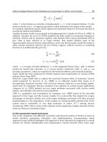
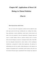
pregnancy, but the very high rate of bioprosthetic SVD in people aged 16–39
years at the time of initial PHV implantation: about 50% at 10 years and 90% at
15 years (Figure 9.1).
46
Furthermore, SVD begins 2–3 years after PHV implan-
tation in this age group. The mortality rate of reoperation was 3.8–8.7% (Table
9.2).
44,45
At 9 years, the rate of SVD of newer porcine valves and the stentless
porcine valves is within the expected range of SVD exhibited with earlier
stented porcine valves, indicating at present that all porcine valves have similar
Table 9.2 Bioprosthesis and pregnancy: late complications (10 years)
Badduke et al.
45
Jamieson et al.
44
Actuarial (%)
SVD 76.7 ± 14 55.3 ± 8.2
Valve-related complication 78.3 ± 12.7
—
Valve-related reoperation 79.7 ± 12.4 59.8 ± 7.8
Non-actuarial (%)
SVD 47.1 50.9
PHV endocarditis 11.8 5.7
Thromboembolism 5.9 5.7
Non-SVD
—
1.9
Sudden death
—
1.9
Total 70.6 66.1
Mortality rate of reoperation (%) 8.7 3.8
Values are expressed as mean ± SD or percentage.
SVD, structural valve deterioration; PHV, prosthetic heart valve.
From Hung and Rahimtoola.
3
110 Chapter 9
100
90
80
70
60
50
40
30
20
10
0
0 1 2 3 4 5 6 7 8 9 10 11 12 13 14 15 16
Follow-up time (years)
Percentage freedom from SVD
Average actuarial rates of SVD
at:
2–3 years:
5 years:
10 years:
15 years:
2–3%
4%
44%
90%
Figure 9.1 Structural valve deterioration (SVD) of aortic valve replacement porcine
bioprosthesis (Stanford University): age 16–39 years at time of implantation. (Adapted
from Yun et al.
46
)
rates of SVD.
48
Although the rate of SVD at 10 and 15 years is usually pointed
out to the patient as a reason for use of a bioprostheses, careful review of the
data shows that after insertion of porcine bioprostheses in people aged 16–39
years SVD begins as early as 2 years and is about 10–15% at 5 years (Figure 9.1).
Pregnancy by itself may be associated with SVD; the average rate was 24%
(Table 9.3), which is partly accounted for by the expected early rate of SVD in
young people
45
(Figure 9.1).
In addition, most women who have undergone an initial valve replacement
for rheumatic valve disease have mitral valve disease and, thus, have mitral
porcine bioprostheses, which have earlier onset of SVD and an overall incidence
of SVD that is greater than with AVR. As a result, the incidence of SVD will be
greater than cited above for AVR. Moreover, many are still in sinus rhythm
when they have their children but may eventually develop atrial fibrillation,
and will then require anticoagulant treatment. Patients with mitral valve dis-
ease have or develop left atrial enlargement and/or left atrial hypertension,
which may result in thromboemboli, and also atrial fibrillation, which further
increases the risks of thromboemboli.
31
Thus, use of a bioprosthesis is also asso-
ciated with an incidence of emboli, which is similar in patients not taking anti-
coagulants to that in patients with mechanical prosthetic valves who are taking
Artificial heart valves 111
Table 9.3 Bioprostheses and pregnancy: early structural valve deterioration (SVD)
References No. of patients No. of cases of Percentage Comments
early SVD
Born et al.
17
20 4 20 Needed reoperation
during pregnancy or in
puerperium
Bartolotti et al.
48
7229<3 months after
delivery
Salazar et al.
13
5 3 60 During pregnancy and
7–12 months after
pregnancy
Badduke et al.
45
17 2 12 Reoperation 3–10
months after
pregnancy
Hanania et al.
19
42 5 12
a
4–36 months after
delivery
Sbarouni and 49 17 35
b
During pregnancy or
Oakley
11
soon after delivery
Total 140 33 24
a
Porcine valves
b
Mainly porcine and few biological valves.
From Hung and Rahimtoola.
3
Table 9.4 Reoperation for structural valve deterioration of biological valves
Bioprostheses/Homografts
• May be needed:
—
even before first pregnancy
—
during or soon after pregnancy
—
with increasing frequency up to 10–15 years
• Are associated with:
—
morbidity
—
mortality (babies/children will be without a biological mother)
• ≥two to four reoperations may be needed over the woman’s lifetime
anticoagulants. Increased risk of thromboembolism may be indicated by spon-
taneous echocardiographic contrast ‘smoke’ within the left atrium.
47,49
In summary, three important issues need to be considered before a biopros-
thetic PHV is implanted in a young woman before pregnancy (Table 9.4).
Sbarouni and Oakley have asked: ‘Why should young women be singled out for
obligatory re-operation with this attendant risk?
11,31
112 Chapter 9
Pericardial bioprosthesis
There are limited data on pericardial bioprostheses in people aged 16–40 years
at the time of the PHV. One non-peer-reviewed article probably shows some-
what lower rates of SVD than porcine bioprostheses.
51
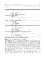
Homografts (allografts)
Homografts (allografts) have the same rate of SVD as porcine bioprostheses
50
(Figure 9.2). Data are not available for pregnant women and there are signifi-
cant concerns with the use of homografts (Table 9.5 and see Table 9.4).
Autografts (pulmonary autograft for aortic valve replacement
[Ross principle])
The Ross principle, first described by Donald Ross in 1967, involves two valve
replacements for one valve disease.
53,54
It is a more complex and more difficult
procedure, but does have some advantages, e.g. when inserted in children, the
valve increases in size as the child grows. Of eight women who had fourteen
pregnancies after receiving a pulmonary autograft, one woman developed
Table 9.5 Key issues with regard to homograft valves
• More difficult to insert (requires reimplantation of coronary arteries)
• Perioperative myocardial infarction: about six % in those without associated coronary artery
disease (CAD)
• Rate of structural valve deterioration (SVD) similar to bioprostheses
• More expensive than bioprostheses
• ≥two to four reoperations may be needed over the woman’s lifetime
• Reoperation(s) more difficult; requires repeat reimplantation(s) of coronary arteries. This is
also true of reoperation on stentless bioprostheses
0
0
20
40
60
80
100
510152025
Time since valve replacement (years)
25 years
35 years
Age at implant
Freedom from SVD
Figure 9.2 Structural valve deterioration (SVD) of homograft: aortic valve replacement
by age at time of implant. (Adapted from Svensson et al.
51
and Takkenberg et al.
52
)
Artificial heart valves 113
dilated cardiomyopathy (?peripartum cardiomyopathy) 6 months after deliv-
ering, one developed obstruction of the unsupported fascial pulmonary valve
and one developed acute endocarditis of the freeze-dried aortic homograft,
which had been inserted in the pulmonary position. The remaining five pa-
tients were well at last follow-up.
A review of this procedure showed the following:
• Risk for thromboemboli was 0–1.2% per year.
• Risk of infective endocarditis was 0–1.2%.
• Reoperation within the first 6 months was 0, 1.5, 3.8 and 10% in four differ-
ent studies.
• Late reoperation rates ranged from 0.4% to 1.5% per year.
• There is also a risk of rheumatic valvulitis in the autograft in those who have
rheumatic heart valve disease.
55,56
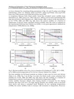
A recent study from Europe of patients, whose average age was 27 years at the
time of the Ross principle operation, showed that the incidence of autograft dys-
function, defined as the development of moderate or severe aortic regurgita-
tion, was 15% at 5 years and 25% at 7 years (Figure 9.3). In addition, associated
aortic root dilatation in the young is about 58% at 7 years (Figure 9.4).
The only studies with a follow-up of >10 years are from Ross’s group.
57,58–60
The freedom from autograft replacement ranged from 48.5 ± 13.7% at 19 years
to 85% at 20 years;
3
the most likely explanation for this wide range is selection
of patients reported in these four studies. In the series from the National Heart
Hospital:
58
• The operative mortality rate was 13%.
• In operative survivors (i.e. excluding operative mortality), late mortality rate
was 40.5% and actuarially determined mortality rate at 15 and 20 years was
25% and 39%, respectively.
• Actuarially determined freedom from autograft replacement was 75% at 20
years.
100
90
80
70
60
50
40
30
20
10
0
012345678
Autograft dysfunction
Time
at: 5 years 15±5%
7 years 25±8%
Freedom (%)
Patients at risk:
78 67 56 35 23 10 7
Patient no.
Age at operation:
91
27±10 years
(range 6–49)
Figure 9.3 Ross principle: freedom from autograft dysfunction. (Adapted from Luciani
et al.
61
)
114 Chapter 9
100
90
80
70
60
50
40
30
20
10
0
012345678
Years
Age at operation: 27±10 years
(range 6–49)
Patients at risk:
80 68 55 33 22 11 6
Freedom (%)
Risk factors for autograft dilatation
Cox proportional hazard
Age
Preoperative sinus Valsalva diameter
Root replacement technique
Pericardial strip buttressing
Beta factor
–0.07
0.24
2.80
–2.61
Standard error
0.04
0.12
1.27
1.33
P
0.05
0.02
0.03
0.04
Figure 9.4 Ross principle: freedom from dilatation. (Adapted from Luciani et al.
61
)
From a patient’s perspective, experience and skill in the performance of the
Ross principle procedure is not as widely available when compared with that
available for bioprostheses. In fact, Ross stated in an editorial from 2000 that the
Ross procedure should be renamed the Ross principle because what is being
performed today is very different from what he originally described.
54
Reoperation to replace autografts may be more difficult (see Table 9.5). There
is a need to replace the autograft as well as the aortic root and to repeat reim-
plantation of the coronary arteries. During the reoperation, homografts in the
pulmonic position may also need to be replaced (Table 9.6).
Management strategies
The management of young women with valvular heart disease (VHD) who are
contemplating a future pregnancy, the choice of PHV if one is necessary and the
management of such patients during pregnancy were outlined by Hung and
Rahimtoola in 2003 (Figure 9.5).
3
The choice of a PHV should be a joint decision
by the patient, cardiologist and cardiac surgeon. In young women with a PHV,
the importance of very early diagnosis of subsequent pregnancy needs to em-
phasized to the patient (Figure 9.6). It should be repeatedly emphasized to the
patient that, if she misses a menstrual period and there is a possibility of a preg-
nancy, she should be tested immediately for pregnancy. If she is pregnant,
Artificial heart valves 115
immediate consultation and joint care of the patient with a cardiologist and
perinatologist should be sought.
31
If the woman has a mechanical prosthetic
heart valve, then during weeks 6–12 of the pregnancy and before any type of
delivery, warfarin should be discontinued and intravenous unfractionated he-
parin given. Warfarin crosses the placenta, the fetus is anticoagulated and there
Figure 9.5 Young women with valvular heart disease (VHD) requiring prosthetic heart
valve (PHV) and considering pregnancy. (Reproduced with permission from Hung and
Rahimtoola.
3
)
Table 9.6 Key issues concerning the Ross principle
• Two-valve replacements for one-valve disease
• Structural valve deterioration of homograft in the pulmonary position
• Rate of autograft dysfunction in young people about 25% at 7 years
• Rate of associated aortic root dilatation in young people about 58% at 7 years
• Very early autograft dysfunction in some patients (an incidence of up to 10%)
• Experience and skill not as widely available when compared with that available for
bioprosthetic implantation
• Reoperation to replace autograft may be more difficult. Needs:
—
replacement of autograft
—
replacement of aortic root
—
repeat reimplantation of coronary arteries
—
homograft in pulmonary position may also need to be replaced
116 Chapter 9
is a risk of intracranial hemorrhage during a vaginal delivery. Therefore, it is rec-
ommended to use intravenous unfractionated heparin in the last 4 weeks of
pregnancy, which is discontinued before delivery.
33
Alternatively, there is the
option for elective cesarean section.
16
If the patient has a biological PHV, there is
a need for early diagnosis of SVD. Patients with aortic or mitral regurgitation
(AR and MR, respectively) can cope with the volume load of pregnancy better
than patients with severe valve stenosis, because the reduction of systemic
vascular resistance during pregnancy favors a reduction in AR and MR. The
volume load associated with pregnancy is not well tolerated in the presence
of a severe valve stenosis (aortic stenosis defined as valve area ≤1.0 cm
2
;
≤0.6 cm
2
/m
2
) or mitral stenosis (mitral valve area ≤1.5 cm
2
).
Peripartum antimicrobial therapy
Prophylactic antibiotics
In patients with native VHD, the indications for antibiotic prophylaxis are the
same as in the non-pregnant state to cover dental or other procedures or condi-
tions likely to cause Gram-positive bacteraemia.
62
The American Heart Association (AHA) position paper
62
and the subsequent
American College of Cardiology (ACC)/AHA guidelines
63
do not recommend
Figure 9.6 Young women with prosthetic valves and currently pregnant a Blood beta
human chorionic gonadotropin. (Reproduced with permission from Hung and
Rahimtoola.
3
)
Artificial heart valves 117
routine antibiotic prophylaxis in patients with VHD undergoing uncomplicated
vaginal delivery, unless bleeding and tearing would occur, or with a Caesarean
section, unless infection is suspected.
62
The AHA advises that antibiotics are in-
dicated for high risk patients with PHVs, a previous history of endocarditis, com-
plex congenital heart disease or a surgically constructed systemic-pulmonary
conduit (Table 9.7).
62
The Task Force on Infective Endocarditis of the European Society of Cardiol-
ogy (ESC). Guidelines on Prevention, Diagnosis and Treatment recommends
prophylaxis only in patients with high or moderate risk (so PHVs) undergoing
gynaecological procedures in the presence of infection
64
but many practitioners
routinely provide antibiotics.
The Task Force on Management of Cardiovascular Diseases During Pregnan-
cy of the ESC advises that prophylaxis is indicated in patients with PHVs or pre-
vious endocarditis and may be chosen for anticipated normal delivery in other
patients because complications are unpredictable. Antibiotics should be given
before surgical delivery or cardiac surgery (Table 9.8).
65
Table 9.7 American Heart Association recommendations for patients at high risk:
cardiac conditions in which antimicrobial prophylaxis is indicated*
• Prosthetic heart valves
• Complex congenital cyanotic heart disease
• Previous infective endocarditis
• Surgically constructed systemic or pulmonary conduits
• Acquired valvular heart diseases
• Mitral valve prolapse with valvular regurgitation or severe valve thickening
• Non-cyanotic congenital heart diseases ( except for secundum-type atrial septal defect)
including bicuspid aortic valves
• Hypertrophic cardiomyopathy
*Adapted from Dajani et al.
62
Table 9.8 The Task Force on the Management of Cardiovascular Diseases During
Pregnancy of the ESC*
• Antibiotic prophylaxis is discretionary for anticipated normal delivery but should be given to
patients with prosthetic heart valves or a history of endocarditis.
• Antibiotic prophylaxis may be chosen in other patients with anticipated normal delivery
because complications are unpredictable.
• Antibiotics should be given to patients at risk of endocarditis before surgical intervention,
cesarean delivery or cardiac surgery.
*Adapted from Oakley et al.
62
The AHA official recommendations on prevention of infective endocarditis
62
advise antibiotic
prophylaxis for normal delivery in patients with PHVs or previous endocarditis but The ACC/AHA
Guidelines on management of VHD
63
state that antibiotics are optional in high risk patients
(Eds.).
The incidence of bacteremia after normal delivery has been reported as be-
tween 0 and 5% and tends to include many different organisms.
30,62,63
More-
over, in clinical practice, it is not possible to guarantee that bleeding/tearing of
the vagina/perineum will not occur and, therefore, we recommend routine an-
tibiotic prophylaxis for delivery in all patients at risk of infective endocarditis.
Conclusions
Mechanical valves
• Patients with mechanical valves need close monitoring of warfarin therapy
during pregnancy. Substitution of warfarin with intravenous unfractionated
heparin in the first 6–12 weeks is associated with a low rate of warfarin em-
bryopathy. The initiation of heparin therapy is clinically most feasible and
practical at 4–6 weeks of pregnancy. Women who need <5 mg warfarin may
be at low risk for fetal warfarin embryopathy and may receive warfarin
throughout pregnancy, but more data are needed. Substitution of warfarin
with intravenous unfractionated heparin in the last 2 weeks of pregnancy is
associated with a reduced rate of bleeding in the baby during vaginal delivery
and in the mother with vaginal delivery or with cesarean section.
• Subcutaneous heparin, LMWH and direct thrombin inhibitors cannot be
recommended at the present time for use in these patients.
• If anticoagulation is needed, the use of LMWH is of concern because the FDA
has cited the occurrence of both teratogenic and non-teratogenic effects with
the use of LMWH. More data, including randomized trials, are needed.
Biological valves
• Both men and women aged 16–39 years at the time of bioprosthetic PHV im-
plantation are at risk of SVD, which begins 2–3 years after valve replacement:
at 10–15 years, the rate of SVD is very high (50–90%). Porcine bioprostheses
have a risk of early SVD during or shortly after the end of pregnancy. More-
over, at 10 years there is a high rate of SVD (55%–77%) and valve-related
reoperation (60–80%).
• One has to balance the risks of SVD and its consequences to the mother and
family in those who receive a bioprosthetic PHV against the small risk of war-
farin embryopathy in the fetus in those women who receive a mechanical PHV.
• There are limited data on the use of pericardial bioprostheses.
• There are limited data in patients who had received a homograft.
• More data are needed in patients who received a pulmonary autograft
procedure according to the Ross principle.
references
1 DiSaia PJ. Pregnancy and delivery of a patient with a Starr–Edwards mitral valve
prosthesis: report of a case. Obstet Gynecol 1966;28:469–72.
2 Hall JG. Embryopathy associated with oral anticoagulant therapy. Birth Defects
1965;12:133–40.
118 Chapter 9
3 Hung L, Rahimtoola SH. Prosthetic heart valves and pregnancy. Circulation
2003;107:1240–6.
4 Hall JAG, Paul RM, Wilson KM. Maternal and fetal sequelae of anticoagulation dur-
ing pregnancy. Am J Med 1980;68:122–40.
5 Sahul WL, Emery H, Hall JG. Chondrodysplasia punctata and maternal warfarin use
during pregnancy. Am J Dis Child 1975;129:362.
6 Ben Ismail M, Abid F, Trabelsi S, Taktak M, Fekih M. Cardiac valve prostheses, anti-
coagulation and pregnancy. Br Heart J 1986;55:101–5.
7 Chen WWC, Chau CS, Lee PK et al. Pregnancy in patients with prosthetic heart-
valves: an experience with 45 pregnancies. Q J Med 1982;51:358–65.
8 Larrea JL, Nunez L, Reque JA et al. Pregnancy and mechanical valve prosthesis:
a high-risk situation for the mother and the fetus. Ann Thorac Surg 1983;36:
459–63.
9 Salazar E, Izaguirre R, Verdejo J et al. Failure of adjusted doses of subcutaneous he-
parin to prevent thrombo-embolic phenomena in pregnant patients with mechanical
cardiac valve prosthesis. J Am Coll Cardiol 1996;27:1698–703.
10 Iturbe-Alessio I, del Carmen Fonseca M, Mutchinik O et al. Risks of anticoagulant
therapy in pregnant women with artificial heart valves. N Engl J Med 1986;315:
1390–3.
11 Sbarouni E, Oakley CM. Outcome of pregnancy in women with valve prosthesis.
Br Heart J 1994;71:196–201.
12 Chong MKB, Harvey D, Deswiet M. Follow-up study of children whose mothers were
treated with warfarin during pregnancy. Br J Obstet Gynaecol 1984;91:1070–3.
13 Salazar E, Zajarias A, Gutierrez N et al. The problem of cardiac valve prosthesis, anti-
coagulant, and pregnancy. Circulation 1984;70(suppl 1):169–77.
14 Pavunkumar P, Venugopal P, Kaul U et al. Pregnancy in patients with prosthetic car-
diac valve: a 10-year experience. Scand J Thorac Cardiovasc Surg 1988;22:9–22.
15 Sareli P, England MJ, Berk MR et al. Maternal and fetal sequelae of anticoagulation
during pregnancy in patients with mechanical heart valve prostheses. J Am Coll
Cardiol 1989;63:1462–5.
16 Cotrufo M, de Luca TSL, Calabro R et al. Coumadin anticoagulation during preg-
nancy in patients with mechanical valve prostheses. Eur J Cardiothorac Surg
1991;5:300–5.
17 Born D, Martinez EE, Almeida PAM et al. Pregnancy in patients with prosthetic heart
valves: the effects of anticoagulation on mother, fetus, and neonate. Am Heart J
1992;124:413–17.
18 Wong V, Cheng CH, Chan KC. Fetal and neonatal outcome of exposure to anticoagu-
lants during pregnancy. Am J Med Genet 1993;45:17–21.
19 Hanania G, Thomas D, Michel PL et al. Pregnancy in patients with valvular prosthe-
ses
—
retrospective cooperative study in France (155 cases). J Arch Mal Coeur Vaiss
1994;87:429–437.
20 Vitale N, Feo MD, DeSanto LS et al. Dose dependent fetal complications of warfarin in
pregnant women with mechanical heart valves. J Am Coll Cardiol 1999;33:1637–41
21 LeSanto LS, Romano G, Corte AD et al. Mitral mechanical replacement in young
rheumatic women: Analysis of long-term survival, valve-related complications, and
pregnancy outcomes over a 3,707 patient-year follow-up. J Thorac Cardiovas Surg
2005;130:13–19.
22 Chan WC, Anand S, Ginsberg JS. Anticoagulation of pregnant women with mechan-
ical valves: a systematic review of the literature. Arch Intern Med 2000;160:191–6.
Artificial heart valves 119
23 Brill-Edwards P, Ginsberg JS, Johnston M, Hirsh J. Establishing a therapeutic range
for heparin therapy. Ann Intern Med 1993; 119:104–9.
24 Ginsberg JS, Kowalchuk G, Hirsh J et al. Heparin therapy during pregnancy
—
risks to
the fetus and mother. Arch Intern Med 1989;149:2233–6.
25 Ginsberg JS, Barron WM. Pregnancy and prosthetic heart valves. Lancet
1994;344:1170–2.
26 Oakley CM. Anticoagulants in pregnancy.Br Heart J 1995;74:107–11.
27 Ginsberg JS, Hirsh J. Use of anticoagulants during pregnancy. Chest 1989;95:
156S–60S.
28 Ginsberg JS, Hirsh J. Anticoagulants during pregnancy. Annu Rev Med 1989;40:79–86.
29 Ginsberg JS, Barron WM. Pregnancy and prosthetic heart valves. Lancet
1994;344:1170–2.
30 Ginsberg JS, Greer I, Hirsh J. Use of anti-thrombotic agents during pregnancy. Chest
2001;119:122S–31S
31 Oakley C, ed. Artificial heart valves. In: Heart Disease in Pregnancy. London: BMJ
Publishing Group, 1997: pp 135–46.
32 Ginsberg JS, Hirsh J. Use of anti-thrombotic agents during pregnancy. Chest
1998;114:524S–30S.
33 Salazar E, Zajarias A, Gutiarraz N, Iturbe I. The problem of cardiac valve prostheses:
anticoagulants and pregnancy. Circulation 1984;70:169–77.
34 Whitfield LR, Lefe AS, Levy G. Effect of pregnancy on the relationship between con-
centration and anticoagulation action of heparin. Clin Pharmacol Ther 1983;34:23–8.
35 Bennett GG, Oakley CM. Pregnancy in a patient with mitral valve prosthesis. Lancet
1968;i:616–19.
36 Oakley CM. Clinical and pregnancy perspectives: anticoagulation. Eur Heart J
1995;16:1317–19.
37 Oakley CM. Anticoagulants in pregnancy. Br Heart J 1995;74:107–11.
38 Idir M, Madonna F, Rondant R. Collapse and massive pulmonary edema secondary
to thrombosis of a mitral mechanical heart valve prosthesis during low-molecular
weight heparin therapy. J Heart Valve Dis 1999;8:303–4.
39 FDA Med Watch. Available at: www.fda.gov/medwatch (accessed July 20, 2002).
40 Lee LH, Liauw PC, Ng AS. Low molecular weight heparin for thromboprophylaxis
during pregnancy in 2 patients with mechanical mitral valve replacement. Thromb
Haemost 1996;76:628–30.
41 Rowan JA, McCowan LM, Raudkivi PJ, North RA. Enoxaparin treatment in women
with mechanical heart valves during pregnancy. Am J Obstet Gynecol 2001;185:633–7.
42 Bates SM, Greer IA, Hirsh J, Ginsberg JS. Use of antithrombotic agents during
pregnancy: the Seventh ACCP Conference on Antithrombotic and Thrombolytic
Therapy. Chest 2004;126(suppl 3):627S–44S.
43 Oran B, Lee-Parritz A, Ansell J. Low molecular weight heparin for the prophylaxis of
thromboembolism in women with prosthetic mechanical heart valves during preg-
nancy. Thromb Haemost 2004;92:747–51.
44 Jamieson WRE, Miller DC, Akins CW et al. Pregnancy and bioprosthesis: influence
on structural valve deterioration. Ann Thorac Surg 1995;60:S282–7.
45 Badduke ER, Jamieson RE, Miyashima RT et al. Pregnancy and childbearing in a
population with biologic valvular prostheses. J Thorac Cardiovasc Surg 1991;102:179–86.
46 Yun KL, Miller DC, Moore KA et al. Durability of the Hancock MO bioprosthesis
compared with the standard aortic valve bioprosthesis. Ann Thorac Surg 1995;60:
221–8.
120 Chapter 9
47 Daniel WG, Nellessen U, Schroder E, Normast-Daniel B, Nikutta P, Lichtler PR. Left
atrial spontaneous echo contrast in mitral valve disease: an indicator for increased
thromboembolic risk. J Am Coll Cardiol 1988;11:1204–11.
48 Bartolotti U, Milano A, Massucco A et al. Pregnancy in patients with a porcine valve
bioprosthesis. Am J Cardiol 1982;50:1051–4.
49 Butchart EG, Moreno de la Santa P, Rooney SJ, Lewis PA. The role of risk factors and
trigger factors in cerebrovascular events after mitral valve replacement. J Card Surg
1994;9(suppl):228–36.
50 Grunkemeier GL, Li H-H, Naftel DC et al. Long-term performance of heart valve pros-
thesis. Curr Probl in Cardiol 2000;25:73–156.
51 Svensson LG, Blackstone EH, Cosgrove III OM. Surgical options in young adults with
aortic valve disease. G Curr Probl in Condiol 2003;28:417–79.
52 Takkenberg JJ, van Herwerdeen LA, Eijkema NSMJ et al. Evolution of allograft aortic
valve replacement over 13 years: Results of 275 procedures. Eur J Cardio Thorac Surg
2002;21:683–91.
53 Ross DN. Replacement of the aortic and mitral valves with a pulmonary autograft.
Lancet 1967;2:956–81.
54 Ross DN. The pulmonary autograft: the Ross principle (or Ross procedural confu-
sion). J Heart Valve Dis 2000;9:174–5.
55 Choudhary SK, Mather A, Chandler H et al. Aortic valve replacement with biological
substitute. J Cardiac Surg 1998;13:1–8.
56 Pieters FAA, Al-Halees, Hade L et al. Results of the Ross operation in rheumatic ver-
sus non-rheumatic aortic valve disease. J Heart Valve Dis 2000;9:38–44.
57 Matsuki O, Oldta Y, Ahneida RS et al. Two decades experience with aortic valve re-
placement with pulmonary autograft. J Thoracic Cardiovasc Surg 1988;9••5:705–71.
58 Ross D, Jackson M, Davies J. Pulmonary autograft aortic valve replacement: long-
term results. J Cardiac Surg 1991; 6(suppl):529–33.
59 Ross D, Jackson M, Davies J. The pulmonary autograft: a permanent aortic valve. Eur
J Cardiothorac Surg 1992;6:113–17.
60 Chambers JE, Somerville J, Stone S et al. Pulmonary autograft procedure for aortic
valve disease: long-term results of the pioneer series. Circulation 1997;96:2206–14.
61 Luciani GB, Casali G, Favaro A et al. Fate of aortic not late after Ross operation. Circu-
lation 2003;108(Suppl II):61–7.
62 Dajani AS, Taubert KA, Wilson W et al. Prevention of bacterial endocarditis: recom-
mendations by the American Heart Association. Circulation 1997;96:358–66.
63 Bonow RD, Carabello B, Delern AC et al. ACC/AHA Guidelines for the management
of patients with valvular heart disease. A report of the American college of
Cardiology/American Heart Association Task Force on Practice Guidelines. J Am Coll
Cardiol 1998;
32:1486–588.
64 Horstkotte D, Follak F, Gutsehik E et al. The Task Force on Infective Endocarditis of
the European Society of Cardiology. Guidelines on Prevention, Diagnosis and Treat-
ment of Infective endocarditis. Eur Heart J 2004;25:267–76.
65 Oakley C, Child A, Iung B, Presbitero P, Tornos P, Expert Consensus Document on
Management of Cardiovascular Diseases during Pregnancy, Eur Heart J 2003;24:
761–81.
66 Baker TH, Hubbell R. Reappraisal of asymptomatic puerperal bacteremia. Am J Obstet
GynecoI 1967;97:575–6.
Artificial heart valves 121
CHAPTER 10
Management of
pregnancy in Marfan
syndrome, Ehlers–Danlos
syndrome and other heritable
connective tissue disorders
Lilian J Meijboom, Barbara JM Mulder
The major heritable disorders of connective tissue that may cause problems in
obstetric management include Marfan syndrome, Ehlers–Danlos syndrome,
osteogenesis imperfecta, pseudoxanthoma elasticum and achondroplasia.
1
Al-
though individually rare, together they form an important group, requiring
cooperative management by several specialists during pregnancy. Improved
medical and surgical management permits affected women to reach child-
bearing age, but good advice should begin during family or individual counsel-
ing sessions, well before child bearing starts. The genetic risk, the possibilities of
prenatal diagnosis and the obstetric risk to women should also be discussed later
with the prospective parents and, if pregnancy is contraindicated, the alterna-
tives of childlessness, adoption or ovum donation should be discussed.
Marfan syndrome
Marfan syndrome is an autosomal dominantly inherited connective tissue dis-
order with an estimated incidence of 1 in 5000. The syndrome involves many
systems but the prominent manifestations are of skeletal, ocular and cardiovas-
cular origin.
2
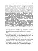
Aortic dilatation and dissection are the major causes of morbidity
and mortality (Figure 10.1).
3,4
Marfan syndrome is the result of a mutation in
the fibrillin gene on chromosome 15.
5
Genotype–phenotype correlations in
Marfan syndrome have been complicated by the large number of unique muta-
tions reported, as well as by clinical heterogeneity among individuals with the
same mutation.
6,7
As a result of the intragenic heterogeneity, molecular genet-
ic screening is hampered to a considerable extent, and the diagnosis of Marfan
syndrome is still based mainly on clinical major and minor manifestations, as
defined by a council of experts in the field, known as the Ghent nosology.
7,8
A
122
Heart Disease in Pregnancy, Second Edition
Edited by Celia Oakley, Carole A Warnes
Copyright © 2007 by Blackwell Publishing
definite diagnosis requires occurrence of major manifestations in two different
categories, and involvement (presence of criteria) of a third category (Table
10.1). In clinical practice diagnosis should be established by a multidisciplinary
team.
For women with Marfan syndrome, pregnancy presents a twofold problem:
50% risk of transmission of Marfan syndrome to the fetus and aortic dissection
or progression of aortic dilatation in the mother.
Prenatal screening
If one parent has Marfan syndrome then there is a 50% chance in each preg-
nancy that the child (male or female) will inherit the dominant gene. In
25–30% of patients the syndrome arises as a spontaneous mutation in either
the ovum or the sperm. If unaffected parents have such a child, the risk of re-
currence in a subsequent pregnancy is the population prevalence (1 in 5000)
and is negligible.
Currently pre-implantation diagnosis and prenatal diagnosis for Marfan
syndrome are generally limited to those families in which the mutation in the
Marfan syndrome and other heritable connective tissue disorders 123
Figure 10.1 Magnetic resonance angiography of a
dilated aortic root in a patient with Marfan
syndrome.
124 Chapter 10
Table 10.1 Diagnostic criteria for Marfan syndrome
Category Major criteria Minor criteria
Family history Independent diagnosis in parent, None
child, sibling
Genetics Mutation FBN-1 None
Cardiovascular Aortic root dilatation Mitral valve prolapse
Dissection of ascending aorta Calcification of the mitral valve
(<40 years)
Dilatation of pulmonary artery
Dilatation/dissection of
descending aorta
Ocular Ectopia lentis (Two needed):
Flat cornea
Myopia
Elongated globe
Skeletal * Pectus excavatum needing surgery Moderate pectus excavatum
Pectus carinatum High narrowly arched palate
Pes planus Typical face
Wrist and thumb sign Joint hypermobility
Scoliosis >20° or spondylolisthesis
Arm span–height ratio >1.05
Protrusio acetabulae (radiograph,
MRI)
Diminished extension elbows
(<170°)
Pulmonary Spontaneous pneumothorax
Apical bullae
Skin Unexplained stretch marks
(striae)
Recurrent or incisional herniae
Central nervous Lumbosacral dural ectasia (CT or
system MRI)
*Presence of at least four of the manifestations listed under ‘Major criteria’ are necessary for the
skeletal system to be classified as major feature. Presence of at least two of the manifestations
listed under ‘Major criteria’ and at least two of the manifestations listed under ‘Minor criteria’
are necessary for the skeletal system to be involved (minor).
CT, computed tomography; MRI, magnetic resonance imaging.
FBN-1 gene is known. Mutation identification can be performed in individual
cases but is time-consuming and, in about 20% of patients with a definite diag-
nosis of Marfan syndrome based on clinical findings, it is not possible to find a
mutation.
9
On the other hand, polymorphism in the FBN-1 gene can be found
without any evidence of the disease.
The major advantage of pre-implantation diagnosis in comparison with
prenatal diagnosis is the possibility of avoiding termination, which can be
extremely distressing for the couples concerned. Another concern with respect
to genetic counseling for prenatal diagnosis is the variability in phenotypic
expressions, even within families. This wide clinical variability and the lack of
clear-cut genotype–phenotype correlations make predictions about clinical
severity difficult.
10
In a recent study two-thirds of patients expressed interest in
using a prenatal test to determine whether their fetus would be affected with
Marfan syndrome.
11
It is unknown in how many of these patients an elective
abortion is performed.
Pregnancy and cardiovascular complications
During pregnancy important maternal cardiovascular changes occur, such as
increases in blood volume, heart rate, stroke volume, cardiac output, left ven-
tricular wall mass and end-diastolic dimensions.
12
In addition, hormonal
changes occur, which lead to histological changes in the aorta. Fragmentation of
the reticulum fibers, a diminished amount of acid mucopolysaccharides and
loss of the normal corrugation of elastic fibers have been observed in the aortic
wall of pregnant patients.
13
So, it has been suggested that both hemodynamic
and hormonal mechanisms play an important role in the increased susceptibil-
ity to aortic dissection in women during pregnancy.
The aortic root diameter above which pregnancy should be discouraged in
women with Marfan syndrome is still a matter of debate. The Canadian guide-
lines recommend that women with an aortic root diameter >44 mm should
strongly be discouraged from becoming pregnant; the European guidelines dis-
courage pregnancy above an aortic root diameter of 40 mm.
14,15
Both guide-
lines were based on three studies in which it became apparent that the risk for
dissection was low in women with minimal cardiac involvement and an aortic
root diameter <40 mm.
16–18
However, in these studies very few patients were
included with aortic root diameters >40 mm.
In a recent prospective study no aortic dissections occurred in patients with-
out previous aortic dissection and an aortic root diameter ≤45 mm.
19
Moreover,
little to no change in aortic root diameter throughout pregnancy was observed.
Only one woman known to have a previous type A dissection developed a type
B dissection during her second pregnancy. So, pregnancy in women with Mar-
fan syndrome seems to be relatively safe up to an aortic root diameter of 45 mm;
however, a completely safe diameter does not exist. Also, in two studies it has
been shown that pregnancy in women with Marfan syndrome has no negative
effect on aortic root growth during long-term follow-up.
18,19
In women with minimal cardiac involvement (aortic root diameter <45 mm,
and no significant aortic or mitral regurgitation) pregnancy is relatively safe;
however they should be told of a 1% risk of aortic dissection or other serious
cardiac complications, such as endocarditis or congestive cardiac failure during
pregnancy.
5,20
Family history and aortic growth should be taken into account
when considering aortic surgery before pregnancy. Women with aortic root di-
ameters >45mm have an increased risk of developing aortic dissection during
Marfan syndrome and other heritable connective tissue disorders 125