Báo cáo y học: "Harmine activates intrinsic and extrinsic pathways of apoptosis in B16F-10 melanom" doc
Bạn đang xem bản rút gọn của tài liệu. Xem và tải ngay bản đầy đủ của tài liệu tại đây (1.45 MB, 8 trang )
RESEARCH Open Access
Harmine activates intrinsic and extrinsic pathways
of apoptosis in B16F-10 melanoma
Thayele Purayil Hamsa and Girija Kuttan
*
Abstract
Background: Harmine is a beta-carboline alkaloid from the plant Peganum harmala. Previous studies found that
harmine inhibited metastasis of B 16F-10 melanoma cells. This study aims to elucidate the role of harmine in
apoptosis of B16F-10 cells.
Methods: B16F-10 melanoma cells were treated in the presence and absence of harmine in vitro. Morphological
changes, cell cycle and expression of various pro and anti- apoptotic genes were analyzed for the study of apoptosis.
Results: Morphological observation and DNA laddering assay showed that harmine treated cells displayed marked
apoptotic characteristics, such as nuclear fragmentation, appearance of apoptotic bodies and DNA laddering
fragment. TUNEL assay and flow cytometric analysis also confirmed apoptosis. Furthermore, RT-PCR analysis
showed that harmine induced apoptosis in B16F-10 melanoma cells by up-regulating Bax and activating Caspase-3,
9 and p53 and down-regulating Bcl-2. Harmine also up-regulated Caspase-8 and Bid, indicating that harmine
affected both extrinsic and intrinsi c pathways of apoptosis. This study also showed inhibitory effects of harmine on
some transcription factors and pro- inflammatory cytokines that protect cell from apoptosis.
Conclusion: Harmine activates both intrinsic and extrinsic pathways of apoptosis and regulates some transcription
factors and pro-inflammatory cytokines.
Background
Apoptosis, programmed cell death, occurs during nor-
mal development and tissue homeostasis or as a
response to cellular insults and oncogenesis [1]. Apopto-
sisinvolvesasequenceofspecific morphological
changes in a dying cell: condensation of the cytoplasm
and nuclear chromatin, followed by breakage of cells
into membrane bound apoptotic bodies containing a
variety of cytoplasmic organelles and nuclear fragments,
which are then engulfed by neighboring cel ls and
macrophages [2].
Apoptosis pathways can generally be divided into sig-
naling via the death receptors (extrinsic) or the mito-
chondria (intrinsic) pathways. Both pathw ays lead to
activation of the members of highly selective prote ases
referred to as ‘Caspases’ [3]. A family of specific cysteine
proteases ubiquitously expressed as inactive zymogens,
Caspases are t he key destructive molecules of apoptosis
and controls all steps of apoptosis; however, in response
to specific death stimuli, caspases are activated in a cas-
cade of auto- stimulation and trans- stimulation [4].
Extrinsic pathways involve a sequential activation of Cas-
pase-8 and 3 which cleaves target proteins, leading to
apoptosis. Intrin sic pathways are directly or indirectly
activated by intrinsic death stimuli such as reactive
oxygen species (ROS), DNA-damaging reagents, resulting
in the release of cytochrome-c and t he activation of Cas-
pase-9 which in turn activates Caspase-3 [3]. Between the
death receptor and the mitochondrial signaling pathways,
the p ro-apoptotic protein Bid serves as a cross-talker
(upon cleavage b y activated Caspase-8) by inducing the
translocation of the pro-apoptotic proteins Bax and/or
Bak to t he mitochondrial membrane [5]. The compo-
nents of the extrinsic and intrinsic pathways are regu-
lated by th e members of a fam ily of proteins called Bcl-2.
Bcl- 2 anti-apoptoti c proteins have been tar gets for antic-
ancer drug development for at least a decade [6].
P53 is a nuclear transcription factor that accumulates
in response to cellular stress, including D NA damage
and oncogene activation. This triggers transcriptional
trans activation o f p53 target genes such as p21, p27,
* Correspondence:
Amala Cancer Research Centre, Amala Nagar, Thrissur, Kerala, India, 680555
Hamsa and Kuttan Chinese Medicine 2011, 6:11
/>© 2011 Hamsa and Kuttan; licensee Bi oMed Central Ltd. This is an Open Access article distributed under the terms of the Creative
Commons Attribution License ( http://creativecommon s.org/licenses/by/2.0), which permits unrestricted use, distribution, and
reproduction in any mediu m, provided the original work is properly cited.
Bax, leading to cell cycle arrest, senescence and/or apop-
tosis [7]. The p53 tumour-suppressor protein can inter-
vene at every major step in apoptotic pathways as a key
regulator of apoptosis and carcinogenesis [8].
Nuclear factor-B (NF-B) signaling pathway is gener-
ally considered as a survival factor that activates expres-
sion of various anti-apoptotic genes such as Bcl-2, Bcl-
xL that block apoptosis [9]. Inhibition of NF-B leads to
down-regulation of the NF-B-regulated anti-apoptotic
proteins, thereby promoting apoptosis [3]. Expressi on of
many pro-inflammatory cytokines is regulated at the
level of transcription by the transcription factor NF-B.
Thus, inhibition of NF-B is an important therapeutic
target for the treatment of cancer [10].
Transcription factors also play a key role in controlling
cell proliferation, cell cycle progression and apoptosis [11].
c-Fos and ATF-2 genes encode a nuclear transcription fac-
tor that induces transcription of a number of other genes
involved in the regulation of cytokine synthesis, cell repli-
cation, cell cycle control and apoptosis. Hypophosphory-
lated or transcriptionally inactive forms of ATF2 reduce
TNF-a expression, resulting in sensitization of melanoma
to treatment via increased apoptosis [12-14]. In respons e
to stress stimuli, ATF-2 activates a variety of gene targ ets
including cyclin A, cyclin D and c-jun which are involved
in oncogenesis in various tissue types [15]. Similarly cyclic
AMP-response element-binding protein (CREB) was
reported to suppress apoptosis, induce cell proliferation
and mediate inflammation and tumour metastasis [16].
Beta-carbolines, a large group of indole alkaloids, are
widely distributed in nature, such as various plants, marine
creatures, insects, mammalians as well as human tissues
and body fluids [17]. Harmine (7-methoxy-1-methyl-9H-
pyrido [3,4-b] indole), originally isolated from the seeds of
Peganum harmala, is a tricyclic compound belonging to
the b -carboline alkaloids. These alkaloids possess a broad
range of pharmacological activities, such as anxiolytic and
behavioral effects [18]. Recent studies demonstrated that
harmine possessed significant anti-tumor potential both in
vitro and in vivo [19], eg significant tumor inhibition in
mice bearing Lewis Lung Cancer, sarcoma180 or Hep-A
tumor [20] and broad cytotoxicity spectrum against
human lung carcinoma cell lines [21].
There have been no reports on the anti-proliferative
and apoptotic activity of harmine on highly metastatic
B16F-10 melanoma cells. Therefore, this study was con-
ducted to explore the critical events leading to apoptosis
in B16F-10 melanoma cells.
Methods
Cells
B16F-10 melanoma cells were obtained from National
Centre for Cell Science (India). The cells were cultured
in Dulbecco’s Modified Eagle’ sMedium(DMEM)
supplemented with 10% FCS (Foetal Calf Serum) and
antibiotics in a humidified incubator at 37°C in 5% CO
2
atmosphere and maintained in continuous exponential
growth by twice-a-week passages.
Chemicals and reagents
Mouse Bcl-2, Caspase-3, 8, 9, Bax, Bid, p53 and GAPDH
primer sequences were obtained from Maxim Biotech
(USA). Harmine was purchased from Sigma (USA).
DMEM was procured from Himedia Laboratory (India).
Cells-c DNA kit was purchased from Ambion (USA).
Transfactor kit was purchased from BD Biosciences
(USA). All other reagents used were of analytical reagent
grade.
Effects of harmine on the viability of B16F-10 melanoma
cells
B16F-10 melanoma cells (5 × 10
3
cells/well) were pla-
ted in 96-well flat bot tomed titer plate and incubated
for 24 hours at 37°C in 5% CO
2
atmosphere. Different
concentrations of harmine (1-100 μg/mL) were added
and incubated furthe r for 48 hours. Before four hours
of completion of incubation, 20 μl 3-4, 5-dimethylthi a-
zol-2-yl)-2, 5-diphenyltetrazolium bromide (MTT)
(5mg/mL) was added [22]. Percentage of viable
cells was determined with an ELISA pla te reader at
570 nm.
Morphological analysis
B16F-10 melanoma cells (5 × 10
3
cells/well) suspended
in DMEM were plated in 96-well flat-botto m titer plate
and incubated for 24 hours at 37°C in 5% CO
2
atmo-
sphere. After 24 hours, various concentrations of ha r-
mine (0.5, 1 and 2 μg/mL) were added to the cells and
incubated further for 48 hours under the same condi-
tions. The cells were then washed twice with PBS
(pH7.4), fixed with 5% formalin and stained with haema-
toxylin and eosin. The cells were observ ed under mi cro-
scope and photographed.
DNA fragmentation analysis
One m illion B16F-10 melanoma cells were treated with
different concentrations of h armine (0.5, 1 and 2 μg/
mL)andincubatedfor24hoursat37°Cin5%CO
2
atmosphere. After incubation, the cells were treated
with 0.1 mL lysis buffer (100 mmol/L Tris-HCl, pH8.0,
containing 0.2% Triton-X100 and 1 mmol/L EDTA) for
10 minutes at -20°C. DNA was extracted according to
the phenol-chloroform method [23], precipitated with
chilled ethanol and re-suspended i n Tris/EDTA buffer
(10mmol/LTris-HCl,pH8.0and1mmol/LEDTA).
DNA samples were separated by electrophoresis in 1%
agarose gels. DNA was stained with ethidium bromide
and photographed under UV light.
Hamsa and Kuttan Chinese Medicine 2011, 6:11
/>Page 2 of 8
TUNEL assay
TUNEL assay was performed to d etect apoptosis via
DNA fragmentation by Apoptag Peroxidase in situ
(Apoptosis detection kit, CHEMICON International,
USA). B16F-10 melanoma cells (5 × 10
3
cells/well)
suspended in D MEM supplemented with 10% FCS,
100 μg/ml streptomycin and penicillin and 2 mmol/L
glutamine were plated in 96-well flat bottom titer plate
and incubated for 24 hours at 37°C in 5% CO
2
atmo-
sphere. After 24 hours, aliquots of harmine (1 and 2 μg/
mL) were added to the cells and incubated further for
48 hours under the same conditions. The cells were
washed in PBS and stained according to the manufac-
turer’s instructions. TUNEL positive cells were counted
as apoptotic cells.
Cell cycle analysis
One million B16F-10 cells suspended in DMEM were
seeded in a culture fl ask and incuba ted for 48 hours at
37°C in CO
2
atmosphere with and withou t harmine.
Treated and untreated cells were harvested, washed with
PBS and fixed with 70% ethanol for 24 hours. The cells
were then centrifuged (420 × g,Remi,India)andthe
pellet was re-suspended in PBS containing propidium
idodide and RNase A. Flow cytometric analysis was per-
formed with the FACS Calibur flow cytometer (Becton
Dick inson, Singapore) using the CycleTEST PLUS DNA
Reagent kit (Becton Dickinson, Singapore) according to
the manufacturer’s instructions.
Effects of harmine on pro-inflammatory cytokines and
GM-CSF levels
B16F-10 melanoma cells (5 × 10
3
cells/well) suspended
in DMEM were plated in 96-well flat-botto m titer plate
and incubated for 24 hours at 37°C in 5% CO
2
atmo-
sphere. Harmine (2 μg/mL) was added to t he cells and
incubated further for 48 hours under the same condi-
tions. The supernatant was used t o estimate the cyto-
kines, namely IL-1b,IL-6,TNF-a and GM-CSF with
specific ELISA kits (Pierce Biotech nology, USA) accor d-
ing to the manufacturer’s instructions.
Effects of harmine on gene expression
To determine the mRNA expression levels of genes
responsible for triggering apoptosis, we carried out a
semi-quantitative reverse transcription polymerase chain
reaction (RT-PCR). B16F-10 cells were cultured with
medium containing only FCS for 24 hours at 37°C in
5% CO
2
atmosphere. Harmine (2 μg/mL per well) was
added to a 96-well flat-bottom titer plate and incubated
for four hours. cDNA was prepared from B16F-10 mela-
noma cells by cells to cDNA™ II kit (Ambion Inc, U.S.
A). Briefly, cells were washed with PBS and heated in
cell lysis buffer (provided in the kit) to release the RNA
into the solution, followed by a heating step to i nacti-
vate endogenous RNases. The genomic DNA was
further degraded by treating with DNase followed by
inactivation o f DNase by heating at 70°C. Reverse tran-
scription was performed at 42°C for 50 minutes in
Moloneymurineleukemiavirusreversetranscriptase
(provided in the kit). Gene expression analysis was per-
formed with PCR. The murine Bcl-2, Caspases-3, 8, 9,
p53, Bid and Bax genes were amplified against GAPDH
standard. Amplified PCR products were subjected to
electrophoresis on a 1.8% agarose gel and stained with
ethidium bromide and photographed under UV light.
Effects of harmine on transcription factors
Nuclear extracts were prepare d according to a pre-
viously described method [24]. B16F-10 cells suspended
in serum free medium were treated with harmine for
two hours at 37°C in 5% CO
2
atmosphere. The cells
were washed twice with PBS and incubated further with
TNF-a (10rg/mL) for 30 minutes to activate cytoplas-
mic transcription factor. The cells were then lysed with
lysis buffer incubated for 15 minutes on ice. The cell
suspension was centrifuged and disrupted using a syr-
inge and centrifuged (10,000-11,000 × g, Remi,India) for
20 minutes. The crude nuclear pellet obtained is sus-
pended in nuclear extraction buffer. Nuclei were dis-
rupted with a fresh syringe, centrifuged and the
super natant was collected. Protein concentrations of the
nuclear extracts were estimated according to the stan-
dard Bradford method and stored at -70°C.
Transcription factor pro filing was performed wit h the
BD Mercury™ Transfactor kit (BD Biosciences, USA).
When nuclear extracts added to the well, DNA will bind
to their consensus sequences in the well. Bound tran-
scription factors in the DNA were detected by specific
primary antibody towards NF-Bp65, NF-Bp50, NF-B
c-Rel, c-Fos, ATF-2 and CREB. A horse radish peroxi-
dase-conjugated secondary antibody was then used to
detect the bound primary antibody. The enzymatic pro-
duct was measured with standard microtiter plate reader
at 655 nm. Percentage inhibition was calculated accord-
ing to the following formula:
% inhibition = 100 –([ODoftreated/ODofcontrol]×100)
where OD is optical density.
Statistical analysis
All data were represe nted as mean ± standard deviation
(SD). Significance levels for comparison of differences
were determined with one way ANOVA, followed by
Dunnet’s Comparison test using Graphpad Instat (ver-
sion 3.00 for W indows 98, G raphPad Software, USA).
Means of the treated groups were compared with that
Hamsa and Kuttan Chinese Medicine 2011, 6:11
/>Page 3 of 8
of the control group and P < 0.05 was considered statis-
tically significant.
Results
Effects of harmine on the viability of B16F-10 melanoma
cells
MTT assay is a standard colorimetric assay for measur-
ing cellular viability. MTT is reduced to purple forma-
zan in mitochondria and is directly relat ed to the
number of viable cells. E ffect of harmine on the viability
of B16F-10 melanoma cells in culture is in Table 1. Har-
mine up to 2 μg/mL, was not directly cytotoxic to B16F-
10 melanoma cells and conc entrations of 0.5, 1 and 2
μg/mL were used for further experiments.
Apoptotic analysis
Harmine induced marked apoptosis in B16F-10 cells.
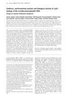
Morphological changes indicating apoptosis (eg mem-
brane blebbing, chromatin condensation, DNA fragmen-
tation, appearance of apoptotic bodies) [25] (Figure 1)
were observed at 1 and 2 μg/mLofharminebynuclear
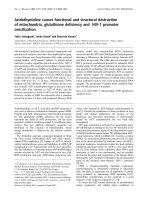
staining. The typical ‘DNA ladder’ was observed on
DNA electrophoresis gel for treated cells at 2 μg/mL
(Figure 2, lane 5). No observable changes were obtained
in the morphology of cells treated with 0.5 μg/mL of
harmine. Moreover, harmine at 1 and 2 μg/mL did not
show any feat ures of apopto sis on normal human umbi-
lical vein endothelial cells (HUVEC) (data not shown).
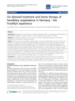
TUNEL assay
Thi s method is used to assay the endonuclease cleavage
products by enzymatically end-labeling the DNA strand
breaks [26]. Terminal transferase was used to add
labeled UTP to the 3’ end of the DNA fragments. As
shown in figu re 3, numerous TUNEL positive cells were
observed when B1 6F-10 cells were treated with harmine
at 1 and 2 μg/mL, indicating apoptotic cell death of
B16F-10 melanoma cells.
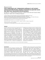
Cell cycle analysis
The effects of the harmine on cell cycle distribution
were determined (Figure 4). Harmine inhibited cell
growth with arrest at G
1
and reduced transition to the S
and G
2
/M phases of the cell cycle. The proportion of
the sub-G
0
/G
1
peak was negligible in the control
(2.32%) cel ls and most cells (79.57%) were in G1 and S
phases due to the high proliferative state of B16F-10 cell
line. Exposure of cells to harmine (1 and 2 μg/mL) for
48 hours resulted in cell accumulation at the sub-G
0
/G
1
phase in a dose-dependent manner. At 1 μg/mL 28.27%
cells were accumulated and 70.41% cells at 2 μg/mL.
Effects of harmine on pro-inflammatory cytokine and GM-
CSF levels
Harmine signi ficantly inhibited the production of pro-
inflammatory cytokines, namely TNF-a,IL-1b,IL-6and
GM-CSF by B16F-10 melanoma cell in culture (Tabl e 2).
Harmine (2 μg/mL) showed maximum inhibition of all
cytokines.
Effects of harmine on gene expression
RT-PCR analysis revealed a significant down regulation
in the expression of Bcl-2 gene compared to control. At
the same time, expression of pro-apoptotic g enes such
as p53, Caspase-3, 8, 9, Bid and Bax were significantly
up-regulated by the treatment with harmine, which indi-
cated the involvement of harmine in both intrinsic and
extrinsic pathways of apoptosis. Cell death mechanism
induced by the harmine in B16F-10 melanoma cells may
be mediated by the activation of these genes controlling
both intrinsic and extrinsic pathways of apoptosis
(Figure 5A).
Effects of harmine on transcription factors
The DNA bound transcription factor was determined
with corresponding primary antibody, which was
detected with horseradish peroxidase-conjugated sec-
ondary antibody. The percentage in hibition in the acti-
vation/translocation NF-B sub units, namely p65, p50
and c-Rel, were 64.07, 70.08 and 41.0 3 respectively after
harmine treatment with. Inhibition in the activation of
other transcription factors such as c-Fos (73.11%), ATF-
2 (63.51%) and CREB (55.59%) were also observed with
harmine treatment (Figure 5B).
Discussion
In the present study, treatment of melanoma cells with
harmine induced morphological changes inclu ding con-
densation of nuclear chromatin, formation of apoptotic
bodies and blebbing of the cell membrane. All these
morphological character istics are biochemical hallmarks
of apoptosis, indicating that apoptosis may play a crucial
role in cell death elicited by the h armine on B16F-10
Table 1 Percentage cell viability of B16F-10 melanoma
cells in culture after treatment with harmine
Concentration (μg/mL) Percentage of viability
1 100
2 100
5 97.88
10 69.63
20 48.41
50 26.26
75 0
100 0
Vehicle (0.1% DMSO) 100
B16F-10 melanoma cells were incubated with different concentrations (1-100
μg/mL) of harmine. Percentage of viability was determined using MTT assay.
Hamsa and Kuttan Chinese Medicine 2011, 6:11
/>Page 4 of 8
melanoma cells. DNA extracts from harmine treated
B16F-10 melanoma cells also showed characteristic lad-
der pattern of discontinuous DNA fragments. Moreover,
presence of pyknotic nuclei (characteristic of cells
undergoing apoptosis [27] was further confirmed with
tunnel assay. MTT assay ruled out necrosis as a
probable cause of cell death in harmine treated cell s as
most of the cells exhibited intact plasma membranes.
P53 is a nuclear transcription factor that accumulates
in response to cellular stress, including D NA damage
and oncogene activation. This triggers transcriptional
transactivation of p53 target genes such as p21, Bax,
leading to cell cycl e arrest, senescence and/or apoptosis
[7]. The mitochondrial death pathway is controlled by
memb ers of the Bcl-2 family, including the anti-ap opto-
tic Bcl-2 and the pro-apoptotic Bax and Bid proteins.
The pro-apoptotic Bcl-2 family members Bax is crucial
in regulating a wide range of apoptotic stimuli [28] and
become activated by Bcl-2 family members that have
only the BH3 doma in, namely Bid [29]. It was reported
that over expression of Bax results in the release of
cytochrome- c from mitochondria to the cytosol and
induction of apoptosis [30] and that the direct incuba-
tion of Bax protein with isolated mitochondria also
induced cytochrome-c release [31]. P53 is a potent acti-
vator of the caspase cascade by stimulating pro-apopto-
tic proteins (Bid and Bax) and promoting the release of
apoptogenic factors (cytoch rome c), leading to Caspase-
9 a ctivation and in turn cleaving effector caspase s such
as Caspase-3 [32]. Expression analysis of mRNA
revealed the apoptotic regulation of various genes in
B16F-10 melanoma cells treated with harmine. E xpres-
sion of pro-apoptotic genes such as P53, Caspase-3, 8
and 9, Bid, Bax was significantly induced at the earlier
phase of treatme nt (4 hours), suggesting that h armine
was an initiator or inducer of the apoptotic mechanism.
Harmine could enhance the activa tion of Bcl-2 family
pro-apoptotic protei ns such as Bax and Bid while it
could also down-regulate the expressions of Bcl-2 in
B16F-10 melanoma cells. Activation of Caspase-8 and
Bid along with other caspases indicates the involvem ent
of harmine in both extrinsic and intrinsic pathways of
apoptosis because Bid serves as a cross-talker upon clea-
vage by activated Caspase-8 by inducing the transloca-
tion of the pro-apoptotic proteins Bax and/or Bak to the
mitochondrial membrane [5]. Tumor apoptosis was
Figure 1 Effect of harmine on the morphology of B16F-10 melanoma cells. Cells treated with harmine show membrane blebbing and
presence of apoptotic bodies (n = 3; 400×).
Figure 2 Effect of harmine on B16F-10 melanoma DNA
integrity. Lane 1- molecular weight marker, Lane 2- DNA from
untreated control cells, Lane 3-DNA from harmine (0.5 μg/mL)
treated cells. Lane 4-DNA from harmine (1 μg/mL) treated cells and
lane 5 -DNA from harmine (2 μg/mL) treated cells (n = 3).
Hamsa and Kuttan Chinese Medicine 2011, 6:11
/>Page 5 of 8
closely associated with its cell cycle arrest. Over expres-
sion of cyclin dependent kinase inhibitors such as p27,
p21 may lead to apoptosis of tumor cells, inhibit their
proliferation and diminish their metastasis [33,34]. The
present study found that harmine caused cell cycle
arrest in G0/G1 phase and showed an evident apoptot ic
sub-G0/G1 peak in B16F10 melanoma cells.
The NF-B protein family encompasses transcription
factors involved in controlling the expressions of genes
cruci al for several important cellular signal transduction
pathways in inflammation, proliferation and in defense
against apoptosis. Constitutive activation of NF-Band
chronic inflammation has a major role in the develop-
ment of most tumors, including leukemia, lymphomas
and s olid tumours. Inhibition of NF-B leads to down-
regulation of the NF-B-regulated anti-apoptotic pro-
teins and other pro-inflammatory cytokines, thereby
promoting apoptotic cell death [35,36]. In this study,
inhibition of the activation of NF-B was probably
attribut ed to the decreased production of pro-inflamma-
tory cytokines in B16F10 melanoma cells.
Genes controlling transcription is deregulated in a
wide range of cancers; thus, targeting proteins that regu-
late signaling pathways for translation and protein
synthesis is a realistic strategy for cancer treatment.
Members of the AP-1 (activator protein-1) family are
necessary for cell cycle progression in several cell sys-
tems and also for cell transformation induced by a vari-
ety of oncogenes, including Src, Ras and Raf [37]. ATF-
2 regulates the transcription of several genes involved in
cytokine synthesis, cell cycle control apoptosis and DNA
repair [38]. Cyclin D1, an important gene for the inte-
gration of proliferative and anti-proliferative signals dur-
ing the G1 phase of the cell cycle, possesses a CRE
Figure 3 TUNEL assay. B16F-10 melanoma cells were treated with harmine for 48 hours and TUNEL assay was performed to detect apoptosis.
TUNEL positive cells were counted as apoptotic cells. (n = 3; 200×).
Figure 4 Effect of harmine on cell cycle progression. B16F-10 melanoma cells w ere treated with harmine for 4 8 h and a nalyzed for
propidium iodide stained-DNA content by flow cytometry. Values indicate the percentage of the cell population at the phase of the cell cycle.
M
1
=G
1
(Diploid), M
2
=G
2
/M (Tetraploid), M
3
= S (Synthetic phase), M
4
= Sub-G
1
phase. The population of cells in the sub-G
0
/G
1
phase
represents cellular fragments due to apoptosis.
Hamsa and Kuttan Chinese Medicine 2011, 6:11
/>Page 6 of 8
element within its promoter region. In murine chondro-
cytes, cyclin D1 is directly activated by ATF-2 while the
levels of activation are reduced in AT F-2-deficient mice.
Cyclin D1 is activated by ATF-2 in proliferating murine
melanoma cells [14]. CREB also regulates the expression
of a repertoire of genes related to cell survival, inflam-
mation and proliferation, such as Bcl-2, Bcl-xL, COX-2
and TNF-a [15]. As these transcription factors are
major negative regulators of apoptosis, their inhibition
by harmine pro motes apoptosis in B16F-10 melanoma
cells.
Conclusion
Harmine activates bo th intrinsic and extrinsic pathways
of apoptosis and regulates some transcription factors
and pro-inflammatory cytokines.
Abbreviations
Bax: Bcl-2 associated X protein; Bid: BH3 interacting domain death agonist;
CREB: cyclic AMP-response element-binding protein; DMEM: Dulbecco’s
Modified Eagle’s Medium; FCS: Foetal Calf Serum; GM-CSF: Granulocyte
monocyte colony stimulating factor; IL: Interleukin; MTT: 3-4, 5-
dimethylthiazol-2-yl)-2, 5-diphenyltetrazolium bromide; NF: Nuclear factor;
ROS: Reactive oxygen species; TNF: Tumour necrosis factor; TUNEL: Terminal
deoxynucleotidyl transferase dUTP nick end labeling
Acknowledgements
The authors express gratitude to Dr Ramadasan Kuttan (Research Director,
Amala Cancer Research Centre) for his valuable suggestions and support in
this study.
Authors’ contributions
GK designed and coordinated the study. TPH carried out the study including
acquisition, analysis and interpretation of the data. Both authors read and
approved the final version of the manuscript.
Competing interests
The authors declare that they have no competing interests.
Received: 12 October 2010 Accepted: 23 March 2011
Published: 23 March 2011
References
1. Pradelli LA, Beneteau M, Ricci JE: Mitochondrial control of caspase-
dependent and -independent cell death. Cell Mol Life Sci 2010,
67:1589-1597.
2. Van Herreweghe F, Festjens N, Declercq W, Vandenabeele P: Tumor
necrosis factor-mediated cell death: to break or to burst, that’s the
question. Cell Mol Life Sci 2010, 67:1567-1579.
3. Li-Weber M: Targeting apoptosis pathways in cancer by Chinese
medicine. Cancer Lett 2010.
Table 2 Effect of harmine on the release of TNF-a, IL-1b, IL-6 and GM CSF by B16F-10 melanoma cells
Cytokine (pg/mL) Harmine
Control 1 μg/mL 2 μg/mL
TNF-a 29.17 ± 5.94 26.18 ± 1.71 20.19 ± 1.12 (P = 0.012)
IL-1b 98.15 ± 2.08 74.48 ± 2.11 (P < 0.001) 61.34 ± 2.34 (P < 0.001)
IL-6 61.43 ± 4.44 40.42 ± 5.19 (P < 0.001) 28.46 ± 2.80 (P < 0.001)
GM-CSF 34.55 ± 1.07 29.55 ± 1.21 (P = 0.01) 22.81 ± 1.19 (P < 0.001)
B16F-10 cells (5 × 10
3
cells) were cultured in the presence of harmine for 48 hours, and level of pro-inflammatory cytokines in the culture supernatant was estimated.
Values are expressed as mean ± SD. Statistical analysis was performed with ANOVA, followed by Dunnet’s test using GraphPad Instat software.
Figure 5 (A) Effect of harmine on gene expression. B16F-10 cel ls were
cultured in the presence and absence of harmine (2 μg/mL) and cDNA
was synthesized and amplified with appropriate primers using PCR. The
mRNA expression levels were normalized with GAPDH (house-keeping
gene). (B) Effect of harmine o n transcription factors. B16F-10 m elanoma
cells were treated with harmine (2 μg/mL) for two hours and then
incubated with TNF-a (10 pg/mL) for 30 minutes. Percentage inhibition in
various transcription factors were me asured by E LISA method.
Hamsa and Kuttan Chinese Medicine 2011, 6:11
/>Page 7 of 8
4. Gyrd-Hansen M, Meier P: IAPs: from caspase inhibitors to modulators of
NF-kappaB, inflammation and cancer. Nat Rev Cancer 2010, 10:561-574.
5. Billen LP, Shamas-Din A, Andrews DW: Bid: a Bax-like BH3 protein.
Oncogene 2008, 27(Suppl 1):S93-104.
6. Hockenbery DM: Targeting mitochondria for cancer therapy. Environ Mol
Mutagen 2010, 51:476-489.
7. Farnebo M, Bykov VJ, Wiman KG: The p53 tumor suppressor: a master
regulator of diverse cellular processes and therapeutic target in cancer.
Biochem Biophys Res Commun 2010, 396:85-89.
8. Vogelstein B, Kinzler KW: Cancer genes and the pathways they control.
Nat Med 2004, 10:789-799.
9. Dutta J, Fan Y, Gupta N, Fan G, Gélinas C: Current insights into the
regulation of programmed cell death by NF-kappaB. Oncogene 2006,
25(51):6800-6816.
10. Ghosh CC, Ramaswami S, Juvekar A, Vu HY, Galdieri L, Davidson D,
Vancurova I: Gene-Specific Repression of Proinflammatory Cytokines in
Stimulated Human Macrophages by Nuclear I{kappa}B{alpha}. J Immunol
2010, 185:3685-3693.
11. Persengiev SP, Green MR: The role of ATF/CREB family members in cell
growth, survival and apoptosis. Apoptosis 2003, 8:225-228.
12. Bhoumik A, Huang TG, Ivanov V, Gangi L, Qiao RF, Woo SL, Chen SH,
Ronai Z: An ATF2-derived peptide sensitizes melanomas to apoptosis
and inhibits their growth and metastasis. J Clin Invest 2002, 110:643-650.
13. Muscella A, Urso L, Calabriso N, Vetrugno C, Rochira A, Storelli C,
Marsigliante S: Anti-apoptotic effects of protein kinase C-delta and c-fos
in cisplatin-treated thyroid cells. Br J Pharmacol 2009, 156:751-763.
14. Ivanov VN, Ronai Z: Down-regulation of tumor necrosis factor alpha
expression by activating transcription factor 2 increases UVC-induced
apoptosis of late-stage melanoma cells. J Biol Chem 1999,
274:14079-14089.
15. Vlahopoulos SA, Logotheti S, Mikas D, Giarika A, Gorgoulis V, Zoumpourlis V:
The role of ATF-2 in oncogenesis. Bioessays 2008, 30:314-327.
16. Aggarwal S, Kim SW, Ryu SH, Chung WC, Koo JS: Growth suppression of
lung cancer cells by targeting cyclic AMP response element-binding
protein. Cancer Res 2008, 68:981-988.
17. Cao R, Chen Q, Hou X, Chen H, Guan H, Ma Y, Peng W, Xu A: Synthesis,
acute toxicities, and antitumor effects of novel 9-substituted beta-
carboline derivatives. Bioorg Med Chem 2004, 12:4613-4623.
18. Murray TD, Berger A: Alcohol withdrawal. Va Med Q
1997, 124:184-187,
189.
19. Perez Martin JM, Labrador V, Fernandez Freire P, Molero ML, Hazen MJ:
Ultrastructural changes induced in HeLa cells after phototoxic treatment
with harmine. J Appl Toxicol 2004, 24:197-201.
20. Chen Q, Chao R, Chen H, Hou X, Yan H, Zhou S, Peng W, Xu A: Antitumor
and neurotoxic effects of novel harmine derivatives and structure-
activity relationship analysis. Int J Cancer 2005, 114:675-682.
21. Abe A, Yamada H: Harmol induces apoptosis by caspase-8 activation
independently of Fas/Fas ligand interaction in human lung carcinoma
H596 cells. Anticancer Drugs 2009, 20:373-381.
22. Cole SP: Rapid chemosensitivity testing of human lung tumor cells using
the MTT assay. Cancer Chemother Pharmacol 1986, 17:259-263.
23. Chomczynski P, Sacchi N: Single-step method for RNA isolation by acid
guanidium thiocyanate-phenol-chloroform extraction. Anal Biochem 1987,
162:156-159.
24. Dignam JD, Lebovitz RM, Roeder RG: Accurate transcription initiation by
RNA polymerase II in a soluble extract from isolated mammalian nuclei.
Nucleic Acids Res 1983, 11:1475-1489.
25. Evan G, Littlewood T: A matter of life and cell death. Science 1998,
281:1317-1322.
26. Kressel M, Groscurth P: Distinction of apoptotic and necrotic cell death
by in situ labelling of fragmented DNA. Cell Tissue Res 1994, 278:549-556.
27. Karin M, Delhase M: The I kappa B kinase (IKK) and NF-kappa B: key
elements of proinflammatory signalling. Semin Immunol 2000, 12:85-98.
28. Wei MC, Zong WX, Cheng EH, Lindsten T, Panoutsakopoulou V, Ross AJ,
Roth KA, MacGregor GR, Thompson CB, Korsmeyer SJ: Proapoptotic BAX
and BAK: a requisite gateway to mitochondrial dysfunction and death.
Science 2001, 292:727-730.
29. Chipuk JE, Green DR: How do BCL-2 proteins induce mitochondrial outer
membrane permeabilization? Trends Cell Biol 2008, 18:157-164.
30. Luo W, Liu J, Li J, Zhang D, Liu M, Addo JK, Patil S, Zhang L, Yu J,
Buolamwini JK, Chen J, Huang C: Anti-cancer effects of JKA97 are
associated with its induction of cell apoptosis via a Bax-dependent and
p53-independent pathway. J Biol Chem 2008, 283:8624-8633.
31. Antonsson B, Montessuit S, Lauper S, Eskes R, Martinou JC: Bax
oligomerization is required for channel-forming activity in liposomes
and to trigger cytochrome c release from mitochondria. Biochem J 2000,
345(Pt 2):271-278.
32. Ooi KL, Tengku Muhammad TS, Lim CH, Sulaiman SF: Apoptotic effects of
Physalis minima L. chloroform extract in human breast carcinoma T-47D
cells mediated by c-myc-, p53-, and caspase-3-dependent pathways.
Integr Cancer Ther 2010, 9:73-83.
33. Cheng JD, Werness BA, Babb JS, Meropol NJ: Paradoxical correlations of
cyclin-dependent kinase inhibitors p21waf1/cip1 and p27kip1 in
metastatic colorectal carcinoma. Clin Cancer Res 1999, 5:1057-1062.
34. Megha T, Lazzi S, Ferrari F, Vatti R, Howard CM, Cevenini G, Leoncini L,
Luzi P, Giordano A, Tosi P: Expression of the G2-M checkpoint regulators
cyclin B1 and P34CDC2 in breast cancer: a correlation with cellular
kinetics. Anticancer Res 1999, 19:163-169.
35. Lawrence T: The nuclear factor NF-kappaB pathway in inflammation. Cold
Spring Harb Perspect Biol 2009, 1:a001651.
36. Piotrowska A, Izykowska I, Podhorska-Okolow M, Zabel M, Dziegiel P: The
structure of NF- kappaB family proteins and their role in apoptosis.
Postepy Hig Med Dosw 2008, 62:64-74.
37. Choi HS, Kang BS, Shim JH, Cho YY, Choi BY, Bode AM, Dong Z: Cot, a
novel kinase of histone H3, induces cellular transformation through up-
regulation of c-fos transcriptional activity. FASEB J 2008, 22:113-126.
38. Xiao L, Rao JN, Zou T, Liu L, Marasa BS, Chen J, Turner DJ, Zhou H,
Gorospe M, Wang JY: Polyamines regulate the stability of activating
transcription factor-2 mRNA through RNA-binding protein HuR in
intestinal epithelial cells. Mol Biol Cell 2007, 18:4579-4590.
doi:10.1186/1749-8546-6-11
Cite this article as: Hamsa and Kuttan: Harmine activates intrinsic and
extrinsic pathways of apoptosis in B16F-10 melanoma. Chinese Medicine
2011 6:11.
Submit your next manuscript to BioMed Central
and take full advantage of:
• Convenient online submission
• Thorough peer review
• No space constraints or color figure charges
• Immediate publication on acceptance
• Inclusion in PubMed, CAS, Scopus and Google Scholar
• Research which is freely available for redistribution
Submit your manuscript at
www.biomedcentral.com/submit
Hamsa and Kuttan Chinese Medicine 2011, 6:11
/>Page 8 of 8