Báo cáo y học: "Clinical review: Imaging in ischaemic stroke – implications for acute management" pot
Bạn đang xem bản rút gọn của tài liệu. Xem và tải ngay bản đầy đủ của tài liệu tại đây (194.94 KB, 9 trang )
Page 1 of 9
(page number not for citation purposes)
Available online />Abstract
Imaging has become a cornerstone of stroke management, trans-
lating pathophysiological knowledge to everyday decision-making.
Plain computed tomography is widely available and remains the
standard for initial assessment: the technique rules out haemor-
rhage, visualizes the occluding thrombus and identifies early tissue
hypodensity and swelling, which have different implications for
thrombolysis. Based on evidence from positron emission
tomography (PET), however, multimodal imaging is increasingly
advocated. Computed tomography perfusion and angiography
provide information on the occlusion site, on recanalization and on
the extent of salvageable tissue. Magnetic resonance-based
diffusion-weighted imaging (DWI) has exquisite sensitivity for acute
ischaemia, however, and there is increasingly robust evidence that
DWI combined with perfusion-weighted magnetic resonance
imaging (PWI) and angiography improves functional outcome by
selecting appropriate patients for thrombolysis (small DWI lesion
but large PWI defect) and by ruling out those who would receive
no benefit or might be harmed (very large DWI lesion, no PWI
defect), especially beyond the 3-hour time window. Combined
DWI–PWI also helps predict malignant oedema formation and
therefore helps guide selection for early brain decompression.
Finally, DWI–PWI is increasingly used for patient selection in
therapeutic trials. Although further methodological developments
are awaited, implementing the individual pathophysiologic
diagnosis based on multimodal imaging is already refining
indications for thrombolysis and offers new opportunities for
management of acute stroke patients.
Introduction
In the present era of thrombolysis, of specialized acute stroke
units and of endovascular and neurosurgical interventions,
imaging has become a cornerstone of modern stroke
management. Imaging of the ischaemic process has taken
centre stage in four key areas: shaping the basic concepts of
stroke pathophysiology; guiding therapeutic approaches that
tackle these concepts; translating this knowledge to everyday
clinical decision-making; and motivating new therapeutic
developments in the field. The present review will briefly
discuss these roles, focusing on recent advances in imaging
that pertain to everyday practice.
Basic concepts
Following occlusion of a major intracranial artery, particularly
the middle cerebral artery (MCA), a gradient of hypoperfusion
emerges in the supplied basal ganglia, white matter and
cortical mantle [1]. Regions suffering the most severe hypo-
perfusion (often in and around the sylvian fissure in proximal
occlusion) rapidly progress to irreversible damage,
representing the ‘ischaemic core’. This tissue exhibits very
low cerebral blood flow (CBF), cerebral blood volume (CBV)
and metabolic rates of oxygen and glucose [2]. The remaining
hypoperfused tissue – with lost autoregulation – is patho-
physiologically divided relative to a well-defined perfusion
threshold into two compartments; namely, the ‘penumbra’
and the ‘oligaemia’.
In the penumbra, oxygen metabolism is preserved relative to
CBF, the oxygen extraction fraction is elevated and often
reaches its theoretical maximum of 100% (severe ‘misery
perfusion’), and the CBV is normal or elevated. Tissue within
the penumbra is functionally impaired and contributes to the
clinical deficit, yet is still viable and hence potentially
salvageable by effective reperfusion. The extent of the
penumbra, however, decreases over time by gradual
recruitment into the core, and as such represents a key target
for therapeutic intervention, albeit with a progressively
shrinking temporal window of opportunity – hence the ‘time is
brain’ rule [3]. This course of events varies from patient to
patient, but up to one-third of patients still exhibit large
volumes of penumbra 18 hours after stroke onset [4].
Review
Clinical review: Imaging in ischaemic stroke – implications for
acute management
Ramez Reda Moustafa and Jean-Claude Baron
Department of Clinical Neurosciences, University of Cambridge, Cambridge CB2 2QQ, UK
Corresponding author: Jean-Claude Baron,
Published: 11 September 2007 Critical Care 2007, 11:227 (doi:10.1186/cc5973)
This article is online at />© 2007 BioMed Central Ltd
ADC = apparent diffusion coefficient; ASPECTS = Alberta Stroke Programme Early CT Score; CBF = cerebral blood flow; CBV = cerebral blood
volume; CT = computed tomography; DWI = diffusion-weighted imaging; FLAIR = fluid-attenuated inversion recovery; MCA = middle cerebral
artery; MR = magnetic resonance; MRI = magnetic resonance imaging; MTT = mean transit time; PET = positron emission tomography; PCT = per-
fusion computed tomography; PWI = perfusion-weighted imaging; rt-PA = recombinant tissue plasminogen activator; TTP = time to peak.
Page 2 of 9
(page number not for citation purposes)
Critical Care Vol 11 No 5 Moustafa and Baron
The oligaemic compartment, on the other hand, suffers a
milder degree of hypoperfusion with normal oxygen consump-
tion and with elevated CBV and oxygen extraction fraction,
and is not normally at risk of infarction [4]. If the occlusion
persists, however, secondary events such as systemic
hypotension, intracranial hypertension or hyperglycaemia may
topple this delicate balance and force the oligaemia into a
penumbral state, and eventually recruitment into the necrotic
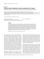
core. Figure 1 illustrates these concepts.
This understanding of the pathophysiology underlies the
urgency of acute stroke management and is the rationale for
approaches, established or still experimental, to rescue the
penumbra, such as reperfusion therapy, neuroprotection,
induced arterial hypertension and oxygen therapy. Besides
being instrumental in this development, imaging in the acute
setting brings these physiological concepts to the bedside
and aims to identify the different tissue compartments
amenable to therapy and to define the potential for recovery
in the individual patient.
Imaging techniques
Plain computed tomography
Despite being surpassed by magnetic resonance imaging
(MRI) in versatility and image quality, plain computed tomo-
graphy (CT) remains the standard tool for initial assessment
in most centres because it is widely available and because
the large thrombolysis trials were all CT-based [5,6]. Apart
from ruling out haemorrhage, early tissue ischaemic changes
can be identified by CT within 3 hours of onset in up to 75%
of patients with MCA stroke [7], yet with moderate
interobserver agreement depending on experience [8]. These
changes comprise: tissue hypodensity, which is associated
with severe reductions in CBF and CBV on perfusion imaging
[9] and whose extent can predict final infarction [10]; and
cortical swelling without hypodensity, which on MRI is
associated with increased CBV, moderate hypoperfusion and
a normal or near-normal apparent diffusion coefficient (ADC),
reflecting salvageable tissue [11].
Early ischaemic changes thus include elements of both the
core and the penumbra. Large parenchymal hypodensity also
statistically predicts the risk of thrombolysis-associated
haemorrhage, hence the widespread notion of withholding
this treatment if it exceeds one-third of the MCA territory [6].
The Alberta Stroke Programme Early CT Score (ASPECTS)
[7] has better interrater reliability in assessing early ischaemic
changes [12], yet this is not independently associated with
poor clinical outcome [13]. Since the ASPECTS combines
swelling and hypodensity, it may not distinguish irreversibly
damaged tissue from viable tissue. A recent study comparing
CT with MRI [14] has confirmed that focal brain swelling
does not always represent infarcted tissue, supporting the
removal of this criterion from the ASPECTS scoring system.
An additional early CT sign in ischaemic stroke is the direct
visualization of the thrombus, seen as increased attenuation
in the transverse M1 segment (hyperdense MCA sign) or in
cross-section within the sylvian fissure (dot sign) [15]. The
specificity of these signs is high, but their sensitivity is
moderate (30–40%) [16], probably because CT cannot
detect fresh fibrin-poor thrombi [17]. In a general stroke
population, the hyperdense MCA sign is associated with poor
prognosis and a risk of thrombolysis-associated haemorrhage
[18], but its resolution is associated with a favourable
outcome. In patients with acute MCA occlusion, however, this
sign has no independent prognostic value [19]. Equivalent
signs have recently been reported on MRI [20].
Plain CT is also very sensitive to intracranial haemorrhage
and subarachnoid haemorrhage. Studies using gradient-
recalled echo T2* MRI, however, have shown that intracranial
haemorrhage can be equally detected with very high
sensitivity even by inexperienced users [21,22], and that fluid-
attenuated inversion recovery (FLAIR) MRI can also
demonstrate subarachnoid haemorrhage equally well [23].
These findings may support the idea of omitting CT as the
initial investigation in acute stroke and proceeding directly to
MRI (see below).
Computed tomography and magnetic resonance
angiography
In the acute setting, CT or magnetic resonance (MR) angio-
graphy can determine the site of occlusion, early recanaliza-
tion and the presence of abnormalities in the proximal arterial
tree such as stenosis, occlusion or dissection, pertaining to
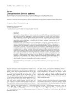
Figure 1
Hypoperfused tissue compartments after acute MCA occlusion and
the consequences of decreasing cerebral perfusion pressure. (a) The
three hypoperfused tissue compartments (the core, the penumbra and
the oligaemia) after acute middle cerebral artery occlusion. A further
compartment with normal perfusion but partially exhausted vascular
reserve (denoted autoregulated) surrounds the oligaemic compartment
(see text). (b) Consequences of decreasing cerebral perfusion
pressure, as a result of, for example, a fall in systemic blood pressure
or an increase in intracranial pressure from vasogenic oedema, on the
four tissue compartments illustrated in (a), showing an enlargement of
the core at the expense of the penumbra, and of the latter into the
oligaemia and autoregulated compartments, with attending clinical
deterioration. The final infarction potentially involves all four
compartments entirely.
Page 3 of 9
(page number not for citation purposes)
the cause of the stroke [24]. These data can usefully inform
the decision to use intravenous thrombolysis or to proceed to
mechanical embolectomy, for example in ‘T occlusion’ of the
carotid termination [25,26]. Unlike CT, time-of-flight MR
angiography is noninvasive, utilizing the intrinsic properties of
moving blood [27]. Although less accurate than contrast-
enhanced MR angiography, this makes the technique
particularly appealing when combined with perfusion-
weighted imaging (PWI) as it avoids the repeated use of a
contrast agent.
Source images from CT angiography can themselves be
used to detect areas of very low CBV, which are comparable
with MRI diffusion-weighted imaging (DWI) lesions [24,28]
and are predictive of subsequent infarction within 6 hours
[29]. The added value is attractive, yet the technique still
needs to be fully validated.
DWI remains by far the most sensitive method of detecting
acute ischaemia [30,31] and can be positive a few minutes
from onset [32], allowing accurate localization and subtyping
of stroke. The DWI signal reflects restriction of the random
motion of water in tissue and the decline of its ADC –
although the exact biological correlates are not completely
understood, this probably involves energy failure and
subsequent cytotoxic oedema [33,34]. In combination with
perfusion imaging, DWI can also be used, albeit cautiously, to
define the ischaemic core and the penumbra [35] (see below).
Multimodal stroke imaging
Largely based on seminal positron emission tomography
(PET) observations [3,4,36], most authorities nowadays
consider that the heterogeneity and complexity of acute
ischaemic stroke necessitates a multimodal approach to
imaging that provides not only structural but also functional
and haemodynamic information to aid the decision-making
process [37]. For CT this approach currently includes plain
CT, CT angiography and perfusion computed tomography
(PCT) [28,38], while in MRI the approach includes a
combination of conventional sequences (such as T1W, T2W
and fluid-attenuated inversion recovery) and T2*W, time-of-
flight MR angiography, DWI and PWI [39].
Perfusion computed tomography
PCT images are acquired in the cine mode after intravenous
injection of an iodinated contrast agent, generating maps of
CBF, CBV as well as mean transit time (MTT) and time to peak
(TTP) [40]. The maps are reproducible, especially when relative
perfusion parameters are used [41], and reportedly have > 90%
sensitivity and specificity for detecting large hemispheric stroke
[42]. Anatomical coverage, however, is typically restricted to
20 mm (two to four slices), reducing sensitivity to stroke not
caused by proximal major artery occlusion [43].
Recent studies on PCT in acute stroke demonstrated that
tissue with CBV < 2 ml/100 g represents the core, while a
relative MTT above 145% of the normal hemisphere best
outlines all at-risk tissue [44]. The penumbra can thus be
estimated as the tissue existing between those two
thresholds. Using this methodology, PCT parameters
correlate very well with MR DWI–PWI and are a good
predictor of the final infarct volume and clinical recovery
[38,41,45,46]. PCT is also potentially useful in decision-
making when the time of onset is unknown, such as with
awakening stroke [47]. In combination with CT angiography,
PCT has comparable utility with that of MR in selecting
patients for thrombolysis [38].
Magnetic resonance diffusion-perfusion imaging
The commonly used dynamic susceptibility-weighted contrast
PWI technique is similar in principle to PCT, and measures
changes in the magnetic field induced by passage of
gadolinium-based contrast in cerebral tissue – but with lesser
accuracy, particularly for CBF. Arterial spin labelling PWI is a
newer technique that avoids the use of a contrast agent
through magnetically labelling the arterial blood entering the
skull and then tracking its motion through the tissue [48]. The
latter technique, however, is less widely available and still
requires further validation in stroke.
Among the generated MRI perfusion maps, TTP and MTT are
preferred for identifying hypoperfused tissue because they
correlate best with tissue fate [49,50]. Comparison of the
perfusion deficit depicted on these maps with the DWI lesion
(assumed to denote the core) yields either a mismatch
pattern (PWI > DWI), a matched lesion pattern (PWI = DWI)
or a reperfusion pattern (DWI > PWI). The mismatch pattern
is taken to indicate the existence of salvageable at-risk tissue
and is found in about 70% of all patients with anterior-
circulation stroke within 6 hours of onset [51]. The pattern’s
presence is strongly associated with proximal MCA occlusion
[51] and its resolution on reperfusion is associated with
neurological recovery [52-54]. Moreover, successful reper-
fusion prevents further expansion of the DWI lesion into the
area of mismatch [55].
The DWI–PWI mismatch can be used to select patients who
are most likely to benefit from thrombolytic therapy [56], and
the mismatch is incorporated into several ongoing thrombo-
lysis trials (see below). It has also been used to show how
variables such as hyperglycaemia [57], haematocrit [58] and
age [59] influence outcome through altering the fate of the
penumbra. DWI has also shown utility in providing a
physiologic endpoint for new therapies such as normobaric
high-flow oxygen [60].
The clinical implications of a matched DWI–PWI pattern are
less clear. In the presence of a large DWI lesion and proximal
MCA occlusion, this pattern appears to accurately predict the
development of a malignant MCA syndrome [61,62]. For
other scenarios where a matched pattern is found, the
evidence is lacking with regard to outcome and with regard
Available online />to whether there is any benefit from instituting thrombolysis or
another specific therapy. The third pattern of normal (or
increased) perfusion with a variable size DWI lesion indicates
recanalization [63], and effectively does not appear to benefit
from thrombolysis (see below).
A number of uncertainties have recently arisen regarding the
pathophysiologic accuracy of the DWI–PWI mismatch
concept. Studies in animals and in humans have documented
the reversibility of DWI lesions and normalization of the ADC,
thus arguing against equivalence of the DWI lesion to the
‘core’ [64,65]. Predictors of such normalization are
thrombolytic therapy and recanalization, particularly within the
3-hour time window [66]. This suggests that the DWI lesion
may include penumbral tissue, as echoed recently using PET
[67,68]. Corresponding uncertainties also exist regarding
PWI, particularly in the selection of parameters for defining
the tissue at risk and in the choice of arterial input function
[49,69]. The DWI–PWI mismatch may thus overestimate the
penumbra by including oligaemic tissue or even normally
perfused but autoregulated tissue that is not at risk [70].
These questions become particularly relevant when defining
the management of matched DWI–PWI lesions, since
response to recanalization depends on whether or not there
still is penumbral tissue. Nevertheless, the DWI–PWI
concept remains a clinically and experimentally useful tool
provided these shortcomings are recognized.
Implications of imaging for thrombolysis
The 3-hour window
Patients treated with intravenous thrombolysis within the first
3 hours after stroke are at least 30% more likely to have little
or no disability at 3 months (number needed to treat = 8)
[5,71]. This is essentially based on selecting patients who
have stroke symptoms that are not rapidly resolving or minor
(NIH stroke scale < 3) with the absence of haemorrhage on
plain CT. Nonetheless, despite the use of clinical exclusion
criteria [72], the treatment carries a risk of around 6–7% of
thrombolysis-associated symptomatic haemorrhage;
therefore, the emerging role of imaging in this acute setting,
beyond exclusion of intracranial haemorrhage and
subarachnoid haemorrhage, is to identify and exclude that
subgroup of patients who are unlikely to benefit and may be
harmed by recombinant tissue plasminogen activator (rt-PA),
in turn reducing the number needed to treat. As already
mentioned, early hypodensity on plain CT >1/3 MCA territory
is associated with thrombolysis-associated haemorrhage.
Nonetheless, this fact is still debated since analysis of the
0–3 hour group in the NINDS cohort does not support this
exclusion on the basis of the extent of early ischaemic
changes alone (that is, including swelling) [73].
Similarly, MR-based studies show that severely reduced
ADC, CBF and CBV are associated with subsequent
haemorrhagic transformation within the infarction [74,75].
These studies, however, do not distinguish symptomatic and
asymptomatic grades of haemorrhagic transformation, and
thus their relevance to clinical outcome is unclear. Another
proposed MRI marker of haemorrhagic transformation is
delayed gadolinium enhancement of cerebrospinal fluid
space on FLAIR [76]. This marker appears only after
reperfusion has been achieved and thus its clinical usefulness
is uncertain. Thomalla and colleagues [77] make the
distinction between haemorrhagic transformation and
parenchymal haemorrhage, arguing that the former is a
clinically irrelevant epiphenomenon whereas the latter is a
direct effect of rt-PA therapy and deserves further investiga-
tion. Finally, T2* MRI can identify microbleeds, which may
also arguably pose a risk of parenchymal haemorrhage after
thrombolysis, yet the evidence for or against this view is still
scarce [78,79].
The constraint of the 3-hour window makes it necessary that
imaging is performed in as short a time as possible. Because
CT provides relatively limited information in early stroke,
multimodal MRI is increasingly being advocated as the
imaging investigation of choice [80]. The main concern, how-
ever, is the possible delay in treatment – up to 20 minutes in
experienced centres [81] – but this may be balanced by the
gain in diagnostic accuracy. Furthermore, shorter door-to-
needle times can probably be achieved through omitting CT,
increasing the familiarity of staff with MRI [82] and tailoring
MRI protocols to suit hyperacute stroke patients [39]. Recent
data thus indeed suggest that MR-based protocols are of
clinical benefit even within the 3-hour window (see below).
Expanding the time window for thrombolysis
For several reasons, including poor public knowledge about
stroke, ineffective delivery of patients to capable centres and
lack of preparedness in many community hospitals, only
about 20% of stroke patients arrive at emergency depart-
ments within the 3-hour window and only 3–8% of eligible
patients currently receive rt-PA therapy, except in a few
regional referral centres [83]. Being able to extend this time
window beyond 3 hours will therefore be extremely important.
A recent meta-analysis of several rt-PA studies has
suggested a potential for a favourable outcome if treatment is
given beyond 3 hours [84], and this motivates ongoing
thrombolysis trials such as IST3 and ECASS3. Indeed, the
pathophysiological model outlined earlier suggests that
reperfusion can be beneficial beyond 3 hours through salvage
of the penumbra in appropriate patients. Efforts are thus
currently directed at adopting acute MR to select suitable
patients beyond the 3-hour window.
The Diffusion and Perfusion Imaging Evaluation for Under-
standing Stroke Evolution (DEFUSE) study used MRI to
evaluate treatment with alteplase 3–6 hours from stroke
onset, and demonstrated a better clinical response among
patients with small DWI and the presence of mismatch on
MR than in other subgroups, including the ‘matched’
DWI–PWI and the small DWI and PWI lesion subgroups
Critical Care Vol 11 No 5 Moustafa and Baron
Page 4 of 9
(page number not for citation purposes)
[85]. The ongoing EPITHET trial [86] further addresses this
question by randomizing patients to alteplase or placebo
3–6 hours after stroke onset regardless of the baseline MRI
findings, testing the hypothesis that in retrospective analysis
patients with mismatch will benefit more than those without.
Studies comparing MRI-based alteplase treatment within
3–6 hours with conventional CT-based treatment within
3 hours have demonstrated similar recanalization rates and
functional outcomes [87,88]. Furthermore, MRI-based
treatment in the timeframe of 0–6 hours also shows similar or
superior safety and efficacy to CT-based treatment within
3 hours, when compared directly [89] or with data from a
meta-analysis [90]. Preliminary findings from pooling of results
of 1,210 patients confirm and amplify these conclusions [91].
MR-based selection has also been used in two studies
testing the new thrombolytic agent desmoteplase. In the
Desmoteplase in Acute Ischemic Stroke trial [92], the
presence of a MR DWI–PWI mismatch of 20% or higher was
used to select patients for thrombolysis in the window of
3–9 hours. A more favourable clinical outcome was demon-
strated in patients who experienced reperfusion than in those
who did not (52.5% versus 24.6%), and the treatment effect
was independent of the duration from onset to treatment.
Similar criteria were also used in the follow-up dose-finding
study [93], with good clinical outcome. Results of the
Desmoteplase in Acute Ischemic Stroke II study are still
awaited. The mismatch concept is also being employed for
selecting suitable candidates in ongoing trials of mechanical
clot retrieval, such as MERCI.
Finally, MRI is also being employed for selecting suitable
candidates in trials of mechanical clot retrieval in posterior
circulation stroke [94] where CT is often unhelpful and the
evidence is much more limited on the use of thrombolysis.
Implications of imaging for other specific
therapies
Neuroprotection
When tested in humans, neuroprotectant agents designed to
delay or prevent the demise of at-risk tissue and thus extend
the therapeutic time window have consistently failed to
produce the effects observed in animal studies. This failure
may be attributed in part to the very limited use of physiologic
imaging in such trials [95], in addition to potential flaws in trial
design, inadequate preclinical data or even the choice of
ineffective compounds.
Despite earlier failures, interest has recently been revived in
normobaric oxygen therapy in acute stroke. In a pilot study
[60], the MRI DWI–PWI mismatch was used to select acute
stroke patients (<12 hours from onset) to receive either
100% oxygen or room air for 8 hours via a face mask.
Oxygen-treated patients improved clinically during therapy
and at 24 hours, and smaller MR diffusion lesions were seen
in this group than in control subjects at early time points.
Moreover, oxygen therapy was associated with an increase in
relative CBF and CBV within the perfusion (MTT) abnormality,
consistent with earlier observations of a vasodilatory
response to hyperoxia in ischaemic brain tissue rather than
the vasoconstriction induced in normal brain tissue [96].
Larger trials using a similar methodology may eventually
establish the usefulness of this simple and widely available
approach to neuroprotection.
Surgical brain decompression
Space-occupying malignant MCA infarctions carry a very
poor prognosis under standard therapy, with a case-fatality
rate approaching 80%. Decompressive surgery, in the form of
wide hemicraniectomy and duraplasty, performed as early as
possible (within 48 hours of stroke onset), has been shown in
pooled randomized trials to not only significantly reduce
mortality by an absolute 50% but also to improve functional
outcome in the survivors, although less impressively [97].
Early decompression probably works not only by preventing
life-threatening herniation and subsequent brainstem
compression, but also by reducing the detrimental effects of
raised intracranial pressure on tissue perfusion pressure,
which can precipitate the penumbra, the oligaemia and even
perhaps the simply autoregulated tissue into irreversible
damage (see Figure 1).
Predicting the development of malignant MCA infarctions as
early as possible, particularly from imaging parameters, is
thus important to allow surgery to be undertaken in time.
Imaging-based predictors such as occlusion of the proximal
MCA, carotid T occlusion, involvement of both the superficial
and deep MCA territories, an inadequate circle of Willis, and
involvement of other vascular territories have modest but
useful value [62,98]. DWI–PWI MR, however, appears of
considerable potential. In one study, a DWI lesion volume
above 145 ml within 14 hours of onset was reported to
predict this fate with 100% sensitivity and 94% specificity
[62]. In another study, a smaller ADC lesion volume (82 ml)
was advocated if imaging was performed within 6 hours [61].
Furthermore, a ratio of the time to peak to ADC lesion volume
< 2.4 and/or an ADC value within the core < 300 mm
2
/s were
also proposed as predictors of malignant MCA infarctions in
the same study. In the DEFUSE study [85], a DWI or PWI
lesion volume >100 ml also accurately predicted malignant
MCA infarctions. There is also some evidence that other
factors such as blood–brain barrier breakdown may be
instrumental in the development of malignant infarction [99].
Hypothermia
Induction of moderate hypothermia (around 33ºC) has also
been considered in the treatment of malignant MCA
infarctions, and some small open studies showed a beneficial
effect on clinical outcome [100,101], although with attendant
risks of pneumonia and a rebound increase in intracranial
pressure on rewarming. The current trend in ongoing trials is
Available online />Page 5 of 9
(page number not for citation purposes)
to go for less dramatic hypothermia (around 35ºC), and use
intravenous infusion of cooling fluid, which seems less
problematic. The Cooling for Acute Ischaemic Brain Damage
study used MRI to show a decrease of infarct growth with
hypothermia and pointed to its possible effectiveness, yet the
small number of patients precluded statistically significant
results [102]. Interestingly, marked resolution of the DWI
lesion has recently been anecdotally reported after
hypothermic treatment [103], thus challenging the inevitable
grim outlook of malignant MCA infarctions and suggesting
that imaging can be used to select potential responders to
such treatment and to monitor treatment effects.
Implications of imaging for general
management
Demonstration of a high oxygen extraction fraction or
DWI–PWI mismatch in the setting of acute stroke implies
that autoregulation of CBF is impaired in the affected
territory. Any lowering of the systemic arterial pressure is
therefore likely to further reduce the cerebral perfusion
pressure and in turn the CBF in the affected tissue, which
can be harmful not only for the penumbra – which may
precipitate into necrosis – but also for the oligaemia, which
may become penumbral (Figure 1). Accordingly, reductions in
systemic arterial pressure in acute ischaemic stroke have
frequently been associated with worse outcome [104]. This
issue is especially important in view of the frequent
occurrence of reactive hypertension in this setting, and is
reflected in recommendations for management of blood
pressure in acute stroke [71]. Conversely, observing hyper-
perfusion, particularly if early oedema is demonstrated by CT
or MRI, may provide a rationale for treating arterial hyper-
tension since some experimental studies suggest that hyperper-
fusion in necrotic tissue may promote the development of
malignant brain swelling.
Conclusions
Physiologic imaging in the acute stroke setting allows the
clinician to visualize each patient’s pathophysiological
situation before aggressive therapy is considered [36]. Based
on the evidence reviewed above, three main patterns of
changes, each with different management implications, can
be encountered. If an early extensive core is documented,
outcome is invariably poor with considerable risk of malignant
MCA infarction, and surgical brain decompression should be
considered. Secondly, when early recanalization (without an
already extensive core) is documented, spontaneous
outcome is invariably good so no aggressive therapy should
be considered. Finally, if substantial penumbra (again without
extensive core) is documented, management should aim at
saving as much penumbra as possible – this pattern includes
the best candidates for thrombolysis, although the risk of
haemorrhagic transformation should be balanced with the
expected benefit. This practical framework is based on
current evidence but remains to be formally supported by
randomized prospective trials.
Imaging has become an integral part of acute stroke care and
the future holds more promise. Considerable evidence is
already accumulating that multimodal CT or MRI, as
compared with plain CT, provides information that is both
useful in clinical trials and in the individual patient, even within
the current 3-hour window. In the future, practical implemen-
tation of PCT with whole-brain coverage, estimation of CBF
by noncontrast arterial spin labelling [48] and of oxygen
extraction fraction based on the principles of blood-oxygen-
level-dependent (BOLD) imaging [105], and, possibly, MR-
based pH imaging [106] may add more dimensions to
imaging of ischaemic stroke. Future advances in physiologic
imaging, such as a readily available means of imaging
selective neuronal loss, translating the knowledge from PET
and single-photon emission CT studies [107,108], would
also further refine our understanding of acute stroke
pathophysiology and treatment.
Competing interests
The authors declare that they have no competing interests.
References
1. Astrup J, Siesjo BK, Symon L: Thresholds in cerebral ischemia –
the ischemic penumbra. Stroke 1981, 12:723-725.
2. Marchal G, Benali K, Iglesias S, Viader F, Derlon JM, Baron JC:
Voxel-based mapping of irreversible ischaemic damage with
PET in acute stroke. Brain 1999, 122(Pt 12):2387-2400.
3. Baron JC, von Kummer R, del Zoppo GJ: Treatment of acute
ischemic stroke. Challenging the concept of a rigid and uni-
versal time window. Stroke 1995, 26:2219-2221.
4. Baron JC: Mapping the ischaemic penumbra with PET: impli-
cations for acute stroke treatment. Cerebrovasc Dis 1999, 9:
193-201.
5. Tissue plasminogen activator for acute ischemic stroke. The
National Institute of Neurological Disorders and Stroke rt-PA
Stroke Study Group. N Engl J Med 1995, 333:1581-1587.
6. Hacke W, Kaste M, Fieschi C, von Kummer R, Davalos A, Meier D,
Larrue V, Bluhmki E, Davis S, Donnan G, et al.: Randomised
double-blind placebo-controlled trial of thrombolytic therapy
with intravenous alteplase in acute ischaemic stroke (ECASS
II). Second European–Australasian Acute Stroke Study Inves-
tigators. Lancet 1998, 352:1245-1251.
7. Barber PA, Demchuk AM, Zhang J, Buchan AM: Validity and reli-
ability of a quantitative computed tomography score in pre-
dicting outcome of hyperacute stroke before thrombolytic
therapy. ASPECTS Study Group. Alberta Stroke Programme
Early CT Score. Lancet 2000, 355:1670-1674.
8. Grotta JC, Chiu D, Lu M, Patel S, Levine SR, Tilley BC, Brott TG,
Haley EC, Jr, Lyden PD, Kothari R, et al.: Agreement and vari-
ability in the interpretation of early CT changes in stroke
patients qualifying for intravenous rtPA therapy. Stroke 1999,
30:1528-1533.
9. Kucinski T, Majumder A, Knab R, Naumann D, Fiehler J, Vaterlein
O, Eckert B, Rother J, Zeumer H: Cerebral perfusion impair-
ment correlates with the decrease of CT density in acute
ischaemic stroke. Neuroradiology 2004, 46:716-722.
Critical Care Vol 11 No 5 Moustafa and Baron
Page 6 of 9
(page number not for citation purposes)
This article is part of a review series on Stroke,
edited by
David Menon.
Other articles in the series can be found online at
/>theme-series.asp?series=CC_Stroke
10. von Kummer R, Bourquain H, Bastianello S, Bozzao L, Manelfe C,
Meier D, Hacke W: Early prediction of irreversible brain
damage after ischemic stroke at CT. Radiology 2001, 219:95-
100.
11. Na DG, Kim EY, Ryoo JW, Lee KH, Roh HG, Kim SS, Song IC,
Chang KH: CT sign of brain swelling without concomitant
parenchymal hypoattenuation: comparison with diffusion- and
perfusion-weighted MR imaging. Radiology 2005, 235:992-
998.
12. Coutts SB, Demchuk AM, Barber PA, Hu WY, Simon JE, Buchan
AM, Hill MD: Interobserver variation of ASPECTS in real time.
Stroke 2004, 35:e103-e105.
13. Patel SC, Levine SR, Tilley BC, Grotta JC, Lu M, Frankel M, Haley
EC, Jr, Brott TG, Broderick JP, Horowitz S, et al.: Lack of clinical
significance of early ischemic changes on computed tomog-
raphy in acute stroke. JAMA 2001, 286:2830-2838.
14. Butcher KS, Lee SB, Parsons MW, Allport L, Fink J, Tress B,
Donnan G, Davis SM: Differential prognosis of isolated cortical
swelling and hypoattenuation on CT in acute stroke. Stroke
2007, 38:941-947.
15. Leary MC, Kidwell CS, Villablanca JP, Starkman S, Jahan R, Duck-
wiler GR, Gobin YP, Sykes S, Gough KJ, Ferguson K, et al.: Vali-
dation of computed tomographic middle cerebral artery ‘dot’
sign: an angiographic correlation study. Stroke 2003, 34:2636-
2640.
16. Barber PA, Demchuk AM, Hill MD, Pexman JH, Hudon ME, Frayne
R, Buchan AM: The probability of middle cerebral artery MRA
flow signal abnormality with quantified CT ischaemic change:
targets for future therapeutic studies. J Neurol Neurosurg Psy-
chiatry 2004, 75:1426-1430.
17. Kirchhof K, Welzel T, Mecke C, Zoubaa S, Sartor K: Differentia-
tion of white, mixed, and red thrombi: value of CT in estima-
tion of the prognosis of thrombolysis phantom study.
Radiology 2003, 228:126-130.
18. Qureshi AI, Ezzeddine MA, Nasar A, Suri MF, Kirmani JF, Janjua N,
Divani AA: Is IV tissue plasminogen activator beneficial in
patients with hyperdense artery sign? Neurology 2006, 66:
1171-1174.
19. von Kummer R, Meyding-Lamade U, Forsting M, Rosin L, Rieke K,
Hacke W, Sartor K: Sensitivity and prognostic value of early CT
in occlusion of the middle cerebral artery trunk. Am J Neurora-
diol 1994, 15:9-15; discussion 16-18.
20. Kim HS, Lee DH, Choi CG, Kim SJ, Suh DC: Progression of
middle cerebral artery susceptibility sign on T2*-weighted
images: its effect on recanalization and clinical outcome after
thrombolysis. Am J Roentgenol 2006, 187:W650-W657.
21. Fiebach JB, Schellinger PD, Gass A, Kucinski T, Siebler M, Vill-
ringer A, Olkers P, Hirsch JG, Heiland S, Wilde P, et al.: Stroke
magnetic resonance imaging is accurate in hyperacute intrac-
erebral hemorrhage: a multicenter study on the validity of
stroke imaging. Stroke 2004, 35:502-506.
22. Kidwell CS, Chalela JA, Saver JL, Starkman S, Hill MD, Demchuk
AM, Butman JA, Patronas N, Alger JR, Latour LL, et al.: Compari-
son of MRI and CT for detection of acute intracerebral hemor-
rhage. JAMA 2004, 292:1823-1830.
23. Noguchi K, Ogawa T, Inugami A, Toyoshima H, Sugawara S,
Hatazawa J, Fujita H, Shimosegawa E, Kanno I, Okudera T, et al.:
Acute subarachnoid hemorrhage: MR imaging with fluid-
attenuated inversion recovery pulse sequences. Radiology
1995, 196:773-777.
24. Ezzeddine MA, Lev MH, McDonald CT, Rordorf G, Oliveira-Filho J,
Aksoy FG, Farkas J, Segal AZ, Schwamm LH, Gonzalez RG, et al.:
CT angiography with whole brain perfused blood volume
imaging: added clinical value in the assessment of acute
stroke. Stroke 2002, 33:959-966.
25. Wunderlich MT, Stolz E, Seidel G, Postert T, Gahn G, Sliwka U,
Goertler M: Conservative medical treatment and intravenous
thrombolysis in acute stroke from carotid T occlusion. Cere-
brovasc Dis 2005, 20:355-361.
26. Jansen O, von Kummer R, Forsting M, Hacke W, Sartor K:
Thrombolytic therapy in acute occlusion of the intracranial
internal carotid artery bifurcation. Am J Neuroradiol 1995, 16:
1977-1986.
27. Graves MJ: Magnetic resonance angiography. Br J Radiol
1997, 70:6-28.
28. Schramm P, Schellinger PD, Klotz E, Kallenberg K, Fiebach JB,
Kulkens S, Heiland S, Knauth M, Sartor K: Comparison of perfu-
sion computed tomography and computed tomography
angiography source images with perfusion-weighted imaging
and diffusion-weighted imaging in patients with acute stroke
of less than 6 hours’ duration. Stroke 2004, 35:1652-1658.
29. Hunter GJ, Hamberg LM, Ponzo JA, Huang-Hellinger FR, Morris
PP, Rabinov J, Farkas J, Lev MH, Schaefer PW, Ogilvy CS, et al.:
Assessment of cerebral perfusion and arterial anatomy in
hyperacute stroke with three-dimensional functional CT: early
clinical results. Am J Neuroradiol 1998, 19:29-37.
30. Fiebach JB, Schellinger PD, Jansen O, Meyer M, Wilde P, Bender
J, Schramm P, Juttler E, Oehler J, Hartmann M, et al.: CT and dif-
fusion-weighted MR imaging in randomized order: diffusion-
weighted imaging results in higher accuracy and lower
interrater variability in the diagnosis of hyperacute ischemic
stroke. Stroke 2002, 33:2206-2210.
31. Lovblad KO, Laubach HJ, Baird AE, Curtin F, Schlaug G, Edelman
RR, Warach S: Clinical experience with diffusion-weighted MR
in patients with acute stroke. Am J Neuroradiol 1998, 19:1061-
1066.
32. Hjort N, Christensen S, Solling C, Ashkanian M, Wu O, Rohl L,
Gyldensted C, Andersen G, Ostergaard L: Ischemic injury
detected by diffusion imaging 11 minutes after stroke. Ann
Neurol 2005, 58:462-465.
33. Busza AL, Allen KL, King MD, van Bruggen N, Williams SR,
Gadian DG: Diffusion-weighted imaging studies of cerebral
ischemia in gerbils. Potential relevance to energy failure.
Stroke 1992, 23:1602-1612.
34. Nicoli F, Lefur Y, Denis B, Ranjeva JP, Confort-Gouny S, Cozzone
PJ: Metabolic counterpart of decreased apparent diffusion
coefficient during hyperacute ischemic stroke: a brain proton
magnetic resonance spectroscopic imaging study. Stroke
2003, 34:e82-e87.
35. Schlaug G, Benfield A, Baird AE, Siewert B, Lovblad KO, Parker
RA, Edelman RR, Warach S: The ischemic penumbra: opera-
tionally defined by diffusion and perfusion MRI. Neurology
1999, 53:1528-1537.
36. Marchal G, Serrati C, Rioux P, Petit-Taboue MC, Viader F, de la
Sayette V, Le Doze F, Lochon P, Derlon JM, Orgogozo JM, et al.:
PET imaging of cerebral perfusion and oxygen consumption
in acute ischaemic stroke: relation to outcome. Lancet 1993,
341:925-927.
37. Muir KW, Buchan A, von Kummer R, Rother J, Baron JC: Imaging
of acute stroke. Lancet Neurol 2006, 5:755-768.
38. Wintermark M, Meuli R, Browaeys P, Reichhart M, Bogousslavsky
J, Schnyder P, Michel P: Comparison of CT perfusion and
angiography and MRI in selecting stroke patients for acute
treatment. Neurology 2007, 68:694-697.
39. U-King-Im JM, Trivedi RA, Graves MJ, Harkness K, Eales H,
Joubert I, Koo B, Antoun N, Warburton EA, Gillard JH, et al.:
Utility of an ultrafast magnetic resonance imaging protocol in
recent and semi-recent strokes. J Neurol Neurosurg Psychiatry
2005, 76:1002-1005.
40. Wintermark M, Sesay M, Barbier E, Borbely K, Dillon WP, East-
wood JD, Glenn TC, Grandin CB, Pedraza S, Soustiel JF, et al.:
Comparative overview of brain perfusion imaging techniques.
Stroke 2005, 36:e83-e99.
41. Muir KW, Halbert HM, Baird TA, McCormick M, Teasdale E:
Visual evaluation of perfusion computed tomography in acute
stroke accurately estimates infarct volume and tissue viability.
J Neurol Neurosurg Psychiatry 2006, 77:334-339.
42. Wintermark M, Fischbein NJ, Smith WS, Ko NU, Quist M, Dillon
WP: Accuracy of dynamic perfusion CT with deconvolution in
detecting acute hemispheric stroke. Am J Neuroradiol 2005,
26:104-112.
43. Maruya J, Yamamoto K, Ozawa T, Nakajima T, Sorimachi T,
Kawasaki T, Tanaka R: Simultaneous multi-section perfusion
CT and CT angiography for the assessment of acute ischemic
stroke. Acta Neurochir (Wien) 2005, 147:383-391; discussion
391-382.
44. Wintermark M, Flanders AE, Velthuis B, Meuli R, van Leeuwen M,
Goldsher D, Pineda C, Serena J, van der Schaaf I, Waaijer A, et
al.: Perfusion-CT assessment of infarct core and penumbra:
receiver operating characteristic curve analysis in 130
patients suspected of acute hemispheric stroke. Stroke 2006,
37:979-985.
45. Wintermark M, Reichhart M, Thiran JP, Maeder P, Chalaron M,
Schnyder P, Bogousslavsky J, Meuli R: Prognostic accuracy of
Available online />Page 7 of 9
(page number not for citation purposes)
cerebral blood flow measurement by perfusion computed
tomography, at the time of emergency room admission, in
acute stroke patients. Ann Neurol 2002, 51:417-432.
46. Eastwood JD, Lev MH, Wintermark M, Fitzek C, Barboriak DP,
Delong DM, Lee TY, Azhari T, Herzau M, Chilukuri VR, et al.: Cor-
relation of early dynamic CT perfusion imaging with whole-
brain MR diffusion and perfusion imaging in acute
hemispheric stroke. Am J Neuroradiol 2003, 24:1869-1875.
47. Hellier KD, Hampton JL, Guadagno JV, Higgins NP, Antoun N,
Day DJ, Gillard JH, Warburton EA, Baron JC: Perfusion CT helps
decision making for thrombolysis when there is no clear time
of onset. J Neurol Neurosurg Psychiatry 2006, 77:417-419.
48. Petersen ET, Zimine I, Ho YC, Golay X: Non-invasive measure-
ment of perfusion: a critical review of arterial spin labelling
techniques. Br J Radiol 2006, 79:688-701.
49. Sobesky J, Zaro Weber O, Lehnhardt FG, Hesselmann V, Thiel A,
Dohmen C, Jacobs A, Neveling M, Heiss WD: Which time-to-
peak threshold best identifies penumbral flow? A comparison
of perfusion-weighted magnetic resonance imaging and
positron emission tomography in acute ischemic stroke.
Stroke 2004, 35:2843-2847.
50. Grandin CB, Duprez TP, Smith AM, Oppenheim C, Peeters A,
Robert AR, Cosnard G: Which MR-derived perfusion parame-
ters are the best predictors of infarct growth in hyperacute
stroke? Comparative study between relative and quantitative
measurements. Radiology 2002, 223:361-370.
51. Barber PA, Davis SM, Darby DG, Desmond PM, Gerraty RP, Yang
Q, Jolley D, Donnan GA, Tress BM: Absent middle cerebral
artery flow predicts the presence and evolution of the
ischemic penumbra. Neurology 1999, 52:1125-1132.
52. Staroselskaya IA, Chaves C, Silver B, Linfante I, Edelman RR,
Caplan L, Warach S, Baird AE: Relationship between magnetic
resonance arterial patency and perfusion–diffusion mismatch
in acute ischemic stroke and its potential clinical use. Arch
Neurol 2001, 58:1069-1074.
53. Singer OC, Du Mesnil De Rochemont R, Foerch C, Stengel A,
Sitzer M, Lanfermann H, Neumann-Haefelin T: Early functional
recovery and the fate of the diffusion/perfusion mismatch in
patients with proximal middle cerebral artery occlusion. Cere-
brovasc Dis 2004, 17:13-20.
54. Baird AE, Lovblad KO, Dashe JF, Connor A, Burzynski C, Schlaug
G, Straroselskaya I, Edelman RR, Warach S: Clinical correla-
tions of diffusion and perfusion lesion volumes in acute
ischemic stroke. Cerebrovasc Dis 2000, 10:441-448.
55. Jansen O, Schellinger P, Fiebach J, Hacke W, Sartor K: Early
recanalisation in acute ischaemic stroke saves tissue at risk
defined by MRI. Lancet 1999, 353:2036-2037.
56. Hjort N, Butcher K, Davis SM, Kidwell CS, Koroshetz WJ, Rother
J, Schellinger PD, Warach S, Ostergaard L: Magnetic resonance
imaging criteria for thrombolysis in acute cerebral infarct.
Stroke 2005, 36:388-397.
57. Baird TA, Parsons MW, Phanh T, Butcher KS, Desmond PM,
Tress BM, Colman PG, Chambers BR, Davis SM: Persistent
poststroke hyperglycemia is independently associated with
infarct expansion and worse clinical outcome. Stroke 2003,
34:2208-2214.
58. Allport LE, Parsons MW, Butcher KS, MacGregor L, Desmond
PM, Tress BM, Davis SM: Elevated hematocrit is associated
with reduced reperfusion and tissue survival in acute stroke.
Neurology 2005, 65:1382-1387.
59. Ay H, Koroshetz WJ, Vangel M, Benner T, Melinosky C, Zhu M,
Menezes N, Lopez CJ, Sorensen AG: Conversion of ischemic
brain tissue into infarction increases with age. Stroke 2005,
36:2632-2636.
60. Singhal AB, Benner T, Roccatagliata L, Koroshetz WJ, Schaefer
PW, Lo EH, Buonanno FS, Gonzalez RG, Sorensen AG: A pilot
study of normobaric oxygen therapy in acute ischemic stroke.
Stroke 2005, 36:797-802.
61. Thomalla GJ, Kucinski T, Schoder V, Fiehler J, Knab R, Zeumer H,
Weiller C, Rother J: Prediction of malignant middle cerebral
artery infarction by early perfusion- and diffusion-weighted
magnetic resonance imaging. Stroke 2003, 34:1892-1899.
62. Oppenheim C, Samson Y, Manai R, Lalam T, Vandamme X,
Crozier S, Srour A, Cornu P, Dormont D, Rancurel G, et al.: Pre-
diction of malignant middle cerebral artery infarction by diffu-
sion-weighted imaging. Stroke 2000, 31:2175-2181.
63. Kidwell CS, Saver JL, Mattiello J, Starkman S, Vinuela F, Duckwiler
G, Gobin YP, Jahan R, Vespa P, Villablanca JP, et al.: Diffu-
sion–perfusion MRI characterization of post-recanalization
hyperperfusion in humans. Neurology 2001, 57:2015-2021.
64. Rother J, de Crespigny AJ, D’Arceuil H, Iwai K, Moseley ME:
Recovery of apparent diffusion coefficient after ischemia-
induced spreading depression relates to cerebral perfusion
gradient. Stroke 1996, 27:980-986; discussion 986-987.
65. Kidwell CS, Saver JL, Mattiello J, Starkman S, Vinuela F, Duckwiler
G, Gobin YP, Jahan R, Vespa P, Kalafut M, et al.: Thrombolytic
reversal of acute human cerebral ischemic injury shown by
diffusion/perfusion magnetic resonance imaging. Ann Neurol
2000, 47:462-469.
66. Fiehler J, Knudsen K, Kucinski T, Kidwell CS, Alger JR, Thomalla
G, Eckert B, Wittkugel O, Weiller C, Zeumer H, et al.: Predictors
of apparent diffusion coefficient normalization in stroke
patients. Stroke 2004, 35:514-519.
67. Guadagno JV, Warburton EA, Aigbirhio FI, Smielewski P, Fryer
TD, Harding S, Price CJ, Gillard JH, Carpenter TA, Baron JC:
Does the acute diffusion-weighted imaging lesion represent
penumbra as well as core? A combined quantitative PET/MRI
voxel-based study. J Cereb Blood Flow Metab 2004, 24:1249-
1254.
68. Guadagno JV, Warburton EA, Jones PS, Day DJ, Aigbirhio FI,
Fryer TD, Harding S, Price CJ, Green HA, Barret O, et al.: How
affected is oxygen metabolism in DWI lesions?: a combined
acute stroke PET–MR study. Neurology 2006, 67:824-829.
69. Rose SE, Janke AL, Griffin M, Finnigan S, Chalk JB: Improved
prediction of final infarct volume using bolus delay-corrected
perfusion-weighted MRI: implications for the ischemic
penumbra. Stroke 2004, 35:2466-2471.
70. Sobesky J, Zaro Weber O, Lehnhardt FG, Hesselmann V, Nevel-
ing M, Jacobs A, Heiss WD: Does the mismatch match the
penumbra? Magnetic resonance imaging and positron emis-
sion tomography in early ischemic stroke. Stroke 2005, 36:
980-985.
71. Khaja AM, Grotta JC: Established treatments for acute
ischaemic stroke. Lancet 2007, 369:319-330.
72. Adams HP, Jr, del Zoppo G, Alberts MJ, Bhatt DL, Brass L, Furlan
A, Grubb RL, Higashida RT, Jauch EC, Kidwell C, et al.: Guide-
lines for the early management of adults with ischemic
stroke: a guideline from the American Heart
Association/American Stroke Association Stroke Council,
Clinical Cardiology Council, Cardiovascular Radiology and
Intervention Council, and the Atherosclerotic Peripheral Vas-
cular Disease and Quality of Care Outcomes in Research
Interdisciplinary Working Groups: the American Academy of
Neurology affirms the value of this guideline as an educa-
tional tool for neurologists. Stroke 2007, 38:1655-1711.
73. Demchuk AM, Hill MD, Barber PA, Silver B, Patel SC, Levine SR:
Importance of early ischemic computed tomography changes
using ASPECTS in NINDS rtPA Stroke Study. Stroke 2005, 36:
2110-2115.
74. Fiehler J, Remmele C, Kucinski T, Rosenkranz M, Thomalla G,
Weiller C, Zeumer H, Rother J: Reperfusion after severe local
perfusion deficit precedes hemorrhagic transformation: an
MRI study in acute stroke patients. Cerebrovasc Dis 2005, 19:
117-124.
75. Alsop DC, Makovetskaya E, Kumar S, Selim M, Schlaug G:
Markedly reduced apparent blood volume on bolus contrast
magnetic resonance imaging as a predictor of hemorrhage
after thrombolytic therapy for acute ischemic stroke. Stroke
2005, 36:746-750.
76. Warach S, Latour LL: Evidence of reperfusion injury, exacer-
bated by thrombolytic therapy, in human focal brain ischemia
using a novel imaging marker of early blood–brain barrier dis-
ruption. Stroke 2004, 35(11 Suppl 1):2659-2661.
77. Thomalla G, Sobesky J, Kohrmann M, Fiebach JB, Fiehler J, Zaro
Weber O, Kruetzelmann A, Kucinski T, Rosenkranz M, Rother J, et
al.: Two tales: hemorrhagic transformation but not parenchy-
mal hemorrhage after thrombolysis is related to severity and
duration of ischemia: MRI study of acute stroke patients
treated with intravenous tissue plasminogen activator within
6 hours. Stroke 2007, 38:313-318.
78. Kakuda W, Thijs VN, Lansberg MG, Bammer R, Wechsler L,
Kemp S, Moseley ME, Marks MP, Albers GW: Clinical impor-
tance of microbleeds in patients receiving IV thrombolysis.
Neurology 2005, 65:1175-1178.
Critical Care Vol 11 No 5 Moustafa and Baron
Page 8 of 9
(page number not for citation purposes)
79. Kidwell CS, Saver JL, Villablanca JP, Duckwiler G, Fredieu A,
Gough K, Leary MC, Starkman S, Gobin YP, Jahan R, et al.: Mag-
netic resonance imaging detection of microbleeds before
thrombolysis: an emerging application. Stroke 2002, 33:95-98.
80. Chalela JA, Kidwell CS, Nentwich LM, Luby M, Butman JA,
Demchuk AM, Hill MD, Patronas N, Latour L, Warach S: Magnetic
resonance imaging and computed tomography in emergency
assessment of patients with suspected acute stroke: a
prospective comparison. Lancet 2007, 369:293-298.
81. Kang DW, Chalela JA, Dunn W, Warach S: MRI screening
before standard tissue plasminogen activator therapy is feasi-
ble and safe. Stroke 2005, 36:1939-1943.
82. Schellinger PD, Jansen O, Fiebach JB, Pohlers O, Ryssel H,
Heiland S, Steiner T, Hacke W, Sartor K: Feasibility and practi-
cality of MR imaging of stroke in the management of hypera-
cute cerebral ischemia. Am J Neuroradiol 2000, 21:1184-1189.
83. Reeves MJ, Arora S, Broderick JP, Frankel M, Heinrich JP, Hicken-
bottom S, Karp H, LaBresh KA, Malarcher A, Mensah G, et al.:
Acute stroke care in the US: results from 4 pilot prototypes of
the Paul Coverdell National Acute Stroke Registry. Stroke
2005, 36:1232-1240.
84. Hacke W, Donnan G, Fieschi C, Kaste M, von Kummer R, Broder-
ick JP, Brott T, Frankel M, Grotta JC, Haley EC, Jr, et al.: Associa-
tion of outcome with early stroke treatment: pooled analysis
of ATLANTIS, ECASS, and NINDS rt-PA stroke trials. Lancet
2004, 363:768-774.
85. Albers GW, Thijs VN, Wechsler L, Kemp S, Schlaug G, Skalabrin
E, Bammer R, Kakuda W, Lansberg MG, Shuaib A, et al.: Mag-
netic resonance imaging profiles predict clinical response to
early reperfusion: the diffusion and perfusion imaging evalua-
tion for understanding stroke evolution (DEFUSE) study. Ann
Neurol 2006, 60:508-517.
86. Butcher KS, Parsons M, MacGregor L, Barber PA, Chalk J, Bladin
C, Levi C, Kimber T, Schultz D, Fink J, et al.: Refining the perfu-
sion–diffusion mismatch hypothesis. Stroke 2005, 36:1153-
1159.
87. Rother J, Schellinger PD, Gass A, Siebler M, Villringer A, Fiebach
JB, Fiehler J, Jansen O, Kucinski T, Schoder V, et al.: Effect of
intravenous thrombolysis on MRI parameters and functional
outcome in acute stroke <6 hours. Stroke 2002, 33:2438-
2445.
88. Ribo M, Molina CA, Rovira A, Quintana M, Delgado P, Montaner J,
Grive E, Arenillas JF, Alvarez-Sabin J: Safety and efficacy of
intravenous tissue plasminogen activator stroke treatment in
the 3- to 6-hour window using multimodal transcranial
Doppler/MRI selection protocol. Stroke 2005, 36:602-606.
89. Kohrmann M, Juttler E, Fiebach JB, Huttner HB, Siebert S,
Schwark C, Ringleb PA, Schellinger PD, Hacke W: MRI versus
CT-based thrombolysis treatment within and beyond the 3 h
time window after stroke onset: a cohort study. Lancet Neurol
2006, 5:661-667.
90. Thomalla G, Schwark C, Sobesky J, Bluhmki E, Fiebach JB,
Fiehler J, Zaro Weber O, Kucinski T, Juettler E, Ringleb PA, et al.:
Outcome and symptomatic bleeding complications of intra-
venous thrombolysis within 6 hours in MRI-selected stroke
patients: comparison of a German multicenter study with the
pooled data of ATLANTIS, ECASS, and NINDS tPA trials.
Stroke 2006, 37:852-858.
91. Schellinger P, Thomalla G, Kohrmann M: MRI-based thromboly-
sis is at least as safe and effective as standard CT-based
treatment: a multicenter study of 1210 patients [abstract].
Stroke 2007, 38:454.
92. Hacke W, Albers G, Al-Rawi Y, Bogousslavsky J, Davalos A,
Eliasziw M, Fischer M, Furlan A, Kaste M, Lees KR, et al.: The
Desmoteplase in Acute Ischemic Stroke Trial (DIAS): a phase
II MRI-based 9-hour window acute stroke thrombolysis trial
with intravenous desmoteplase. Stroke 2005, 36:66-73.
93. Furlan AJ, Eyding D, Albers GW, Al-Rawi Y, Lees KR, Rowley HA,
Sachara C, Soehngen M, Warach S, Hacke W: Dose Escalation
of Desmoteplase for Acute Ischemic Stroke (DEDAS): evi-
dence of safety and efficacy 3 to 9 hours after stroke onset.
Stroke 2006, 37:1227-1231.
94. Ostrem JL, Saver JL, Alger JR, Starkman S, Leary MC, Duckwiler
G, Jahan R, Vespa P, Villablanca JP, Gobin YP, et al.: Acute
basilar artery occlusion: diffusion–perfusion MRI characteriza-
tion of tissue salvage in patients receiving intra-arterial stroke
therapies. Stroke 2004, 35:e30-e34.
95. Warach S, Pettigrew LC, Dashe JF, Pullicino P, Lefkowitz DM,
Sabounjian L, Harnett K, Schwiderski U, Gammans R: Effect of
citicoline on ischemic lesions as measured by diffusion-
weighted magnetic resonance imaging. Citicoline 010 Investi-
gators. Ann Neurol 2000, 48:713-722.
96. Nakajima S, Meyer JS, Amano T, Shaw T, Okabe T, Mortel KF:
Cerebral vasomotor responsiveness during 100% oxygen
inhalation in cerebral ischemia. Arch Neurol 1983, 40:271-276.
97. Vahedi K, Hofmeijer J, Juettler E, Vicaut E, George B, Algra A,
Amelink GJ, Schmiedeck P, Schwab S, Rothwell PM, et al.: Early
decompressive surgery in malignant infarction of the middle
cerebral artery: a pooled analysis of three randomised con-
trolled trials. Lancet Neurol 2007, 6:215-222.
98. Jaramillo A, Gongora-Rivera F, Labreuche J, Hauw JJ, Amarenco
P: Predictors for malignant middle cerebral artery infarctions:
a postmortem analysis. Neurology 2006, 66:815-820.
99. Serena J, Blanco M, Castellanos M, Silva Y, Vivancos J, Moro MA,
Leira R, Lizasoain I, Castillo J, Davalos A: The prediction of
malignant cerebral infarction by molecular brain barrier dis-
ruption markers. Stroke 2005, 36:1921-1926.
100. Schwab S, Georgiadis D, Berrouschot J, Schellinger PD,
Graffagnino C, Mayer SA: Feasibility and safety of moderate
hypothermia after massive hemispheric infarction. Stroke
2001, 32:2033-2035.
101. Krieger DW, De Georgia MA, Abou-Chebl A, Andrefsky JC, Sila
CA, Katzan IL, Mayberg MR, Furlan AJ: Cooling for acute
ischemic brain damage (cool aid): an open pilot study of
induced hypothermia in acute ischemic stroke. Stroke 2001,
32:1847-1854.
102. De Georgia MA, Krieger DW, Abou-Chebl A, Devlin TG, Jauss M,
Davis SM, Koroshetz WJ, Rordorf G, Warach S: Cooling for
Acute Ischemic Brain Damage (COOL AID): a feasibility trial
of endovascular cooling. Neurology 2004, 63:312-317.
103. Berger C, Schramm P, Schwab S: Reduction of diffusion-
weighted MRI lesion volume after early moderate hypother-
mia in ischemic stroke. Stroke 2005, 36:e56-e58.
104. Ahmed N, Nasman P, Wahlgren NG: Effect of intravenous
nimodipine on blood pressure and outcome after acute
stroke. Stroke 2000, 31:1250-1255.
105. Geisler BS, Brandhoff F, Fiehler J, Saager C, Speck O, Rother J,
Zeumer H, Kucinski T: Blood-oxygen-level-dependent MRI
allows metabolic description of tissue at risk in acute stroke
patients. Stroke 2006, 37:1778-1784.
106. Sun PZ, Zhou J, Sun W, Huang J, van Zijl PC: Detection of the
ischemic penumbra using pH-weighted MRI. J Cereb Blood
Flow Metab 2007, 27:1129-1136.
107. Baron JC: How healthy is the acutely reperfused ischemic
penumbra? Cerebrovasc Dis 2005, 20(Suppl 2):25-31.
108. Saur D, Buchert R, Knab R, Weiller C, Rother J: Iomazenil-
single-photon emission computed tomography reveals selec-
tive neuronal loss in magnetic resonance-defined mismatch
areas. Stroke 2006, 37:2713-2719.
Available online />Page 9 of 9
(page number not for citation purposes)