Báo cáo khoa học: "Elevated troponin and myocardial infarction in the intensive care unit: a prospective study" potx
Bạn đang xem bản rút gọn của tài liệu. Xem và tải ngay bản đầy đủ của tài liệu tại đây (191.38 KB, 9 trang )
Open Access
Available online />R636
Vol 9 No 6
Research
Elevated troponin and myocardial infarction in the intensive care
unit: a prospective study
Wendy Lim
1
, Ismael Qushmaq
1
, Deborah J Cook
2
, Mark A Crowther
3
, Diane Heels-Ansdell
4
,
PJ Devereaux
5
and the Troponin T Trials Group
1
Research Fellow, Department of Medicine, McMaster University, Hamilton, Ontario, Canada
2
Professor, Departments of Medicine and Clinical Epidemiology & Biostatistics, McMaster University, Hamilton, Ontario, Canada
3
Associate Professor, Department of Medicine, McMaster University, Hamilton, Ontario, Canada
4
Statistical Analyst, Department Clinical Epidemiology & Biostatistics, McMaster University, Hamilton, Ontario, Canada
5
Assistant Professor, Departments of Medicine and Clinical Epidemiology & Biostatistics, McMaster University, Hamilton, Ontario, Canada
Corresponding author: Deborah J Cook,
Received: 26 Jun 2005 Revisions requested: 16 Aug 2005 Revisions received: 23 Aug 2005 Accepted: 2 Sep 2005 Published: 28 Sep 2005
Critical Care 2005, 9:R636-R644 (DOI 10.1186/cc3816)
This article is online at: />© 2005 Lim et al.; licensee BioMed Central Ltd.
This is an Open Access article distributed under the terms of the Creative Commons Attribution License ( />2.0), which permits unrestricted use, distribution, and reproduction in any medium, provided the original work is properly cited.
Abstract
Introduction Elevated troponin levels indicate myocardial injury
but may occur in critically ill patients without evidence of
myocardial ischemia. An elevated troponin alone cannot
establish a diagnosis of myocardial infarction (MI), yet the
optimal methods for diagnosing MI in the intensive care unit
(ICU) are not established. The study objective was to estimate
the frequency of MI using troponin T measurements, 12-lead
electrocardiograms (ECGs) and echocardiography, and to
examine the association of elevated troponin and MI with ICU
and hospital mortality and length of stay.
Method In this 2-month single centre prospective cohort study,
all consecutive patients admitted to our medical-surgical ICU
were classified in duplicate by two investigators as having MI or
no MI based on troponin, ECGs and echocardiograms obtained
during the ICU stay. The diagnosis of MI was based on an
adaptation of the joint European Society of Cardiology/
American College of Cardiology definition: a typical rise or fall of
an elevated troponin measurement, in addition to ischemic
symptoms, ischemic ECG changes, a coronary artery
intervention, or a new cardiac wall motion abnormality.
Results We screened 117 ICU admissions and enrolled 115
predominantly medical patients. Of these, 93 (80.9%) had at
least one ECG and one troponin; 44 of these 93 (47.3%) had
at least one elevated troponin and 24 (25.8%) had an MI.
Patients with MI had significantly higher mortality in the ICU
(37.5% versus 17.6%; P = 0.050) and hospital (50.0% versus
22.0%; P = 0.010) than those without MI. After adjusting for
Acute Physiology and Chronic Health Evaluation II score and
need for inotropes or vasopressors, MI was an independent
predictor of hospital mortality (odds ratio 3.22, 95% confidence
interval 1.04–9.96). The presence of an elevated troponin
(among those patients in whom troponin was measured) was
not independently predictive of ICU or hospital mortality.
Conclusion In this study, 47% of critically ill patients had an
elevated troponin but only 26% of these met criteria for MI. An
elevated troponin without ischemic ECG changes was not
associated with adverse outcomes; however, MI in the ICU
setting was an independent predictor of hospital mortality.
Introduction
Cardiac troponin is specific to the myocardium, and levels in
the serum rise 3–4 hours after the occurrence of cardiac
symptoms in patients with acute myocardial infarction (MI) [1].
Because of its high sensitivity and specificity, elevated levels
of troponin indicate myocardial damage but not the mecha-
nism of damage. The diagnosis of MI has thus evolved follow-
ing the introduction of routine troponin testing, resulting in the
redefinition of MI by the joint European Society of Cardiology
(ESC)/American College of Cardiology (ACC) in 2000 [2].
Based on this consensus document, any amount of myocardial
damage, as detected by serum troponin and associated with
evidence of ischemia, should be considered an MI.
ACC = American College of Cardiology; APACHE = Acute Physiology and Chronic Health Evaluation; CI = confidence interval; ECG = electrocar-
diogram; ESC = European Society of Cardiology; ICU = intensive care unit; IQR = interquartile range; MI = myocardial infarction; OR = odds ratio.
Critical Care Vol 9 No 6 Lim et al.
R637
It is increasingly recognized that elevated troponin levels occur
in many patients who do not have evidence of flow-limiting cor-
onary artery disease [3,4]. In addition to nonthrombotic car-
diac conditions (myocardial contusion, infiltrative myocardial
diseases), nonthrombotic noncardiac diagnoses (sepsis, pul-
monary embolism, stroke, renal failure) are also associated
with elevated levels of troponin. Given this observation, it is
generally considered inappropriate to use elevated troponin
levels as the only diagnostic criterion for MI [5]. The ESC/ACC
consensus document [2] defines MI not only based on a typi-
cal rise and fall in biomarkers but also requires the presence of
one of the following: ischemic symptoms, electrocardiogram
(ECG) changes consistent with myocardial ischemia, or need
for a coronary artery intervention.
Use of a 12-lead ECG recording is the most common and
widely available method for assessing for the presence of myo-
cardial ischemia in the intensive care unit (ICU). Continuous
ECG recordings and use of cardiac echocardiography for wall
motion abnormalities, while available, have not been well stud-
ied in these patients [6]. Although coronary angiography is
considered the reference standard diagnostic test for coro-
nary heart disease, it is not feasible to perform angiography in
all critically ill patients with elevated troponin levels. Therefore,
because of limitations in other diagnostic tests, the use of
intermittent 12-lead ECG in combination with monitoring tro-
ponin levels is the practical approach that most physicians use
to diagnose MI in the ICU.
In the ICU, the diagnosis of MI is challenging for many reasons.
Symptoms of MI in critically ill patients may be masked by sed-
ative or analgesic medications; these patients are also fre-
quently unable to communicate ischemic symptoms because
of endotracheal intubation or coma. Furthermore, because ele-
vated troponin levels occur in critically ill patients without evi-
dence of myocardial ischemia, the interpretation of an elevated
troponin value is variable among clinicians and is often uncer-
tain. More importantly, elevated troponin levels predict a poor
prognosis in patients with acute coronary syndromes [7-12]
and may also predict adverse outcomes in patients admitted
to the ICU. In a medical ICU, patients with elevated troponin T
or I admitted with nonacute coronary syndrome diagnoses
exhibited a fourfold higher mortality (22.4 versus 5.2%; P <
0.018) [3]. In surgical ICU patients, moderate elevations in tro-
ponin I (0.4–2.0 µg/l) were associated with higher mortality
(rates ranging from 12.4% to 38.4%, depending on the
degree of troponin elevation) than in patients with normal tro-
ponin levels (3.3%); longer hospital and ICU lengths of stay
were also found in patients with elevated troponin [13].
Based on these studies, the association between elevated tro-
ponin and adverse outcomes remains uncertain because uni-
variable analyses were conducted, which does not account for
the likelihood that patients with elevated troponin levels likely
have other reasons for a worse outcome. Studies that used
multivariable analyses are limited but have suggested that an
elevated troponin I level is associated with increased cardiac
events in a medical-surgical ICU [6], and is associated with
increased in-hospital death in ICU patients with exacerbations
of chronic obstructive pulmonary disease [14] and left ven-
tricular dysfunction in patients with septic shock [15].
The interpretation of elevated troponin levels during critical ill-
ness remains unclear. In the ICU some patients with elevated
troponin values and nonspecific ECG changes are considered
to have suffered an MI and are treated with anti-ischemic,
antiplatelet, and anticoagulant therapies, whereas others are
considered to have an alternative explanation for the troponin
rise and do not receive any of these therapies. As a first step
toward exploring these issues, we performed a prospective
cohort study in medical-surgical ICU patients to examine the
frequency of MI, in which the troponin levels were examined in
relation to 12-lead ECGs to diagnose MI. We also examined
whether elevated troponin levels and MI were associated with
the outcomes of hospital and ICU mortality and length of stay.
Materials and methods
Patients
In this prospective cohort study, we included all consecutive
patients admitted to the ICU at St Joseph's Hospital (Hamil-
ton, Ontario, Canada) from 12 July to 12 September 2004. All
aspects of patient management were at the discretion of the
ICU team, which was unaware of the study in order to elimi-
nate any influence on troponin or ECG test ordering. This
study was approved by our institutional research ethics board,
which waived the need for informed consent for this noninter-
ventional audit with no influence on clinical decision making.
Setting
The ICU at St Joseph's Hospital is a 15-bed, university affili-
ated medical-surgical ICU. Although the hospital has a cardiac
care unit for patients with primary cardiac diagnoses or requir-
ing telemetry, such patients also requiring mechanical ventila-
tion and those receiving inotropes or vasopressors are
admitted to the ICU. The ICU is staffed by critical care physi-
cians and physicians in training.
Data collection
We collected all 12-lead ECGs, troponin measurements and
echocardiograms performed during the ICU admission. The
frequency and timing of all troponin measurements, ECGs and
echocardiography was determined by the ICU team based on
their clinical judgment, and appropriate follow up of abnormal
results was also left to the discretion of the ICU team.
Although not mandated, many patients in the ICU have screen-
ing troponin levels and ECG recordings routinely performed
within 24 hours of ICU admission. We also collected informa-
tion on patient demographics, baseline risk factors for cardiac
disease, need for advanced life support (mechanical ventila-
tion, inotropes or vasopressors, and dialysis), cardiac
Available online />R638
medications, ICU and hospital mortality, and length of ICU and
hospital stay.
Electrocardiography
Twelve-lead ECGs (PageWriter, Hewlett-Packard, Palo Alto,
CA, USA) were obtained at the direction of the ICU team as
clinically indicated. ECGs were performed by a technologist
during the day, and by the ICU bedside nurse in emergencies
and during the evenings and weekends. We removed all
patient identifiers from the ECG before their interpretation by
the investigators. To replicate clinical practice, the computer
generated ECG interpretation printed on the ECGs was not
removed.
Troponin measurements
Blood samples for troponin T measurements were drawn into
EDTA tubes, and plasma for sample analysis was obtained fol-
lowing centrifugation of whole blood at 1,500 g for 15 min.
Troponin T was measured using an electrochemilumines-
cence immunoassay (Roche Modular analytics E170 [Elecsys
module] immunoassay analyzer; Roche Diagnostics, Indianap-
olis, IN, USA). The analytical sensitivity (lower detection limit)
of this assay is 0.01 µg/l.
Interpretation of results
Analysis of troponin levels
In accordance with the US National Academy of Clinical Bio-
chemistry draft guidelines on biomarkers of the acute coronary
syndrome and heart failure, a single cutpoint at the lowest ana-
lytical value with 10% coefficient of variation was used [16].
Using these guidelines, cardiac troponin T values above 0.04
µg/l were considered evidence of myocardial necrosis and lev-
els of 0.04 µg/l or less were considered to represent no evi-
dence of myocardial necrosis.
Classification of myocardial infarction
The definition of MI in the ICU was adapted from the joint
ESC/ACC redefinition of acute MI [2]. The consensus docu-
ment defines MI through pathologic findings, or the presence
of a typical rise and gradual fall in troponin or a more rapid rise
and fall in creatine kinase-MB with one of the following:
ischemic symptoms, development of pathologic Q waves on
ECG, ischemic ECG changes (ST-segment elevation or
depression), or a coronary artery intervention.
We made two adaptations to the proposed criteria. First, we
adapted the biomarker criterion because the troponin rise can
be missed in the absence of patient communication, and an
elevated troponin can be discovered following the peak level
and after an event has occurred. In addition, troponin can
remain elevated for up to 14 days [1] and in practice is not
always remeasured to ensure that it is decreasing. Conse-
quently, we accepted either a typical rise or a typical fall in tro-
ponin to satisfy this criterion. Second, although the summary
section of the consensus document did not include imaging
techniques as a criterion for diagnosing MI, it is included in the
text. We introduced the presence of new or presumed new
cardiac wall motion abnormalities on transthoracic echocardi-
ography or radionuclide imaging, in combination with elevated
biomarkers, to diagnose MI in the ICU. Without this additional
echocardiogram criterion, physicians might miss the diagnosis
of MI in patients who have suffered an MI. This may occur
because ICU patients with an elevated troponin are invariably
unable to communicate ischemic symptoms, and some may
have an uninterpretable ECG, a chronic left bundle branch
block, or infarction in a territory of the ECG that has low sen-
sitivity for MI.
Using these a priori criteria, two investigators (IQ, DJC) were
provided with all available 12-lead ECGs and troponin meas-
urements for each patient, and echocardiogram results. With
the investigators blinded to each other's assessments,
patients were then independently classified as having MI or no
MI during their ICU stay [2].
Statistical analysis
We report continuous data as mean and standard deviation or
median and interquartile range (IQR), as appropriate. We
report proportions with 95% confidence intervals (CIs) for
binary data. We compared continuous variables using
unpaired t-tests or the Wilcoxon two-sample rank sum test,
and dichotomous variables using Fisher's exact test. In multi-
variable regression analyses, we adjusted for Acute Physiol-
ogy and Chronic Health Evaluation (APACHE) II score and
advanced life support (mechanical ventilation, inotropes or
vasopressors, and hemodialysis at any time in the ICU) in order
to examine the association between MI and both ICU and hos-
pital mortality. Because troponin is a key component of the
diagnosis of MI but may be increased in conditions other than
MI, we examined the independent additional risk for death
associated with elevated troponin level by including it in a sen-
sitivity analysis in the final model of hospital mortality. Associ-
ations are expressed using odds ratios (ORs) and 95% CIs.
Results
Patients
We screened 117 admissions to the ICU during the study
period. Two patients were admitted twice, and only their first
ICU admission was considered, resulting in enrolment of 115
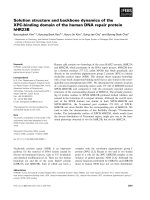
patients in the study. The frequency of ECG recordings and
troponin measurements is shown in Fig. 1. Of the 115
patients, 93 (80.9%) patients had at least one ECG performed
and one troponin measurement during ICU admission, seven
(6.1%) had at least one ECG performed but no troponin meas-
urement, 11 (9.6%) had at least one troponin measurement
but no ECG performed, and four (3.5%) had neither an ECG
performed nor troponin measurement. Patients had a median
of 2 (IQR 1–4) ECGs during their ICU stay. For 23 patients,
28 echocardiograms were performed.
Critical Care Vol 9 No 6 Lim et al.
R639
Patient characteristics are summarized in Table 1. The mean ±
standard deviation age of the patients was 64.1 ± 17.2 years
and they had a APACHE II score of 21.9 ± 9.8; 61 (53.0%)
were female. Most admissions were medical (72.2%), and
most patients (62.6%) were mechanically ventilated (inva-
sively or noninvasively) at the time of enrolment.
Excluding the 22 patients without both a troponin level and an
ECG performed, the frequency of MI based on available ECGs
and troponin measurements was 25.8%, occurring in 24
patients. Twenty-one patients sustained a non-ST-segment
elevation MI whereas three sustained an ST-segment elevation
MI. Patients diagnosed with MI were more likely to have under-
lying insulin-requiring diabetes mellitus and peripheral vascular
disease than were those patients without MI. No patients with
MI had ischemic chest pain symptoms, and no patients with MI
had this diagnosis made based on angiography, percutaneous
coronary intervention, or autopsy. The median (IQR) troponin
level in patients with MI was significantly higher (0.26 µg/l
[0.16–0.72 µg/l]) than in patients without MI (<0.04 µg/l
[<0.04–0.05 µg/l]; P < 0.0001).
Table 2 shows the frequency of morbidity and mortality out-
comes. Both ICU and hospital mortality were significantly
higher in patients diagnosed with MI than in those without MI
(37.5% versus 17.6%, P = 0.05; and 50.0% versus 22.0%, P
= 0.01, respectively). We found no difference between
patients with and without MI with respect to the duration of
mechanical ventilation or duration of ICU or hospital stay.
These outcomes were no different between patients with ST-
segment elevation MI (n = 3) and non-ST-segment elevation
MI (n = 21; data not shown).
Table 3 summarizes factors associated with ICU and hospital
mortality in the univariable and multivariable regression analy-
ses. Factors independently associated with ICU mortality were
APACHE II score (OR 2.70, 95% CI 1.27–5.72) and need for
inotropes or vasopressors (OR 6.12, 95% CI 1.31–28.68).
Factors independently associated with hospital mortality were
APACHE II score (OR 2.37, 95% CI 1.21–4.63), need for ino-
tropes or vasopressors (OR 4.76, 95% CI 1.27–17.82) and a
diagnosis of MI (OR 3.22, 95% CI 1.04–9.96). When troponin
values were added to the latter model, it was not significant in
the multivariable analysis but was significant in the univariable
analysis; for troponin values of 0.04–1.0 µg/l and ≥1.1 µg/l,
the ORs were 2.99 (95% CI 1.23–7.23) and 9.33 (95% CI
1.53–56.93), respectively, compared with the normal refer-
ence range (<0.04 µg/l).
Discussion
Because cardiac troponin is a sensitive and specific measure
of myocardial necrosis, it is the preferred biomarker for use in
the diagnosis of acute MI. Although an elevated troponin indi-
cates myocardial necrosis, it does not always indicate MI.
Thus, in the ICU setting, where elevated troponin is frequently
observed, additional evidence of myocardial ischemia can be
obtained by using a 12-lead ECG. In this single centre pro-
spective cohort study of predominantly medical ICU patients,
47% of critically ill patients had at least one elevated troponin
measurement but only 26% met diagnostic criteria for MI
based on a typical rise or fall in elevated troponin measure-
ments and ischemic changes on a 12-lead ECG, with ECGs
performed as clinically indicated. Patients with MI had signifi-
cantly higher troponin levels than did those without MI.
Patients who were diagnosed with MI had twofold increased
rates of ICU and hospital mortality. The presence of an ele-
vated troponin measurement alone was not associated with
adverse outcomes, but the presence of MI was independently
predictive of hospital mortality.
The incidence and prevalence rates of elevated levels of tro-
ponin cited in the literature vary widely, ranging from 15% to
Figure 1
Electrocardiogram and troponin measurement frequency for enrolled patientsElectrocardiogram and troponin measurement frequency for enrolled patients. ECG, electrocardiogram; MI, myocardial infarction.
Available online />R640
70% of patients [17-19]. Recent studies have examined the
frequency of elevated troponin levels excluding those patients
with underlying coronary heart disease [3]; a 55% prevalence
of elevated levels of troponin was reported, of which 72% of
patients with an elevated troponin did not have flow-limiting
coronary artery disease based on stress echocardiography or
autopsy. The variability in frequency rates observed in our
study and other studies is likely due to the heterogeneous
nature of ICU populations and the threshold at which a tro-
ponin measurement is considered positive. The current tro-
ponin threshold recommended by the ESC/ACC has been
defined for noncritically ill populations, and whether this
threshold differs in the ICU setting is unknown. Furthermore,
the various troponin assays are not standardized and, although
Table 1
Patient clinical characteristics
Total (n = 115) MI (n = 24) No MI (n = 91) P value
Age (years; mean ± SD) 64.1 ± 17.2 62.8 ± 18.3 64.4 ± 17.0 0.69
Sex (n; % female) 61 (53.0) 13 (54.2) 48 (52.7) 1.00
APACHE II score (mean ± SD) 21.9 ± 9.8 26.3 ± 10.4 20.7 ± 9.3 0.01
Medical (n [%]) 83 (72.2) 20 (83.3) 63 (69.2) 0.21
Epidural (n [%]) 12 (10.4) 0 (0.0) 12 (13.2) 0.07
Past medical history (n [%])
Smoker 35 (30.4) 5 (20.8) 30 (33.0) 0.32
Hypertension 59 (51.3) 14 (58.3) 45 (49.5) 0.50
Diabetes (oral agent) 9 (7.8) 3 (12.5) 6 (6.6) 0.39
Diabetes (insulin) 17 (14.8) 10 (41.7) 7 (7.7) <0.001
Hypercholesterolemia 14 (12.2) 2 (8.3) 12 (13.2) 0.73
Angina 15 (13.0) 3 (12.5) 12 (13.2) 1.00
Myocardial infarction 25 (21.7) 9 (37.5) 16 (17.6) 0.05
Congestive heart failure 14 (12.2) 5 (20.8) 9 (9.9) 0.17
Peripheral vascular disease 10 (8.7) 7 (29.2) 3 (3.3) <0.001
Transient ischemic attack 4 (3.5) 2 (8.3) 2 (2.2) 0.19
Stroke 8 (7.0) 3 (12.5) 5 (5.5) 0.36
Baseline life support interventions (n [%])
Ventilation
Invasive mechanical ventilation 67 (58.3) 16 (66.7) 51 (56.0) 0.49
Noninvasive mechanical ventilation 5 (4.3) 2 (8.3) 3 (3.3) 0.28
Inotropes and vasopressors
Epinephrine 2 (1.7) 1 (4.2) 1 (1.1) 0.38
Dopamine 10 (8.7) 3 (12.5) 7 (7.7) 0.43
Norepinephrine 19 (16.5) 5 (20.8) 14 (15.4) 0.54
Dobutamine 4 (3.5) 2 (8.3) 2 (2.2) 0.19
Phenylephrine 18 (15.7) 9 (37.5) 9 (9.9) 0.003
Vasopressin 5 (4.3) 3 (12.5) 2 (2.2) 0.06
Hemodialysis
Intermittent dialysis 14 (12.2) 4 (16.7) 10 (11.0) 0.49
Continuous renal replacement therapy 0 (0.0) 0 (0.0) 0 (0.0) -
APACHE, Acute Physiology and Chronic Health Evaluation; MI, myocardial infarction; SD, standard deviation.
Critical Care Vol 9 No 6 Lim et al.
R641
it is recommended that levels exceeding the 99th percentile
be considered positive, this level varies according to manufac-
turer [20].
It is important to recognize that although a considerable
number of ICU patients have elevated troponin measurements,
and elevated troponin measurements are specific for myocar-
dial necrosis, troponin itself does not distinguish between
ischemic and nonischemic etiologies of myocardial injury.
Interpretation of elevated troponin levels in the ICU must be
considered in the context of the patient's symptoms (fre-
quently limited in the ICU) or correlated with ECG findings or
other imaging modalities. Most studies have examined ele-
vated troponin levels in the critically ill in isolation, and it is
unclear what proportion of patients have actually suffered an
MI. One study evaluated troponin with ECGs in 34 consecu-
tive critically ill patients who were mechanically ventilated and
underwent thoracic or vascular surgery [21]. It found that 11
patients (32%) had elevated troponin levels, and ECGs were
available in 10 patients. Four patients (12%) had ST-segment
elevation or depression, meeting criteria for MI; three patients
had nonspecific changes and three had no ECG changes.
Another study used continuous 12-lead telemetry monitoring
in 76 patients admitted with noncardiac conditions [6]. An
elevated troponin level was found in 12 patients (15.8%), and
six of these patients had transient ischemic events (mainly ST-
segment depression) on telemetry.
The importance of a diagnosis that does not alter a patient's
prognosis is questionable. Therefore, potentially the most
important reason to identify critically ill patients with elevated
troponin as having an MI or not having an MI is that the prog-
nosis of these patients may be different. In the noncritically ill
population, elevated troponin levels are an independent prog-
nostic marker for short-term and long-term outcomes in
patients with acute coronary syndromes. In the ICU several
studies have reported that elevated troponin levels are associ-
ated with adverse outcomes. Elevated troponin I is a predictor
of mortality in medical-surgical ICU patients [17-19], including
those without acute coronary syndromes [3], and in ICU
patients with early sepsis [22], acute exacerbations of chronic
obstructive pulmonary disease [14], pulmonary embolism [23]
and following cardiac surgery [24-26]. In surgical ICU
patients, troponin is a predictor of mortality and longer length
of ICU and hospital stay [13]. However, most studies have
examined troponin alone and did not examine prognosis in
relation to those patients who had associated ECG changes
(i.e. patients with MI).
One retrospective study examined the degree of troponin ele-
vation in relation to prognosis [13], and among the patients
with recognized MI mortality was 13.6% in those with moder-
ate elevations of troponin I (2.0–10.0 µg/l) and 32.4% in
patients with troponin I above 10.0 µg/l. Like our study, this
was limited in that there was no screening; it had a retrospec-
tive design, and it was unclear how the diagnosis of MI was
made. Although we found that MI was predictive of hospital
mortality, it was not predictive of other morbidity outcomes,
including the duration of mechanical ventilation and ICU and
hospital stays. This may be attributable to the relatively small
number of patients included in our study or it may be an arte-
fact of the distribution of some early deaths in this cohort. Sim-
ilarly, predictors of ICU and hospital mortality, including need
for life-saving therapies (hemodialysis, mechanical ventilation),
were not significant in the multivariable analysis, which may
relate to the distribution of risk factors (hemodialysis being
infrequent and mechanical ventilation being common) in a
study of this size.
Identification of those critically ill patients with MI has several
treatment implications. In noncritically ill patients, patients with
elevated troponin levels and acute MI benefit from antithrom-
botic therapy [27-30]. Critically ill patients who also have ele-
vated troponin and acute MI would also be expected to benefit
from these therapies, but those patients who have elevated
troponin without MI may not benefit and in fact may be harmed.
Furthermore, critically ill patients with ST-segment elevation MI
should be distinguished from those with non-ST-segment ele-
vation MI because the former warrants urgent revasculariza-
tion (or thrombolysis, although this is not always an option for
ICU patients). We did not detect differences in outcome in
patients diagnosed with ST-segment elevation or non-ST-seg-
ment elevation MI, although our analysis was underpowered to
detect such differences.
Table 2
Frequency of morbidity and mortality outcomes
MI (n = 24) No MI (n = 91) P value
Duration of mechanical ventilation (days; median [IQR]) 2 (1–6) 2 (0–5) 0.32
Duration of ICU stay (days; median [IQR]) 4.5 (2–8) 4 (2–6) 0.62
ICU mortality (n [%]) 9 (37.5) 16 (17.6) 0.05
Duration of hospital stay (days; median [IQR]) 18 (4.5–41) 12 (7–21) 0.34
Hospital mortality (n [%]) 12 (50.0) 20 (22.0) 0.01
ICU, intensive care unit; IQR, interquartile range; MI, myocardial infarction.
Available online />R642
Not only has recognition of MI been poorly studied but also the
impact of antithrombotic and anti-ischemic agents has not
been well documented in these patients. In one study, surgical
ICU patients with moderate elevations in troponin I (2.0–10.0
µg/l, and not necessarily diagnosed with MI) who were treated
with β-blockers and aspirin were reported to have lower mor-
tality than patients with the same range of troponin elevation
who did not receive these therapies [13]. However, findings
from this retrospective study should be cautiously interpreted
because selection bias might have resulted in patients who
were less critically ill receiving β-blockers and aspirin (e.g.
patients without a coagulopathy and not requiring β-agonist
infusions).
The strengths of our study include use of a priori definitions for
MI, and duplicate assessment by two independent investiga-
tors to classify events. Determination not only of patients with
elevated troponin levels but also of those with MI provides rel-
evant information not previously reported. However, there are
several important limitations to the study. First, although ECG
has been reported to be more sensitive for detecting myocar-
dial ischemia than conventional ICU monitoring [30], the ECG
itself has limitations. The conventional ECG is not very sensi-
tive for detecting infarction in certain locations (posterior) [32],
and not all patients who have myocardial necrosis exhibit ECG
changes [2]. In addition, uninterpretable ECGs occur among
patients who are pacemaker dependent or have left bundle
branch blocks, in whom acute changes cannot be detected
using a standard 12-lead ECG. A second limitation is that
systematic screening of all patients with troponin and ECG
recordings was not performed in this study, and hence we
cannot determine the true prevalence and incidence of ele-
vated troponin and MI. Finally, there is currently no consensus
on the appropriate diagnosis of MI in critically ill patients,
whose ability to communicate may be severely limited and in
whom diagnostic tests have not been vigorously evaluated.
Our results may have differed if we required two or more ele-
vated troponin measurements to meet the biomarker criterion,
or if we required two or more ECGs demonstrating evolving
changes [5].
The utility of screening for MI in the ICU population has not
been studied. In view of the prognostic and possible therapeu-
tic implications of establishing a diagnosis of MI in the critically
ill, use of noninvasive tests – including troponin and ECG –
may be a reasonable approach. In contrast to the noncritically
ill population, limitations in the ability of patients to communi-
cate ischemic symptoms and the use of vasopressors and
mechanical ventilation are unique to the ICU and may require
the use of alternate methods of diagnosis. However, we can-
not recommend for or against systematic troponin screening
based on our study. The appropriate use of these tests and
other diagnostic methods, including echocardiography in a
screening mode, must be properly evaluated in well designed
prospective studies.
Conclusion
In summary, elevated troponin levels are common in critically ill
patients, but not all patients with elevated levels have MI.
Almost half of the patients admitted to this general medical-
surgical ICU (consisting mainly of medical patients) had ele-
vated troponins during their ICU stay, and approximately 26%
of patients were found to have an MI. Patients diagnosed with
MI in the ICU based on elevated troponin levels and ischemic
ECG changes had higher ICU and hospital mortality, and MI
was independently predictive of hospital mortality. However,
we did not find that an elevated troponin alone (among those
patients who had troponin measurements during their ICU
admission) was associated with adverse outcomes. Screening
with troponin levels in the ICU should not be done in isolation
because ECG is necessary to interpret abnormal levels. Future
studies should determine the prognostic importance of ele-
vated troponin in the ICU by more comprehensively examining
other diagnoses and their consequences, and by evaluating
the role of other modalities (i.e. echocardiography, radionu-
clide imaging, magnetic resonance imaging) to diagnose MI.
Second, the role of antithrombotic and anti-ischemic agents in
Table 3
Predictors of intensive care unit and hospital mortality
Predictor ICU mortality (OR [95% CI]) Hospital mortality (OR [95% CI])
Univariable Multivariable Univariable Multivariable
APACHE II score (10-point increment) 3.92 (2.08–7.40) 2.70 (1.27–5.72) 3.10 (1.82–5.30) 2.37 (1.21–4.63)
Mechanical ventilation 6.14 (1.71–21.97) 0.69 (0.11–4.18) 3.66 (1.36–9.82) 0.56 (0.13–2.46)
Inotropes or vasopressors 8.23 (2.95–23.00) 6.12 (1.31–28.68) 6.10 (2.50–14.89) 4.76 (1.27–17.82)
Hemodialysis 1.58 (0.54–4.62) 0.63 (0.16–2.42) 1.38 (0.50–3.81) 0.52 (0.14–1.87)
MI 2.81 (1.05–7.55) 2.25 (0.65–7.77) 3.55 (1.39–9.10) 3.22 (1.04–9.96)
This table summarizes the relation between mortality and APACHE II score, MI, and each of the three types of advanced life support at any time
during the ICU stay (mechanical ventilation: 61.7% of patients; inotropes or vasopressors: 38.3% of patients; hemodialysis: 18.3% of patients).
APACHE, Acute Physiology and Chronic Health Evaluation; CI, confidence interval; ICU, intensive care unit; MI, myocardial infarction; OR, odds
ratio.
Critical Care Vol 9 No 6 Lim et al.
R643
critically ill patients with troponin elevation requires evaluation
in large prospective randomized controlled trials to determine
the appropriate management of these patients.
Competing interests
The author(s) declare that they have no competing interests.
Authors' contributions
WL, DC, MC, PD, and IQ obtaining funding for the study. DC,
IQ, and WL conceived and designed the study. IQ, WL, and
DC collected data. DH-A, WL, and DC conducted statistical
analysis. WL, DC and DH-A drafted the report, and PD, MC
and IQ critically revised the manuscript. DC was guarantor.
Acknowledgements
This study was funded by a grant from the Regional Medical Associates
of McMaster University, Canada, and the Father Sean O'Sullivan
Research Center of St. Joseph's Hospital in Hamilton. We thank Andrea
Tkaczyk, Laura Donahoe, Jill Hancock and Ellen McDonald for their help
with data collection, and Kristina Lutz for her help with the data entry.
WL is a Clinical Scholar with a Graduate Scholarship from the Canadian
Institutes of Health Research, DJC is a Research Chair of the Canadian
Institutes for Health Research, MAC holds a Career Investigator Award
from the Heart and Stroke Foundation of Canada, and PJD holds a Sen-
ior Research Fellowship Award of the Canadian Institutes of Health
Research.
References
1. Katus HA, Remppis A, Looser S, Hallermeier K, Scheffold T, Kubler
W: Enzyme linked immuno assay of cardiac troponin T for the
detection of acute myocardial infarction in patients. J Mol Cell
Cardiol 1989, 21:1349-1353.
2. Alpert JS, Thygesen K, Antman E, Bassand JP: Myocardial infarc-
tion redefined – a consensus document of the Joint European
Society of Cardiology/American College of Cardiology Com-
mittee for the redefinition of myocardial infarction. J Am Coll
Cardiol 2000, 36:959-969.
3. Ammann P, Maggiorini M, Bertel O, Haenseler E, Joller-Jemelka HI,
Oechslin E, Minder EI, Rickli H, Fehr T: Troponin as a risk factor
for mortality in critically ill patients without acute coronary
syndromes. J Am Coll Cardiol 2003, 41:2004-2009.
4. Ammann P, Fehr T, Minder EI, Gunter C, Bertel O: Elevation of
troponin I in sepsis and septic shock. Intensive Care Med
2001, 27:965-969.
5. Luepker RV, Apple FS, Christenson RH, Crow RS, Fortmann SP,
Goff D, Goldberg RJ, Hand MM, Jaffe AS, Julian DG, et al.: Case
definitions for acute coronary heart disease in epidemiology
and clinical research studies: a statement from the AHA Coun-
cil on Epidemiology and Prevention; AHA Statistics Commit-
tee; World Heart Federation Council on Epidemiology and
Prevention; the European Society of Cardiology Working
Group on Epidemiology and Prevention; Centers for Disease
Control and Prevention; and the National Heart, Lung, and
Blood Institute. Circulation 2003, 108:2543-2549.
6. Booker KJ, Holm K, Drew BJ, Lanuza DM, Hicks FD, Carrigan T,
Wright M, Moran J: Frequency and outcomes of transient myo-
cardial ischemia in critically ill adults admitted for noncardiac
conditions. Am J Crit Care 2003, 12:508-517.
7. Antman EM, Tanasijevic MJ, Thompson B, Schactman M, McCabe
CH, Cannon CP, Fischer GA, Fung AY, Thompson C, Wybenga D,
Braunwald E: Cardiac-specific troponin I levels to predict the
risk of mortality in patients with acute coronary syndromes. N
Engl J Med 1996, 335:1342-1349.
8. Stubbs P, Collinson P, Moseley D, Greenwood T, Noble M: Pro-
spective study of the role of cardiac troponin T in patients
admitted with unstable angina. BMJ 1996, 313:262-264.
9. Lindahl B, Venge P, Wallentin L: Relation between troponin T
and the risk of subsequent cardiac events in unstable coro-
nary artery disease. The FRISC study group. Circulation 1996,
93:1651-1657.
10. Galvani M, Ottani F, Ferrini D, Ladenson JH, Destro A, Baccos D,
Rusticali F, Jaffe AS: Prognostic influence of elevated values of
cardiac troponin I in patients with unstable angina. Circulation
1997, 95:2053-2059.
11. Newby LK, Christenson RH, Ohman EM, Armstrong PW, Thomp-
son TD, Lee KL, Hamm CW, Katus HA, Cianciolo C, Granger CB,
et al.: Value of serial troponin T measures for early and late risk
stratification in patients with acute coronary syndromes. The
GUSTO-IIa Investigators. Circulation 1998, 98:1853-1859.
12. Hamm CW, Ravkilde J, Gerhardt W, Jorgensen P, Peheim E,
Ljungdahl L, Goldmann B, Katus HA: The prognostic value of
serum troponin T in unstable angina. N Engl J Med 1992,
327:146-150.
13. Relos RP, Hasinoff IK, Beilman GJ: Moderately elevated serum
troponin concentrations are associated with increased mor-
bidity and mortality rates in surgical intensive care unit
patients. Crit Care Med 2003, 31:2598-2603.
14. Baillard C, Boussarsar M, Fosse JP, Girou E, Le Toumelin P,
Cracco C, Jaber S, Cohen Y, Brochard L: Cardiac troponin I in
patients with severe excerbation of chronic obstructive pulmo-
nary disease. Intensive Care Med 2003, 29:584-589.
15. ver Elst KM, Spapen HD, Nguyen DN, Garbar C, Huyghens LP,
Gorus FK: Cardiac troponins I and T are biological markers of
left ventricular dysfunction in septic shock. Clin Chem 2000,
46:650-657.
16. Wu AH, Apple FS, Gibler WB, Jesse RL, Warshaw MM, Valdes R
Jr: National Academy of Clinical Biochemistry Standards of
Laboratory Practice: recommendations for the use of cardiac
markers in coronary artery diseases. Clin Chem 1999,
45:1104-1121.
17. Guest TM, Ramanathan AV, Tuteur PG, Schechtman KB, Laden-
son JH, Jaffe AS: Myocardial injury in critically ill patients. A fre-
quently unrecognised complication. JAMA 1995,
273:1945-1949.
18. Kollef MH, Ladenson JH, Eisenberg PR: Clinically recognized
cardiac dysfunction: an independent determinant of mortality
among critically ill patients. Is there a role for serial measure-
ment of cardiac troponin I? Chest 1997, 111:1340-1347.
19. Noble JS, Reid AM, Jordan LV, Glen AC, Davidson JA: Troponin I
and myocardial injury in the ICU. Br J Anaesth 1999, 82:41-46.
20. Scirica BM, Morrow DA: Troponins in acute coronary
syndromes. Prog Cardiovasc Dis 2004, 47:177-188.
Key messages
• The diagnosis of MI in the ICU is usually dependent on
elevated troponin levels and ischemic changes on a 12-
lead ECG or a new wall motion abnormality on echocar-
diography, because ICU patients are usually unable to
communicate chest pain symptoms as a result of admin-
istration of narcotics or sedatives, or decreased level of
consciousness.
• In this single centre cohort study, 47% of predominantly
medical critically ill patients had at least one elevated
troponin measurement, but only 26% of these patients
had MI.
• An elevated troponin level alone is not an independent
predictor of ICU or hospital mortality.
• Patients with MI had a significant twofold increase in
risk for ICU and hospital mortality compared with
patients without MI.
• After adjusting for APACHE II score and inotrope or
vasopressor use, development of MI in the ICU setting
was an independent predictor of hospital mortality.
Available online />R644
21. Klein Gunnewiek JM, van de Leur JJ: Elevated troponin T concen-
trations in critically ill patients. Intensive Care Med 2003,
29:2317-2322.
22. Spies C, Haude V, Fitzner R, Schroder K, Overbeck M, Runkel N,
Schaffertzik W: Serum cardiac troponin T as a prognostic
marker in early sepsis. Chest 1998, 113:1055-1063.
23. La Vecchia L, Ottani F, Favero L, Spadaro GL, Rubboli A, Boanno
C, Mezzena G, Fontanelli A, Jaffe AS: Increased cardiac troponin
I on admission predicts in-hospital mortality in acute pulmo-
nary embolism. Heart 2004, 90:633-637.
24. Lehrke S, Steen H, Sievers HH, Peters H, Opitz A, Muller-Bardorff
M, Wiegand UK, Katus HA, Giannitsis E: Cardiac troponin T for
prediction of short- and long-term morbidity and mortality
after elective open heart surgery. Clin Chem 2004,
50:1560-1567.
25. Lyon WJ, Baker RA, Andrew MJ, Tirimacco R, White GH, Knight
JL: Relationship between elevated preoperative troponin T and
adverse outcomes following cardiac surgery. ANZ J Surg
2003, 73:40-44.
26. Baggish AL, MacGillivray TE, Hoffman W, Newell JB, Lewan-
drowski KB, Lee-Lewandrowski E, Anwaruddin S, Siebert U,
Januzzi JL: Postoperative troponin-T predicts prolonged inten-
sive care unit length of stay following cardiac surgery. Crit
Care Med 2004, 32:1866-1871.
27. ISIS-2 (Second International Study of Infarct Survival) Collabora-
tive Group: Randomised trial of intravenous streptokinase, oral
aspirin, both, or neither among 17,187 cases of suspected
acute myocardial infarction: ISIS-2. Lancet 1988, 2:349-360.
28. Antiplatelet Trialists' Collaboration: Collaborative meta-analysis
of randomized trials of antiplatelet therapy for prevention of
death, myocardial infarction, and stroke in high-risk patients.
BMJ 2002, 324:71-86.
29. Assessment of the Safety and Efficacy of a New Thrombolytic Reg-
imen (ASSENT)-3 Investigators: Efficacy and safety of tenect-
eplase in combination with enoxaparin, abciximab, or
unfractionated heparin: the ASSENT-3 randomised trial in
acute myocardial infarction. Lancet 2001, 358:605-613.
30. Lindahl B, Diderholm E, Lagerquist B, Venge P, Wallentin L:
Effects on mortality of long-term treatent with low molecular
weight heparin in relation to troponin T level and ECG findings
– a FRISC 2 substudy. Eur Heart J 2000:521.
31. Martinez EA, Kim LJ, Faraday N, Rosenfeld B, Bass EB, Perler BA,
Williams GM, Dorman T, Pronovost PJ: Sensitivity of routine
intensive care unit surveillance for detecting myocardial
ischemia. Crit Care Med 2003, 31:2302-2308.
32. Chow T-C: Electrocardiography in Clinical Practice: Adult and
Pediatric 4th edition. Philadelphia: WB Saunders Company; 1996.