test điện tâm đồ phần
Bạn đang xem bản rút gọn của tài liệu. Xem và tải ngay bản đầy đủ của tài liệu tại đây (838.87 KB, 20 trang )
test ®iÖn tim ®å
•
LVH & PVCs: Precordial Leads-KH
•
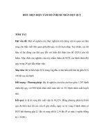
Left Atrial Enlargement-KH
•
Left atrial enlargement is illustrated by increased P wave duration in lead II, top ECG, and
by the prominent negative P terminal force in lead V1, bottom tracing
II
V1
•
LVH - Best seen in the frontal plane leads!-KH
Lewis Index: 1) R in aVL >11 mm
2) R in I + S in III >25mm
3) (RI+SIII) - (RIII+SI) >17mm
•
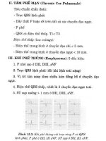
Right Atrial Enlargement (RAE) & Right Ventricular Hypertrophy
(RVH)-KH
•
RAE is recognized by the tall (>2.5mm) P waves in leads II, III, aVF. RVH is likely because of right
axis deviation (+100 degrees)
Left Atrial Abnormality & 1st degree AV Block-KH
Sóng P rộng (>0,12s) và có khía ở DII, DIII; hai pha ở chuyển đạo V1 Tất cả các tiêu chuẩn
cho nhĩ trái không bình th ờng hoặc dầy nhĩ (LAE). Khoảng PR > 0,2s: Block AV cấp I.
•
Severe RVH
- Trôc P râ (+150 degrees)
- D¹ng qR ë V1, R/S ë V1 > 1; S/R ë V6 > 1
- ST chªnh dèc xuèng ë c¸c chuyÓn ®¹o tr íc tim ph¶i
•
LVH: Limb Lead Criteria-KH
•
In this example of LVH, the precordial leads don't meet the usual voltage criteria or exhibit
significant ST segment abnormalities. The frontal plane leads, however, show voltage criteria for
LVH and significant ST segment depression in leads with tall R waves. The voltage criteria include
1) R in aVL >11 mm; 2) R in I + S in III >25mm; and 3) (RI+SIII) - (RIII+SI) >17mm (Lewis Index).
•
Left Atrial Enlargement: Leads II and V1-KH
•
RAE & RVH-KH
•
Left Atrial Abnormality & 1st Degree AV Block: Leads II and V1-KH
- P > 0,12s vµ cã khÝa ë DII; hai pha ë chuyÓn ®¹o V1
- Kho¶ng PR > 0,2s
•
Left Atrial Enlargement & Nonspecific ST-T Wave Abnormalities-KH
•
LAE is best seen in V1 with a prominent negative (posterior) component measuring 1mm wide and
1mm deep. There are also diffuse nonspecific ST-T wave abnormalities which must be correlated
with the patient's clinical status. Poor R wave progression in leads V1-V3, another nonspecific
finding, is also present
•
LVH and Many PVCs-KH
•
The combination of voltage criteria (SV2 + RV6 >35mm) and ST-T abnormalities in V5-6
are definitive for LVH. There may also be LAE as evidenced by the prominent negative P
terminal force in lead V1. Isolated PVCs and a PVC couplet are also present.
•
Right Axis Deviation & RAE (P Pulmonale): Leads I, II, III-KH
•
LVH: Limb Lead Criteria-KH
Lewis Index: 1) R in aVL >11 mm
2) R in I + S in III >25mm
3) (RI+SIII) - (RIII+SI) >17mm
•
LVH: Strain pattern + Left Atrial Enlargement-KH
- SV2 + RV5 >35mm
- Sãng P réng (>0.12s) vµ cã khÝa ë DII, DIII; hai pha ë chuyÓn ®¹o V1
•
RVH with Right Axis Deviation
•
Note the qR pattern in right precordial leads. This suggests right ventricular pressures greater than
left ventricular pressures. The persistent S waves in lateral precordial leads and the RAD are other
finding in RVH.
•
LVH with "Strain"-KH
- SV2 + RV5 >35mm
- Lewis Index: 1) R in aVL >11 mm
2) R in I + S in III >25mm
3) (RI+SIII) - (RIII+SI) >17mm
•
Right Ventricular Hypertrophy (RVH) & Right Atrial Enlargement
(RAE)-KH
•
In this case of severe pulmonary hypertension, RVH is recognized by the prominent anterior forces
(tall R waves in V1-2), right axis deviation (+110 degrees), and "P pulmonale" (i.e., right atrial
enlargement). RAE is best seen in the frontal plane leads; the P waves in lead II are >2.5mm in
amplitude
Xin tr©n träng c¸m ¬n !