Báo cáo y học: "nfluence of atorvastatin on coronary calcifications and myocardial perfusion defects in systemic lupus erythematosus patients: a prospective, randomized, double-masked, placebo-controlled study" docx
Bạn đang xem bản rút gọn của tài liệu. Xem và tải ngay bản đầy đủ của tài liệu tại đây (537.57 KB, 9 trang )
RESEARC H ARTIC LE Open Access
Influence of atorvastatin on coronary
calcifications and myocardial perfusion defects in
systemic lupus erythematosus patients:
a prospective, randomized, double-masked,
placebo-controlled study
Wojciech Plazak
1*
, Krzysztof Gryga
2
, Hanna Dziedzic
1
, Lidia Tomkiewicz-Pajak
1
, Malgorzata Konieczynska
3
,
Piotr Podolec
1
and Jacek Musial
2
Abstract
Introduction: Mortality in systemic lupus erythematosus (SLE) patients is influenced by an increased occurrence of
severe cardiovascular complications. Statins have been proven to protect a wide spectrum of SLE patients from
these compl ications. This study was conducted to determine the possible efficacy of atorvastatin in SLE patients as
assessed by m ulti-detector computed tomography (MDCT)-based coronary calcium scoring and single photon
emission computed tomography (SPECT) of the myocardium.
Methods: Sixty SLE patients in stable clinical conditions were randomized to receive either atorvastatin (40 mg
daily; n = 28) or placebo (n = 32). Clinical and biochemical evaluation together with MDCT-based coronary calcium
scoring and SPECT studies (Tc-99 m sestamibi) were performed at the time of randomization and after 1 year of
treatment.
Results: At randomization, SPECT revealed perfusion defects at rest in 22 (36.7%) patients and exercise-induced
defects in 8 (13.3%), whereas MDCT revealed coronary calcifications in 15 subjects (25%). Coronary calcium deposits
increased after 1 year in the placebo group (plaque volume change from 35.2 ± 44.9 to 62.9 ± 72.4, P < 0.05;
calcium score from 32.1 ± 39.1 to 59.5 ± 64.4; P < 0.05), but not in the atorvastatin group (plaque volume 54.5 ±
62.4 vs. 51.0 ± 47.6, P not significant; calcium score 44.8 ± 50.6 vs. 54.9 ± 62.5, P not significant). The atorvastatin
group showed a decrease in total serum cholesterol (from 5.1 ± 1.2 to 4.4 ± 0.7 mmol/L, P < 0.05), LDL cholesterol
(2.9 ± 1.0 to 2.3 ± 0.6 mmol/L, P < 0.05), triglycerides (1.6 ± 0.6 to 1.2 ± 0.5 mmol/L, P < 0.05), and C-reactive
protein (CRP) (4.4 ± 4.1 to 2.7 ± 1.7 mg/L, P < 0.05). There was no change in the mean Systemic Lupus
Erythematosus Disease Activity Index (SLEDAI) score in patients from both groups. Perfusion defects observed at
randomization showed no change after one year treatment with atorvastatin.
Conclusions: In SLE patients 40 mg of atorvastatin daily for 1 year led to a decrease in serum lipids and CRP
levels. Additionally the progression of atherosclerosis, as assessed by MDCT-based coronary calcium scoring, is
restrained by atorvastatin treatment. The value of statin treatment in patients with SLE free from cardiovascular
disease clinical symptoms should be addressed in large, prospective clinical trials.
Keywords: systemic lupus erythematosus, autoimmune diseases, coronary calcification, accelerated atherosclerosis,
MDCT, perfusion scintigraphy, statins
* Correspondence:
1
Department of Cardiac and Vascular Diseases, the John Paul II Hospital,
Jagiellonian University Medical College, Pradnicka Str 80, 31-202 Krakow,
Poland
Full list of author information is available at the end of the article
Plazak et al. Arthritis Research & Therapy 2011, 13:R117
/>© 2011 Plazak e t al.; licensee BioMed Central Ltd. This is an open access article distributed under the terms of the Creative Commons
Attribution License http://creativecommons.o rg/licenses/by/2.0), which permits unrestrict ed use, distribution, and reproduction in any
medium, provided the origina l work is properly cited.
Introduction
Systemic lupus erythematosus (SLE) is a generalized
autoimmune disease, in which diffuse, chronic inflam-
matory reactions play an important pathogenic role.
Contemporary mortality of SLE patients is mainly due
to severe cardiovascular complications [1]. Suggested
factors that may influence accelerated arteriosclerosis
include a generalized, chronic inflammation and corti-
costeroid usage [2]. The relation between increased
levels of inflammatory cytokines and life-threatening
cardiovascular episodes has been well-documented [3].
However, the optimal strategy for the prevention of
atherosclerosis in SLE patients is not established.
Statins, HMG-CoA reductase inhibitors, are widely
used in the treatment of hyperlipidemia and prevention
against cardiovascular disease. In the general population,
large randomized controlled trials have demonstrated
their beneficial effects in hypercholesterolemia treatment
[4], as well as primary and secondary preven tion of cor-
onary artery disease [5-7] with the regression of estab-
lished coronary atherosclerosis [8]. Interestingly, the
magnitude of the protection and decrease in mor tality
afforded by statins cannot be explained entirely by their
cholesterol-lowering effect. It has been shown, among
others, that statins exert strong anti-inflammatory action
[9] and ameliorate endothelialdysfunction,protecting
from inflammation-induced endothelial injury [10,11].
Statins are recommended for patients with SLE at high
cardiovascular risk with diagnosed coronary artery dis-
ease, but these recommendations are based on the
extrapolation of the results obtained in non-SLE popula-
tions [12-16]. There has been little evidence for the
effectiveness of statins in cardiovascular symptom-free
SLE patients. Implementation of multi-detector com-
puted tomography (MDCT) and single photon emission
computerized tomography (SPECT) allows for a non-
invasive evaluation of coronary atherosclerosis and myo-
cardial perfusion abnormalities, and enables the assess-
ment of statin influence on coronary artery structural
changes and heart function.
This study was conducted to determine the effect of
atorvastatin treatment on MDCT-based coronary cal-
cium sco ring and SPECT-assessed myocardial perfusion
abnormalities in SLE patients free of clinical symptoms
of cardiovascular disease.
Materials and methods
The study was performed in 60 consecutive patients
treated for systemic SLE in the Department of Internal
Medicine, Jagiellonian University Medical College, Kra-
kow. All patients fulfilled at least four American College
of Rheumatology classification criteria for SLE [17,18]
and were in stable clinical conditions (no need for
immunosuppressive therapy intensification, i.e. current
immunosuppressive drug dose increase or introduction
of an additional immunosuppressive drug within the
past three months). Patients with known cancer, clinical
symptoms of coronary heart disease or heart failure
(New York Heart Association III or IV class), renal fail-
ure (creatinine clearanc e < 30 ml/min), and/ or respira-
tory failure were excluded from the study.
Atorvastatin was chosen for this study because of its
superiority over two o ther statins (simvas tatin and pra-
vastatin) in the inhibition of atherosclerosis shown by
two large clinical trials [19,20]. We chose, however, a
daily dose of 40 mg to limit treatment-associa ted
adverse events.
Patients were randomized (random option in Micro-
soft Excel software, Qumak Secom SA, Warsaw, Poland)
to atorvastatin (40 mg, in the evening) or placebo group.
Placebo group received shape and color- matched pla-
cebo tablets at the same time. The duration of the study
was one year. All parameters described below were
assessed at randomization and after one year of treat-
ment by medical staff, unaware of the type of treatment.
The S PECT study (ECAM Gamma Camera, Siemens,
Munich, Germany) was performed at rest and during
exercise in a two-day protocol. At the first day, at near
maximal stress, a 25 to 40 mCi dose of Tc-99 m sesta-
mibi was injected (actual patient do se was modified tak-
ing into account patients weight) and exercise continued
for one additional minute after injection. Tc-99 m sesta-
mibi SPECT imaging was begun 15 to 30 minutes later.
On the second day rest examinations were performed.
SPECT was performed using a circular 180° acquisition
for 60 projections at 20 seconds per projection. Myocar-
dial perfusion was assessed in 17 left ventricle myocar-
dial segments. The number of segments with persistent
or exerc ise-induced perfusion defects were assessed by
visual interpretation.
Coronary calcium scoring was performed using a mul-
tidetector CT imager (Somatom Definition, Siemens,
Munich, Germany). The images were ECG triggered
with 3 mm thick sections obtained covering the whole
heart. Coronary artery calcifications were defined as
lesions with attenuation greater than 130 HU in more
than four adjacent pixels. For the quantification of cor-
onary calcium 3D Leonardo application (Siemens,
Munich, Germany) was used. The number of athero-
sclerotic plaques in particular coronary arteries and its
volume were assessed. The Agatson calcium score was
calculated [21].
Laboratory tests included determination of serum anti-
nuclear antibodies (ANA) presence, t heir titer (in direct
immunofluorescence; Hep-2 cells; Euroimmun GmbH,
Lubeck, Germany) and type (immunoblotting; Euroline
Plazak et al. Arthritis Research & Therapy 2011, 13:R117
/>Page 2 of 9
System, Euroimmun GmbH, Lubeck, Germany), serum
concentrations of C-reactive protein (CRP), and comple-
ment C3c and C4 components by nephelometry (Sie-
mens, Munich, Germany).
In addition, serum levels of anticardiolipin (aCL) and
antib2GPI antibodies (of both, IgG and IgM class) were
measured using home-made ELISA with the Sapporo
standard for antib2GPI an tibody measurements (HCAL
for IgG, EY2C9 for IgM), as previously described [22].
The values exceeding 99
th
percentile of a healthy popu-
lation sample were considered positive.
Lupus anticoagulant (LA) was determined in accor-
dance with the three-step procedure recommended by
the International Society on Thrombosis and Haemosta-
sis [23].
Statistical analysis was performed using Statistica Six
Sigma software (StatSoft, Krakow, Poland). All numeri-
cal data were expressed as mean values ± standard
deviations, as median values or as proportions. Continu-
ous variables were compared using a t-test. Chi-square
test was used to examine differences in proportions.
The level for statistical significance was predetermined
at P < 0.05.
Before the study, an informed consent was obtained
from each patient. The study protocol conforms to the
ethical guidelines of the 1975 Declaration of Helsinki.
The study was approved by the Ethical Committee of
the Jagiellonian University in Krakow, Poland.
Results
The study group consisted of 54 (90%) females and 6
(10%) males, aged 20 to 73 years (mean 41.8 years).
Twenty eight patients formed the atorvastatin group and
32 patients belonged to the placebo group. Three sub-
jects were previously diagnosed with antiphospholipid
syndrome (APS) based on the revised APS classification
criteria [24]. One of these three suffered from an objec-
tively confirmed pulmonary embolism. ECG recordings
were normal in all the patients. Results of peripheral
blood count, serum sodium, potassium, glucose, creati-
nine, and urinalysis were all normal. Systemic Lupus
Erythematosus Disease Act ivity Index (SLEDAI) score
[25] at randomization ranged from 0 to 20 (median 4).
The main complaints at inclusion were arthralgias and
main laboratory abnormalities - low complement levels
and increased ANA titers (four patients were ANA
negative; Table 1). Immunosuppressive treatment
included: methylprednisolone in 32 (53.3%) subjects (≤ 4
mg for clinical stability m aintenance), prednisone in 2
(3.3%), chloroquine derivate in 5 (8.3%), azathioprine in
4 (6.7%), cyclophosphamide in 3 (5%), and methotrexate
in 2 (3.3%). The other 12 patients did not use any
immunosuppressive drugs in the past 12 months of
observation. Other treatments included angiotensin con-
verting enzyme inhibitors in 4 (6.7%) subjects, bet a
blockers in 3 (5%) and calcium c hannel blockers in 2
(3.3%). APS patients were treated with anticoagulant
(warfarin, two patients) or antiplatelet therapy (aspirin,
one patient). The above described pharmac otheraphy
remained unchanged during the one-year treatment
period.
Baseline characteristics of the study patients b y pla-
cebo/atorvastatin group is shown in Table 2.
During the entire observation period, pathologic
results of SPE CT or MDCT were found in 37 ( 61.6%)
out of 60 patients examined.
At randomization, SPECT study revealed myocardial
perfusion abnormalities in 30 (50.0%) patients, persistent
defects in 22 (36.7%) patients, and exercise-induced
defects in 8 (13.3%). The number of myo cardial seg-
ments with persistent defects ranged from two to five
(median three), and with exercise-induced defec ts from
one to four (media n three). Perfusion abnormaliti es
were observed predominantly in the region supplied by
the left an terior descending artery (22 patients, 73%),
but also in the right coronary artery (three patient s,
10%) or left anterior descending together with right or
circumflex arteries (five patients, 17%). Out of 30
Table 1 Autoantibodies and other laboratory parameters in SLE patients at randomization
Range (mean ± SD) Number (%) of patients with out-of-range values
ANA (titer) 0-1/20480 56 (93.3%)
C3c (g/l) 0.43-1.39 (0.90 ± 0.25) 32 (53.3%)
C4 (g/l) 0.02-0.26 (0.13 ± 0.05) 16 (26.7%)
LA - 11 (18.3%)
aCL IgG (RU/ml) 0.68-121.56 (14.4 ± 20.3) 20 (33.3%)
aCL IgM (RU/ml) 1.62-52.93 (12.1 ± 10.6) 26 (43.3%)
antib2GPI IgG (RU/ml) 0.16-95.33 (3.8 ± 15.3) 8 (13.3%)
antib2GPI IgM (RU/ml) 0.14-21.66 (2.2 ± 3.7) 24 (40%)
aCL, anticardiolipin antibodies; ANA, antinuclear antibodies; antib2GPI, antib2-glycoprotein I antibodies; LA, lupus anticoagulant; SD, standard deviation; SLE,
systemic lupus erythematosus.
Abnormal low levels for C3c: < 0.9 g/l, for C4: < 0.1 g/l. Cut-off value for aCL IgG: > 20 RU/ml, aCL IgM: > 30 RU/ml, antib2GPI IgG: > 3 RU/ml, antib2GPI IgM >
2.6 RU/ml.
Plazak et al. Arthritis Research & Therapy 2011, 13:R117
/>Page 3 of 9
patients with perfusion abnormalities, in 21 (70%) the
typical signs of ischemia (horizontal or dow n-slope ST
depression ≥ 0.1 mV) were visible in ECG recording s
during exercise.
At randomization, MDCT revealed coronary calcifica-
tions in 15 (25%) patients. The number of atherosclero-
tic calcified plaques ranged from 2 to 13 (median 3), its
volume 4 to 156.4 mm
3
(mean 45.5 ± 58.6). Calcium
scores ranged from 2 to 138.9 (mean 39.9 ± 50.9). Calci-
fications were present in left anterior descending artery
(eight patients, 53%), right coronary artery (two patients,
13%), left anterior descending with right coronary ar tery
(one patient, 7%) or all three arteries (four patients,
27%).
Of the group of patients with any pathology in SPECT
or MDCT at baseline (n = 36, 100%), myocardial perfu-
sion abnormalities accompanied by the presence of cor-
onary calcifications were present in nine (25%) patients.
In 21 (58%) patients, SPECT study was abnormal despite
the l ack of coronary calcifications (calcium score = 0).
On the other hand, in six (17%) patients with mild cal-
cium deposits (two to three plaques, calc ium score 4.4
to 35.1 (mean 14.8 ± 14.2)) SPECT study did not show
any perfusion defects.
During one-year observation progressi on of athero-
sclerosis was observed only in the placebo group (Table
3). Out of nine patients with coronary plaques at rando-
mization, the increase of plaque volume (> 10 mm
3
)
after one year was observed in five (55.6%). In one
patient free of calcium deposits at randomization, new
plaques appeared after one year. As a result, the mean
coronary plaque volume and calcium score increased
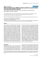
significantly (Table 3). An example of atherosclerosis
progression in a patient from the placebo group is
shown in Figure 1.
In the atorvastatin group, t here was no increase of
plaque volume (> 10 mm
3
) in any of the six patients
with deposits found at randomization. Also, the mean
coronary plaque volume and calcium score did not
change (Table 3).
The number of patients with perfusion defects and the
number of myocardial segments with pe rsistent or exer-
cise-induced defects in the SPECT study remained
unchanged during one-year observation in neither group
of patients studied (Table 4).
After one year of treatment, total serum cholesterol
decreased promptly by 13%, low-density lipoprotein
(LDL) cholesterol by 21%, triglycerides by 25% and CRP
concentration by 39% in the atorvastatin group, but
remained unchanged in the placebo group (Table 5).
There was no change in the activity of alanine amino-
transferase (ALT) and aspartate aminotransferase (AST)
nor creatine phosphokinase (CPK) in either group,
except for one patient in the placebo group (Table 5).
There was no need for atorvastatin discontinuation in
any of the patients.
Mean value of the SLEDAI score remained unchanged
in both groups (Table 5). During the t reatment period,
SLE flare (SLEDAI increase ≥ 3) was observed in two
patients from the atorvastatin group and in one from
the placebo group. In two atorvastatin group patients,
theSLEDAIincrease(from8to12andfrom4to8
points) resulted solely from the onset of hematuria. In
one patient from the placebo group, SLEDAI increase
Table 2 Baseline characteristics of the study patients by group
Placebo group
(n = 32)
Atorvastatin group
(n = 28)
P
Age (years) 41.4 ± 12.4 41.8 ± 13.4 ns
Gender (females/males) 30/2 24/4 ns
Arterial hypertension (n (%)) 2 (6.3%) 1 (3.6%) ns
Diabetes mellitus (n (%)) 0 (0%) 0 (0%) ns
Obesity (n (%)) 0 (0%) 0 (0%) ns
Tobacco smoking (n (%)) 1 (3.1%) 1 (3.6%) ns
Total cholesterol (mmol/l) 4.5 ± 0.8 5.1 ± 1.2 ns
LDL cholesterol (mmol/l) 2.6 ± 0.8 2.9 ± 1.0 ns
HDL cholesterol (mmol/l) 1.4 ± 0.3 1.4 ± 0.3 ns
Triglycerides (mmol/l) 1.2 ± 0.5 1.6 ± 0.6 < 0.05
CRP (mg/l) 4.0 ± 8.9 4.4 ± 4.1 ns
Number of patients with plaques in MDCT (n (%)) 9 (28.1%) 6 (21.4%) ns
Plaque volume (mm
3
) 35.2 ± 44.9 54.5 ± 62.4 ns
Calcium score 32.1 ± 39.1 44.8 ± 50.6 ns
Number of patients with perfusion defects in SPECT 18 (56.3%) 12 (42.9%) ns
Number of underperfused myocardial segments (median) 3 (9.4%) 3 (10.7%) ns
CRP, C-reactive protein; HDL, high-density lipoprotein; LDL, low-density lipoprotein; MDCT, multi-detector computed tomography; ns, not significant; SPECT, single
photon emission computed tomography.
Plazak et al. Arthritis Research & Therapy 2011, 13:R117
/>Page 4 of 9
(from 4 to 10 points) resulted from the onset of both
hematuria and pyuria.
Discussion
The major finding of this study is the inhibition of
atherosclerosis progression by atorvastatin in SLE
patients as evidenced by MDCT-based calcium scoring.
Toourknowledge,itisthefirstreportshowingsucha
beneficial effect of statin therapy in this population at
high risk of life-t hreatening cardiovascular complica-
tions. The volume of coronary calcified plaques was
stable in the active-treatment group, and increased sig-
nificantly in the placebo group. At the same time, cor-
onary calcium score increased significantly in the
placebo group only.
Patients with SLE suffer from premature atherosclero-
sis. Our study supports previously published data on
high frequency of myocardial perfusion defects in SLE
patients as demonstrated by th e SPECT study [ 26,27].
Perfusion defects were present in 50% of cases, despite
normal ECG recordings at rest and lack of any clinical
symptoms of myocardial ischemia. P redominantly per-
sistent perfusion abnormalities were detected. In most
of the patients, the number of underperfused left ventri-
cle segments was low. However, it has been already
established that the presence of even small perfusion
defects in the SPECT study strongly affects prognosis
[28,29]. Beside the presence of myocardial perfusion
defects, 25% of our asymptomatic SLE patients showed
calcified atherosclerotic changes in their coronary
arteries. It is the most frequent localization of such
changes in SLE, as shown in another study of 50 SLE
patients, where the frequency of atherosclerotic plaques
observed in MDCT were the highest in coronary arteries
(42% of patients with calcifications), followed by carotid
arteries (24% of patients with calcifications) [30]. A
study of 157 SLE patients showed that in subjects with
the mean age of 40 years - comparable with the age of
our patients - the frequency of coronary artery calcifica-
tionsis30to40%[31].Thispercentageisrelatively
higher than in the general population: in the study of
35,388 subjects calcium scores above 10 were observed
in only 10% of cases, and calcium scores above 100 in
2% [32]. Coronary calcium deposits provide an indepen-
dent indication of a short- and long-term risk of cardiac
events, even in patients with normal SPECT results
[33-35].
Our results support also the published data showing
higher frequenc y of myocardial perfusion abnormalities
detected by SPECT as compared with the frequency of
coronary calcium deposits detected by MDCT in SLE
population [26,27,31]. This might be partially explained
by the fact that antiphospholipid antibodies are asso-
ciated with thrombotic events in coronary beds, rather
than with subclinical atheroscleros is [36]. Thrombosis in
coronary arteries leads to perfusion defects detectable by
SPECT, but not by MDCT. Calcified plaques may
develop in time at the basis of thrombi or may f orm
due to endothelium dysfunction. In the present study
the patients with perfusion abnormalities despite the
lack of coronary calcifications were observed. On the
other hand, small coronary plaques may have no influ-
ence on the perfusion: the patients with normal perfu-
sion despite small calcium deposits in the arteries were
also observed.
The inhibition of atherosclerosis progression in SLE
patients by atorvastatin seems o f major importance for
their prognosis. In a seven-year prospective follow-up
studyinagroupof1,126otherwisehealthysubjects,
Chang et al. showed that the risk of myocardial infarc-
tion or the need for revascularization correlated with
the patients calcium score and occurred at higher fre-
quency in subjects with cal cium score above 100 [33].
In our study, the mean value of the calcium score was
lower (39.9 ± 50.9) and the follow-up period much
shorter, but the significant progression of atherosclerosis
Table 3 Coronary calcium score, number and volume of coronary plaques in SLE patients from the placebo group and
atorvastatin group at randomization and after one year of treatment
At randomization After one year P
Placebo group, n =32
Number of patients with plaques 9 (28.1%) 10 (31.3%) ns
Plaque volume (mm
3
) 35.2 ± 44.9 62.9 ± 72.4 < 0.05
Number of plaques 2-13 (median 4) 1-12 (median 5) ns
Calcium score 32.1 ± 39.1 59.5 ± 54.4 < 0.05
Atorvastatin group, n =28
Number of patients with plaques 6 (21.4%) 6 (21.4%) ns
Plaque volume (mm
3
) 54.5 ± 62.4 51.0 ± 47.6 ns
Number of plaques 2-4 (median 2) 1-8 (median 2) ns
Calcium score 44.8 ± 50.6 54.9 ± 62.5 ns
ns, not significant; SLE, systemic lupus erythematosus.
Plazak et al. Arthritis Research & Therapy 2011, 13:R117
/>Page 5 of 9
in the placebo group (increase of mean calcium score by
85.4% during one year) may have important clinical
implications for patients’ future.
Atorvastatin did not influence myocardial perfusion as
assessed by SPECT. Calcium deposits in coronary
arteries revealed by MDCT were obviously too small to
result in any significant persistent or exercise-induced
perfusion defects.
It has been shown that statins e xert not only anti-
lipid, but also marked anti-inflammatory effects [9].
Accordingly, in our study serum concentrations of total
cholesterol, LDL cholesterol, and triglycerides all
decreased after atorvastatin treatment. Importantly, this
was accompanied by the decrease in CRP despite an
unchanged immunosuppressive therapy. It was pre-
viously shown that the magnitude of protection and the
decrease in mortality afforded by statins cannot be
entirely explained b y their cholesterol-lowering effect
[10]. A large study of 3,745 patients showed that
patients who have low CRP levels after statin therapy
have better clinical outcome than those with higher
CRP levels, regardless of the resultant level of LDL cho-
lesterol decrease [9]. The ability of atorvastati n to l ower
CRP concentrations shown in this study is of major
importance for SLE patients, as an ongoing chronic
inflammation presents as the major mechanism of sys-
temic SLE complications.
Recently, the Lupus Atherosclerosis Prevention Study
has been completed [37], based on the methodology
similar to that described above. The authors found a
greater increase in coronary artery calcium score in the
placebo group, but due to a calcium score increase
observed also in the atorvastatin group, the int er-group
change was not statistically significant. There was, how-
ever, a significant difference in favor of atorvastatin in
the proportion of patients i n whom carotid intima-
media thickness improved, stayed the same, or got
worse. Surprisingly , during follow up, a greater decrease
of CRP level was observed in the placebo group as com-
pared with the atorvastatin group.
Statin therapy in SLE may b e complicated by the
reported cases of statin-induced lupus-like syndrome
[38-40]. Pathogenic mechanisms may include increased
cellular apoptosis induced by statins [41] and/or direct
immunomodulatory effect of statins on T lymphocytes
[42]. In our patients, no changes typical of any statin-
related adver se events were observed. Liver enzyme and
CPK levels were normal in all active-treated subjects.
There was also no other a dverse effects that would
require discontinuation of therapy.
(
a
)
(b)
aorta
aorta
Figure 1 The examples of multi-detector computed
tomography in a patient from the (a) placebo group at
randomization and (b) after one year. a) At randomization, two
calcified plaques are seen in left anterior descending artery (red
colour) and one calcified plaque in circumflex artery (blue colour).
Plaques volume 156.4 mm
3
, calcium score 138.9. b) After one year,
the volume of previously observed plaques increased with the new
calcification in distal part of left anterior descending artery. Plaques
volume 223 mm
3
, calcium score 202.5.
Plazak et al. Arthritis Research & Therapy 2011, 13:R117
/>Page 6 of 9
Our results may have important implications for the
management of SLE patients, because the presence of
atherosclerotic plaques detected by MDCT and myo-
cardial perfusion defects detected by SPECT are strong
predictors of death in other populations of patients
[28,29,33-35]. Possible beneficial effects of statin
treatment on prognosis of SLE patients should, how-
ever, be addressed in future large prospective clinical
trials.
Limitations of the study
Although the most commonly used marker o f coronary
atherosclerosis is calcium scoring, we also measured the
volume of calcified plaques in coronary arteries. This is
because a major limitation of Agatson calcium score
estimation is the measurement of calcium deposits area
and density measurement of the calcium (Hounsfield
units, HU) itself. The density is assessed using the
weigh ting factor in a stepwise manner, that is not linear
or continuous: for calcium measures 130 to 200 HU the
density score is one, for 200 to 300 HU the density
score is two, etc. [21]. The refore, small HU difference
may yield a m ajor Agatson score difference. Also, its
reproducibility is limited to ± 15 to 20%.
Although coron ary calcified plaques are proved to be
responsible for myocardial ischemia a nd myocardial
infarction, the other mechanisms of coronary flow
abnormalities in SLE population sho uld also be under-
lined. Endothelial damage and/or microthrombosis in
coronary bed related to antiphospholipid autoantibodies
[36,43,44] was discussed above.
Conclusions
The SPECT study showed myocardial perfusion defects
in 50% of SLE patients despite normal ECG recordings
and lack of clinical symptoms of myocardial ischemia.
In addition, 25% of patients showed atherosclerotic pla-
ques in coronary arteries.
Treatment with atorvastatin lead not only to the
decrease of serum lipids and CRP levels, but also to the
limitation of atherosclerosis p rogression as assessed by
MDCT-based calcium scoring. The def inite value of sta-
tin therapy in SLE patients free of clinical symptoms of
Table 4 Persistent and exercise-induced myocardial perfusion defects in SLE patients from placebo group and
atorvastatin group at randomization and after one year of treatment
At randomization After one year P
Placebo group, n =32
Number of patients with
persistent perfusion defects
14 (43.8%) 11 (34.3%) ns
Number of persistently underperfused segments 2-5 (median 3) 3-6 (median 3) ns
Number of patients with exercise-induced perfusion defects 4 (12.5%) 6 (18.8%) ns
Number of underperfused
myocardial segments at exercise
1-4 (median 3) 2-3 (median 3) ns
Atorvastatin group, n =28
Number of patients with persistent perfusion defects 8 (28.6%) 8 (28.6%) ns
Number of persistently underperfused segments 1-5 (median 3) 2-6 (median 3) ns
Number of patients with exercise-induced perfusion defects 4 (14.3%) 5 (17.9%) ns
Number of underperfused myocardial segments at exercise 2-4 (median 3) 3-6 (median 3) ns
ns, not significant; SLE, systemic lupus erythematosus.
Table 5 Biochemical data and SLEDAI score in SLE
patients from atorvastatin group and from placebo
group at randomization and after one year of treatment
At randomization After one year P
Atorvastatin group, n =28
Total cholesterol (mmol/l) 5.1 ± 1.2 4.4 ± 0.7 < 0.05
LDL cholestrol (mmol/l) 2.9 ± 1.0 2.3 ± 0.6 < 0.05
HDL cholesterol (mmol/l) 1.4 ± 0.3 1.4 ± 0.3 ns
Triglycerides (mmol/l) 1.6 ± 0.6 1.2 ± 0.5 < 0.05
CRP (mg/l) 4.4 ± 4.1 2.7 ± 1.7 < 0.05
ALT (IU/l) 23.9 ± 6.7 22.4 ± 6.9 ns
AST (IU/l) 22.9 ± 3.7 31.5 ± 6.2 ns
CPK (IU/l) 70.0 ± 78.2 62.9 ± 47.2 ns
SLEDAI 2-20 (median 4) 0-20 (median 4) ns
Placebo group, n =32
Total cholesterol (mmol/l) 4.5 ± 0.8 4.5 ± 0.7 ns
LDL cholestrol (mmol/l) 2.6 ± 0.8 2.6 ± 0.8 ns
HDL cholesterol (mmol/l) 1.4 ± 0.3 1.4 ± 0.3 ns
Triglycerides (mmol/l) 1.2 ± 0.5 1.3 ± 0.6 ns
CRP (mg/l) 4.0 ± 8.9 3.9 ± 5.1 ns
ALT (IU/l) 27.1 ± 8.6 39.1 ± 51.4* ns
AST (IU/l) 26.1 ± 6.2 40.2 ± 56.6* ns
CPK (IU/l) 53.2 ± 37.5 71.2 ± 57.2 ns
SLEDAI 0-12 (median 4) 0-12 (median 2) ns
* in one patient increased ALT (248 IU/l) and AST (273 IU/l) levels were
observed
CRP, C-reactive protein; ALT, alanine aminotransferase; AST, aspartate
aminotransferase ; CPK, creatine phosphokinase; HDL, high-density lipoprotein;
LDL, low-density lipoprotein; ns, not significant; SLE, systemic lupus
erythematosus; SLEDAI, Systemic Lupus Erythematosus Disease Activity Index.
Plazak et al. Arthritis Research & Therapy 2011, 13:R117
/>Page 7 of 9
cardiovascular disease should be ad dressed in l arge pro-
spective clinical trials.
Abbreviations
aCL: anticardiolipin antibodies; ANA: antinuclear antibodies; ALT: alanine
aminotransferase; APS: antiphospholipid syndrome; AST: aspartate
aminotransferase; CPK: creatine phosphokinase; CRP: C-reactive protein;
ELISA: enzyme linked immunosorbent assay; LA: lupus anticoagulant; LDL:
low-density lipoprotein; MDCT: multi-detector computed tomography; SLE:
systemic lupus erythematosus; SLEDAI: Systemic Lupus Erythematosus
Disease Activity Index; SPECT: single photon emission computed
tomography.
Acknowledgements
This study was supported by a grant No N40201231/0460 from the Polish
Ministry of Science and Higher Education.
Author details
1
Department of Cardiac and Vascular Diseases, the John Paul II Hospital,
Jagiellonian University Medical College, Pradnicka Str 80, 31-202 Krakow,
Poland.
2
Department of Internal Medicine, Jagiellonian University Medical
College, Skawinska Str 8, 31-066 Krakow, Poland.
3
Center for Diagnosis,
Prevention and Telemedicine, the John Paul II Hospital, Jagiellonian
University Medical College, Pradnicka Str 80, 31-202 Krakow, Poland.
Authors’ contributions
WP was responsible for the study concept and design, acquisition, analysis
and interpretation of the data, and manuscript preparation. KG, HD, LTP, and
MK acquired and analyzed the data. PP and JM were responsible for data
interpretation and manuscript preparation. All authors read and approved
the final version of the manuscript.
Competing interests
The authors declare that they have no competing interests.
Received: 13 January 2011 Revised: 9 May 2011 Accepted: 20 July 2011
Published: 20 July 2011
References
1. Bruce IN: “Not only but also": factors that contribute to accelerated
atherosclerosis and premature coronary heart disease in systemic lupus
erythematosus. Rheumatology 2005, 44:1492-1502.
2. Lopez-Pedrera Ch, Aguirre MA, Barbarroja N, Cuadrado MJ: Accelerated
atherosclerosis in systemic lupus erythematosus: role of
proinflammatory cytokines and therapeutic approaches. J Biomed
Biotechnol 2010, pii: 607084.
3. Pons-Estel GJ, Gonzales LA, Zhang J, Burgos Pl, Reveille JD, Vila LM,
Alarcon GS: Predictors of cardiovascular damage in patients with
systemic lupus erythematosus: data from LUMINA (LXVIII), a multicenter
US cohort. Rheumatology 2009, 48:817-822.
4. ALLHAT-LLT Officers and Coordinators: Major outcomes in moderately-
hypercholesterolemic, hypertensive patients randomized to pravastatin
vs. usual care: the antihypertensive and lipid-lowering treatment to
prevent heart attack trial (ALLHAT-LLT). JAMA 2002, 288:2998-3007.
5. Shepherd J, Cobbe SM, Ford I, Isles CG, Lorimer AR, MacFarlane PW,
McKillop JH, Packard CJ: Prevention of coronary heart disease with
pravastatin in men with hypercholesterolemia. West of Scotland
Coronary Prevention Study Group. N Engl J Med 1995, 333:1301-1307.
6. Ray KK, Cannon Ch, McCabe C, Cairns R, Tonkin A, Sacks F, Jackson G,
Braunwald E: Early and late benefits of high-dose atorvastatin in patients
with acute coronary syndromes. J Am Coll Cardiol 2005, 46:1405-1410.
7. Schwartz G, Olsson A, Ezekowitz M, Ganz P, Oliver M, Waters D, Zeiher A,
Chaitman B, Leslie S, Stern T: Effect of atorvastatin on early recurrent
ischaemic events in acute coronary syndromes. The MIRACL study: a
randomized controlled trial. JAMA 2001, 285:1711-1718.
8. Nissen SE, Nicholls SJ, Sipahi I, Libby P, Raichlen JS, Ballantyne CM,
Davignon J, Erbel R, Fruchart JC, Tardif JC, Schoenhagen P, Crowe T, Cain V,
Wolski K, Goormastic M, Tuzcu EM: Effect of very high-intensity statin
therapy on regression of coronary atherosclerosis: the ASTEROID trial.
JAMA 2006, 295:1556-1565.
9. Ridker PM, Cannon Ch, Morrow D, Rifai N, Rose L, McCabe C, Pfeffer M,
Braunwald E: C-reactive protein levels and outcomes after statin therapy.
New Engl J Med 2005, 352:20-28.
10. Masumoto A, Hirooka Y, Hironaga K, Eshima K, Setoguchi S, Egashira K,
Takeshita A: Effect of pravastatin on endothelial function in patients with
coronary artery disease (cholesterol-independent effect of pravastatin).
Am J Cardiol 2001, 88:1291-1294.
11. Mason JC, Ahmed Z, Mankoff R, Lidington EA, Ahmad S, Bhatia V,
Kinderlerer A, Randi AM, Haskard DO: Statin-induced expression of decay-
accelerating factor protects vascular endothelium against complement-
induced injury. Circ Res 2002, 91:696-703.
12. Giri S, Parke AL, Waters DD: Controlling cardiovascular risk factors in
systemic lupus erythematosus. J Musculoskel Med 1998, 15:42-52.
13. Urowitz MB, Gladman DD: How to improve morbidity and mortality in
systemic lupus erythematosus. Rheumatology (Oxford) 2000, 39:238-244.
14. Samon JE, Roman MJ:
Accelerated atherosclerosis in systemic lupus
erythematosus: implications for patient management. Curr Opin
Rheumatol 2001, 13:341-344.
15. Noel B: Risks and benefits of statins in lupus erythematosus. Arch Intern
Med 2004, 164:107-108.
16. Bruce IN: Cardiovascular disease in lupus patients: should all patients be
treated with statins and aspirin? Best Pract Res Clin Rheumatol 2005,
19:823-838.
17. Smolen J, Weisman M: Connective tissue disorders. In Rheumatology.
Edited by: Hochberg M, Silman A, Smolen J, Weinblatt M, Weisman M.
Philadelphia, Mosby Elsevier; 2008:1205-1485.
18. Smith EL, Shmerling RH: The American College of Rheumatology criteria
for the classification of systemic lupus erythematosus: strengths,
weaknesses, and opportunities for improvement. Lupus 1999, 8:586-595.
19. Smilde TJ, van Wissen S, Wollersheim H, Trip MD, Kastelein JJ,
Stalenhoef AF: Effect of aggressive versus conventional lipid lowering on
atherosclerosis progression in familial hypercholesterolaemia (ASAP): a
prospective, randomized, double-blind study. Lancet 2001, 357:577-581.
20. Taylor AJ, Kent SM, Flaherty PJ, Coyle LC, Markwood TT, Vemalis MN:
ARBITER: arterial biology for the investigation of the treatment effects of
reducing cholesterol. A randomized trial comparing the effects of
atorvastatin and pravastatin on carotid intima media thickness.
Circulation 2002, 106:2055-2060.
21. Agatson AS, Janowitz WR, Hildner FJ, Zusmer NR, Viamonte M Jr, Detrano R:
Quantification of coronary artery calcium using ultrafast computed
tomography. J Am Coll Cardiol 1990, 15:827-832.
22. Swadzba J, de Clerck LS, Stevens WJ, Bridts CH, van Cotthem KA, Musial J,
Jankowski M, Szczeklik A: Anticardiolipin antibodies, anti-β2-glycoprotein
I, antiprothrombin antibodies and lupus anticoagulant in patients with
systemic lupus erythematosus with a history of thrombosis. J Rheumatol
1997, 24:1710-1715.
23. Pengo V, Tripodi A, Reber G, Rand JH, Ortel TL, Galli M, De Groot PG:
Update of the guidelines for lupus anticoagulant detection.
Subcommittee on Lupus Anticoagulant/Antiphospholipid Antibody of
the Scientific and Standarisation Committee of the International Society
on Thrombosis and Haemostasis. J Thromb Haemost 2009, 7:1737-1740.
24. Miyakis S, Lockshin MD, Atsumi T, Branch DW, Brey RL, Cervera R,
Derksen RH, De Groot PG, Koike T, Meroni PL, Reber G, Shoenfeld Y,
Tincani A, Vlachoyiannopoulos PG, Krilis SA: International consensus
statement on an update of the classification criteria for definite
antiphospholipid syndrome (APS). J Thromb Heamost 2006, 4:295-306.
25. Permarheum SLEDAI Calculator. [ />html].
26. Lin CC, Ding HJ, Chen YW, Wang JJ, Ho ST, Kao A: Usefulness of
technetium-99 m sestamibi myocardial perfusion SPECT in detection of
cardiovascular involvement in patients with systemic lupus
erythematosus or systemic sclerosis. Int J Cardiol 2003, 92:157-161.
27. Lin JJ, Hsu HB, Sun SS, Wang JJ, Ho ST, Kao CH: Single Photon Emission
Computed Tomography of technetium-99 m tetrofosmin myocardial
perfusion imaging in patients with systemic lupus erythematosus - a
preliminary report. Jpn Heart J 2003, 44:83-89.
28. Vanzetto G, Ormezzano O, Fagret D, Comet M, Denis B, Machecourt J: Long
term additive prognostic value of thalium-201 myocardial perfusion
imaging over clinical and exercise stress test in low to intermediate risk
Plazak et al. Arthritis Research & Therapy 2011, 13:R117
/>Page 8 of 9
patients: study in 1137 patients with 6-year follow-up. Circulation 1999,
100:1521-1527.
29. Hachamovitch R, Berman DS, Shaw LJ, Kiat H, Cohen I, Cabico JA,
Friedman J, Diamond GA: Incremental prognostic value of myocardial
perfusion single photon emission computed tomography for the
prediction of cardiac death: differential stratification for risk of cardiac
death and myocardial infarction. Circulation 1998, 97:535-543.
30. Yiu KH, Wang S, Mok MY, Ooi GC, Khong PL, Mak KF, Lam KF, Lau CS,
Tse HF: Pattern of arterial calcification in patients with systemic lupus
erythematosus. J Rheumatol 2009, 36:2212-2217.
31. Kao AH, Wasko MCM, Krishnaswami S, Wagner J, Edmundowicz D, Shaw P,
Cunningham AL, Danchenko N, Sutton-Tyrrell K, Tracy RP, Kuller LH,
Manzi S: C-reactive protein and coronary artery calcium in asymptomatic
women with systemic lupus erythematosus or rheumatoid arthritis. Am J
Cardiol 2008, 102:755-760.
32. Raggi P, Gongora M, Gopal A, Callister T, Budoff M, Shaw L: Coronary
artery calcium to predict all-cause mortality in elderly men and women.
J Am Coll Cardiol 2008, 52:17-23.
33. Chang SM, Nabi F, Xu J, Peterson LE, Achari A, Pratt CM, Mahmarian JJ: The
coronary artery calcium score and stress myocardial perfusion imaging
provide independent and complementary prediction of cardiac risk. J
Am Coll Cardiol 2009, 54:1872-1882.
34. Polonsky TS, McClelland RL, Jorgensen NW, Bild DE, Burke GL, Guerci AD,
Greenland P: Coronary artery calcium score and risk classification for
coronary heart disease prediction. JAMA 2010, 303:1610-1616.
35. Uebleis C, Becker A, Griesshammer I, Cumming P, Becker C, Schmidt M,
Barterstein P, Hacker M: Stable coronary artery disease: prognostic value
of myocardial perfusion SPECT in relation to coronary calcium scoring -
long-term follow-up. Radiology 2009, 252:682-690.
36. Petri M: The lupus anticoagulant is a risk factor for myocardial infarction
(but not atherosclerosis): Hopkins Lupus Cohort. Thromb Res 2004,
114:593-595.
37. Petri M, Kiani A, Post W, Christopher-Stine L, Madger L: Lupus
Atherosclerosis Prevention Study (LAPS). Ann Rheum Dis 2011, 70:760-765.
38. Bannwarth B, Miremont G, Papapietro PM: Lupuslike syndrome associated
with simvastatin. Arch Intern Med 1992, 152:1093.
39. Hanson J, Bossingham D: Lupus-like syndrome associated with
simvastatin. Lancet 1998, 352:1070.
40. Srivastana M, Rencic A, Diglio G, Santana H, Bonitz P, Watson R, Ha E,
Anhalt GJ, Provost TT, Nousari CH: Drug-induced, Ro/SSA-positive
cutaneous lupus erythematosus. Arch Dermatol 2003, 139:45-49.
41. Noel B:
Statins and lupus erythematosus. Rheumatology 2004, 43:397-398.
42. Kwak B, Mulhaupt F, Myit S, Mach F: Statins as a newly recognized type of
immunomodulator. Nature Med 2000, 6:1399-1400.
43. Long BR, Leya F: The role of antiphospholipid syndrome in
cardiovascular disease. Hematol Oncol Clin N Am 2008, 22:79-94.
44. Alexanderson E, Gomez-Leon A, Vargas A, Romero JL, Sierra Fernandez C,
Rodriguez Valero M, Garcia Rojas L, Meave A, Amigo MC: Myocardial
ischaemia in patients with primary APS: a
13
N-ammonia PET assessment.
Rheumatology 2008, 47:894-896.
doi:10.1186/ar3402
Cite this article as: Plazak et al.: Influence of atorvastatin on coronary
calcifications and myocardial perfusion defects in systemic lupus
erythematosus patients: a prospective, randomized, double-masked,
placebo-controlled study. Arthritis Research & Therapy 2011 13:R117.
Submit your next manuscript to BioMed Central
and take full advantage of:
• Convenient online submission
• Thorough peer review
• No space constraints or color figure charges
• Immediate publication on acceptance
• Inclusion in PubMed, CAS, Scopus and Google Scholar
• Research which is freely available for redistribution
Submit your manuscript at
www.biomedcentral.com/submit
Plazak et al. Arthritis Research & Therapy 2011, 13:R117
/>Page 9 of 9