A TEXTBOOK OF POSTPARTUM HEMORRHAGE - PART 2 potx
Bạn đang xem bản rút gọn của tài liệu. Xem và tải ngay bản đầy đủ của tài liệu tại đây (1.72 MB, 50 trang )
total there were 266 cases of postpartum hemor
-
rhage, representing a near-miss postpartum
hemorrhage rate of 7.9/1000 deliveries.
Prual and colleagues examined severe mater
-
nal morbidity from direct obstetric causes in
West Africa between 1994 and 1996
32
. A severe
obstetric event was defined as prepartum,
peripartum or postpartum hemorrhage leading
to blood transfusion, or hospitalization for more
than 4 days or to hysterectomy. A total of 1307
severe maternal morbidity events were identi-
fied, with obstetric hemorrhage representing
the largest group involving 601 cases, 342 of
which were postpartum hemorrhage. The near-
miss obstetric hemorrhage rate was 30.5 (CI
28.1–33.0)/1000 live births and the near-miss
postpartum hemorrhage rate was 17.4 (CI
15.6–19.3)/1000 live births.
The Pretoria region of South Africa has
used the same definition of ‘near miss’ for
over 5 years, allowing comparison of temporal
changes
33
. Rates per 1000 births for near misses
plus maternal deaths over 5 years from severe
postpartum hemorrhage are shown in Table 4.
These rates are not dissimilar to those in
Canada or the UK.
ETIOLOGY AND PRECIPITATING
FACTORS
Causes of primary postpartum
hemorrhage
In recent years, individual authors and aca
-
demic groups have used the Four Ts pneu
-
monic to provide a simplistic categorization of
the causes of postpartum hemorrhage. This is
shown in Table 5
34
.
Uterine atony
Uterine atony, the most common cause of
postpartum hemorrhage, is reported in 70%
of cases
34
. It can occur after normal vaginal
delivery, instrumental vaginal delivery and
abdominal delivery. A large cohort study found
an incidence of uterine atony after primary
Cesarean section of 1416/23 390 (6%)
35
. Multi
-
ple linear regression analysis demonstrates the
following factors as being independently associ
-
ated with risk of uterine atony: multiple gesta
-
tion (odds ratio (OR) 2.40, 95% CI 1.95–2.93),
Hispanic race (OR 2.21, 95% CI 1.90–2.57),
induced or augmented labor for > 18 h (OR
2.23, 95% CI 1.92–2.60), infant birth weight
> 4500 g (OR 2.05, 95% CI 1.53–2.69), and
clinically diagnosed chorioamnionitis (OR 1.80,
95% CI 1.55–2.09).
Surprisingly, it is much more difficult to find
comparable studies of risk factors for uterine
23
Vital statistics
Number
of cases
(1991–2000)
Rate per 1000
deliveries
(95% CI)
Rate per 1000
deliveries
(1991–1993)
Rate per 1000
deliveries
(1998–2000)
Relative
risk
(95% CI)*
PPH requiring
transfusion
2317 0.91 (0.87–0.95) 1.27 0.63 0.5 (0.44–0.55)
PPH requiring
hysterectomy
892 0.35 (0.33–0.37) 0.26 0.46 1.76 (1.48–2.08)
*The 1991–1993 period was the reference period
Ta bl e 3
Postpartum hemorrhage (PPH) rates in Canada 1991–2000. Adapted from Wu Wen
30
1997–99 2000 2001 2002
Rate/1000 births 0.96 1.37 2.38 2.28
Ta bl e 4 Rates per 1000 births for near misses plus
maternal deaths from severe postpartum hemor
-
rhage in Pretoria. Adapted from Pattinson et al.
33
Tone – uterine atony
Trauma – of any part of the genital tract, inverted
uterus
Tissue – retained placenta, invasive placenta
Thrombin – coagulopathy
Ta bl e 5 The Four Ts of postpartum hemorrhage
(from ALSO
34
)
45
Z:\Sapiens Publishing\A5211 - Postpartum Hemorrhage\Make-up\Postpartum Hemorrhage - Voucher Proofs #T.vp
30 August 2006 14:19:10
Color profile: Generic CMYK printer profile
Composite Default screen
atony in women achieving vaginal delivery. A
single center, case-control study from Pakistan
reporting on women who had either assisted or
non-assisted vaginal delivery found only two
factors had a strong association with uterine
atony: gestational diabetes mellitus (OR 7.6,
95% CI 6.9–9.0) and prolonged second stage
of labor in multiparas (OR 4.0, 95% CI
3.1–5.0)
36
. They found no association with
high parity, age, pre-eclampsia, augmentation
of labor, antenatal anemia and a history of poor
maternal or perinatal outcomes.
Trauma
Trauma is reported to be the primary cause of
postpartum hemorrhage in 20% of cases
34
(see
also Chapter 9). Genital tract trauma at delivery
is associated with an odds ratio of 1.7 (95% CI
1.4–2.1) for postpartum hemorrhage (measured
blood loss > 1000 ml)
37
. Similar results were
found in a Dutch study with a reported OR of
1.82 (CI 1.01–3.28) for postpartum hemor-
rhage (≥ 1000 ml) with perineal trauma ≥ first-
degree tears
38
. Trauma to the broad ligament,
uterine rupture, cervical and vaginal tears and
perineal tears are all associated with increased
blood loss at normal vaginal delivery.
Inversion of the uterus is a rare cause of
postpartum hemorrhage (see Chapter 9). The
incidence of inversion varies from 1 in 1584
deliveries in Pakistan
39
to around 1 in 25 000
deliveries in the USA, UK and Norway
40
. Blood
loss at delivery with a uterine inversion is usually
at least 1000 ml
41
, with 65% of uterine inver
-
sions being complicated by postpartum hemor
-
rhage and 47.5% requiring blood transfusion in
a large series of 40 cases
42
.
Tissue
Retained placenta accounts for approximately
10% of all cases of postpartum hemorrhage
34
.
Effective uterine contraction to aid hemostasis
requires complete expulsion of the placenta.
Most retained placentas can be removed manu
-
ally, but rarely the conditions of placenta per
-
creta, increta, and accreta may be responsible for
placental retention (see Chapters 24 and 36).
Retained placenta occurs after 0.5–3% of deliv
-
eries
43
. Several case–control and cohort studies
show that retained placenta is associated with
increased blood loss and increased need for
blood transfusion. Stones and colleagues
reported that retained placenta had a RR of 5.15
(99% CI 3.36–7.87) for blood loss ≥ 1000 ml
within the first 24 h of delivery
44
.Baisandcol
-
leagues found an incidence of 1.8% for retained
placenta in Holland
38
. Using multiple regression,
these authors determined that retained placenta
was associated with an OR of 7.83 (95% CI
3.78–16.22) and 11.73 (95% CI 5.67–24.1) for
postpartum hemorrhage of ≥ 500 ml and
postpartum hemorrhage ≥ 1000 ml, respectively.
In addition, retained placenta was found to have
an OR of 21.7 (95% CI 8.9–53.2) for red cell
transfusion in this Dutch cohort.
Tanberg and colleagues reported an inci
-
dence of retained placentas of 0.6% in a large
Norwegian cohort of 24 750 deliveries and
showed that hemoglobin fell by a mean of
3.4 g/dl in the retained placental group com-
pared to no fall in the controls
45
. In addition,
blood transfusion was required in 10% of the
retained placental group but only 0.5% of the
control group. A similar incidence of retained
placenta was found in a Saudi Arabian case–
control study which demonstrated increased
blood loss in women with a retained placenta
(mean 437 ml) compared with controls (mean
263 ml)
46
. A large study from Aberdeen of over
36 000 women reported postpartum hemor
-
rhage in 21.3% of women with retained pla
-
centa compared to 3.5% in vaginal deliveries
without retained placenta
47
. Both studies con
-
firmed that women with a history of retained
placenta have an increased risk of recurrence
in subsequent pregnancies
46,47
. In the study by
Adelusi and colleagues, 6.1% of the patients
with retained placenta had a prior history of
retained placenta, compared to none in their
control group of normal vaginal deliveries
46
.
Placental accreta is a rare and serious compli
-
cation, occurring in about 0.001–0.05% of all
deliveries
48,49
. Makhseed and colleagues found
an increasing risk for accreta with increasing
numbers of Cesarean sections (OR 4.11, 95%
CI 0.83–19.34) after one previous Cesarean
section and an OR of 30.25 (95% CI 9.9–92.4)
after two previous Cesarean sections, compared
with no previous Cesarean section. Kastner
and colleagues found that placenta accreta was
24
POSTPARTUM HEMORRHAGE
46
Z:\Sapiens Publishing\A5211 - Postpartum Hemorrhage\Make-up\Postpartum Hemorrhage - Voucher Proofs #T.vp
30 August 2006 14:19:10
Color profile: Generic CMYK printer profile
Composite Default screen
implicated in 49% of their 48 cases of emer
-
gency hysterectomy
50
. Zaki and co-workers
found an incidence of 0.05% of placenta accreta
in a population of 23 000 women
49
. They found
that rates of postpartum hemorrhage and emer
-
gency hysterectomy were higher in the accreta
group compared to the placenta previa group
undergoing Cesarean section. Postpartum hem
-
orrhage occurred in 91.7% of the accreta group
compared to 18.4% of the previa group (OR
48.9, 95% CI 5.93–403.25), whereas 50% of
accreta cases required emergency hysterectomy
compared to 2% in the previa group (OR 48,
95% CI 7.93–290.48). Within the accreta
group, 75% of patients had a previous history of
Cesarean section, compared to 27.5% in the
previa group (OR 7.9, 95% CI 1.98–31.34).
Thrombin
Disorders of the clotting cascade and platelet
dysfunction are the cause of postpartum hemor-
rhage in 1% of cases
34
. Known associations with
coagulation failure include placental abruption,
pre-eclampsia, septicemia and intrauterine
sepsis (see Chapter 44), retained dead fetus,
amniotic fluid embolus, incompatible blood
transfusion, abortion with hypertonic saline and
existing coagulation abnormalities
4,51,52
(see
Chapter 25).
ANTENATAL RISK FACTORS FOR
PRIMARY POSTPARTUM
HEMORRHAGE
Age
Increasing maternal age appears to be an inde
-
pendent risk factor for postpartum hemorrhage.
In Japan, Ohkuchi and colleagues studied
10 053 consecutive women who delivered a
singleton infant
53
. Excessive blood loss (≥ 90th
centile) was defined separately for vaginal and
Cesarean deliveries (615 ml and 1531 ml,
respectively). On multivariate analysis, age ≥ 35
years was an independent risk factor for post
-
partum hemorrhage in vaginal deliveries (OR
1.5, 95% CI 1.2–1.9) and Cesarean deliveries
(OR 1.8, 95% CI 1.2–2.7). In Nigeria, Tsu
reported that advanced maternal age (≥ 35
years) was associated with an adjusted RR of 3.0
(95% CI 1.3–7.3) for postpartum hemorrhage
(defined as visual estimation of ≥ 600 ml)
54
.
Ijaiya and co-workers in Nigeria found that the
risk of postpartum hemorrhage in women > 35
years was two-fold higher compared to women
< 25 years, although no consideration of con
-
founding was made in this study
55
. Rates of
obstetric hysterectomy have also been reported
to increase with age; Okogbenin and colleagues
in Nigeria reported an increase from 0.1% at 20
years to 0.7% at ≥ 40 years
56
. However, others
have found no relationship between delaying
childbirth and postpartum hemorrhage
57
.
Ethnicity
Several studies have examined whether ethnic
-
ity is a factor for postpartum hemorrhage.
Magann and co-workers, using a definition of
postpartum hemorrhage of measured blood loss
> 1000 ml and/or need for transfusion
37
, found
Asian race to be a risk factor (OR 1.8, 95%
CI 1.4–2.2)). Other studies have observed
similar findings in Asians
58
(OR 1.73, 95% CI
1.20–2.49) and Hispanic races (OR 1.66, 95%
CI 1.02–2.69)
58
(OR for hematocrit < 26%,
3.99, 95% CI 0.59–9.26)
59
.
Body mass index
Women who are obese have higher rates of
intrapartum and postpartum complications.
Usha and colleagues performed a population-
based observational study of 60 167 deliveries
in South Glamorgan, UK; women with a body
mass index (BMI) > 30 had an OR of 1.5 (95%
CI 1.2–1.8) for blood loss > 500 ml, compared
to women with a BMI of 20–30
60
. Stones and
colleagues reported a RR for major obstetric
hemorrhage of 1.64 (95% CI 1.24–2.17) when
the BMI was 27+
44
.
Parity
Although grand multiparity has traditionally
been thought of as risk factor for postpartum
hemorrhage, Stones and colleagues and
Selo-Ojeme did not demonstrate any relation
between grand multiparity and major obstetric
hemorrhage
44,61
. This observation was con
-
firmed in a large Australian study which used
25
Vital statistics
47
Z:\Sapiens Publishing\A5211 - Postpartum Hemorrhage\Make-up\Postpartum Hemorrhage - Voucher Proofs #T.vp
30 August 2006 14:19:10
Color profile: Generic CMYK printer profile
Composite Default screen
multivariate logistic regression analysis and
found no association between grand multiparity
(≥ five previous births) and postpartum hemor
-
rhage (> 500 ml)
62
. Tsu reported an association
with low parity (0–1 previous birth) with
adjusted RR without intrapartum factors of
1.7 (95% CI 1.1–2.7) and adjusted RR with
intrapartum factors of 1.5 (95% CI 0.95–2.5)
but not with grand multiparity (defined as five
or more births)
54
. Ohkuchi also found primi
-
parity to be associated with excessive blood loss
at vaginal delivery (OR 1.6, 95% CI 1.4–1.9)
53
.
Studies from Pakistan
63
and Nigeria
55
have
reported an association between grand multi
-
parity and postpartum hemorrhage, but both
studies failed to account for other confounding
factors such as maternal age.
Other medical conditions
Several medical conditions are associated with
postpartum hemorrhage. Women with type II
diabetes mellitus have an increased incidence of
postpartum hemorrhage of > 500 ml (34%)
compared to the non-diabetic population
(6%)
64,65
. Connective tissue disorders such as
Marfans and Ehlers-Danlos syndrome have also
been associated with postpartum hemor-
rhage
66,67
. Blood loss at delivery is also
increased with inherited coagulopathies
52
. The
most common inherited hemorrhagic disorder
is von Willebrand’s disease, with a reported
prevalence of between 1 and 3%. Most (70%)
have Type 1 disease characterized by low
plasma levels of factor VIII, von Willebrand fac
-
tor antigen, and von Willebrand factor activity.
Less common inherited bleeding disorders
include carriage of hemophilia A (factor VIII
deficiency) or hemophilia B (factor IX defi
-
ciency) and factor XI deficiency. In their review,
Economaides and colleagues suggest that the
risks of primary postpartum hemorrhage in
patients with von Willebrand’s disease, factor
XI deficiency, and carriers of hemophilia are
22%, 16%, and 18.5%, respectively, compared
with 5% in the general obstetric population
52
.
James also reviewed the numerous case series
and the more limited case–control studies of
women with bleeding disorders and came to
similar conclusions
68
(see Chapter 25).
Prolonged pregnancy
A large Danish cohort study compared a post-
term group (gestational age ≥ 42 weeks or
more) of 77 956 singleton deliveries and a term
group of 34 140 singleton spontaneous deliver
-
ies
69
. Adjusted odds ratio for postpartum
hemorrhage was 1.37 (95% CI 1.28–1.46),
suggesting an association between prolonged
pregnancy and postpartum hemorrhage.
Fetal macrosomia
Several studies confirm that fetal macrosomia is
associated with postpartum hemorrhage. Jolly
and colleagues examined 350 311 completed
singleton pregnancies in London
70
. Linear
regression analysis suggested that a birth weight
> 4 kg was better at predicting maternal mor
-
bidity than birth weight > 90th centile. Post
-
partum hemorrhage was increased in women
with fetal macrosomia (OR 2.01; 95% CI
1.93–2.10). In a large cohort of 146 526
mother–infant pairs in California, Stotland and
co-workers also demonstrated an adjusted OR
for postpartum hemorrhage of 1.69 (95% CI
1.58–1.82) in infants of 4000–4499 g compared
to 2.15 (95% CI 1.86–2.48) and 2.03 (95% CI
1.33–3.09) with weights of 4500–4999 g and
≥ 5000 g, respectively
71
. In Nigeria, a case–
control study of 351 infants weighing > 4 kg
with 6563 term infants found an incidence
of postpartum hemorrhage of 8.3% and
2.1%, respectively
72
. Bais and colleagues, in
their Dutch study, also demonstrated an
increase in risk for postpartum hemorrhage
(≥ 500 ml) and severe postpartum hemorrhage
(≥ 1000 ml) with infants with weights ≥ 4kg
(OR 2.11, 95% CI 1.62–2.76 and 2.55, 95%
CI 1.5–4.18)
38
.
Multiple pregnancies
Epidemiological studies suggest twins and
higher-order pregnancies are at increased risk for
postpartum hemorrhage. Walker and co-workers
conducted a retrospective cohort study involving
165 188 singleton pregnancies and 44 674 multi
-
ple pregnancies in Canada
73
. Multiple pregnan
-
cies were associated with an increased risk for
postpartum hemorrhage (RR 1.88, 95% CI
26
POSTPARTUM HEMORRHAGE
48
Z:\Sapiens Publishing\A5211 - Postpartum Hemorrhage\Make-up\Postpartum Hemorrhage - Voucher Proofs #T.vp
30 August 2006 14:19:11
Color profile: Generic CMYK printer profile
Composite Default screen
1.81–1.95), hysterectomy (RR 2.29, 95% CI
1.66–3.16) and blood transfusion (RR 1.67,
95% CI 1.13–2.46). Several other studies have
estimated the RR of postpartum hemorrhage
associated with multiple pregnancies to be
between 3.0 and 4.5
44,58,74
. Bais and colleagues,
in a Dutch population-based cohort study of
3464 women, used multiple regression analysis
and found that the OR for postpartum hemor
-
rhage ≥ 500 ml for multiple pregnancy was 2.6
(95% CI 1.06–-6.39)
38
. Albrecht and co-workers
conducted a retrospective review of 57 triplet
deliveries and found an incidence of 12.3% for
postpartum hemorrhage requiring transfusion
75
,
and a case series of 71 quadruplet pregnancies
conducted by Collins and colleagues estimated
that the frequency of postpartum hemorrhage
and transfusion to be 21% (95% CI 11–31%)
and 13% 95% CI 5–21%), respectively
76
.
MagannandcolleaguesdemonstratedanORfor
postpartum hemorrhage of 2.2 (95% CI 1.5–3.2)
in multiple pregnancies
37
, and Stones and col-
leagues showed a relative risk of 4.46 (95% CI
3.01–6.61) for obstetric hemorrhage with
multiple pregnancies
44
.
Fibroids
Obstetric textbooks suggest that leiomyomas
can be a cause of postpartum hemorrhage. This
is mainly based on case reports
77
, but one
cohort study of 10 000 women in Japan found
that women with leiomyomas had an OR of 1.9
(95% CI 1.2–3.1) and 3.6 (95% CI 2.0–6.3) for
excessive blood loss at vaginal and Cesarean
delivery, respectively
53
.
Antepartum hemorrhage
Antepartum hemorrhage has been linked to
postpartum hemorrhage risk with an OR of 1.8
(95% CI 1.3–2.3)
37
. Stones and co-workers
found a RR for major obstetric hemorrhage
(> 1000 ml) of 12.6 (95% CI 7.61–20.9), 13.1
(95% CI 7.47–23) and 11.3 (95% CI
3.36–38.1) for proven abruption, previa with
bleeding, and previa with no bleeding, respec
-
tively
44
. Ohkuchi and colleagues, in their
10 000 women, demonstrated that a low-lying
placenta was associated with odds ratios of 4.4
(95% CI 2.2–8.6) and 3.3 (95% CI 1.4–7.9) for
excess blood loss at the time of vaginal and
Cesarean delivery, respectively
53
. This study
also reported that placenta previa was associ
-
ated with an OR of 6.3 (95% CI 4.0–9.9) for
excessive blood loss at Cesarean delivery.
Previous history of postpartum
hemorrhage
Magann and colleagues found previous post
-
partum hemorrhage to be associated with
an increased risk for subsequent postpartum
hemorrhage (OR 2.2, 95% CI 1.7–2.9)
37
.
Previous Cesarean delivery
The Japanese study demonstrated an odds ratio
of 3.1 (95% CI 2.1–4.4) for excessive blood loss
at vaginal delivery in women with a previous
Cesarean section
53
.
INTRAPARTUM RISK FACTORS
FOR PRIMARY POSTPARTUM
HEMORRHAGE
Induction of labor
Meta-analysis of trials of induction of labor at or
beyond term indicates that induction does not
increase Cesarean section or operative vaginal
delivery rates
78
. However, this meta-analysis did
not examine blood loss at delivery. Epidemio
-
logical studies suggest a link between induction
of labor and postpartum hemorrhage. Brinsden
and colleagues reviewed 3674 normal deliveries
and found that the incidence of postpartum
hemorrhage was increased after induction of
labor
79
; among primipara, the incidence was
nearly twice that of spontaneous labor, even
when only normal deliveries were considered.
The study of Magann and colleagues suggested
an OR of 1.5 (95% CI 1.2–1.7) for postpartum
hemorrhage after induction of labor
37
and Bais
and co-workers found an OR of 1.74 (95% CI
1.06–2.87) for severe postpartum hemorrhage
of > 1000 ml after induction of labor
38
.
Tylleskar and colleagues performed a pro
-
spective, randomized, control trial of term
induction of labor with amniotomy plus
oxytocin versus waiting for spontaneous labor
in 84 women and found no difference in the
27
Vital statistics
49
Z:\Sapiens Publishing\A5211 - Postpartum Hemorrhage\Make-up\Postpartum Hemorrhage - Voucher Proofs #T.vp
06 September 2006 16:51:58
Color profile: Generic CMYK printer profile
Composite Default screen
amount of bleeding at the third stage
80
.A
Cochrane review
81
of amniotomy versus vaginal
prostaglandin for induction of labor reported
no difference in postpartum hemorrhage rates.
Another Cochrane
82
review of amniotomy plus
intravenous oxytocin included only one
placebo-controlled trial, but no data on post
-
partum hemorrhage were reported. This review
compared amniotomy plus intravenous oxy
-
tocin against vaginal prostaglandin (two trials,
160 women) and found a higher rate of
postpartum hemorrhage in the amniotomy/
oxytocin group (13.8% vs. 2.5% respectively,
RR 5.5, 95% CI 1.26–24.07)
82
.
A review of intravenous oxytocin alone for
cervical ripening
83
found no difference in
postpartum hemorrhage rates compared to the
placebo/expectant management group (three
trials, 2611 women; RR 1.24, 95% CI
0.85–1.81) or vaginal PGE
2
(four trials, 2792
women; RR 1.02, 95% CI 0.75–-1.4). Use of
mechanical methods to induce labor
84
was not
associated with any difference in postpartum
hemorrhage rates when compared to placebo
(one study, 240 women, RR 0.46, 95% CI
0.09–2.31), prostaglandin vaginal PGE
2
(one
study, 60 women, RR 3.0, 95% CI 0.33–27.24),
intracervical PGE
2
(three studies, 3339 women,
RR 0.91, 95% CI 0.40–2.11), misoprostol (one
study, 248 women, RR 2.34, 95% CI
0.46–11.85) or to oxytocinon alone (one study,
60 patients, RR 1.0, 95% CI 0.22–4.56).
Meta-analysis
85
of trials of membrane sweep
-
ing for induction of labor found a reduction in
postpartum hemorrhage compared to no inter
-
vention (three trials, 278 women, RR 0.31, 95%
CI 0.11–0.89). A review of oral misoprostol for
induction of labor
86
did not include any trial
that compared this agent with placebo. How
-
ever, one trial reported in this review, involving
692 women and using PGE
2
in the control arm,
found no difference in postpartum hemorrhage
rate (RR 0.98, 95% CI 0.73–1.31). Other
reviews of induction of labor methods have
reported no difference in postpartum hemor
-
rhage rates between vaginal misoprostol when
compared to placebo (two trials, 107 women,
RR 0.91, 95% CI 0.13–6.37)
87
, vaginal prosta
-
glandins (five trials, 1002 women, RR 0.88,
95% CI 0.63–1.22), intracervical prosta
-
glandins (two trials, 172 women, RR 1.62, 95%
CI 0.22–12.19), or with oxytocin (two trials,
245 women, RR 0.51, 95% CI 0.16–1.66).
Finally, a review of vaginal PGE
2
for induction
of labor suggested an increased risk of post
-
partum hemorrhage compared to placebo
88
(eight studies, 3437 women, RR 1.44, 95% CI
1.01–2.05).
Duration of labor
First stage
Compared with the second stage of labor, lim
-
ited evidence is available regarding the influence
of the duration of the first stage of labor on
postpartum hemorrhage
89
. Magann and col
-
leagues defined a prolonged first stage of labor
as a latent phase of > 20 h in nulliparous and
> 14 h in multiparous and/or an active phase of
< 1.2 cm per hour in nulliparous and < 1.4 cm
in multiparous patients
37
. These investigators
found an OR of 1.6 for prolonged first stage of
labor but the 95% CI ranged from 1 to 1.6.
Second stage
Several large studies have explored the relation-
ship between the length of the second stage
and adverse maternal and neonatal outcomes.
Cohen analyzed obstetric data from 4403
nulliparas and found an increase in postpartum
hemorrhage rate after more than 3 h in the
second stage
90
. He attributed this to the
increased need for mid-forceps delivery. A large
retrospective study involving 25 069 women in
spontaneous labor at term with a cephalic pre
-
sentation found that second-stage duration had
a significant independent association with the
risk of postpartum hemorrhage
91
. A more recent
retrospective cohort study of 15 759 nulliparous
term, cephalic singleton births in San Francisco
divided the second stage of labor into 1-h inter
-
vals
92
. Postpartum hemorrhage was defined as
estimated blood loss of > 500 ml after vaginal
delivery or > 1000 ml after Cesarean delivery.
The frequency of postpartum hemorrhage
increased from 7.1% when the second stage
lasted 0–1 h to 30.9% when it lasted > 4 h. The
risk for postpartum hemorrhage with a second
stage of > 3 h remained statistically significant
when controlled for confounders (including
28
POSTPARTUM HEMORRHAGE
50
Z:\Sapiens Publishing\A5211 - Postpartum Hemorrhage\Make-up\Postpartum Hemorrhage - Voucher Proofs #T.vp
30 August 2006 14:19:12
Color profile: Generic CMYK printer profile
Composite Default screen
operative vaginal delivery, episiotomy, birth
weight and fetal position) (OR 1.48, 95% CI
1.24–1.78). Myles and colleagues examined
6791 cephalic singleton births and found that
the incidence of postpartum hemorrhage was
2.3% in women experiencing a second stage
< 2 h compared to 6.2% in women with a
longer second stage
93
. Janni and co-workers
compared 952 women with a singleton cephalic
pregnancy after 34 weeks’ gestation with a ‘nor
-
mal’ second stage to 248 women with a second
stage > 2 h
94
. The median difference between
intrapartum and postpartum hemoglobin levels
was lower in the normal group (−0.79 g/dl)
compared to the prolonged second-stage group
(−1.84 g/dl) Multivariate binary logistic regres
-
sion confirmed duration of the second stage as
an independent predictor of postpartum hemor
-
rhage (RR 2.3, 95% CI 1.6–3.3). Magann and
colleagues also found an OR of 1.6 (95% CI
1.1–2.1) for prolonged second stage
37
.
Third stage
Strong evidence indicates that, despite the use of
active management, prolongation of the third
stage of labor increases the risk for postpartum
hemorrhage. Combs and colleagues studied
12 979 singleton, vaginal deliveries and found
that the median duration of the third stage was
6 min (interquartile range 4–10 min)
95
.The
incidence of postpartum hemorrhage and blood
transfusion remaining constant until the third
stage reached 30 min (3.3% of deliveries). There
-
after, it increased progressively, reaching a pla
-
teau at 75 min
95
. Dombrowski and colleagues
studied the third stage in 45 852 singleton deliv
-
eries ≥ 20 weeks’ gestation
96
. Postpartum hem
-
orrhage was defined as an estimated blood loss
≥ 500 ml. At all gestational ages, the frequency
of postpartum hemorrhage increased with in
-
creasing duration of the third stage, reaching the
peak at 40 min. Magann and colleagues per
-
formed a prospective observational study of 6588
vaginal deliveries
97
. Postpartum hemorrhage was
defined as a blood loss > 1000 ml or hemodyna
-
mic instability requiring blood transfusion. Post
-
partum hemorrhage risk was significant (and
increased in a dose-related fashion with time) at
10 min (OR 2.1, 95% CI 1.6–2.6), 20 min (OR
4.3, 95% CI 3.3–5.5) and at 30 min (OR 6.2,
95% CI 4.6–8.2). Using receiver operating char
-
acteristic (ROC) curves, the best predictor for
postpartum hemorrhage was a third stage of
≥ 18 min
97
. Similarly, a Dutch population-based
cohort study of 3464 nulliparous women sugges
-
ted that a third stage of ≥ 30 min was associated
with a blood loss of ≥ 500 ml (OR 2.61, 95% CI
1.83–3.72) and ≥ 1000 ml (OR 4.90, 95%
CI 2.89–8.32)
38
. Blood loss was determined by
a combination of measurement and visual
estimation.
Analgesia
A retrospective case–control study involving
1056 and 6261 women with and without epi
-
dural analgesia, respectively, found that use of
epidural analgesia was associated with intrapar
-
tum hemorrhage > 500 ml
98
. Magann and col
-
leagues also found an OR of 1.3 for postpartum
hemorrhage with epidural analgesia, but the 95%
CI extended from 1 to 1.6
37
. However, if Cesar-
ean delivery is required, regional analgesia is
superior to general anesthesia in reducing blood
loss, according to evidence from one random-
ized, controlled trial involving 341 women
99
.
Delivery method
The NICE guideline of the UK on Cesarean sec-
tion examined maternal morbidity in a compari
-
sonofplannedCesareansectionwithplanned
vaginal birth from available randomized, con
-
trolledtrialsonanintention-to-treatbasis
100
.
For maternal obstetric hemorrhage (defined as
blood loss > 1000 ml), an absolute risk of 0.5%
for planned Cesarean section and 0.7% for vagi
-
nal birth (RR 0.8, 95% CI 0.4–4.4) was
reported, suggesting there is no difference in risk.
Magann and colleagues examined the inci
-
dence and risk factors for postpartum hemor
-
rhage in 1844 elective Cesarean sections and
2933 non-elective Cesarean sections
101
.Twocri
-
teria were used to define postpartum hemor
-
rhage: measured blood loss > 1000 ml and/or
need for blood transfusion and measured blood
loss > 1500 ml and/or need for blood trans
-
fusion. Six percent of all Cesarean deliveries
were complicated by a blood loss > 1000 ml.
The postpartum hemorrhage rates for elective
Cesarean section (blood loss > 1000 ml –
29
Vital statistics
51
Z:\Sapiens Publishing\A5211 - Postpartum Hemorrhage\Make-up\Postpartum Hemorrhage - Voucher Proofs #T.vp
30 August 2006 14:19:12
Color profile: Generic CMYK printer profile
Composite Default screen
4.84%, blood loss > 1500 ml – 1.9%) were
lower than for non-elective Cesarean delivery
(6.75% and 3.04%, respectively). During the
4-year period of this study, there were 13 868
vaginal deliveries with a postpartum hemorrhage
rate of 5.15% (blood loss > 1000 ml) and 2.4%
(blood loss > 1500 ml)
101
. No data on operative
vaginal delivery rate were reported. Although the
postpartum hemorrhage rate was higher in
women undergoing non-elective Cesarean deliv
-
ery than after vaginal delivery, the difference in
rate for elective Cesarean delivery was not statis
-
tically significant different. Using linear regres
-
sion, risk factors for postpartum hemorrhage at
elective Cesarean delivery were leiomyomas, pla
-
centa previa, preterm birth and general anesthe
-
sia. For non-elective Cesarean delivery, risk
factors were blood disorders, retained placenta,
antepartum transfusion, antepartum/intra
-
partum hemorrhage, placenta previa, general
anesthesia, and macrosomia.
Combs and colleagues performed a case–
control study involving 3052 Cesarean deliver-
ies
102
. They reported a postpartum hemorrhage
incidence (based on fall in hematocrit and/or
need for blood transfusion) of 6.4% for Cesar-
ean delivery, similar to Magann and colleagues.
However, Combs and colleagues did not differ-
entiate elective from non-elective deliveries.
This group also examined 9598 vaginal
deliveries and found an overall incidence of
postpartum hemorrhage of 3.9%
58
. Using
multiple linear regression, they reported an
adjusted OR of 1.66 (95% CI 1.06–2.60) for
forceps or vacuum extraction use, suggesting
that operative vaginal delivery is associated with
postpartum hemorrhage. In addition, the use of
sequential instruments (forceps after unsuccess
-
ful vacuum extraction) to achieve vaginal
delivery is a further risk factor (OR 1.9, 95%
CI 1.1–3.2)
37
or relative risk of 1.6 (95% CI,
1.3–2.0)
103
for postpartum hemorrhage.
Episiotomy
A Cochrane review argues for restrictive use
of episiotomy because this policy is associated
with fewer complications
104
. Surprisingly, this
meta-analysis does not address the question of
postpartum hemorrhage incidence with episio
-
tomy. Iatrogenic trauma by the indiscriminate
use of a mid-line or mediolateral episiotomy is
associated with increased blood loss and post
-
partum hemorrhage in most studies, with blood
loss increases of between 300 and 600 ml com
-
pared with no episiotomy
105,106
. Stones and
colleagues reported a relative risk of 2.06 (95%
CI 1.36–3.11) for postpartum hemorrhage
when episiotomy occurred
44
. Bais and co-
workers reported similar results with an OR of
2.18 (95% CI 1.68–-2.81)
38
, and Combs and
colleagues reported that a mediolateral episio
-
tomy is associated with an odds ratio of 4.67
(95% CI 2.59–-8.43) for postpartum hemor
-
rhage
58
. However, one recent randomized, con
-
trolled trial of the use of episiotomy when
perineal tears appear imminent suggested no
difference in postpartum hemorrhage rates
107
.
Chorioamnionitis
Several studies have reported an increased risk
for postpartum hemorrhage in the presence
of chorioamnionitis, ORs ranging from 1.3
(95% CI 1.1–1.7) at vaginal birth
37
to 2.69
(95% CI 1.44–5.03) at Cesarean section
102
(see
Chapter 44).
CONCLUSIONS
Postpartum hemorrhage remains an extremely
important cause of maternal mortality and mor
-
bidity throughout the world. Sadly substandard
care continues to contribute to mortality and
morbidity from postpartum hemorrhage, regard
-
less of the country in which death takes place.
Major obstetric hemorrhage complicates
around 10% of live births and is responsible
for 28% of direct deaths, globally. Marked dif
-
ferences exist between countries; in the UK
there are five deaths per million maternities,
whereas the figure is 100 times higher in parts of
Africa. Severe obstetric hemorrhage is increas
-
ingly used as a measure of quality of health care
in women. In the UK, severe obstetric hemor
-
rhage occurs in three to seven cases per 1000
livebirths, with postpartum hemorrhage impli
-
cated in 70% of cases. In contrast, rates as high
as 30.5 per 1000 livebirths are reported in parts
of Africa, with postpartum hemorrhage rates of
17.4 per 1000.
30
POSTPARTUM HEMORRHAGE
52
Z:\Sapiens Publishing\A5211 - Postpartum Hemorrhage\Make-up\Postpartum Hemorrhage - Voucher Proofs #T.vp
30 August 2006 14:19:12
Color profile: Generic CMYK printer profile
Composite Default screen
References
1. Park EH, Sachs BP. Postpartum hemorrhage
and other problems of the third stage. In James
DK, Steer PJ, Weiner CP, Gonik B, eds. High
Risk Pregnancy: Management Options. London:
WB Saunders, 1999;1231–46
2. Pritchard JA, Baldwin RM, Dickey JC, et al.
Red blood cell loss and changes in apparent
blood volume during and following vaginal
delivery, caesarean section and caesarean
section plus total hysterectomy. Am J Obstet
Gynecol 1962;84:1271
3. Brace V, Penney GC. Scottish Confidential
Audit of Severe Maternal Morbidity: First
Annual Report 2003. 22, 5–31. 2005.
Aberdeen, Scottish Programme for Clinical
Effectiveness in Reproductive Health
4. Griffiths D, Howell C. Massive obstetric
haemorrhage. In Johanson R, Cox C, Grady K,
Howell C, eds. Managing Obstetric Emergencies
and Trauma (MOET) course manual. London:
RCOG Press, 2003:151–62
5. Grady K, Cox C. Shock. In Johanson R, Cox
C, Grady K, Howell C, eds. Managing Obstetric
Emergencies and Trauma. London: RCOG
Press, 2003:81–90
6. Gulmezoglu AM, Hofmeyr GJ. Prevention and
treatment of postpartum haemorrhage. In
MacLean AB, Neilson J, eds. Maternal
Morbidity and Mortality. London: RCOG Press,
2002:241–51
7. Strand RT, da Silva F, Bergstrom S. Use of
cholera beds in the delivery room: a simple and
appropriate method for direct measurement of
postpartum bleeding. Trop Doctor 2003;33:
215–16
8. Chua S, Ho LM, Vanaja K, Nordstrom L, Roy
AC, Arulkumaran S. Validation of a laboratory
method of measuring postpartum blood loss.
Gynecol Obstet Invest 1998;46:31–3
9. Duthie SJ, Ven D, Yung GL, Guang DZ, Chan
SY, Ma HK. Discrepancy between laboratory
determination and visual estimation of blood
loss during normal delivery. Eur J Obstet
Gynecol Reprod Biol 1991;38:119–24
10. Glover P. Blood loss at delivery: how accurate is
your estimation? Aust J Midwifery 2003;16:21–4
11. Higgins PG. Measuring nurses’ accuracy of
estimating blood loss. J Adv Nursing 1982;7:
157–62
12. Newton M, Mosey LM, Egli GE, Gifford WB,
Hull CT. Blood loss during and immediately
after delivery. Obstet Gynecol 1961;17:9–18
13. Hill JA, Fadel HE, Nelson MC, Nelson RM,
Nelson GH. Blood loss at vaginal delivery.
South Med J 1986;79:188–192.
14. Prasertcharoensuk W, Swadpanich U, Lumbi
-
ganon P. Accuracy of the blood loss estimation
in the third stage of labor. Int J Gynaecol Obstet
2000;71:69–70
15. Razvi K, Chua S, Arulkumaran S, Ratnam SS.
A comparison between visual estimation and
laboratory determination of blood loss during
the third stage of labour. Aust N Z J Obstet
Gynaecol 1996;36:152–4
16. Irons DW, Sriskandabalan P, Bullough CH. A
simple alternative to parenteral oxytocics for the
third stage of labor. Int J Gynaecol Obstet 1994;
46:15–18
17. Prendiville WJ, Elbourne D, McDonald S.
Active versus expectant management in the
third stage of labour (update of Cochrane Data
-
base Syst Rev. 2000;(2):CD000007; PMID:
10796082). (Review). Cochrane Database of
Systematic Reviews 2000;CD000007
18. Grimes DA, Schulz KF. Bias and causal
associations in observational research. Lancet
2002;359:248–52
19. Katz MH. Multivariable analysis: A practical
guide for clinicians. Cambridge: Cambridge
University Press, 1999
20. Grimes DA, Schulz KF. Clinical research
in obstetrics and gynecology: a Baedeker for
busy clinicians. Obstet Gynecol Survey 2002;57:
S35–S53
21. Mamdani M, Sykora K, Li P,
et al. Reader’s
guide to critical appraisal of cohort studies. 2.
Assessing potential for confounding. BMJ
2005;330:960–2
22. Anonymous. Introduction. In Lewis G, ed.
Why Mothers Die 2000–2002. London: RCOG,
2004:1–24
23. Hall M. Haemorrhage. In Lewis G, ed. Why
Mothers Die 2000–2002. London: RCOG Press,
2004:86–93
24. Chang J, Elam-Evans LD, Berg CJ, et al.
Pregnancy-related mortality surveillance,
United States, 1991–1999. CDC. http//www.
cdc.gov/mmwr/preview/mmwrhtml/ss5202a1.
htm 52(SS02), 1–8. 2003
25. Berg CJ, Atrash HK, Koonin LM, Tucker M.
Pregnancy-related mortality in the United
States, 1987–1990. Obstet Gynecol 1996;88:
161–7
26. Bouvier-Colle MH, Varnoux N, Breart G.
Maternal deaths and substandard care: the
results of a confidential survey in France.
31
Vital statistics
53
Z:\Sapiens Publishing\A5211 - Postpartum Hemorrhage\Make-up\Postpartum Hemorrhage - Voucher Proofs #T.vp
30 August 2006 14:19:12
Color profile: Generic CMYK printer profile
Composite Default screen
Medical Experts Committee. Eur J Obstet
Gynecol Reproduct Biol 1995;58:3–7
27. Bouvier-Colle MH, Ouedraogo C, Dumont A,
et al. Maternal mortality in West Africa. Rates,
causes and substandard care from a prospective
survey. Acta Obstet Gynecol Scand 2001;80:
113–19
28. Spies CA, Bam RH, Cronje HS, Schoon MG,
Wiid M, Niemand I. Maternal deaths in
Bloemfontein, South Africa, 1986–1992. South
Afri Med J 1995;85:753–5
29. Waterstone M, Bewley S, Wolfe C. Incidence
and predictors of severe obstetric morbidity:
case–control study. BMJ 2001;322:1089–93
30. Wen SW, Huang L, Liston R, et al. Severe
maternal morbidity in Canada, 1991–2001.
Can Med Assoc J 2005;173:759–64
31. Filippi V, Ronsmans C, Gohou V, et al.
Maternity wards or emergency obstetric rooms?
Incidence of near-miss events in African
hospitals. Acta Obstet Gynecol Scand 2005;84:
11–16
32. Prual A, Bouvier-Colle MH, de Bernis L, Breart
G. Severe maternal morbidity from direct
obstetric causes in West Africa: incidence and
case fatality rates. Bull WHO 2000;78: 593–602
33. Pattinson RC, Hall M. Near misses: a useful
adjunct to maternal death enquiries. Br Med
Bull 2003;67:231–43
34. Anderson J, Etches D, Smith D. Postpartum
haemorrhage. In Damos JR, Eisinger SH, eds.
Advanced Life Support in Obstetrics (ALSO)
provider course manual. Kansas: American
Academy of Family Physicians, 2000:1–15
35. Rouse DJ, Leindecker S, Landon M, et al. The
MFMU Cesarean Registry: uterine atony after
primary cesarean delivery. Am J Obstet Gynecol
2005;193:1056–60
36. Feerasta SH, Motiei A, Motiwala S, Zuberi
NF. Uterine atony at a tertiary care hospital in
Pakistan: a risk factor analysis. J Pak Med Assoc
2000;50:132–6
37. Magann EF, Evans S, Hutchinson M, Collins
R, Howard BC, Morrison JC. Postpartum
hemorrhage after vaginal birth: an analysis of
risk factors. S Med J 2005;98:419–22
38. Bais JM, Eskes M, Pel M, Bonsel GJ, Bleker
OP. Postpartum haemorrhage in nulliparous
women: incidence and risk factors in low and
high risk women. A Dutch population-based
cohort study on standard (> or = 500 ml) and
severe (> or = 1000 ml) postpartum haemor
-
rhage. Eur J Obstet Gynecol Reprod Biol
2004;115:166–72
39. Hussain M, Jabeen T, Liaquat N, Noorani K,
Bhutta SZ. Acute puerperal uterine inversion.
J Coll Phys Surg–Pakistan 2004;14:215–17
40. Milenkovic M, Kahn J. Inversion of the uterus:
a serious complication at childbirth. Acta Obstet
Gynecol Scand 2005;84:95–6
41. Beringer RM, Patteril M. Puerperal uterine
inversion and shock. Br J Anaesthes 2004;
92:439–41
42. Baskett TF. Acute uterine inversion: a review of
40 cases. J Obstet Gynaecol Can 2002;24:953–6
43. Weeks AD, Mirembe FM. The retained pla
-
centa – new insights into an old problem. Eur J
Obstet Gynecol Reprod Biol 2002;102:109–10
44. Stones RW, Paterson CM, Saunders NJ. Risk
factors for major obstetric haemorrhage. Eur J
Obstet Gynecol Reprod Biol 1993;48:15–18
45. Tandberg A, Albrechtsen S, Iversen OE.
Manual removal of the placenta. Incidence and
clinical significance. Acta Obstet Gynecol Scand
1999;78:33–6
46. Adelusi B, Soltan MH, Chowdhury N,
Kangave D. Risk of retained placenta: multi-
variate approach. Acta Obstet Gynecol Scand
1997;76:414–18
47. Hall MH, Halliwell R, Carr-Hill R. Concomi-
tant and repeated happenings of complications
of the third stage of labour. Br J Obstet Gynaecol
1985;92:732–8
48. Makhseed M, el-Tomi N, Moussa M. A retro-
spective analysis of pathological placental
implantation – site and penetration. Int J
Gynaecol Obstet 1994;47:127–34
49. Zaki ZM, Bahar AM, Ali ME, Albar HA,
Gerais MA. Risk factors and morbidity in
patients with placenta previa accreta compared
to placenta previa non-accreta. Acta Obstet
Gynecol Scand 1998;77:391–4
50. Kastner ES, Figueroa R, Garry D, Maulik D.
Emergency peripartum hysterectomy: experi
-
ence at a community teaching hospital. Obstet
Gynecol 2002;99:971–5
51. Walker ID, Walker JJ, Colvin BT, Letsky EA,
Rivers R, Stevens R. Investigation and manage
-
ment of haemorrhagic disorders in pregnancy.
Haemostasis and Thrombosis Task Force.
J Clin Pathol 1994;47:100–8
52. Economides DL, Kadir RA, Lee CA. Inherited
bleeding disorders in obstetrics and gynaecol
-
ogy. Br J Obstet Gynaecol 1999;106:5–13
53. Ohkuchi A, Onagawa T, Usui R, et al. Effect of
maternal age on blood loss during parturition:
a retrospective multivariate analysis of 10,053
cases. J Perinat Med 2003;31:209–15
32
POSTPARTUM HEMORRHAGE
54
Z:\Sapiens Publishing\A5211 - Postpartum Hemorrhage\Make-up\Postpartum Hemorrhage - Voucher Proofs #T.vp
30 August 2006 14:19:13
Color profile: Generic CMYK printer profile
Composite Default screen
54. Tsu VD. Postpartum haemorrhage in Zimba
-
bwe: a risk factor analysis. Br J Obstet Gynaecol
1993;100:327–33
55. Ijaiya MA, Aboyeji AP, Abubakar D. Analysis
of 348 consecutive cases of primary postpartum
haemorrhage at a tertiary hospital in Nigeria.
J Obstet Gynaecol 2003;23:374–7
56. Okogbenin SA, Gharoro EP, Otoide VO,
Okonta PI. Obstetric hysterectomy: fifteen
years’ experience in a Nigerian tertiary centre.
J Obstet Gynaecol 2003;23:356–9
57. Roberts CL, Algert CS, March LM. Delayed
childbearing – are there any risks? Med J Aust
1994;160:539–44
58. Combs CA, Murphy EL, Laros RK Jr. Factors
associated with postpartum hemorrhage with
vaginal birth. Obstet Gynecol 1991;77:69–76
59. Petersen LA, Lindner DS, Kleiber CM,
Zimmerman MB, Hinton AT, Yankowitz J.
Factors that predict low hematocrit levels in the
postpartum patient after vaginal delivery. Am
J Obstet Gynecol 2002;186:737–44
60. Usha KT, Hemmadi S, Bethel J, Evans J.
Outcome of pregnancy in a woman with an
increased body mass index. Br J Obstet Gynaecol
2005;112:768–72
61. Selo-Ojeme DO, Okonofua FE. Risk factors
for primary postpartum haemorrhage. A case
control study. Arch Gynecol Obstet 1997;259:
179–87
62. Humphrey MD. Is grand multiparity an inde-
pendent predictor of pregnancy risk? A retro-
spective observational study. Med J Aust 2003;
179:294–6
63. Munim S, Rahbar MH, Rizvi M, Mushtaq N.
The effect of grandmultiparity on pregnancy
related complications: the Aga Khan University
experience. J Pak Med Assoc 2000;50:54–8
64. Dunne F, Brydon P, Smith K, Gee H.
Pregnancy in women with Type 2 diabetes: 12
years outcome data 1990–2002. Diabet Med
2003;20:734–8
65. Dunne F. Type 2 diabetes and pregnancy.
Semin Fetal Neonat Med 2005;10:333–9
66. Rahman J, Rahman FZ, Rahman W,
al-Suleiman SA, Rahman MS. Obstetric and
gynecologic complications in women with Mar
-
fan syndrome. J Reprod Med 2003;48: 723–8
67. Lind J, Wallenburg HC. Pregnancy and the
Ehlers-Danlos syndrome: a retrospective study
in a Dutch population. Acta Obstet Gynecol
Scand 2002;81:293–300
68. James AH. More than menorrhagia: a review of
the obstetric and gynaecological manifestations
of bleeding disorders. Haemophilia 2005;11:
295–307
69. Olesen AW, Westergaard JG, Olsen J. Perinatal
and maternal complications related to post
-
term delivery: a national register-based study,
1978–1993. Am J Obstet Gynecol 2003;189:
222–7
70. Jolly MC, Sebire NJ, Harris JP, Regan L,
Robinson S. Risk factors for macrosomia and
its clinical consequences: a study of 350,311
pregnancies. Eur J Obstet Gynecol Reprod Biol
2003;111:9–14
71. Stotland NE, Caughey AB, Breed EM, Escobar
GJ. Risk factors and obstetric complications
associated with macrosomia. Int J Gynaecol
Obstet 2004;87:220–6
72. Fakeye O. The incidence, sociobiological fac
-
tors and obstetric complications associated with
large infants at Ilorin, Nigeria. Int J Gynaecol
Obstet 1988;27:343–7
73. Walker MC, Murphy KE, Pan S, Yang Q, Wen
SW. Adverse maternal outcomes in multifetal
pregnancies. Br J Obstet Gynaecol 2004;111:
1294–6
74. Klapholz H. Blood transfusion in contemporary
obstetric practice. Obstet Gynecol 1990;75:
940–3
75. Albrecht JL, Tomich PG. The maternal and
neonatal outcome of triplet gestations. Am J
Obstet Gynecol 1996;174:1551–6
76. Collins MS, Bleyl JA. Seventy-one quadruplet
pregnancies: management and outcome. Am J
Obstet Gynecol 1990;162:1384–91
77. Akrivis C, Varras M, Bellou A, Kitsiou E,
Stefanaki S, Antoniou N. Primary postpartum
haemorrhage due to a large submucosal non
-
pedunculated uterine leiomyoma: a case report
and review of the literature. Clin Exp Obstet
Gynecol 2003;30:156–8
78. Crowley P. Interventions for preventing or
improving the outcome of delivery at or beyond
term (Review). Cochrane Database of Systematic
Reviews 2000;CD000170
79. Brinsden PR, Clark AD. Postpartum haemor
-
rhage after induced and spontaneous labour. Br
Med J 1978;2:855–6
80. Tylleskar J, Finnstrom O, Leijon I, Hedenskog
S, Ryden G. Spontaneous labor and elective
induction – a prospective randomized study.
I. Effects on mother and fetus. Acta Obstet
Gynecol Scand 1979;58:513–18
81. Bricker L, Luckas M. Amniotomy alone for
induction of labour. (Review). Cochrane Data
-
base of Systematic Reviews 2000;CD002862
33
Vital statistics
55
Z:\Sapiens Publishing\A5211 - Postpartum Hemorrhage\Make-up\Postpartum Hemorrhage - Voucher Proofs #T.vp
30 August 2006 14:19:13
Color profile: Generic CMYK printer profile
Composite Default screen
82. Howarth GR, Botha DJ. Amniotomy plus
intravenous oxytocin for induction of labour.
(Review). Cochrane Database of Systematic
Reviews 2001;CD003250
83. Kelly AJ, Tan B. Intravenous oxytocin alone
for cervical ripening and induction of labour.
(Review). Cochrane Database of Systematic
Reviews 2001;CD003246
84. Boulvain M, Kelly A, Lohse C, Stan C, Irion O.
Mechanical methods for induction of labour.
(Review). Cochrane Database of Systematic
Reviews 2001;CD001233
85. Boulvain M, Stan C, Irion O. Membrane
sweeping for induction of labour.[update of
Cochrane Database Syst Rev. 2001;(2):
CD000451; PMID: 11405964]. (Review).
Cochrane Database of Systematic Reviews
2001;CD000451
86. Alfirevic Z. Oral misoprostol for induction
of labour.[update of Cochrane Database Syst
Rev. 2000;(4):CD001338; PMID: 11034716].
(Review). Cochrane Database of Systematic
Reviews 2001;CD001338
87. Hofmeyr GJ, Gulmezoglu AM. Vaginal miso-
prostol for cervical ripening and induction
of labour.[update of Cochrane Database Syst
Rev. 2001;(3):CD000941; PMID: 11686970].
(Review). Cochrane Database of Systematic
Reviews 1905;CD000941
88. Kelly AJ, Kavanagh J, Thomas J. Vaginal
prostaglandin (PGE2 and PGF2a) for induc-
tion of labour at term.[update of Cochrane
Database Syst Rev. 2001;(2):CD003101;
PMID: 11406078]. (Review). Cochrane
Database of Systematic Reviews 2001;CD003101
89. Mahon TR, Chazotte C, Cohen WR. Short
labor: characteristics and outcome. Obstet
Gynecol 1994;84:47–51
90. Cohen WR. Influence of the duration of second
stage labor on perinatal outcome and puerperal
morbidity. Obstet Gynecol 1977;49:266–9
91. Saunders NS, Paterson CM, Wadsworth J.
Neonatal and maternal morbidity in relation to
the length of the second stage of labour. Br J
Obstet Gynaecol 1992;99:381–5
92. Cheng YW, Hopkins LM, Caughey AB. How
long is too long: Does a prolonged second stage
of labor in nulliparous women affect maternal
and neonatal outcomes? Am J Obstet Gynecol
2004;191:933–8
93. Myles TD, Santolaya J. Maternal and neonatal
outcomes in patients with a prolonged second
stage of labor. Obstet Gynecol 2003;102:52–8
94. Janni W, Schiessl B, Peschers U, et al. The
prognostic impact of a prolonged second stage
of labor on maternal and fetal outcome. Acta
Obstet Gynecol Scand 2002;81:214–21
95. Combs CA, Laros RK Jr. Prolonged third stage
of labor: morbidity and risk factors. Obstet
Gynecol 1991;77:863–7
96. Dombrowski MP, Bottoms SF, Saleh AA,
Hurd WW, Romero R. Third stage of labor:
analysis of duration and clinical practice. Am J
Obstet Gynecol 1995;172:1279–84
97. Magann EF, Evans S, Chauhan SP, Lanneau
G, Fisk AD, Morrison JC. The length of the
third stage of labor and the risk of postpartum
hemorrhage. Obstet Gynecol 2005;105:290–3
98. Ploeckinger B, Ulm MR, Chalubinski K,
Gruber W. Epidural anaesthesia in labour:
influence on surgical delivery rates, intrapartum
fever and blood loss. Gynecol Obstet Invest
1995;39:24–7
99. Lertakyamanee J, Chinachoti T, Tritrakarn T,
Muangkasem J, Somboonnanonda A, Kolatat
T. Comparison of general and regional anesthe
-
sia for cesarean section: success rate, blood loss
and satisfaction from a randomized trial. J Med
Assoc Thailand 1999;82:672–80
100. Anonymous. Women – centred care. In
National Collaborating Centre for Women’s
and Children’s Health, ed. Caesarean Section.
London: RCOG Press, 2004:20–5
101. Magann EF, Evans S, Hutchinson M, Collins
R, Lanneau G, Morrison JC. Postpartum
hemorrhage after cesarean delivery: an analysis
of risk factors. S Med J 2005;98:681–5
102. Combs CA, Murphy EL, Laros RK Jr. Factors
associated with hemorrhage in cesarean deliver
-
ies.
Obstet Gynecol 1991;77:77–82
103. Gardella C, Taylor M, Benedetti T, Hitti J,
Critchlow C. The effect of sequential use of
vacuum and forceps for assisted vaginal delivery
on neonatal and maternal outcomes. Am J
Obstet Gynecol 2001;185:896–902
104. Carroli G, Belizan J. Episiotomy for vaginal
birth. (Review). Cochrane Database of Systematic
Reviews 2000;CD000081
105. Myers–Helfgott MG, Helfgott AW. Routine
use of episiotomy in modern obstetrics. Should
it be performed?. Obstet Gynecol Clin N Am
1999;26:305–25
106. House MJ, Cario G, Jones MH. Episiotomy
and the perineum: A random controlled trial. J
Obstet Gynaecol 1986;7:107–10
107. Dannecker C, Hillemanns P, Strauss A,
Hasbargen U, Hepp H, Anthuber C.
Episiotomy and perineal tears presumed to be
imminent: randomized controlled trial. Acta
Obstet Gynecol Scand 2004;83:364–8
34
POSTPARTUM HEMORRHAGE
56
Z:\Sapiens Publishing\A5211 - Postpartum Hemorrhage\Make-up\Postpartum Hemorrhage - Voucher Proofs #T.vp
30 August 2006 14:19:13
Color profile: Generic CMYK printer profile
Composite Default screen
4
PITFALLS IN ASSESSING BLOOD LOSS
AND DECISION TO TRANSFER
B. S. Kodkany and R. J. Derman
INTRODUCTION
Pregnancy and childbirth involve health risks,
even for women without any pre-existing health
problems
1–7
. Obstetric hemorrhage is the single
most important cause of maternal death. Of
great importance is the inaccurate assessment of
blood loss that may result in significant adverse
sequelae. Underestimation leads to delayed
treatment and overestimation to unnecessary
and costly interventions. It is axiomatic that
postpartum hemorrhage occurs unpredictably
and no parturient is immune from it. Simply
stated, postpartum hemorrhage is an equal
opportunity killer
8
. Unlike uterine rupture
which can precede death by 24 h and
antepartum hemorrhage which may lead to
death in half that time, postpartum hemorrhage
can be lethal in as little as 2 h.
The common definitions of postpartum
hemorrhage are described in Chapter 2. Tradi
-
tionally, blood loss after delivery is visually
estimated, with wide variations in accuracy.
The importance of accurately measuring vaginal
blood loss at delivery was stressed by Williams
as early as 1919
9
. The birth attendant grossly
makes a quantitative estimate; however, the
associated amount of loss is often far greater
than appreciated by visual estimation alone
10
.
In the past, quantitative methods for estimat
-
ing vaginal blood loss included direct collection
of blood into bedpans or plastic bags; gravi
-
metric methods wherein pads were weighed
before and after use and the difference in the
weight used to determine the amount of blood
lost; determination of changes in blood indices
before and after delivery; the acid hematin
method, by which blood in the sponges and
pads was mixed with a solution that converted
hemoglobin to acid hematin or cyanmethemo
-
globin, which in turn was measured by a
colorimeter; plasma volume determinations
before and after delivery using radioactive tracer
elements; and, finally, measuring blood loss by
using
51
Cr-tagged erythrocytes.
None of these methods was ever adopted in
clinical practice because of their complicated
nature or due to the effort, expense and time
required to obtain results before beginning
interventions. Thus, visual estimation, inaccu-
rate as it may be, continues to be used clinically.
Published studies, in which investigators care-
fully quantified blood loss after delivery, repeat-
edly indicate that clinical estimates of blood
loss are notoriously unreliable, with a tendency
to underestimate the incidence of postpartum
hemorrhage by 30–50%
1
. As a result, numerous
authorities have advocated a more objective
approach to the diagnosis of postpartum
hemorrhage. Although many studies address
this issue, accurate measurement of blood loss
by an ideal method remains a gray area.
NORMAL BLOOD LOSS DURING
DELIVERY
Investigators report a range of average blood
loss during vaginal delivery. For example, at the
low end it has been reported as 343 ml in 1000
consecutive term vaginal deliveries, 339 ml and
490 ml, respectively, in two separate studies of
100 and 123 patients using the acid hematin
spectrophotometric method, and a 450-ml
average blood loss in 123 deliveries using
chromium-labeled red blood cells
10–13
. Despite
such variations, it is now generally accepted
that the average blood loss during delivery is
35
57
Z:\Sapiens Publishing\A5211 - Postpartum Hemorrhage\Make-up\Postpartum Hemorrhage - Voucher Proofs #T.vp
30 August 2006 14:19:13
Color profile: Generic CMYK printer profile
Composite Default screen
between 400 and 500 ml, whereas most
Cesarean births loose about 1000 ml
14
. Unfor
-
tunately, these values are reflective of hospital-
based data, primarily among women in the
developed world.
PHYSIOLOGICAL ADAPTATIONS IN
PREGNANCY
Antepartum adaptations for physiologic blood
loss at delivery include a 42% increase in plasma
volume and a 24% increase in red blood cell
volume by the third trimester
15
. Women who
develop pre-eclampsia either experience little or
no expansion over non-pregnant levels or lose
during the third trimester what gain had been
accrued early in gestation
16
. In severe pre-
eclampsia, the blood volume frequently fails to
expand and is similar to that in a non-pregnant
woman
17
. Hemoconcentration is a hallmark
of eclampsia with increased sensitivity to
even normal blood loss at delivery
18
. Women
so afflicted are relatively less prepared to
withstand blood loss and may develop life-
threatening hypovolemia with smaller amounts
of hemorrhage
16
.
Progressively complicated deliveries are
accompanied by greater degrees of blood
loss: vaginal delivery (500 ml), Cesarean
section (1000 ml), repeat Cesarean section
plus hysterectomy (1500 ml), and emergency
hysterectomy (3500 ml)
19–21
.
Some of the factors leading to increased
blood loss in the third stage of labor are as
follows
22–24
:
(1) Mean vaginal blood loss is higher in
multiparae than in primiparae;
(2) In primiparae, forceps delivery is associated
with greater blood loss than spontaneous
delivery; this is related to the episiotomies
and other injuries to the genital tract;
(3) Patients with an episiotomy and a laceration
lose significantly more blood than those
without such insult. Episiotomies contrib
-
ute 154 ml to the average blood loss
25
.
However, forceps delivery does not appear
to contribute to blood loss per se; any excess
bleeding in this instance is due to the
episiotomy that is almost always required.
DIAGNOSIS OF POSTPARTUM
HEMORRHAGE
Over the years, different methods have been
used for estimation of blood loss; these can be
classified as clinical or quantitative methods and
are delineated below.
Clinical methods
Clinical estimation remains the primary means
to diagnose the extent of bleeding and to direct
interventional therapy in obstetric practice.
Examples include internal hemorrhage due to
ruptured tubal pregnancy, ruptured uterus, and
the concealed variety of abruptio placentae. The
classification of hemorrhage can be based on
a graded physiological response to the loss of
circulating blood volume (Table 1)
26,27
. This
scheme has worked well in the initial manage
-
ment of trauma patients. Knowing that the
blood volume of a pregnant woman is 8.5–9%
of her weight, one is able to quickly approx
-
imate blood loss based on changes in pulse,
36
POSTPARTUM HEMORRHAGE
Class I Class II Class III Class IV
% Blood loss
Pulse (beats/min)
Systolic blood pressure
(mmHg)
Mean arterial pressure
(mmHg)
Tissue perfusion
15
normal
normal
80–90
postural
hypotension
20–25
100
normal
80–90
peripheral
vasoconstriction
30–35
120
70–80
50–70
pallor, restlessness,
oliguria
40
140
60
50
collapse, anuria,
air hunger
Ta bl e 1 Classes of hemorrhage
58
Z:\Sapiens Publishing\A5211 - Postpartum Hemorrhage\Make-up\Postpartum Hemorrhage - Voucher Proofs #T.vp
30 August 2006 14:19:13
Color profile: Generic CMYK printer profile
Composite Default screen
systolic blood pressure and mean arterial
pressure. Thus, the failure to respond to the
initial administration of 3000 ml of crystalloid
would suggest a Class II hemorrhage with loss
greater than 20–30% of the total blood volume
or acute ongoing bleeding
26,27
. A systolic blood
pressure below 100 mmHg and a pulse rate
above 100 beats/min are late signs of depleted
blood volume and indicate commencing failure
of compensatory mechanisms
28
, whereas acute
blood loss might not be reflected by a decrease
in hematocrit or hemoglobin level for 4 h
or more
26,27
. The importance of diagnosis at a
Class I stage cannot be too strongly emphasized
as women can progress into Class II rapidly.
At level III, unless intervention is rapid
and appropriate, women may progress to
irreversible shock.
Quantitative methods
Visual assessment
The standard method of observation used for
the measurement of blood loss is relatively
straightforward and requires no expenditure
8
.
Despite its inaccuracy and variation from one
care-giver to the next, birth attendants correlate
it with clinical signs. A review of the records of
32 799 deliveries at a large municipal hospital
during the decade of 1963–1972 found an inci
-
dence of postpartum hemorrhage of 4.7/1000
live births or 0.47%. This was extremely low
compared to stated rates in the literature, and
the author concluded that many cases of post
-
partum hemorrhage were not recorded due to
underestimation of blood loss
29
.
The accuracy of this method can be
improved by standardization and training. The
observer needs to be trained in determining the
blood loss using a single collecting container
and fixed-sized gauze pads of size 10 × 10 cm.
Simulated scenarios with known measured
blood volume need to be created and calibrated
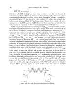
visually (see Figure 1).
Another method of calculation is by allowing
blood to drain into a fixed collecting container
(Figure 2) for estimation at the end of 1 h.
Blood losses on the delivery table, garments and
floor should also be assessed. At the end of 1 h,
the total amount of blood lost is estimated by
totaling up the blood in the container, in the
sponges and secondary blood spillage on the
delivery table, garments and floor. How often
such calculation is utilized is unknown, but
failure to do so undoubtedly contributes to
underestimation.
Direct collection of blood into bedpan or plastic bags
This approach was used in the World Health
Organization (WHO) multicenter, randomized
trial of misoprostol in the management of the
third stage of labor
30
. In this trial, blood loss was
measured from the time of delivery until the
mother was transferred to postnatal care. Imme
-
diately after the cord was clamped and cut, the
blood collection was started by passing a flat
bedpan under the buttocks of a woman deliver
-
ing in a bed or putting in place an unsoiled sheet
for a woman delivering on a delivery table.
37
Assessment of blood loss and decision to transfer
Figure 1 Soakage characteristics of 10 × 10 cm
pads
Figure 2 Blood drained into a fixed collecting
container
59
Z:\Sapiens Publishing\A5211 - Postpartum Hemorrhage\Make-up\Postpartum Hemorrhage - Voucher Proofs #T.vp
30 August 2006 14:19:18
Color profile: Generic CMYK printer profile
Composite Default screen
Blood collection and measurement continued
until the third stage of the labor was completed
and the woman was transferred to the postnatal
ward. This period was generally up to 1 h
postpartum. At that time, the collected blood
was poured into a standard measuring jar
provided by WHO and its volume measured.
To simplify the procedure for measurement
of blood loss, any available small gauze swabs
soaked with blood were put into the measuring
jar and included in the measurement together
with the blood and clots. A validity study was
performed before the trial to assess the effect of
adding the gauze swabs on the estimation
of blood loss and was found to result in an
approximately 10% increase in the blood loss
measurement.
Gravimetric method
This method involves weighing sponges before
and after use. The difference in weight provides
a rough estimate of blood loss.
Determination of changes in hematocrit and
hemoglobin
The changes in values before and after delivery
of the hematocrit and hemoglobin levels provide
quantitative measurements of blood loss, as
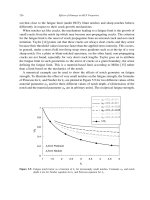
depicted in Figure 3.
Acid hematin method
This method is based on collected blood being
mixed with a standardized solution which
converts hemoglobin to acid hematin or cyan
-
methemoglobin. This in turn can be measured
by a spectrophotometer or colorimeter. Spec
-
trophotometric analysis can be performed by
the methods described below
9,31
:
(1) Preparation of standard Two milliliters of
peripheral blood are collected pre-delivery.
The blood standard is prepared with 0.1 ml
of the patient’s peripheral blood in 9.9 ml of
5% sodium hydroxide solution. The optical
density (OD) is read at 550 nm after
30 min;
(2) Preparation of sample The collected sample
is added to 2 liters of 5% sodium hydroxide
and let stand for 15 min. One ml of the
filtrate is diluted 10 times in 5% sodium
hydroxide and left to stand for another
15 min. The optical density (OD) is read
with a spectrophotometer at 550 nm at
30 min after the addition of sodium
hydroxide to the sample;
(3) Calculations
OD sample ml
OD blood standard
Blood volum
××
×
=
2000 10
100
e
loss
Plasma volume changes
The plasma volume can be determined before
and after delivery using radioactive tracer
elements.
Measurement of tagged erythrocytes
Blood loss can be measured by using
51
Cr-tagged erythrocytes
13
.
Failures of each method
Visual assessment
The major advantage of this method is that it
is a real-time assessment and enables the birth
attendant to correlate findings, on an individu
-
alized basis, with the clinical presentation.
However, significant differences between
clinical estimates and actual measurements
have been consistently demonstrated in several
38
POSTPARTUM HEMORRHAGE
Hemoglobin (g/dl)
Postpartum day
Postpartum hemoglobin
Visual
BRASSS-V
Both groups
11.2
11.0
10.8
10.6
10.4
10.2
10.0
9.8
9.6
01 23 5
Figure 3 Postpartum hemoglobin changes
60
Z:\Sapiens Publishing\A5211 - Postpartum Hemorrhage\Make-up\Postpartum Hemorrhage - Voucher Proofs #T.vp
06 September 2006 16:50:19
Color profile: Generic CMYK printer profile
Composite Default screen
studies
28
. The most common error is under
-
estimation of blood lost, with an average error
of 46% when estimates at the time of delivery
are compared with more precise measurements.
As might be expected, observers tend to give
median or average estimate of blood loss. When
losses were large, they were most often under
-
estimated and, when the losses were less than
average, they tended to be overestimated
11
.
Standardized visual estimation
In an attempt to rectify this error, the use of a
standardized visual estimation can be employed
as a simple method to be routinely practiced in
low-resource setting, albeit based on training
the providers and standardization of the pads
(size and quality) used during delivery. The
accuracy of estimated blood loss is not depend
-
ent upon age or the clinical experience of the
provider
32–35
. Teaching this tool significantly
reduced the error in blood loss estimation for
inexperienced as well as experienced clinicians.
Of particular clinical importance is a reduction
in underestimation of blood loss in the face of
greater degrees of measured blood loss; this has
the strongest potential to reduce hemorrhage-
related morbidity and mortality
36
.
Collection in pan or plastic bags
The errors in estimating blood loss arise from
failure to collect or note all the blood in stained
linen, incomplete extraction from the collection
device, ignoring maternal blood within the
placenta (approximately 153 ml), confusion
related to the mixing of blood contaminated
with amniotic fluid and urine, and technical
inaccuracies associated with transfer of the
collection to a measuring device.
Gravimetric methods
The gravimetric method requires the weighing
of materials such as soaked pads on a scale and
subtracting the known weights of these materi
-
als to determine the blood loss
37
. Inaccuracies
can arise at several steps in this procedure,
including lack of international standardization
of size and weight of gauze, sponges and pads.
Use of blood indices and spectrophotometric
measurement of hemoglobin
The first study reporting on measurement of
blood loss during surgical procedures employed
the colorimetric technique, which required that
hemoglobin be washed from surgical materials
in a blender and measured in a colorimeter
38
.
Clearly, this is impractical in obstetric practice.
Routine hematocrit determination, on the other
hand, is possible if the equipment is available.
However, routine postpartum hematocrits are
unnecessary in clinically stable patients with an
estimated blood loss of less than 500 ml. After
delivery associated with an average blood loss,
the hematocrit drops moderately for 3–4 days,
followed by an increase. The peak drop may be
appreciated on day 2 or day 3 postpartum
39
.By
days 5–7, the postpartum hematocrit will be
similar to the prelabor hematocrit
15
. Should
the postpartum hematocrit be lower than the
prelabor hematocrit, the blood loss may have
been larger than appreciated
40
.
Plasma volume changes and measurement of
tagged erythrocytes
Blood volume estimation using dye-dilution or
radioisotope dilution techniques is more diffi-
cult and requires special equipment and serial
measurements
41,42
. Measurement of erythrocytes
appears to be more consistent than estimates of
plasma volume secondary to physiological hemo
-
dilution causing a fluid overload of approxi
-
mately 1080–1680 ml in pregnancy
14
. Significant
cardiovascular changes occur immediately post
-
partum. The cardiac output remains elevated for
24 h, blood pressure declines initially and then
stabilizes on postpartum day 2. Maternal physio
-
logical changes of hemodilution lead to reduced
hemoglobin and hematocrit values, reflecting
the importance of timing of the measurement
43
.
In the majority of patients
44
, no single timed
hemoglobin or hematocrit determination in the
first 24 h postpartum will detect the peak.
BRASSS-V DRAPE: BLOOD LOSS
COLLECTION TOOL
A randomized, placebo-controlled trial to test
the use of oral misoprostol was conducted to
39
Assessment of blood loss and decision to transfer
61
Z:\Sapiens Publishing\A5211 - Postpartum Hemorrhage\Make-up\Postpartum Hemorrhage - Voucher Proofs #T.vp
30 August 2006 14:19:18
Color profile: Generic CMYK printer profile
Composite Default screen
reduce the incidence of acute postpartum
hemorrhage and hence maternal morbidity and
mortality in women delivering in rural villages
(away from major hospitals) within Belgaum
District, Karnataka, India. The intervention
was delivered by local health-care workers. A
critical component of this trial was the develop
-
ment of a specially designed low-cost ‘calibrated
plastic blood collection drape’ that would objec
-
tively measure the amount of blood collected
in the immediate postpartum period. The
BRASSS-V drape was developed by the
NICHD-funded Global Network UMKC/
JNMC/UIC collaborative team to specifically
estimate postpartum blood loss
45,46
. (The name
‘BRASSS-V’ was coined by adding the first let
-
ter of the names of the seven collaborators who
developed the drape.) The drape has a cali
-
brated and funneled collecting pouch, incorpo
-
rated within a plastic sheet that is placed under
the buttocks of the patient immediately after the
delivery of the baby. The upper end of the sheet
has a belt, which is loosely tied around the
woman’s abdomen to optimize blood collection,
particularly for deliveries performed on the floor
or on a flat surface at homes or in rural primitive
health posts. This simple tool not only has
the potential for a more accurate detection of
postpartum blood loss, but we hypothesize that
this approach will lead to earlier interventions,
with an ultimate goal of decreasing maternal
morbidity and mortality due to postpartum
hemorrhage. Since most developing countries
use some form of under-buttock sheet, either at
home, in the health center or in hospitals, drape
substitution is acceptable and relatively simple.
The BRASSS-V calibrated drape used for
objective estimation of blood loss is shown in
Figures 4 and 5.
Results of three studies conducted at JNMC,
Belgaum, Karnataka, India
4,7
strongly suggest
that the BRASSS-V drape is an accurate and
practical tool to measure blood loss occurring in
the third stage of labor. While, among women
with little blood loss, the ranges of blood loss
were similar in both visual and drape assess
-
ment, the actual visual assessment amount was
considerably less compared with the calibrated
drape values (Table 2 and Figure 6). This
observation further underscores the inaccuracy
of the visual estimation method as described
in the literature, whereas differences between
the drape and spectrophotometry values were
found to be 37.15 ml, with the drape having the
higher value (an average error of 16.1%). The
drape measured blood loss equally and as
40
POSTPARTUM HEMORRHAGE
Figure 4 BRASSS-V blood collection drape with
calibrated receptacle
Figure 5 Collection of blood using BRASSS-V
blood collection drape with calibrated receptacle
62
Z:\Sapiens Publishing\A5211 - Postpartum Hemorrhage\Make-up\Postpartum Hemorrhage - Voucher Proofs #T.vp
30 August 2006 14:19:22
Color profile: Generic CMYK printer profile
Composite Default screen
efficiently as gold-standard spectrophotometry
(Pearson’s correlation coefficient of 0.928;
p = 0.01, Table 3).
Use of the drape diagnosed postpartum
hemorrhage four times as often as the visual
estimate. A larger validation study is presently
underway at the University of Missouri at
Kansas City School of Medicine. In addition,
the drape is being tested in a number of inter
-
national settings including Tibet, Vietnam,
Egypt, Ecuador, Brazil and Argentina. Based on
the Indian experience, it appears to have great
potential for training delivery attendants to
determine postpartum blood loss in an accurate
and timely manner. The drape, apart from
being an objective tool for measurement of
postpartum blood loss, also provided a hygienic
delivery surface while permitting early manage
-
ment and referral. Residents and nurses in
hospital settings and the nurse midwives who
used the BRASSS-V drape during home deliv
-
ery all found it to be a very useful tool to
measure blood loss after delivery and for early
diagnosis of postpartum hemorrhage; it also led
to earlier transfer from rural areas to the higher
facility. The women who delivered at home and
their family members also appreciated the use-
fulness of the drape for easy disposal of body
fluids after birth
45
.
A similar approach has been used in another
recently reported study
48
. A plastic collecting
bag put under the pelvis of the mother just after
delivery can serve as a quantitative and objective
method of measuring blood loss. The study goal
was to assess sensitivity, specificity, positive
predictive value and negative predictive value,
including correlation between the bag’s volume
and hemoglobin and hematocrit variation. The
authors conclude that the collecting pelvis bag is
a rapid and precise procedure with which to
diagnose postpartum hemorrhage in the deliv
-
ery room. It also enables a visual and quantita
-
tive non-subjective estimation of blood loss.
Because of its simplicity and very low cost, the
pelvis collecting bag may have applicability as a
routine preventive measure.
Accurate measurement of blood loss at deliv
-
ery as a means of early detection of postpartum
hemorrhage is necessary for several reasons, not
the least of which is the fact that oxytocic
agents, while an important component for
addressing the third stage of labor, do not
address many factors related to postpartum
41
Assessment of blood loss and decision to transfer
Blood loss (ml)
Visual
(n = 61)
Drape
(n = 62)
All cases
(n = 123)
Mean ± standard deviation
Range
203.11 ± 147.49
50–950
302.82 ± 173.28
50–975
253.37 ± 168.86
50–975
Ta bl e 2 Distribution of blood loss
47
29
12
25
2
8
0
5
10
15
20
25
30
35
40
45
50
No. of cases
<
250 ml 250–500 ml >
500 ml
Visual Drape
Figure 6 Number of cases detected for specific
blood loss (p < 0.01). The calibrated drape more
accurately determined true blood loss when
≥ 250 ml and more accurately estimated overall
levels
Blood loss (ml)
Drape-measured Spectrometry
Mean ± standard
deviation
Range
225 ± 96.10
100–350
187.84 ± 61.79
93.19–285.98
Ta bl e 3 Comparison between drape-measured and
spectrometrically analyzed blood loss
63
Z:\Sapiens Publishing\A5211 - Postpartum Hemorrhage\Make-up\Postpartum Hemorrhage - Voucher Proofs #T.vp
30 August 2006 14:19:22
Color profile: Generic CMYK printer profile
Composite Default screen
hemorrhage in resource-poor areas. Trauma of
the birth canal during delivery and retained
placental fragments are important causes of
postpartum hemorrhage and may occur more
often than previously reported. Visual assess
-
ment of blood loss in the presence of a
contracted uterus may diagnose traumatic post
-
partum hemorrhage late and therefore result
in delayed referrals. In India and many other
developing nations, at least half of all births take
place in rural areas. Most of these deliveries are
conducted by indigenous health-care providers
such as dais (traditional birth attendants) or
auxiliary nurse midwives having varying levels
of training. Blood loss appears to be commonly
underestimated, as visual assessment is the only
means available to the birth attendant to make
this diagnosis. The clinical symptoms of blood
loss (low blood pressure, fast pulse, pallor and
sweating, signs of hypovolemia and impending
shock) are often the primary indicators for inter
-
vention. However, relying on the onset of such
symptoms may lead to delayed intervention,
resulting in increased rates of morbidity and
mortality. As other quantitative methods
employed have both practical and technical lim
-
itations, the employment of simple tools, such
as the BRASSS-V under-buttock blood collec
-
tion drape with a calibrated receptacle, can be
effectively employed for objectively assessing
the blood loss. It is likely to be of great utility to
the midwife/birth attendant and thus help to
ensure more timely and accurate patient man
-
agement. Having identified excessive blood loss,
corrective measures can be taken at the earliest
time, thus improving outcomes associated with
postpartum hemorrhage.
ACKNOWLEDGEMENTS
Our sincere thanks to Dr Shivaprasad S.
Goudar, Professor of Physiology & Research,
Coordinator Global Network for Women’s and
Children’s Health Research Site 8, and Dr
Kamal Patil, Associate Professor of Obstetrics &
Gynecology, JNMC for invaluable assistance
in the preparation of this manuscript. We also
acknowledge the contribution of Dr Kuldeep
Wagh and Dr B. V. Laxmi, residents in
the Department of Obstetrics & Gynecology,
JNMC for participating in the validation study
and to Dr A. Patel for her contributions to the
design of the BRASSS-V drape.
References
1. Starr A. The Safe Motherhood Agenda: Priorities for
the Next Decade. New York: Inter-agency Group
for Safe Motherhood, Family Care International,
1997
2. Reduction of maternal mortality. A joint WHO/
UNFPA/UNICEF/World Bank Statement.
1999 />publications/reduction_of_maternal_mortality/
reduction_of_maternal_mortality_contents.htm
3. Abou Zahr C. Antepartum and postpartum hem
-
orrhage. In Murray CJL, Lopez AD, eds. Health
Dimensions of Sex and Reproduction. Boston:
Harvard University Press, 1998
4. Berg CJ, Atrash HK, Koonin LM, Tucker M.
Pregnancy-related mortality in United States,
1987–1990. Obstet Gynecol 1996;88:161–7
5. Hogberg U, Innala E, Sandstorm A. Maternal
mortality in Sweden, 1980–1988. Obstet Gynecol
1994;84:240–4
42
POSTPARTUM HEMORRHAGE
2
8
123
Visual Drape Total
Figure 7 Number of cases of postpartum
hemorrhage (PPH) detected for specific blood loss
(p < 0.01). The calibrated drape diagnosed PPH at
a rate four times that of the visual estimate method
64
Z:\Sapiens Publishing\A5211 - Postpartum Hemorrhage\Make-up\Postpartum Hemorrhage - Voucher Proofs #T.vp
30 August 2006 14:19:23
Color profile: Generic CMYK printer profile
Composite Default screen
6. Razum O, Jahn A, Blettner M, Reitmaier P.
Trends in maternal mortality ratio among
women of German and non-German nationality
in west Germany, 1980–1996. Int J Epidemiol
1999;28:919–24
7. Dildy GA. Postpartum Hemorrhage. Washington,
DC: American College of Obstetricians and
Gynecologists, 1998
8. Maine D. Safe Motherhood Programs: Options
and Issues. Columbia University: Center for
Population & Family Health, 1993:42
9. Williams JW. The tolerance of freshly delivered
women to excessive loss of blood. Am J Obstet
Gynecol 1919;90:1
10. Duthie SJ, Ven D, Yung GL, Guang DZ, Chan
SY, Ma HK. Discrepancy between laboratory
determination and visual estimation of blood loss
during normal delivery. Eur J Obstet Gynecol
Reprod Biol 1990;38:119–24
11. Newton M, Mosey IM, Egli GE, Gifford WB,
Hull CT. Blood loss during and immediately
after delivery. Obstet Gynecol 1961;17:9–18
12. Newton M. Postpartum hemorrhage. Am J
Obstet Gynecol 1966;94:711–16
13. Gahres EE, Albert SN, Dodek SM. Intrapartum
blood loss measured with Cr51-tagged erythro-
cytes. Obstet Gynecol 1962;19:455–62
14. Nelson GH, Ashford CB, Williamson R. Method
for calculating blood loss at vaginal delivery.
South Med J 1981;74:550–2
15. Chesley LC. Plasma and red cell volumes
during pregnancy. Am J Obstet Gynecol 1972;
112:440–50
16. Knuppel RA, Hatangadi SB. Acute hyper
-
tension related to hemorrhage in obstetric
patients. Obstet Gynecol Clin N Am 1995;22:
111–29
17. Gabbe SG, Niebyl JR, Simpson JL, eds. Obstet
-
rics: Normal and Problem Pregnancies, 4th edn.
Churchill Livingstone, 2001
18. Cunningham FG, Gilstrap LC, Gant NF, et al.,
eds. Williams Obstetrics, 21st edn. McGraw-Hill,
2001
19. Pritchard JA, Baldwin RM, Dickey JC, et al.
Blood volume changes in pregnancy and the
puerperium. II. Red blood cell loss and change
in apparent blood volume during and following
vaginal delivery, cesarean section, and cesarean
section plus total hysterectomy. Am J Obstet
Gynecol 1962;84:1272–82
20. Clark SL, Yeh SY, Phelan JP, et al. Emergency
hysterectomy for obstetric hemorrhage. Obstet
Gynecol 1984;64:376–80
21. Waters EG. Surgical management of postpartum
hemorrhage with particular reference to ligation
of uterine arteries. Am J Obstet Gynecol 1952;64:
1143–8
22. Combs CA, Murphy EL, Laros RK Jr. Factors
associated with hemorrhage in cesarean deliver
-
ies. Obstet Gynecol 1991;77:77–82
23. Calkins LA. Factors governing blood loss in the
third stage of labor. Am J Obstet Gynecol 1929;
17:578
24. Hill JA, Fadel HE, Nelson MC, Nelson RM,
Nelson GH. Blood loss at vaginal delivery. South
Med J 1986;79:188–92
25. Qubil LD, Saski A. Episiotomy blood loss. Am J
Obstet Gynecol 1947;54:51
26. Spoerel WE, Heagy FC. The use of blood vol
-
ume determination for the evaluation of blood
loss during operation. Can J Surg 1962;5:25–32
27. Arulkumaran S, Symonds IB, Fowlie A. Massive
obstetric hemorrhage. In Oxford Handbook of
Obstetrics & Gynaecology. Oxford: Oxford
University Press, 2003:399
28. Brant HA. Precise estimation of postpartum
haemorrhage: difficulties and importance. Br
Med J 1967;1:398–400
29. Hester JD. Postpartum hemorrhage, and
re-evaluation of uterine packing. Obstet Gynecol
1975;45:501–4
30. Gulmezoglu AM, Villar J, Ngoc NT, et al. WHO
Multicentre randomized trial of misoprostol in
the management of the third stage of labour.
Lancet 2001;358:689–95
31. Chua S, Ho LM, Vanaja K, Nordstrom L, Roy
AC, Arulkumaran S. Validation of a laboratory
method of measuring postpartum blood loss.
Gynecol Obstet Invest 1998;46:31–3
32. Dildy GA, Paine AR, George NC, Velasco C.
Estimating blood loss: can teaching significantly
improve visual estimation? Obstet Gynecol 2004;
104:601–6
33. Grant JM. Treating postpartum haemorrhage.
Br J Obstet Gynaecol 1997;104:vii
34. Patton K, Funk DL, McErlean M, Bartfield JM.
Accuracy of estimation of external blood loss by
EMS personnel. J Trauma 2001;50:13–20
35. Meiser A, Casagranda O, Skipka G, Laubenthal
H. Quantification of blood loss. How precise
is visual estimation and what does its accuracy
depend on? Anaesthesist 2001;50:13–20
36. Luegenbiehl DL, Debra L. Improving visual
estimation of blood volume on peripads. MCN
Am J Matern Child Nurs 1997;22:294–8
37. Buchman MI. Blood loss during gynecological
operations. Am J Obstet Gynecol 1953;65:53–64
38. Gatch WD, Little WD. Amount of blood lost
during some of the more common operations.
JAMA 1924;83:1075–6
43
Assessment of blood loss and decision to transfer
65
Z:\Sapiens Publishing\A5211 - Postpartum Hemorrhage\Make-up\Postpartum Hemorrhage - Voucher Proofs #T.vp
30 August 2006 14:19:23
Color profile: Generic CMYK printer profile
Composite Default screen
39. Maruta S. The observation of the maternal
haemodynamics during labour and cesarean
section. Nippon Sanka Fujionka Gakkai Zasshi
1982;34:776–84
40. Pritchard JA, Baldwin RM, Dickey JC, Wiggins
KM. Blood volume changes in pregnancy and
the puerperium. Am J Obstet Gynecol 1962;84:
1271
41. Quinlivan WLG, Brock JA, Sullivan H. Blood
volume changes and blood loss associated with
labor. Correlation of changes in blood volume
measured by
131
I-albumin and Evans blue dye,
with measured blood loss. Am J Obstet Gynecol
1970;6:843–9
42. Ueland K. Maternal cardiovascular dynamics.
VII. Intra-partum blood volume changes. Am J
Obstet Gynecol 1976;126:671–7
43. Robson SC, Boys RJ, Hunter S, Dunlop W.
Maternal hemodynamics after normal delivery
and delivery complicated by postpartum hemor
-
rhage. Obstet Gynecol 1989;74:234–9
44. Nelson GH. Consideration of blood loss at
delivery as a percentage of estimated blood
volume. Am J Obstet Gynecol 1980;138:1117
45. Kodkany BS, Derman RJ, Goudar SS, et al.
Initiating a novel therapy in preventing
postpartum hemorrhage in rural India: a
joint collaboration between the United States
and India. Int J Fertil Women Med 2004;49:
91–6
46. Geller SE, Patel A, Naik VA, et al. Conduct
-
ing international collaborative research in devel
-
oping nations. Int J Gynaecol Obstet 2004;87:
267–71
47. Patel A, Goudar SS, Geller SE, et al. Drape esti
-
mation versus visual assessment for estimating
postpartum hemorrhage. Int J Gynaecol Obstet
2006;93:220–4
48. Tourne G, Collet F, Lasnier P, Seffert P.
Usefulness of a collecting bag for the diagnosis of
post-partum hemorrhage. J Gynecol Obstet Biol
Reprod (Paris) 2004;33:229–34
44
POSTPARTUM HEMORRHAGE
66
Z:\Sapiens Publishing\A5211 - Postpartum Hemorrhage\Make-up\Postpartum Hemorrhage - Voucher Proofs #T.vp
30 August 2006 14:19:23
Color profile: Generic CMYK printer profile
Composite Default screen
5
ASSESSING AND REPLENISHING LOST VOLUME
J. G. L. Cockings and C. S. Waldmann
INTRODUCTION
Classically, shock is defined as a state of inade
-
quate tissue perfusion for the metabolic needs of
the patient. This state of inadequate blood flow
may manifest clinically as tachycardia, pallor,
oliguria, the development of lactic acidosis and
altered mental status.
Shock is either hypovolemic, cardiogenic,
anaphylactic or cytotoxic. Hypovolemic shock
classically associated with postpartum hemor-
rhage is due to loss of circulating blood volume.
Hypotension is often present in severe cases, but
is a late sign and is a poor guide to the volume of
blood lost, as pregnancy is accompanied by an
alteration of cardiovascular physiology and the
response to blood loss and its management may
differ to the non-pregnant situation. Maternal
blood volume increases, total red cell mass also
increases but to a lesser extent, systemic vascu-
lar resistance is reduced, and cardiac output
becomes more dependent on body position.
Massive postpartum hemorrhage accounts for
35% of obstetric admissions to intensive care in
the UK
1,2
. These patients demand rapid assess
-
ment and judicious replenishment of lost circu
-
lating volume, albeit within the context of the
compensatory effects of hypovolemic shock and
the physiological changes seen in late pregnancy.
PHYSIOLOGY
The normal circulating blood volume for a
healthy non-pregnant adult is 70 ml/kg, or 7.5%
of body weight. Cardiac output is 4–6 l/min,
and the non-pregnant adult systemic vascular
resistance is 10–15 mmHg/l/min (900–1200
dyne.s/cm
5
). Maternal blood volume increases
during pregnancy to 40% above baseline by the
30th week, with an accompanying but smaller
(20–30%) increase in red cell volume. Cardiac
output increases to 50% above pre-pregnancy
levels by the 24th week. Systemic blood pres
-
sure is more variable in healthy uncomplicated
pregnancy, with a small fall in the first and sec
-
ond trimesters, but a return to pre-pregnancy
levels by the third. Resting heart rate increases
progressively in the first and second trimesters
to 15–20 beats per minute above pre-pregnant
levels. In addition to these changes, other
changes also take place in the autoregulation of
intravascular volume and the circulation, both
of which affects the body’s response to blood
loss. Examples include a blunted response to
angiotensin II, which may in part be due to
an increased production of nitric oxide
3
,a
decreased tolerance to postural changes and an
increased cardiac noradrenaline turnover
4,5
.
Circulating volume, clinical signs of hypo-
volemia and the body’s ability to compensate for
volume loss are also all affected by pregnancy-
related diseases and their treatment, the effects
of which continue on into the early postpartum
period. Pre-eclampsia, for example, causes a
contracted effective arterial blood volume com
-
pared with the normal peripartum state. Vascu
-
lar reactivity is increased, and widely used drugs
such as hydralazine and magnesium compro
-
mise the body’s ability to produce compensa
-
tory vasoconstriction in the face of hemorrhage.
Indeed, it appears that there is a failure to
increase plasma volume and reduce systemic
vascular resistance in pre-eclampsia, due to
inadequate trophoblastic invasion into the spiral
arteries of the uterus
5
. Pre-eclamptic patients
thus have an increased tendency to develop pul
-
monary edema during volume replacement due
to many factors, including increased capillary
permeability, hypoalbuminemia and left ven
-
tricular dysfunction
6
.
45
67
Z:\Sapiens Publishing\A5211 - Postpartum Hemorrhage\Make-up\Postpartum Hemorrhage - Voucher Proofs #T.vp
30 August 2006 14:19:23
Color profile: Generic CMYK printer profile
Composite Default screen
Normal delivery results in predictable losses
of 300–500 ml blood volume for vaginal deliver
-
ies and 750–1000 ml for Cesarean section births
(see Chapter 4). However, in addition to blood
lost from the body, a substantial amount of
blood is also redirected into the systemic circu
-
lation, often referred to as the autotransfusion
effect. This results in an increase in cardiac out
-
put by as much as 80%. The effect persists in
uncomplicated patients, gradually returning to
non-pregnant levels at 2–3 weeks
5
.
ASSESSMENT OF CIRCULATING
BLOOD VOLUME
Young healthy adults can compensate for the
loss of large volumes from the circulation with
few obvious external signs. Accurate assessment
of blood loss can be difficult for the experienced
as well as the inexperienced examiner, as
described in Chapter 4.
In cases of hemorrhage symptoms often pre-
cede signs. These include unexplained anxiety
and restlessness, the feeling of breathlessness
(with or without an increased respiratory rate),
and a sensation of being cold or generally
unwell. For healthy, non-pregnant adults, hypo-
volemia and associated signs can be divided into
four stages (Table 1). These range from the
largely undetectable stage 1 with less than 15%
loss of volume, to the severe life-threatening
stage when more than 40% has been lost.
Unfortunately, comparable tables for early and
late pregnancy and the immediate postpartum
period have not been compiled, but the signs
follow a similar pattern.
The most important principle in the treat
-
ment of postpartum hemorrhage is early recog
-
nition and prompt correction of lost circulating
volume, together with simultaneous medical
and/or surgical intervention to prevent further
loss. Early recognition of life-threatening physi
-
ological derangements can be improved by the
use of early-warning scoring systems.
Recording physiological observations at
regular intervals has long been routine practice
in hospitals. Early-warning scores derived from
simple routine physiological recordings can
identify patients with greater risk of critical
illness and mortality. Such scores can be used
to flag the early but sometimes subtle signs of
concealed but largely compensated hemorrhage
in the early postpartum patient and have been
recently recommended for use by the Confiden
-
tial Enquiry into Maternal and Child Health
report of 2004
7
. These scores use the physiolog-
ical parameters most likely to detect impending
life-threatening compromise. These usually
comprise respiratory rate, heart rate, systolic
blood pressure, temperature and mental aware-
ness. Each variable is assigned a weighted score
and the total score is the sum of these. This
allows a trigger value for ward staff to call for
assistance from intensive care or other senior
staff. Such systems have been shown to be
reproducible and effective at predicting the
likelihood of progressing on to critical illness.
They are well suited to the early detection of the
46
POSTPARTUM HEMORRHAGE
Classification Class 1 Class 2 Class 3 Class 4
Blood loss
(% volume lost)
Conscious state
Respiratory rate
Complexion
Extremities
Capillary refill
Pulse rate
Systolic blood pressure
Urine output
10–15%
Alert, mild
thirst
normal
normal
normal
normal
normal
normal
normal
15–30%
anxious and
restless
mildly elevated
pale
cool
slow (> 2 s)
normal
normal
reduced
30–40%
agitated or
confused
raised
pale
pale and cool
slow (> 2 s)
elevated
normal or slightly low
reduced
> 40%
drowsy, confused or
unconscious
raised
marked pallor or gray
cold
minimal or absent
fast but thready
hypotensive
oligoanuric
Modified from Baskett PJF. ABC of major trauma. Management of hypovolaemic shock. BMJ 1990;300:
1453–7
Ta bl e 1
Stages of shock
68
Z:\Sapiens Publishing\A5211 - Postpartum Hemorrhage\Make-up\Postpartum Hemorrhage - Voucher Proofs #T.vp
30 August 2006 14:19:24
Color profile: Generic CMYK printer profile
Composite Default screen
often subtle signs of unappreciated blood loss
and can be easily introduced. Altered normal
physiology in late pregnancy and the early
postpartum period demands that these scores,
usually derived from general surgical or medical
patients, be modified for this population as
shown in Table 2.
Once the possibility of intravascular deple
-
tion has been raised, a prompt clinical assess
-
ment is urgent, as the clinical condition of the
patient can change rapidly. Clinical assessment,
in association with non-invasive and invasive
monitoring where appropriate, must be made
by senior clinicians (if available), with special
attention to repeated assessment at frequent
intervals to detect the problem as early as
possible. If senior clinicians are not available,
they should be notified as described in the
protocols in Chapters 22 and 50.
Clinical examination is performed simulta
-
neously with incident-related history taking.
This history may elicit the more obvious
features of shock such as overt blood loss
and pain, but may also elicit the more subtle
features such as general malaise, anxiety and
restlessness, a poorly defined sense of doom and
breathlessness. Physical examination is directed
to the fundamental areas of vital function, the
conscious state and airway protection, the ade
-
quacy of respiratory function, oxygenation and
circulation. In particular, the following should
be assessed and documented:
(1) Early stages of shock are associated with
restlessness and agitation, sometimes with
a heightened sense of thirst, but these
progress to drowsiness when around 30% of
blood volume is lost. Loss of consciousness
is a very late sign, with significant risk of
imminent death.
(2) Tachypnea is an early sign, partly driven
initially by the anxiety, but is an independ
-
ent sign, and the respiratory rate increases
with progressive blood loss and will usually
exceed 20 breaths/min when 30% of blood
volume is lost.
(3) Oxygenation becomes harder to assess
clinically as peripheral pallor becomes more
marked, and the pulse oximeter becomes
less reliable as peripheral perfusion
becomes weaker.
(4) A fall in the jugular venous pressure occurs
reasonably early, but is partly compensated
47
Assessing and replenishing lost volume
Score
3 2 1 0 1 2 3
Respiratory rate (bpm)
Pulse rate (bpm)
Systolic blood pressure
(mmHg)
Diastolic blood pressure
(mmHg)
Conscious level
Urine hourly (ml/h)
or in 24 h
<70
unresponsive
0
<8
<40
71–80
responds
to pain
<30
(< 720 ml)
40–50
81–100
responds
to voice
<45
(< 1000 ml)
9–18
51–100
101–164
<95
alert
>45
(> 1000 ml)
19–25
101–110
165–200
95–104
irritated
26–30
111–129
> 200
> 105
>30
> 129
Final score = sum of individual scores at any one time
Action:
Score 0 or 1 Repeat observations when appropriate for clinical scenario
Score 2 Inform midwife in charge, repeat in 15 min
Score 3 Inform midwife in charge, obstetric registrar and duty anesthetist
Score ≥ 4 As above but the consultant obstetrician should be informed
Consider informing duty consultant anesthetist and intensive care team
Ta bl e 2
Modified early obstetric warning system. Reproduced with permission by Dr R Jones, Consultant
Anaesthetist, Royal Berkshire Hospital, UK, from unpublished work in progress
69
Z:\Sapiens Publishing\A5211 - Postpartum Hemorrhage\Make-up\Postpartum Hemorrhage - Voucher Proofs #T.vp
30 August 2006 14:19:24
Color profile: Generic CMYK printer profile
Composite Default screen