Báo cáo khoa học: "Variation in the Level of Grain Defect Light Flecks and Spots on Cattle Hides" pps
Bạn đang xem bản rút gọn của tài liệu. Xem và tải ngay bản đầy đủ của tài liệu tại đây (71.14 KB, 8 trang )
Nafstad O, Grønstøl H: Variation in the level of grain defect light flecks and spots
on cattle hides. Acta vet. scand. 2001, 42, 91-98. – The occurrence of hide damage
light flecks and spots was determined on tanned hides from 28 herds during a period of
8 to 12 months. Light flecks and spots are described as small areas of grain loss up to 3
mm in diameter that are seen on dyed crust cattle leather. Damage was found on 75.8%
of all hides. The neck and shoulders were the anatomical region with the highest preva-
lence of damage. Sixty-eight per cent of all hides had light flecks and spots in this re-
gion. The forelimbs and dewlap were the anatomical region with the second highest oc-
currence with a prevalence of 39.1%. This distribution corresponded to the known
distribution of lice in cattle. No significant differences were observed in age, sex, prev-
alence of lice in the herd assessed in March or infestations with different lice species.
The frequency of light flecks and spots varied significantly during the year. The fre-
quency was highest in the late winter and early spring, decreased significantly during the
summer and was lowest in the autumn. This variation supported the importance of lice
in the development of light flecks and spots and suggested a relatively long healing pe-
riod for the damages induced by lice.
leather; damage; lice.
Acta vet. scand. 2001, 42, 91-98.
Acta vet. scand. vol. 42 no. 1, 2001
Variation in the Level of Grain Defect Light Flecks
and Spots on Cattle Hides
By O. Nafstad and H. Grønstøl
Department of Large Animal Clinical Sciences, Norwegian School of Veterinary Science, Oslo, Norway.
Introduction
Several ectoparasites can be responsible for
damage of cattle hides (Tancouse 1986). Some
of the damage is very specific, such as grub
damage caused by warble fly larvae, but most
ectoparasite damage is more nonspecific. In re-
cent years, light flecks and spots have been con-
sidered the main damage caused by ectopara-
sites. Webster & Bugby (1990) found a signi-
ficant association between both biting lice
(Damalinia (Bovicola) bovis (Linneaus 1758))
and sucking lice (Linognathus vituli (Linneaus
1758)) and light flecks and spots on leather.
These investigators described light flecks and
spots as small areas of grain loss up to 3 mm in
diameter that are seen on dyed crust leather
(Webster & Bugby 1990). This damage was re-
ported to be a major problem for the hide indus-
try in Great Britain for the first time in 1983 and
has later been reported to be a problem in Hol-
land, Scandinavia, USA and New Zealand
(Webster & Bugby 1990). Tanners from Nordic
countries found light flecks and spots on 50%-
55% of Norwegian cattle hides in 1991, and the
problem was estimated to cost the Norwegian
cattle industry 24-25 mill NOK each year
(Dørum personal communication). The aim of
this paper is to describe the variation in the fre-
quency of light flecks and spots damage on the
hides from cattle not treated for ectoparasites.
Materials and methods
Design
A prospective cohort study was performed in
33 herds during a period of two and a half years
from 1. January 1994 to 30. June 1996, with an-
imals leaving or entering the herds at any time.
Twenty-eight of the herds were treated for lice
between September and December 1994. Five
of the herds took part in a pilot study for the
eradication programme in December 1993.
Hides were collected from all herds during the
whole study, in order to assess the frequency of
damage in hides from untreated and treated an-
imals. The results in the present paper are based
on hides collected from the 28 herds in the main
group before the start of the lice eradication
programme.
The herds
The selection criteria, the prevalence of lice in
the herds and the clinical examination proce-
dure used in the herds are described previously
(Nafstad & Grønstøl 2001). D. bovis was
present in 27 of 28 herds and in 27% of the an-
imals. L. vituli was present in 11 of 28 herds and
in 5% of the animals. At least one of these louse
species was present in all the herds. In a few of
the herds, other ectoparasite species known to
affect the hide quality were also present. Four of
the herds had clinical signs of tail mange
(Chorioptes bovis (Herning 1845)), and the di-
agnosis was confirmed in laboratory examina-
tion. Two of the herds were in the distribution
area of Ixodes ricinus (Linnaeus 1758). A total
of 368 hides sampled from the period before
treatment were included. The mean number of
hides from each herd was 13.1 with a variation
from 2 to 21.
Examination of the hides
The hides were tanned in a commercial tannery
and evaluated as aniline dyed crust leather.
The leather was chrome tanned and vegetable
retanned. The process in the tannery was based
on splitting the hides along the back line. Four
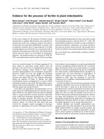
anatomical regions in every half of the hides
were evaluated separately. The regions were
chosen according to the known distribution of
lice on the skin. The 4 regions were the neck
and shoulder, the forelimb and dewlap, the back
and the rump, hindlimb, side and belly (Fig. 1).
Thus 8 registrations were made on each hide.
Each evaluation was based on the number of
identifiable flecks and spots according to the
following scale:
Score 0: No damage.
Score 1: Slight damage, with 1-2 light flecks or
spots per 100 cm
2
.
Score 2: Some damage, with 3-5 light flecks or
spots per 100 cm
2
.
Score 3: Severe damage, with more than 5 light
flecks or spots per 100 cm
2
.
Light flecks and spots were defined as small ar-
eas of grain loss up to 3 mm in diameter (We b -
ster & Bugby 1990).
Statistical methods
All 8 evaluations from 1 hide were used to de-
92 O. Nafstad & H. Grønstøl
Acta vet. scand. vol. 42 no. 1, 2001
Figure 1. The 4 anatomical regions of the hide
which were evaluated after tanning.
termine an overall parameter termed the maxi-
mum score. The maximum score was defined as
the highest single evaluation in 1 hide. In the
statistical analyses the ectoparasite diagnoses
were defined at herd level. Total lice prevalence
was defined as the prevalence of both D. bovis
and L. vituli. All analyses were made in Statis-
tical Analysis System (SAS Institute Inc. 1989).
A Spearman rank correlation test was used for
testing seasonal variations. Otherwise, statisti-
cal hypothesis testing was undertaken using t-
test. The statistical testing was based on the fre-
quency of hides without damage (maximum
score 0).
Results
The total frequency of light flecks and spots
distributed in different anatomical regions is
presented in Table 1. Light flecks and spots
were detected in 75.8% of all hides. The neck
and shoulders were the region with the highest
frequency of damage. Light flecks and spots in
this anatomical region were detected in 67.9%
of all hides.
Effects of age and sex
The frequencies of light flecks and spots as-
sessed on the basis of sex and age are given in
Tables 2 and 3. There were no significant differ-
ences between cows and bulls or between the
different age classes for any of the sexes. Cows
were significantly older at the time of slaughter
than bulls, but this difference had no effect on
the frequency of light flecks and spots.
Defects on cattle hides 93
Acta vet. scand. vol. 42 no. 1, 2001
Table 1. Frequency of hides with light flecks and spots and the frequency of damage within anatomical regions
(n= 368).
Mac. score
0123
All regions 24.2% 37.2% 31.5% 7.3%
Neck and shoulders 32.1% 40.0% 23.9% 4.1%
Forelimbs and dewlap 60.9% 24.5% 11.4% 3.3%
Back 67.9% 15.2% 13.0% 3.8%
Rump, hindlimbs, sides and belly 84.5% 13.3% 2.2% 0.0%
Score 0 - No damage
Score 1 - Slight damage, with 1-2 light flecks or spots per 100 cm
2
.
Score 2 - Some damage, with 3-5 light flecks or spots per 100 cm
2
.
Score 3 - Severe damage, with more than 5 light flecks or spots per 100 cm
2
.
Table 2. Frequency of hide damage in cows divided in age classes at the time of slaughter.
Age in months
Number of Mac. score
hides
0123
< 31.0 52 23.1% 48.8% 25.0% 3.9%
31.0-42.3 57 24.6% 38.6% 31.6% 5.3%
42.4-54.3 38 21.1% 36.8% 36.8% 5.3%
>54.3 59 30.5% 28.8% 32.2% 8.5%
All 206 25.2% 38.0% 31.1% 5.8%
Effects of high and low prevalence of lice as-
sessed in March
The herds were divided into 2 equal groups ac-
cording to the total prevalence of lice, and hides
from the 2 groups were compared. The results
are presented in Table 4. The prevalence of lice,
as assessed in March, had no significant effect
on the frequency of damaged hides from the
herds during the year. Statistical analyses
showed the same result when the same analyses
were performed according to the prevalence of
just biting lice alone or the prevalence of all lice
in animals older than 12 months.
Effects of biting lice compared with the effect of
mixed infections
Hides from herds with only D. bovis and hides
from herds with mixed infections of D. bovis
and L. vituli were compared in Table 5. There
was no significant difference in the frequency
of light flecks and spots between hides from
these 2 groups.
Seasonal variations in the frequency of light
flecks and spots
The seasonal variation in the frequency of light
flecks and spots is shown in Table 6. The fre-
quency of hides without damage varied signifi-
cantly through the year.
Discussion
Webster & Bugby (1990) and Bugby et al.
(1990) described light spots as small areas of
grain loss of 1-3 mm in diameter and light
flecks as spots of less than 1 mm in diameter
seen on dyed crust bovine leather. These inves-
tigators found a significant connection between
the occurrence of the 2 types of damage and
concluded that the 2 defects had a common
cause.
94 O. Nafstad & H. Grønstøl
Acta vet. scand. vol. 42 no. 1, 2001
Table 3. Frequency of hide damage in bulls divided in age classes at the time of slaughter.
Age in months
Number of Mac. score
hides
0123
< 18.4 57 26.3% 36.8% 22.8% 14.0%
18.4-20.3 36 19.4% 38.9% 36.1% 5.6%
20.4-23.2 19 42.1% 31.6% 15.8% 10.5%
>23.2 50 14.0% 38.0% 42.0% 6.0%
All 162 22.2% 36.9% 31.5% 9.4%
Table 4. Frequency of hide damage according to lice prevalence in the herd in March 1994.
Number Average Mac. score
of prevalence of
hides lice in March
0123
Hides from herds with 186 17.7% 25.8% 39.8% 29.6% 4.8%
lowest prevalence.
Hides from herds with 182 41.8% 23.1% 36.8% 30.8% 9.3%
highest prevalence.
Light flecks and spots were found on 75% of all
hides in this study. In 1991, Nordic tanners
found lice related damage on 50%-55% of all
Norwegian hides (Dørum, personal communi-
cation). This investigation was based on a com-
mercial evaluation of finished leather and is not
directly comparable to the present investiga-
tion. The results in the present study were based
on the number of flecks and spots found on
dyed crust leather. Older damage could have re-
ceived a different assessment if the hides had
been examined at a later stage in the tanning
process. The finishing of the leather may mask
small and very superficial grain damage. Thus,
given this different assessment, these 2 reports
suggest about the same level of light flecks and
spots. The quality of the hides in the present
study should, in the light of the selection crite-
ria, be comparable to the average of Norwegian
cattle hides. In a smaller survey from April
1998, light flecks and spots were present on
44% of the hides (Nafstad, unpublished data).
Two hundred hides from abattoirs all over the
country were included in this study. The ani-
mals were slaughtered in March and April
when the lice population is at its highest. The
hides were tanned and examined after the same
procedure as in the main study. The use of in-
secticides increased significantly from 1990 to
1996 (Bredal & Grave 1998), which probably
explain the improvement in hide quality ob-
served in the 1998 survey.
The neck and shoulders were the anatomical re-
gion with the highest prevalence of light flecks
and spots, followed by the forelimbs and dew-
lap and the back. This distribution corre-
sponded well with the predilection sites for lice
(Chalmers & Charleston 1980 a, DeVaney et al.
Defects on cattle hides 95
Acta vet. scand. vol. 42 no. 1, 2001
Table 5. Frequency of hide damage in herds infested with biting lice compared with herds infested with biting
lice and sucking lice.
Number Number Mac. score
of of
herds hides
0123
Biting lice infections 18 189 23.8% 35.5% 35.5% 5.3%
Mixed infections* 12 179 24.6% 39.1% 26.8% 9.5%
*One herd with only sucking lice infestaton.
Table 6. Seasonal variations in the frequency of damaged hides.
Months
Number of Mac. score
hides
0123
Jan. Feb. 83 14.5% 31.3% 39.8% 14.5%
Mar. Apr. 65 13.9% 40.0% 35.4% 10.8%
May. Jun. 75 28.0% 44.0% 26.7% 1.3%
Jul. Aug. 70 24.3% 37.1% 32.9% 5.7%
Sep. Oct. 64 35.9% 39.1% 20.3% 4.7%
Nov. Dec. 11 63.6% 9.1% 27.3% 0.0%
p<0.001 (Spearman correlation test)
1988) and supported the hypothesis that lice are
the main cause of light flecks and spots. Dam-
age associated with other ectoparasites such as
Demodex bovis (Stiles 1892), or I. ricinus
would have given a different distribution. Ticks
mostly affect the belly region and demodicosis
the forelimbs and dewlap (Urquart et al. 1987,
Wall & Shearer 1997).
Tancouse (1986) and Baker & Oormadzi
(1978) stated that severe lice infestations were
responsible for hide and leather damage caused
by secondary bacterial infections or scratching
behaviour. Bugby et al. (1990) found an associ-
ation between lice and light flecks and spots and
suggested that inflammation caused by the lice
infestations was the most probable mechanism
responsible for this damage. Light flecks and
spots were reported to be a major quality pro-
blem for cattle hides in Great Britain around
1983. This increase in light flecks and spots
took place at the same time as the national erad-
ication programme for warble fly was altered.
Systematic treatment with organophosphorous
insecticides was changed to a combination of
movement restrictions and treatment in affected
districts only (Webster & Bugby 1990). A side
effect of the systematic treatment was control
of lice, and the lice population probably in-
creased as a result of the changes in the eradica-
tion programme.
Light flecks and spots or similar damage have
also been associated with various tick species
and Psoroptes ovis (Hering 1838) infestations
in cattle (George et al. 1986, Everett et al. 1977,
Rotz et al. 1983). With the exception of I. rici-
nus, these parasites do not occur in Norway and
can not explain the present level of light flecks
and spots. It has been suggested that stable fly
Stomoxys calcitrans (Linnaeus 1758) can cause
light flecks and spots (Bugby personal commu-
nication). However, more work is needed to de-
termine whether stable fly may cause light
flecks and spots, in addition to lice, under Nor-
wegian circumstances. In an experimental
study, Rotz et al. (1983) found no obvious
change in the leather to be caused by D. bovis
and only dilated hair pores to be caused by L. vi-
tuli. In the same study, these authors found in-
flammatory reactions in the raw hides similar to
the changes reported by Webster & Bugby
(1990). This discrepancy suggested that the tan-
ning method influences the grain damage
shown after tanning.
The results in the present study did not vary
with sex and age of the animals. This result was
surprising given the age distribution of lice in-
fections. Calves and young animals have the
highest prevalence and heaviest infestations of
lice (Chalmers & Charleston 1980 b, Chistens-
son et al. 1994, Nafstad 1998), and conse-
quently a significantly higher occurrence of
light flecks and spots among calves and young
animals could be expected. For the same rea-
son, the difference between cow and bull in the
average age at slaughter should have given a
difference in occurrence of light flecks and
spots. The absence of the expected age varia-
tion in the occurrence of the damage may have
been influenced by a long and not exactly
known healing period for the damage. Chris-
tensson et al. (1994) suggested a healing period
of more than 12 months. Bugby et al. (1990)
found only very slight damage 9 weeks after
treatment of lice. The expected age variation
may also have been disturbed by light flecks
and spots caused by ectoparasites other than
lice.
The frequency of light flecks and spots showed
no significant association with the prevalence
of lice in the herds in March. The reason for this
lack of association may be that one examination
did not give a representative description of the
lice situation in the herd and the fact that light
flecks and spots also may be caused by other ec-
toparasites.
Hides from herds with mixed lice infections
96 O. Nafstad & H. Grønstøl
Acta vet. scand. vol. 42 no. 1, 2001
had the same prevalence of light flecks and
spots as hides from herds with only biting lice
infections. This result corresponded with the
result of Bugby & Webster (1994), who found
no additive effects of mixed infections com-
pared with only biting lice on the frequency of
light flecks and spots. These results showed the
importance of D. bovis for the development of
light flecks and spots.
The seasonal distribution of the lice population
is well known. The population increases during
the winter, decreases with the shedding of the
winter coat in the spring and remains low dur-
ing the summer (Gojmerac 1959, Scharff 1962,
Chalmers & Charleston 1980 b, Geden et al.
1990). The seasonal variation in the frequency
of light flecks and spots show a pattern that can
be related to the variation in the lice population
during the year. The frequency of light flecks
and spots was high in late winter and early
spring, and the frequency decreased slowly dur-
ing the spring, summer and autumn. Thus, these
observations indicate a relative long healing pe-
riod, but perhaps not as long as suggested by
Christensson et al. (1994).
References
Baker KP, Oormadzi H: The probable cause of the
multiple linear scratch defect of cattle hides in
Ireland. J. Soc. Leath. Tech. Chem. 1978, 62,
103-107.
Bredal W, Grave K: Foreskrivningsmønsteret av
antiparasittære midler til bruk på produksjonsdyr
i Norge i perioden 1990-1996 (Prescription of
antiparasitices used in food animal production in
Norway in the period 1990-1996). Husdyrfor-
søksmøtet, Ås 1998.
Bugby A, Webster RM, Tichener RN: Light spot and
fleck, part 2, animal infestation studies. Labora-
tory report 186. British Leather Confederation,
Northampton, 1990.
Chalmers K, Charleston WAG: Cattle lice in New
Zealand: observations on the biology and ecology
of Damalinia bovis and Linognathus vituli. N. Z.
vet. J. 1980a, 28, 214-216.
Chalmers K, Charleston WAG: Cattle lice in New
Zealand: observations on prevalence, distribution
and seasonal patterns of infestation. N. Z. vet. J.
1980b, 28, 198-200.
Christensson D, Gyllensvaan C, Skiøldebrand E, Vir-
ing S: Løss på nøtkreatur i Sverige - en inventer-
ing (Lice in Swedish cattle - a survey). Svensk
Vet. -Tidn. 1994, 46, 119-121.
DeVaney JA, Rowe LD, Craig TM: Density and distri-
bution of three species of lice on calves
in Central Texas. Southwest. Entomol. 1988, 13,
125-130.
Everett AL, Miller RW, Gladney WJ, Hannigan MV:
Effects of some important ectoparasites on the
grain quality of cattle hide leather. J. Amer.
Leath. Chem. Ass. 1977, 72, 6-23.
Geden CJ, Rutz DA, Bishop DR: Cattle lice (Ano-
plura, Mallophaga) in New York: Seasonal
population changes, effects of housing type on infes-
tations of calves, and sampling efficiency. J. econ.
Entomol. 1990, 83, 1435-1438.
George JE, Wright FC, Guillot FS, Buechler PR: Ob-
servations on the possible relationship between
psoroptic mange of cattle and white spot damage
on leather. J. Amer. Leath. Chem. Ass. 1986, 81,
296-304.
Gojmerac WL, Dicke RJ, Allen NN: Factors affecting
the biology of cattle lice. J. econ. Entomol. 1959,
52, 79-82.
Nafstad O, Grønstøl H: Eradication of lice in cattle.
Acta Vet. Scand. 2001, 42, 81-89.
Nafstad O: Forekomsten av lus hos norske storfe
(Prevalence of lice in Norwegian cattle). Norsk
Vet T. 1998, 110, 261-265.
Rotz A, Mumcuoglu Y, Pohlenz JFL, Suter M, Bros-
sard M, Barth D: Experimentelle Infestation von
Rindern mit Ektoparasiten und deren Einflub auf
die Lederqualität (Experimental infestation of
cattle with ectoparasites and their effect on
leather quality). Zbl. Vet. -Med. 1983, 30, 397-
407.
SAS Institute Inc: Guide for personal computers. Ver-
sion 6. Edition, Cary, NC, 1989.
Scharff DK: An investigation of the cattle louse prob-
lem. J. econ. Entomol. 1962, 55, 684-688.
Tancous JJ: Skin, hide and leather defects. Tanners'
Council Research Laboratory, Cincinnati, Ohio,
1986.
Urquhart GM, Armour J, Duncan JL, Dunn AM, Jen-
nings FW: Veterinary parasitology. Longman, Es-
sex, 1987.
Wall R, Shearer D: Veterinary entomology. Chapman
& Hall, London, 1997.
Defects on cattle hides 97
Acta vet. scand. vol. 42 no. 1, 2001
Webster RM, Bugby A: Light spot and fleck grain de-
fects of economic importance to the UK leather
industry, part 1, identification of causal agent.
Laboratory report 184. British Leather Confeder-
ation, Northampton, 1990.
Sammendrag
Variasjoner i forekomsten av narvfeilen lyse flekker
og prikker på storfehuder.
Hudene fra 28 storfebesetninger ble samlet inn gjen-
nom en periode på 8 til 12 måneder for å kartlegge
forekomsten av skaden lyse flekker og prikker på hu-
dene etter garving. Totalt inngikk 368 huder i
undersøkelsen. Skaden lyse flekker og prikker er de-
finert som kratre i overflaten eller narven på storfelær
med en diameter på inntil 3 mm. Hudene ble krom-
garvet og vegetabilsk ettergarvet til annilinlær og
undersøkt før siste overflatebehandling av læret. Ska-
den lyse flekker og prikker ble påvist 75,8% av alle
huder. Nakke og skulder var den anatomiske region
med høyest forekomst av skaden (67,9%) etterfulgt
av bog og dogglapp (39,1%). Dette utbredelses-
mønsteret samsvarer godt med predileksjonsstedene
for lus hos storfe. Det var ingen signifikant kjønns-
eller aldersforskjell i utbredelsen av skaden. Det var
heller ingen signifikant forskjell i forekomsten av
skader på hudene mellom besetninger som hadde en
høy prevalens av lus mål i mars måned og beset-
ninger som hadde en lav prevalens av lus på dette
tidspunktet. Besetninger som var infisert av både
blodlus (Linognathus vituli) og pelslus (Damalinia
bovis) hadde samme forekomst av skader som beset-
ninger som hadde rene pelslusinfeksjoner. Det var en
signifikant sesongmessig variasjon i forekomsten av
skaden lyse flekker og prikker, forekomsten var
høyest hos dyr slaktet fra januar til april, deretter ble
kvaliteten gravis bedre utover sommeren og høsten.
På bakgrunn av disse resultatene konkluderes det
med at det er en sikker sammenheng mellom lus og
skaden lyse flekker og prikker på læret. Pelslus (D.
bovis) er en viktigere årsak enn blodlus (L. vituli), på
bakgrunn av at det ikke var noen tilleggseffekt av
blandede infeksjoner sammenlignet med rene pels-
lusinfeksjoner. Den aldersuavhengige forekomsten
av skaden sammenhold med kjent kunnskap om at
lus i særlig grad forekommer hos kalver og ungdyr,
indikerer en lang avhelingstid. På den andre siden
kan den sesongmessige variasjonen indikere at en
delvis avheling kan inntre i løpet av 2 til 3 måneder.
98 O. Nafstad & H. Grønstøl
Acta vet. scand. vol. 42 no. 1, 2001
(Received February 1, 2000; accepted September 26, 2000).
Reprints may be obtained from: O. Nafstad, Norwegian Meat Research Centre, P.O. Box, 396 Økern, 0513 Oslo,
Norway. E-mail: , tel: +47 22 09 23 42, fax: +47 22 22 00 16.