Báo cáo y học: "Circulating surfactant protein -D is low and correlates negatively with systemic inflammation in early, untreated rheumatoid arthritis" pps
Bạn đang xem bản rút gọn của tài liệu. Xem và tải ngay bản đầy đủ của tài liệu tại đây (373.43 KB, 9 trang )
RESEARC H ARTIC LE Open Access
Circulating surfactant protein -D is low and
correlates negatively with systemic inflammation
in early, untreated rheumatoid arthritis
Anne Friesgaard Christensen
1*
, Grith Lykke Sørensen
2
, Kim Hørslev-Petersen
3
, Uffe Holmskov
2
,
Hanne Merete Lindegaard
1
, Kirsten Junker
2
, Merete Lund Hetland
4
, Kristian Stengaard-Pedersen
5
, Søren Jacobsen
6
,
Tine Lottenburger
3
, Torkell Ellingsen
5
, Lis Smedegaard Andersen
3
, Ib Hansen
5
, Henrik Skjødt
4
,
Jens Kristian Pedersen
3
, Ulrik Birk Lauridsen
4
, Anders Svendsen
1
, Ulrik Tarp
5
, Jan Pødenphant
7
, Aage Vestergaard
8
,
Anne Grethe Jurik
9
, Mikkel Østergaard
5
, Peter Junker
1
Abstract
Introduction: Surfactant protein D (SP-D) is a collectin with immuno-regulatory functions, which may depend on
oligomerization. Anti-microbial and anti-inflammatory properties have been attributed to multimeric SP-D variants,
while trimeric subunits per se have been suggested to enhance inflammation. Previously, we reported low
circulating SP-D in early rheumatoid arthritis (RA), and the present investigation aims to extend these data by serial
SP-D serum measurements, studies on synovial fluid, SP-D size distribution and genotyping in patients with early
RA.
Methods: One-hundred-and-sixty disease-modifying antirheumatic drug (DMARD) naïve RA patients with disease
duration less than six mont hs were studied prospectively for four years (CIMESTRA (Ciclosporine, Methotrexate,
Steroid in RA) trial) including disease activity measures (C-reactive protein, joint counts and Health Assessment
Questionnaire (HAQ) score), autoantibodies, x-ray findings and SP-D. SP-D was quantified by enzyme-linked
immunosorbent assay (ELISA) and molecular size distribution was assessed by gel filtration chromatography.
Further, SP-D Met11Thr single nucleotide polymorphism (SNP) analysis was performed.
Results: Serum SP-D was significantly lower in RA patients at baseline compared with healthy controls (P < 0.001).
SP-D increased slightly during follow-up (P < 0.001), but was still subnormal at four years after adjustment for
confounders (P < 0.001). SP-D in synovial fluid was up to 2.5-fold lower than in serum. While multimeric variants
were detected in serum, SP-D in synovial fluid comprised trimeric subunits only. There were no significant
associations between genotype distribution and SP-D. Baseline SP-D was inversely associated to CRP and HAQ
score. A similar relationship was observed regarding temporal changes in SP-D and CRP (zero to four years). SP-D
was not associated to x-ray findings.
Conclusions: This stud y confirms that circulating SP-D is persistently subnormal in early and untreated RA despite
a favourable therapeutic response obtained during four years of follow-up. SP-D correlated negatively to disease
activity measures, but was not correlated with x-ray progression or SP-D genotype. These observations suggest that
SP-D is implicated in RA pathogenesis at the protein level. The exclusive presence of trimeric SP-D in affected
joints may contribute to the maintenance of joint inflammation.
Trial registration: (j.nr NCT00209859).
* Correspondence:
1
Department of Rheumatology, Odense University Hospital, Sdr. Boulevard
29, DK-5000 Odense C, Denmark and Institute of Clinical Research, University
of Southern Denmark, Winsloewparken 19, DK-5000 Odense C, Denmark
Christensen et al. Arthritis Research & Therapy 2010, 12:R39
/>© 2010 Christensen e t al.; licensee BioMed Central Ltd. This is an ope n access article distribu ted under the terms of the Creative
Commons At tribution License ( 2.0), which permits unrestricted use, distribution, and
reproduction in any medium, pr ovided the origina l work is properly cite d.
Introduction
Within recent years, search for innate immune system
abnormalitie s in rheumatoid arthritis (RA) has attracted
considerable attention [1]. Thus, low serum levels of
mannan-binding lectin (MBL) have been associated with
increased risk of early disease onset and severity of RA
[2,3]. Likewise, variant MBL alleles have been associated
with an unf avourable disease course [4,5]. Recently, we
reported that the serum level of another collectin, sur-
factant protein D (SP-D), is decreased in newly-diag-
nosed, untreated RA [6]. In that study comprising
45 DMARD naïve patients, systemic SP-D was not sig-
nificantly associated to conventional measures of disease
activity such as C-reactive protein and joint counts [6].
Collectins are pattern recognition molecules, which
preferentially bind to carbohydrate moieties expressed
on a variety of pathogens (pathogen associated molecu-
lar patterns (PAMPs)), thereby enhancing aggregation,
opsonisation or MBL-mediated complement activation
[7]. SP-D has a complex qu aternary structure in which
monomers are assembled into tetramers forming dode-
camers or higher order multimers [8,9]. Multimeric SP-
D is suggested to have anti-microbial properties [10-13].
The function of natural trimeric subunit SP-D is not
known in detail, but it seems to be devoid of anti-
inflammatory activity [10-13]. SP-D is primarily synthe-
sized by the respiratory epithelium (type II epithelial
cells and Clara cells) [14,15], but is also expressed in a
variety of extra-pulmonary epithelia [16]. SP-D has been
detected in various body fluids including serum, synovial
fluid, lacrimal and broncho-alveolar lavage liquid
[17-22]. A common polymorphism in the SP-D gene on
chromosome 10, Met11Thr, resulting in either methio-
nine or threonine at residue 11, is a major determinant
for the serum concentration and multimerization of SP-
D [13,22]. The Thr11-variant is associat ed with reduced
oligomerization, reduced binding capacity of microbes
and low serum levels in healthy subjects [13].
The present investigation extends our prev ious obser-
vation by readdres sing the possible association between
SP-D and the Met11Thr polymorphism in early,
untreated RA, and by studying the correlation between
SP-D and disease activity measures and radiographic
progression during a four-year interventional study on
DMARD naïve patients w ith RA of recent onset. In
addition, we compared the SP-D molecular size distribu-
tion in synovial fluid and corresponding sera.
Materials and methods
Patients and controls
One-hundred-and-sixty RA patients were included in
the multicenter, randomized, double-blinded, parallel-
group, placebo-controlled CIMESTRA trial [23,24].
Briefly, patients fulfilled the American College of Rheu-
matology 1987 revised criteria for RA [25]. Further, the
patients appeared with active disease less than six
months, less than or equal to two swollen joints at base-
line, and were aged 18 to 75 years [23,24]. Health
Assessment Questionnaire (HAQ score, 0 to 3) [26],
Visual Analogue Scale (0 to 10) (VAS pain, global and
doctor) and Disease Activity Score in 28 joints (DAS28)
[27] were calculated. Fourteen-hundred-and-seventy-six
healthy twin-individuals aged 18 to 67 years served as
controls [22]. The trial was approved by the local ethics
committee (j. nr M1959-98) and fulfilled the Declaration
of Helsinki and the International Conference on Harmo-
nisation 1996 revised guidelines for Good Clinical Prac-
tice (j.nr NCT00209859). Signed informed consent was
obtained from all study participants.
Treatment strategy
The treatment protocol compared methotrexate (MTX)
plus cyclosporine vs. MTX plus placebo. During the first
eight weeks patients were assessed f ortnightly and every
four weeks thereafter. Subsequently, whenever synovitis
was present MTX dose was escalated by 2.5 mg from
7.5 mg/week to maximum 20 mg/week followed by a
stepwise cyclosporine/placebo-cyclosporine increment
(0.5 mg/kg) every four weeks from 2.5 mg/kg to maxi-
mum 4.0 mg/kg. In addition, intra-articular betametha-
sone (7 mg/l) was injected into swollen joints at any visit
(maximum four joints or 4 ml per visit). During the sec-
ond year, hydroxychloroquine (200 mg/day) was added
and cyclosporine/placebo was tapered to zero, while
MTX was continued [23,24]. During the open exte nsion
study from three to four years the t reatment strategy
continued to aim at tight synovitis control. Oral gluco-
corticoids were allowed in the open extension study.
Laboratory measures
Serum was obtained from routinely drawn non-fasting
blood samples collected between 08.00 a.m. to 2.00 p.m.
Samples were allo wed to clot at r oom temperature fol-
lowed by centrifugation at 3,000 × g for 10 minutes.
Sera were stored at -80°C.
SP-D was measured at baseline, after two weeks, one
and six months, and after one, two, three and four years
using a five-layered sandwich ELISA as previously
described [19]. In c ontrols, SP-D was only measured at
baseline. All analyses were done in duplicate and serial
samples from the same patient were analyzed simulta-
neously. The inter-assay coefficients of variation were
3.5 and 3.8% for low (367 ng/ml) and high (2,470 ng/
ml) quality controls, respectively, and the intra-assay
coefficients of variation were 1.7% for both quality con-
trols. C-reactive protein (CRP) (mg/l) and erythrocyte
Christensen et al. Arthritis Research & Therapy 2010, 12:R39
/>Page 2 of 9
sedimentation rate (ESR) (mm/hour) were assayed by
standard methods. IgM-rheumatoid factor (IgM-RF)
(cut-off level < 16 IU/ml) and anti-CCP (cut-off level <
24 U/ml) (Euro Diagnostica AB, Malmö, Sweden) were
measured by ELISA as previously described [28-30].
Radiographic analysis
Radiographs of hands, wrists, and forefeet were obtained
at baseline (n = 155), and annually thereafter. After four
years 137 radiographs were available, but only
133 patients had radiographs available at baseline and at
four years. Radiographs were scored according to Sharp-
van der Heijde by an independent senior radiologist
who was aware of the sequence of x-ray recordings [31].
The annual estimated progression rate in total Sharp-
van der Heijde Score (TS S), Joint Space Narrowi ng
score (JSN) and erosion score (ES) was calcul ated
according to disease duration and TSS, JSN and ES at
baseline for each patient [32]. Radiographic progression
was defined as the smallest detectable difference from
baseline (= one unit).
Synovial fluid
Corresponding serum and synovial fluid samples were
available from 20 R A patients with joint effusions before
treatment. S ynovial fluid was coll ected by aseptic techni-
que before injection of glucocorticoid and stored at -80°C.
Before analysis, t he samples were centrifuged 30 minutes
at 400 × g and subsequently the supernatant was incu-
bated four hour at 37°C with bovine testic ular hyalur oni-
dase (Sigma H3884, St Louis, MO, USA) to reduce
viscosity (2 μl hyaluronidase (1 mg/ml in 0.2 M TRIS,
0.1 M sodium acetate, pH 7.0) to 300 μl synovial fluid).
Sub sequentl y, th ey were centrifuged at 20.000 × g for 10
minutes at 4°C. The supernatant was assayed for SP-D by
ELISA. The possible trapping of SP-D in the synovial fluid
pellet was studied by incubating the pellet with ethylene-
diaminetetraacetic acid (EDTA) 0.52 M in a TRIS-buffered
saline (TBS) buffer (pH 7.4) at 37°C in 30 minutes fol-
lowed by centrifugation in four minutes at 20.000 × g and
4°C. A total of 50 μl of the resulting supernatant was
re-calcified with 60 μl of 1 M CaCl
2
, and pH was adjusted
to 7.9 by adding 28.5 μl 1 M TRIS pH 8.6 prior to analysis.
Gel filtration chromatography
Gel filtration chromatography was done on available
synovial fluid samples (n = 11) and corresponding sera.
Hyaluronidase-treated samples (200 μl) were applied to
an analytical Superose 6 column connected to a fast-
performance liquid chromatography system (former
Amersham Biosciences, now GE Healthcare, Uppsale,
Sweden) using TBS (pH 7.4) containing 10 mM EDTA
and 0.05% emulphogen as eluent at a flow rate o f
24 ml/hr. Fractions of 0.2 ml were collected and
quantified by the SP-D ELISA. SP-D was eluted as two
structurally different forms with high and low molecular
weight (SP-D multimers (fraction 10 to 18) and SP-D
trimers (fraction 24 to 38)). Size chromatography on
healthy serum followed by SDS-PAGE and Western
blotting has yielded protein bands at > 250 kDa for mul-
timeric SP-D, and 90 kDa, 43 kDa and 40 kDa for tri-
meric SP-D [13,19].
Genotyping
GenomicDNAwasisolatedfromEDTAstabilized
whole blood. Applied Biosystems (Assay-by-design)
(Foster City, California, USA) designed primers and
probes for the non-synonymous substitutions of DNA-
bases of the SP-D gene resulting in the Met/Thr variant.
The genotyping procedure has been described previously
[13]. Human leucocyte antigen (HLA)-DRB1 genotyping
for shared epitope (SE) was performed by polymerase
chain reaction-based sequenc e-specific oligonucleotide
probing, as described elsewhere [ 33,34]. Herein, we
define the shared epitope as the presence of HLA-
DRB1*04 and/or HLADRB1*01 and/or HLADRB1*10.
Statistical analysis
All statistical analyses were conducted using STATA
version 9.2 (StataCorp, College Station, Texas, USA).
Comparisons between groups were done by Mann-
Whitney U-te st or Fischer’s Exact Test, and if analysing
more than two groups, Kruskal-Wallis test was used.
Spearman Rank Correlation analysis was applied when
appropriate. Comparison between patients and controls
was performed using linear regression models, where
control twins were clustered in pairs. Linear regression
was also a pplied in the prospective analysis of SP-D in
RA patients, where repeated measurements in the indi-
vidual patient were clustered. We used logistic regres-
sion to assess whether baseline SP-D could predict
radiographic progression after four years with adjust-
ment for gender, age, smoking, anti-CCP and radio-
graphic status at baseline. Robust estimation of standard
error was calculated. To approximate a normal distribu-
tion, SP-D was logarithmically transformed when used
as continuous, dependent variable in linear regression
analyses.
One individual from each heal thy twin pair was used
for genotype and allele frequency estimation. The geno-
type frequencies were tested for Hardy-Weinberg equili-
brium by (c
2
-analysis. Comparisons of genotype and
allele frequencies in patients and controls were per-
formed by logistic regression with adjustment for gender
and age or by Fishers Exact test.
Since SP-D did not differ between treatment arms,
data from all RA patients were pooled. Analysis was by
intention-to treat (N = 142). Completers’ analysis was
Christensen et al. Arthritis Research & Therapy 2010, 12:R39
/>Page 3 of 9
also performed and gave similar results (data not
shown). Results are presented as median (95% confi-
dence interval) if not otherwise stated. P -values ≤ 0.05
and P ≤ 0.01 were considered significant with single and
multiple testings, respectively.
Results
RA patients and controls
Of 160 patients included, 61 (38%) did not complete the
four-year protocol. The reasons for drop-out were
adverse events (11), treatment failure (10), patients’
request (13) and other (27). Fifty-six (35%) left the study
during the first two years. Patients who dropped out did
not differ from completers with regard to demographic
and clinical variables at baseline (data not shown). At
baseline one patient had serum SP-D of 8,106 ng/ml.
This patient subsequently developed severe pulmonary
fibrosis and was excluded from the statistical analyses.
The demographic characteristics of the RA patients at
baseline and the control population are shown i n
Table 1. Among the 142 patients included in the inten-
tion-to-treat analyses, all data for composite disease
activity measur es were available in 134 individuals.
Seventy-eight percent, 66% and 69% had achieved
ACR50, ACR70 and DAS28 < 2.6 after four years.
Including patients with radiographs available at both
baseline and after four years (N = 133), 53%, 23% and
49% progressed radiographically according to TSS, JSN
and ES score, respectively. Of note, however, radio-
graphic progression at four-year follow-up was small in
terms of Sharp/van der Heijde units (median (iqr): TSS
2 (0 to 7) to 5 (0 to 11), JSN 0 (0 to 2) to 0 (0 to 4) and
ES 2 (0 to 5) to 3 (0 to 8)).
Serum SP-D in RA
Baseline SP-D in RA patients was 693 ng/ml (649; 770)
vs. 913 ng/ml (879; 945) i n controls (P < 0.001). This
difference persisted after adjustment for age, gender and
current smoking status (P < 0.001) and was also present
at four years after adjustment for confounders (P <
0.001). Compared to baseline, SP-D had increased in RA
patients at four years (893 ng/ml [810; 1013] vs. 693 ng/
ml [649; 770], P < 0.001) even when adjusting for gen-
der, age and smoking status (p < 0.001). However, at
four years, SP-D was still lower in RA patients as c om-
pared to c ontrols with a djustment for c onfounders (P <
0.001). There was no significant correlation between age
and SP-D in the RA population (rho = 0.06, P =0.42).
Likewise, there was no significant g ender difference
among RA patients. In contrast, SP-D increased signifi-
cantly with age in healthy subjects (rho = 0.21, P <
0.001), and control males had significantly higher levels
of SP-D compared to females (Tab le 2). Both RA and
control smokers had significantly higher SP-D than
non-smokers (Table 2). Disease activity markers and
HAQ score were inversely c orrelated to SP-D at baseline
(CRP: rho = -0.30, P < 0.001, DAS28: rho = -0.23, P =
0.003 and HAQ: r ho = -0.21, P = 0.008). No significant
difference in SP-D at baseline was observed between
patients with respect to anti-CCP, IgM-RF status or any
SE present (P =0.50,P = 0.14, and P = 0.24, respec-
tively). Furthermore, SP-D did not differ between smok-
ing SE positive vs. non-smoking SE positive patients (P
=0.13).
Table 1 Demographic characteristics of RA patients at baseline and healthy controls
Characteristics RA patients (N = 160) Controls (N = 1476) P-value
Gender f/m (%women) 107/53 (67%) 761/715 (52%) P < 0.001
Age in years 53(42 to 63) 38 (29 to 46) P < 0.001
Current smokers (%) 57 (36%) 482 (33%) P = 0.42
Disease duration (months) 3.5 (2.7 to 5.0) - -
IgM-rheumatoid factor positive (%) 103 (65%) - -
Anti-CCP positive (%) 93 (58%) - -
Any SE present (%) 116 (73%) - -
Median (inter-quartile range)
Comparison between groups was carried out using Mann-Whitney U-test and Fischers Exact tes t
Anti-CCP, antibodies against cyclic citrullinated peptides; SE, shared epitopes; RA, rheumatoid arthritis
Table 2 Baseline surfactant protein D in serum (ng/ml) in
smokers and non-smokers and according to gender in
patients and controls
RA-patients Controls
Men 760 (665;1059) 967 (921;1024)
Women 674 (613;759) 852 (818;902)
P-value* 0.09 < 0.001
Smokers 850 (686;1014)
(n = 57)
1187 (1099;1293) (n = 482)
Non-smokers 671 (604;738)
(n = 101)
827 (802;852) (n = 991)
P-value* 0.03 < 0.001
Median [95% CI], *Mann-Whitney U-test
RA, rheumatoid arthritis
Christensen et al. Arthritis Research & Therapy 2010, 12:R39
/>Page 4 of 9
The CRP change from baseline to four years (Δ)cor-
related inversely to the SP-D change (ΔCRP vs. ΔSP-D,
rho = -0.39 and P < 0.001). We found no association
between SP-D and radiographic data including estimated
annual progression rate (data not shown). Baseline SP-D
did not predict radiographic progression (Total Sharp
score) at four years (P = 0.46)
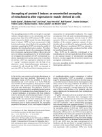
SP-D in synovial fluid and corresponding sera
Synovial fluid was obtained from 20 patients at baseline.
Median SP-D in synovial fluid was 275 ng/ml (221; 29 9).
SP-D in corresponding sera was 678 ng/ml (592; 829). SP-
D in synovial fluid and serum levels correlated significantly
(rho = 0.69, P < 0.001 ), Figure 1. Synovial fluid SP-D was
not significantly associated with sex, age, CRP, autoantibo-
dies, any SE or radiographic findings (data no t shown).
There was no detectable SP-D in the debris enriched pel-
lets resulting from centrifugation of the synovial fluid.
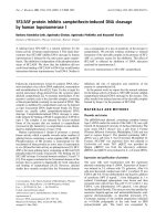
Results from the gel filtration chroma tography are out-
lined in Figure 2. Multimeric SP-D was barely detectable
in synovial fluid as compared to serum, where both multi-
meric and trimeric molecular variant SP-D (trimeric subu-
nits) were detected.
Genetic SP-D variation in RA
The Met11Thr polymorphism was in Hardy-Weinberg
equilibrium in bot h RA and controls (data not shown).
The distribution of genotypes and allele frequencies is
presented in Table 4. When adjusting for gender and
age, there was no overrepresentation of Thr11Thr in RA
patients as compared with controls (Table 3). Circulat-
ing SP-D did not differ between genotypes in R A
patients, whereas healthy individuals with the Thr11Thr
genotype appeared with the lowest level as previously
reported [22]. The genotypes were not associated with
specific disease features including DAS28, CRP, joint
counts, auto-antibodies, HAQ or x-ray findings (data
not shown). The Met11Thr allelic variation could
neither predict x-ray progression nor disease activity
outcome after four years and the size distribution of SP-
D in synovial fluid did not differ between genotypes
(data not shown).
Discussion
Based on the structural similarity between SP-D and
MBL and our preliminary report on low circulating SP-
D in RA [6], this investigation was conducted to study
thepossibleroleofSP-DasdiseasemodifierinRA.
While confirming that SP-D in serum is significantly
decreased in newly-diagnosed, untreated RA sufferers,
we also found an inverse correlation between SP-D and
measures of disease activity at baseline. Although SP-D
increased significantly during follow-up, it remained
subnormal at four years.
The cause of low SP-D in RA is uncertain and differ-
ent mechanisms m ay be involved. Altered SP-D
Figure 1 Scatter plot of SP-D in serum and synovial fluid at baseline (n = 20). Fitted values are depicted by the line. SP-D, surfactant
protein D.
Christensen et al. Arthritis Research & Therapy 2010, 12:R39
/>Page 5 of 9
expression due to genotype abnormalities should be
considered. Thus, in healthy subjects the Thr11-variant
is associated with low SP-D in the circulation [22].
In the previous study by Hoegh et al [6], the Thr11
variant tended to be overrepresented in RA patients as
compared to controls. This trend was no t confirmed in
the present study. Thus, a clear ge netic contribution to
low SP-D in RA cannot be identified in this study. How-
ever, a possible genetic cont ribution to low SP-D in RA
cannot be completely disregarded from this study due to
the limited sample size. Moreover, it should be borne in
mind, that focusing at only one polymorphism in the
analysis of gene patterns and serum SP-D, may underes-
timate the significance of a genetic association, which is
better represented by haplotype blocks [35].
Decreased SP-D in RA could be attributable to
increased clearance from the circulation, for example, by
deposition in inflamed tissues or complex formation
with, for example, microbial or cellular waste [36,37].
Thus, cells undergoing apoptosis express auto-antigens,
which may lead to auto-antibody formation [38]. Both
in vitro and in vivo experiments have indicated that
Figure 2 Size exclusion chromatography of SP-D in serum and synovial fluid. Mean curves of 11 corres ponding serum and synovial fluid
samples. SP-D was eluted as two structurally different forms (SP-D multimers (fraction 10 to 18) and SP-D trimers (fraction 24 to 38)). SP-D,
surfactant protein D.
Table 3 Distribution of the SP-D Met11Thr genotype and allele frequencies and corresponding SP-D serum levels
(median (95% CI))
N(%) of RA patients SP-D ng/ml*
RA patients
N(%) of controls SP-D ng/ml*
Controls
P-value** Odds ratio***
Genotype:
Met11/Met11 41 (27.3) 724 (636; 1,123) 152 (35.8) 1,081 (996; 1,252) P = 0.16 1.0 (ref)
Thr11/Thr11 27 (18.0) 750 (603; 834) 77 (18.1) 896 (788; 955) 1.3 (0.73; 2.4)
Met11/Thr11 82 (54.7) 660 (563; 761) 196 (46.1) 925 (845;1,023) 1.6 (0.97; 2.6)
Allele:
Met11 164 (54.7) 500 (58.8) P = 0.22 1.0 (ref)
Thr11 136 (45.3) 350 (41.2) 1.2 (0.9;1.6)
* Kruskal-Wallis test: RA patients: P = 0.13 and controls: P = 0.0023
** Distribution, P-value calculated using Fishers’ Exact test
*** Odds ratio (95% CI) calculated using logistic regression with health status as the dependent variable and genotype/allele, gender and age as independent
variables.
CI, confidence interval; SP-D, surfactant protein -D; RA, rheumatoid arthritis; Ref, reference
Christensen et al. Arthritis Research & Therapy 2010, 12:R39
/>Page 6 of 9
SP-D enhances clearance of DNA and apoptotic cells by
macrophages, thereby reducing anti-dsDNA antibody
generation [36,39,40]. Such a scavenger mechanism for
SP-D in RA is supported by the inverse association
between SP-D and disease activity measures and by the
gradual SP-D increase during treatment. The inverse
association of SP-D and inflammatory signs and the lack
of association between SP-D and erosive progression
after four years indicate, that subnormal SP-D is primar-
ily linked t o systemic inflammation. According to this,
depressed systemic SP-D may contribute to persistent
low-grade, subclinical joint inflammation as evidenced
by MRI and ultrasonic findings [41,42].
In order to further elucidate the possible role of SP-D in
joint inflammation, we quantifi ed SP-D in p aired serum
and synovial samples and studied the molecular size distri-
bution in serum and synovial fluid. We found a SP-D
serum:synovial fluid ratio at approximately 3:1, which indi-
cates that SP-D reaches the joint cavity by diffusion (bulk
flow) [43]. The diffusion capacity for proteins across the
synovial membrane in rheumatoid arthritis depends on
the degree of synovial inflammation and molecular size
[43-45]. While both multimeric and trimeric subunit SP-D
were present in serum, only trimeric forms could be
demonstrated in synovial fluid. This further supports that
diffusion is the major source of SP-D in the joint cavity
although local degradation of the molecule cannot be
excluded. Knowledge about the biologic properties of tri-
meric SP-D is incomplete. However, previous studies have
indicated that trimers interact preferentially with sp ecific
microbes, microbial compounds or endogenous lipopro-
teins [19,46] implying that trimeric SP-D may possess spe-
cialized functions as compared with multimeric SP-D.
Previously, Gardai et al proposed a model for dual inflam-
matory activity of SP-D. In the absence of microbial
ligands and cell debris, binding of SP-D to macrophages
by the CRD region was suggested to be anti-inflammatory
by blocking p38 mitogen-activated protein kinases (p38
MAPK) [47]. By contrast, binding of microbial constitu-
ents to the CRD region of SP-D would lead to a pro-
inflammatory response [47]. Recently, it was shown that
posttranslational nitrosylation of cystein residues in the
N-terminus of SP-D (SNO-SP-D) caused by inflammation
resulted in disruption of multimeric SP-D into nitrosylated
trimers. This modified trimeric SP-D variant would subse-
quently initiate a pro-inflammatory response via calreticu-
lin/CD91 receptor interaction and activate p38 MAPK
[48]. Inflammatory signalling resulting in p38 phosphory-
lation has been identified as an important determinant of
synovitis severity [49]. Thus, in theory the dominance of
low molecular weight SP-D in synovial fluid observed in
the present study may contribute to the maintenance of
joint inflammation in RA.
SP-D in serum is suggested to originate primarily from
pulmonary leakage [50]. It has previously been demon-
strated that smoking increases SP-D in serum [22]. Our
findings demonstrate that this also applies to RA
patients implying that smoking is a confounder that
should be corrected for in the statistical analysis. It has
been hypothesized that anti- CCP antibodies can be trig-
gered by smoking through citrullination of lung proteins
in SE carriers [51]. We found no correlation between
circulating SP-D and SE status in smoking and non-
smoking RA patients.
When interpreting the present results, the relatively
large number of drop-outs should be considered. How-
ever, there was no difference with respect to baseline
characteristics between completers and non-completers
and the intention to treat analysis included a large
majority of the cohort.
SP-D did not correlate to age in RA patients, but
tended to be higher in males compared to females. By
contrast, SP-D was significantly higher in control males
as compared to females, and SP-D c orrelated positively
with age. This disparity may be due to the different
sizes of the RA and control populations and the relative
overrepresentation of females in the RA cohort. Due to
the difference in age distri bution in the two populations
and rather few controls aged above 50 years we used
logistic regression with adjustment for gender and age
instead of regular frequency matching in comparisons
between controls and patients.
Conclusions
Circulating SP-D is subnormal at disease onset and after
four years treatment in RA. T here were no SP-D
Met11Thr associations with RA disease activity or sub-
normal SP-D. While SP-D did not correlate with x-ray
progression, we found an inverse association between
SP-D and disease activity markers suggesting tha t low
systemic SP-D is involved in the initiation or mainte-
nance of synovitis. Whereas both multimeric and tri-
meric SP-D variants occurred in serum, only low
molecular forms were detected in synovial fluid where it
may contribute to joint inflammation. Overall, this study
suggests that SP-D is implicated in RA pathogenesis at
the protein level.
Abbreviations
Anti-CCP: antibodies against cyclic citrullinated peptides; CI: confid ence
interval; CIMESTRA: Ciclosporine, Methotrexate, Steroid in RA; CRP: c-reactive
protein; DAS: disease activity score; DMARD : disease modifying anti-
rheumatic drug; ES: erosion score; HAQ: health assessment questionnaire;
HLA: human leukocyte antigen; IgM-RF: IgM-rheumatoid factor; JSN: Joint
Space Narrowing score; MBL: mannan-binding lectin; MTX: methotrexate; RA:
rheumatoid arthritis; SE: shared epitopes; SNP: single nucleotide
polymorphism; SP-D: surfactant protein -D; TBS: TRIS-buffered saline; TSS:
total Sharp-van der Heijde Score; VAS: visual analogue scale.
Christensen et al. Arthritis Research & Therapy 2010, 12:R39
/>Page 7 of 9
Acknowledgements
We thank the study of metabolic syndrome and related components
(GEMINAKAR) for providing serum and DNA control samples. In addition, we
appreciate the expert laboratory assistance by Professor Peter Garred at
Department of Clinical Immunology at Rigshospitalet, Copenhagen
University Hospital, Denmark and Niels Heegaard, MD, DmSc at Department
of Biochemistry and Immunology, Statens Serum Institut, Denmark, Professor
C. Bendixen and A. Høj, MSc, PhD at the Department of Animal Breeding
and Genetics, Danish Institute of Agricultural Sciences, Tjele, Denmark, for
doing the SNP analyses.
This study was supported by The Danish Rheumatism Association, Region of
Southern Denmark, Institute of Clinical Research at the University of
Southern Denmark, The A.P. Møller Foundation for the Advancement of
Medical Science, Guldsmed A.L. & D. Rasmussens Mindefond and Else
Poulsens Mindelegat.
Author details
1
Department of Rheumatology, Odense University Hospital, Sdr. Boulevard
29, DK-5000 Odense C, Denmark and Institute of Clinical Research, University
of Southern Denmark, Winsloewparken 19, DK-5000 Odense C, Denmark.
2
Medical Biotechnology Centre, University of Southern Denmark,
Winsloewparken 25, DK-5000 Odense C, Denmark.
3
Department of
Rheumatology, Rheumatism Hospital, Toldbodgade 3, DK-6300 Graasten,
Denmark.
4
Department of Rheumatology, Copenhagen University Hospitals,
Hvidovre and Glostrup, Kettegaards Alle 30, DK-2650 Hvidovre, Denmark.
5
Department of Rheumatology, Aarhus University Hospital, Noerrebrogade
44, DK-8000 Aarhus C, Denmark.
6
Department of Rheumatology,
Copenhagen University Hospital, Rigshospitalet, Blegdamsvej 9, DK-2100
Copenhagen, Denmark.
7
Department of Rheumatology, Copenhagen
University Hospitals, Herlev and Gentofte, Niels Andersens Vej 65, DK-2900
Hellerup, Denmark.
8
Department of Radiology, Copenhagen University
Hospital, Hvidovre, Kettegaards Alle 30, DK-2650 Hvidovre, Denmark.
9
Department of Radiology, Aarhus University Hospital, Noerrebrogade 44,
DK-8000 Aarhus C, Denmark.
Authors’ contributions
All authors contributed to the design of the study, and the acquisition and
interpretation of data. AFC performed the statistical analysis. AFC, PJ and GL
drafted the manuscript. KJ carried out the immunoassays and gel filtration
chromatography. AGJ and AV evaluated the x-ray data. All authors read and
approved the final manuscript.
Competing interests
The authors declare that they have no competing interests.
Received: 26 August 2009 Revisions requested: 23 October 2009
Revised: 11 January 2010 Accepted: 8 March 2010
Published: 8 March 2010
References
1. Arend WP: The innate immune system in rheumatoid arthritis. Arthritis
Rheum 2001, 44:2224-2234.
2. Garred P, Madsen HO, Marquart H, Hansen TM, Sørensen SF, Petersen J,
Volck B, Svejgaard A, Graudal NA, Rudd PM, Dwek RA, Sim RB, Andersen V:
Two edged role of mannose binding lectin in rheumatoid arthritis: a
cross sectional study. J Rheumatol 2000, 27 :26-34.
3. Saevarsdottir S, Vikingsdottir T, Vikingsson A, M anfredsdottir V, Geirsson AJ,
Valdimarsson H: Low Mannose Binding Lectin Predicts Poor Prognosis in
Patients with Early Rheumatoid Arthritis. A Prospective Study.
J Rheumatol 2001, 28:728-734.
4. Graudal NA, Madsen HO, Tarp U, Svejgaard A, Jurik AG, Graudal HK,
Garred P: The association of variant mannose-binding lectin genotypes
with radiographic outcome in rheumatoid arthritis. Arthritis Rheum 2000,
43:515-521.
5. Jacobsen S, Madsen HO, Klarlund M, Jensen T, Skjodt H, Jensen KE,
Svejgaard A, Garred P, TIRA Group: The influence of mannose binding
lectin polymorpisms on disease outcome in early polyarthritis.
J Rheumatol 2001, 28:935-942.
6. Hoegh SV, Lindegaard HM, Sorensen GL, Hoj A, Bendixen C, Junker P,
Holmskov U: Circulating Surfactant Protein D is Decreased in Early
Rheumatoid Arthritis: A 1-year Prospective Study. Scand J Immunol 2008,
67:71-76.
7. Holmskov U: Collectins and collectin receptors in innate immunity. APMIS
Suppl 2000, 100:1-59.
8. Holmskov U, Thiel S, Jensenius JC: Collectins and ficolins: Humoral Lectins
of the Innate Immune Defense. Annu Rev Immunol 2003, 21:547-578.
9. Crouch E, Chang D, Rust K, Persson A, Heuser J: Recombinant pulmonary
surfactant protein D. Post-translational modification and molecular
assembly. J Biol Chem 1994, 269:15808-15813.
10. Bufler P, Schmidt B, Schikor D, Bauernfeind A, Crouch EC, Griese M:
Surfactant Protein A and D Differently Regulate the Immune Response
to Nonmucoid Pseudomonas aeruginosa and Its Lipopolysaccharide. Am
J Respir Cell Mol Biol 2003, 28:249-256.
11. Hartshorn KL, White MR, Tecle T, Tornoe I, Sorensen GL, Crouch EC,
Holmskov U: Reduced influenza viral neutralizing activity of natural
human trimers of surfactant protein D. Respir Res 2007, 8:9.
12. Hartshorn KL, Crouch E, White MR, Colamussi ML, Kakkanatt A, Tauber B,
Shepherd V, Sastry KN: Pulmonary surfactant proteins A and D enhance
neutrophil uptake of bacteria. Am J Physiol 1998, 274:L958-L969.
13. Leth-Larsen R, Garred P, Jensenius H, Meschi J, Hartshorn K, Madsen J,
Tornoe I, Madsen HO, Sorensen G, Crouch E, Holmskov U: A common
polymorphism in the SFTPD gene influences assembly, function, and
concentration of surfactant protein D. J Immunol 2005, 174:1532-1538.
14. Crouch E, Parghi D, Kuan SF, Persson A: Surfactant protein D: subcellular
localization in nonciliated bronchiolar epithelial cells. Am J Physiol 1992,
263:L60-L66.
15. Stahlman MT, Gray ME, Hull WM, Whitsett JA: Immunolocalization of
surfactant Protein-D (SP-D) in human fetal, newborn, and adult tissues.
J Histochem Cytochem 2002, 50:651-660.
16. Madsen J, Kliem A, Tornoe I, Skjodt K, Koch C, Holmskov U: Localization of
lung surfactant protein D on mucosal surfaces in human tissues.
J Immunol 2000, 164:5866-5870.
17. Honda Y, Kuroki Y, Matsuura E, Nagae H, Takahashi H, Akino T, Abe S:
Pulmonary surfactant protein D in sera and bronchoalveolar lavage
fluids. Am J Respir Crit Care Med 1995, 152:1860-1866.
18. Kankavi O: Increased expression of surfactant protein A and D in
rheumatoid arthritic synovial fluid. Croat Med J 2006, 47:155-161.
19. Leth-Larsen R, Nordenbaek C, Tornoe I, Moeller V, Schlosser A, Koch C,
Teisner B, Junker P, Holmskov U: Surfactant protein D (SP-D) serum levels
in patients with community-acquired pneumonia. Clin Immunol 2003,
108:29-37.
20. Nagae H, Takahashi H, Kuroki Y, Honda Y, Nagata A, Ogasawara Y, Abe S,
Akino T: Enzyme-linked immunosorbent assay using F(ab’)2 fragment for
the detection of human pulmonary surfactant protein D in sera. Clin
Chim Acta 1997, 266:157-171.
21. Ni M, Evans DJ, Hawgood S, Anders EM, Sack RA, Fleiszig SMJ: Surfactant
protein D is present in human tear fluid and the cornea and inhibits
epithelial cell invasion by pseudomonas aeruginosa. Infect Immun 2005,
73:2147-2156.
22. Sorensen GL, Hjelmborg J, Kyvik KO, Fenger M, Hoj A, Bendixen C,
Sorensen T, Holmskov U: Genetic and environmental influences of
surfactant protein D serum levels. Am J Physiol Lung Cell Mol Physiol 2006,
290:L1010-L1017.
23. Hetland ML, Stengaard-Pedersen K, Junker P, Lottenburger T, Ellingsen T,
Andersen LS, Hansen I, Skjødt H, Pedersen JK, Lauridsen UB, Svendsen A,
Tarp U, Pødenphant J, Hansen G, Lindegaard H, de Carvalho A,
Østergaard M, Hørslev-Petersen K, the Cimestra Study Group: Combination
treatment with methotrexate, ciclosporine, and intraarticular
betamethasone compared with methotrexate and intraarticular
betamethasone in early active rheumatoid arthritis. Arthritis Rheum 2006,
54:1401-1409.
24. Hetland ML, Stengaard-Pedersen K, Junker P, Lottenburger T, Hansen I,
Andersen LS, Tarp U, Svendsen A, Pedersen JK, Skjodt H, Lauridsen UB,
Ellingsen T, Hansen G, Lindegaard H, Vestergaard A, Jurik AG, Ostergaard M,
Horslev-Petersen K: Aggressive combination therapy with intraarticular
glucocorticoid injections and conventional DMARDs in early rheumatoid
arthritis Two Year Clinical and Radiographic Results From The CIMESTRA
Study. Ann Rheum Dis 2008, 67:815-822.
25. Arnett FC, Edworthy SM, Bloch DA, McShane DJ, Fries JF, Cooper NS,
Healey LA, Kaplan SR, Liang MH, Luthra HS, et al: The American
Christensen et al. Arthritis Research & Therapy 2010, 12:R39
/>Page 8 of 9
Rheumatism Association 1987 revised criteria for the classification of
rheumatoid arthritis. Arthritis Rheum 1988, 31:315-324.
26. Thorsen H, Hansen TM, Mckenna SP, Sørensen SF, Whalley D: Adaption into
Danish of the stanford health assessment questionnaire (HAQ) and the
rheumatoid arthritis quality of life scale (RaQol). Scand J Rheumatol 2001,
30:103-109.
27. Prevoo MLL, van ‘T Hof MA, Kuper HH, van Leeuwen MA, Putte van de LBA,
van Riel PL: Modified disease activity scores that include twenty-eight-
joint counts. Arthritis Rheum 1995, 38:44-48.
28. Høier-Madsen M, Nielsen LP, Møller S: Determination of IgM rheumatoid
factors by enzyme-linked immunosorbent assay (ELISA). Ugeskr Laeger
1986, 148:2018-2021.
29. Nishimura K, Sugiyama D, Kogata Y, Tsuji G, Nakazawa T, Kawano S, Saigo K,
Morinobu A, Koshiba M, Kuntz KM, Kamae I, Kumagai S: Meta-analysis:
Diagnostic Accuracy of Anti-Cyclic Citrullinated Peptide Antibody and
Rheumatoid Factor for Rheumatoid Arthritis. Ann Intern Med 2007,
146:797-808.
30. Vasiliauskiene L, Wiik A, Hoier-Madsen M: Prevalence and clinical
significance of antikeratin antibodies and other serological markers in
Lithuanian patients with rheumatoid arthritis. Ann Rheum Dis 2001,
60:459-466.
31. Heijde van der D: How to read radiographs according to Sharp/van der
Heijde method. J Rheumatol 2000, 27:261-263.
32. Bathon JM, Martin RW, Fleischmann RM, Tesser JR, Schiff MH, Keystone EC,
Genovese MC, Wasko M, Moreland LW, Weaver AL, Markenson J, Finck BK:
A Comparison of Etanercept and Methotrexate in Patients with Early
Rheumatoid Arthritis. N Engl J Med 2000, 343:1586-1593.
33. Hetland M, Ejbjerg B, Horslev-Petersen K, Jacobsen S, Vestergaard A, Jurik A,
Stengaard-Pedersen K, Junker P, Lottenburger T, Hansen I, Andersen LS,
Tarp U, Skjodt H, Pedersen J, Majgaard O, Svendsen AJ, Ellingsen T,
Lindegaard HM, Christensen AF, Vallo J, Torfing T, Narvestad E, Thomsen HS,
Ostergaard M, CIMESTRA study group: MRI bone oedema is the strongest
predictor of subsequent radiographic progression in early rheumatoid
arthritis. Results from a 2 year randomized controlled trial (CIMESTRA).
Ann Rheum Dis 2009, 68:384-390.
34. Kimura A, Sasazuki T: Eleventh International Histocompatibility workshop.
HLA 1991 Oxford: Oxford University PressTsuji K, Aizawa M, Sasazuki M 1992.
35. Heidinger K, Konig IR, Bohnert A, Kleinsteiber A, Hilgendorff A, Gortner L,
Ziegler A, Chakraborty T, Bein G: Polymorphisms in the human surfactant
protein-D (SFTPD) gene: strong evidence that serum levels of surfactant
protein-D (SP-D) are genetically influenced. Immunogenetics 2005, 57:1-7.
36. Palaniyar N, Clark H, Nadesalingam J, Hawgood S, Reid KB: Surfactant
protein d binds genomic DNA and apoptotic cells, and enhances their
clearance, in vivo. Ann NY Acad Sci 2003, 1010:471-475.
37. Palaniyar N, Nadesalingam J, Clark H, Shih MJ, Dodds AW, Reid KB: Nucleic
acid is a novel ligand for innate, immune pattern recognition collectins
surfactant proteins A and D and mannose-binding lectin. J Biol Chem
2004, 279:N32728-32736.
38. Liu G, Wu C, Wu Y, Zhao Y: Phagocytosis of apoptotic cells and immune
regulation. Scand J Immunol
2006, 64:1-9.
39. Palaniyar N, Clark H, Nadesalingam J, Shih MJ, Hawgood S, Reid KB: Innate
immune collectin surfactant protein D enhances the clearance of DNA
by macrophages and minimizes anti-DNA antibody generation. J
Immunol 2005, 174:I7352-7358.
40. Vandivier RW, Ogden CA, Fadok VA, Hoffmann PR, Brown KK, Botto M,
Walport MJ, Fisher JH, Henson PM, Greene KE: Role of Surfactant Proteins
A, D, and C1q in the Clearance of Apoptotic Cells In Vivo and In Vitro:
Calreticulin and CD91 as a Common Collectin Receptor Complex. J
Immunol 2002, 169:3978-3986.
41. Brown AK, Quinn MA, Karim Z, Conaghan PG, Peterfy CG, Hensor E,
Wakefield RJ, O’Connor PJ, Emery P: Presence of significant synovitis in
rheumatoid arthritis patients with disease-modifying antirheumatic
drug-induced clinical remission. Arthritis Rheum 2006, 54:3761-3773.
42. Brown AK, Conaghan PG, Karim Z, Quinn MA, Ikeda K, Peterfy CG, Hensor E,
Wakefield RJ, O’Connor PJ, Emery P: An explanation for the apparent
dissociation between clinical examination and continued structural
detoriation in rheumatoid arthritis. Arthritis Rheum 2008, 58:2958-2967.
43. Kushner I, Somerville JA: Permeability of human synovial membrane to
plasma proteins. Relationship to molecular size and inflammation.
Arthritis Rheum 1971, 14:560-570.
44. Pejovic M, Stankovic A, Mitrovic DR: Determination of the apparent
synovial permeability in the knee joint of patients suffering from
osteoarthritis and rheumatoid arthritis. Br J Rheumatol 1995, 34:520-524.
45. Simkin PA, Pizzorno JE: Synovial permeability in rheumatoid arthritis.
Arthritis Rheum 1979, 22:689-696.
46. Sorensen GL, Hoegh SV, Leth-Larsen R, Thomsen TH, Floridon C, Smith K,
Kejling K, Tornoe I, Crouch EC, Holmskov U: Multimeric and trimeric
subunit SP-D are inconvertible structures with distinct ligand interaction.
Mol Immunol 2009, 10.1016/j.molimm.2009.06.005.
47. Gardai SJ, Xiao YQ, Dickinson M, Nick JA, Voelker DR, Greene KE,
Henson PM: By binding SIRP[alpha] or Calreticulin/CD91, lung collectins
act as dual function surveillance molecules to suppress or enhance
inflammation. Cell 2003, 115:13-23.
48. Guo CJ, Atochina-Vasserman EN, Abramova E, Foley JP, Zaman A, Crouch E,
Beers MF, Savani RC, Gow AJ: S-nitrosylation of surfactant protein-D
controls inflammatory function. PLoS Biol 2008, 6 :e266.
49. Schett G, Zwerina J, Firestein GS: The p38 mitogen-activated protein
kinase (MAPK) pathway in rheumatoid arthritis. Ann Rheum Dis 2008,
67:909-916.
50. Hermans CEDR, Bernard ALFR: Lung epithelium-specific proteins.
characteristics and potential applications as markers. Am J Respir Crit Care
Med 1999, 159:646-678.
51. Klareskog L, Stolt P, Lundberg K, Källberg H, Bengtsson C, Grunewald J,
Rönnelid J, Harris HE, Ulfgren AK, Rantapää-Dahlqvist S, Eklund A,
Padyukov L, Alfredsson L, the EIRA study group:
A new model for an
etiology of rheumatoid arthritis: Smoking may trigger HLA-DR (shared
epitope)-restricted immune reactions to autoantigens modified by
citrullination. Arthritis Rheum 2006, 54:38-46.
doi:10.1186/ar2948
Cite this article as: Christensen et al.: Circulating surfactant protein -D is
low and correlates negatively with systemic inflammation in early,
untreated rheumatoid arthritis. Arthritis Research & Therapy 2010 12:R39.
Submit your next manuscript to BioMed Central
and take full advantage of:
• Convenient online submission
• Thorough peer review
• No space constraints or color figure charges
• Immediate publication on acceptance
• Inclusion in PubMed, CAS, Scopus and Google Scholar
• Research which is freely available for redistribution
Submit your manuscript at
www.biomedcentral.com/submit
Christensen et al. Arthritis Research & Therapy 2010, 12:R39
/>Page 9 of 9