Báo cáo y học: "Inflammatory changes in the airways of mice caused by cigarette smoke exposure are only partially reversed after smoking cessation" docx
Bạn đang xem bản rút gọn của tài liệu. Xem và tải ngay bản đầy đủ của tài liệu tại đây (2.83 MB, 11 trang )
RESEARC H Open Access
Inflammatory changes in the airways of mice
caused by cigarette smoke exposure are only
partially reversed after smoking cessation
Saskia Braber
*
, Paul AJ Henricks, Frans P Nijkamp, Aletta D Kraneveld, Gert Folkerts
Abstract
Background: Tobacco smoking irritates and damages the respiratory tract and contributes to a higher risk of
developing lung emphysema. At present, smoking cessation is the only effective treatment for reducing the
progression of lung emphysema, however, there is hardly anything known about the effects of smoking cessation
on cytokine and chemokine levels in the airways. To the best of our knowledge, this is the first reported in vivo
study in which cytokine profiles were determined after cessation of cigarette smoke exposure.
Methods: The severity of airway remodeling and inflammation was studied by analyzing alveolar enlargement,
heart hypertrophy, inflammatory cells in the bronchoalveolar lavage fluid (BALF) and lung tissue and by
determining the cytokine and chemokine profiles in the BALF of A/J mice exposed to cigarette smoke for
20 weeks and 8 weeks after smoking cessation.
Results: The alveolar enlargement and right ventricle heart hypertrophy found in smoke-exposed mice remained
unchanged after smoking cessation. Although the neutrophilic inflammation in the BALF of cigarette smoke-
exposed animals was reduced after smoking cessation, a sustained inflammation in the lung tissue was observed.
The elevated cytokine (IL-1a and TNF-a) and chemokine (CCL2 and CCL3) levels in the BALF of smoke-exposed
mice returned to basal levels after smoking cessation, while the increased IL-12 levels did not return to its basal
level. The cigarette smoke-enhanced VEGF levels did not significantly change after smoking cessation. Moreover, IL-
10 levels were reduced in the BALF of smoke-exposed mice and these levels were still significantly decreased after
smoking cessation compared to the control animals.
Conclusion: The inflammatory changes in the airways caused by cigarette smoke exposure were only partially
reversed after smoking cessation. Although smoking cessation should be the first step in reducing the progression
of lung emphysema, additional medication could be provided to tackle the sustained airway inflammation.
Introduction
There are currently more than 1.3 billion tobacco smo-
kers worldwide accord ing to the World Health Organi-
zation (WHO) [1]. Cigarette smoke contains more than
4000 hazardous chemical compounds, of which 200 are
highly toxic [2]. It is generally accepted that cigarette
smoking is the most important risk factor for the devel-
opment and progression of chronic obstructive pulmon-
ary disease (COPD) and accounts for about 80% of
COPD cases [3,4]. COPD, a term referring to two lung
diseases: chronic bronchitis and emphysema, is charac-
terized by an airflow limitation that is not fully reversi-
ble. The airflow limitation is usually both progressive
and associated with an abnormal inflammatory response
of the lungs to noxious particles or gases [5]. Pulmonary
hypertension and right ventricular failure are also often
associated with COPD [6,7]. Since a chronic airway
inflammation with alveolar wall destruction and airw ay
remodeling is central to the pathogenesis of COPD, it is
not surprising that several types of inflammatory cells
play a role in this condition [8]. Increased numbers of
macrophages and neutrophils are observed in sputum
andbronchoalveolarlavagefluid(BALF)ofCOPD
patients [9-11]. In addition, COPD patients have
* Correspondence:
Division of Pharmacology, Utrecht Institute for Pharmaceutical Sciences,
Faculty of Science, Utrecht University, Utrecht, The Netherlands
Braber et al. Respiratory Research 2010, 11:99
/>© 2010 Braber et al; licensee BioMed Central Ltd. This is an Open Access article distributed under the terms of the Creative Commons
Attribution License ( censes/by/2.0), which permits unrestricted use, distribution, and reproduction in
any medium, provid ed the original wor k i s properly cited.
elevated levels of T-lymphocytes, in particular CD8+
cells, in lung parenchyma and airways [11-14]. Migra-
tion and activation of inflammatory cells to the lung is
regulated by the release of different mediators, including
proteases, cytokines and chemokines secreted by a vari-
ety of inflammatory and resident ce lls. These mediators
contribute to the chronic inflammatory process with tis-
sue damage and repair processes seen in emphysema
[15,16]. Seve ral cytokines a nd chemokines h ave been
implicated in the airway inflammation in COPD.
Increased levels of interleukin-8 (IL-8), interleukin-12
(IL-12), tumour-necrosis factor-a (TNF-a), monocyte
chemotactic protein-1 (MCP-1; CCL-2), and macro-
phage inflammatory protein-1a (MIP-1a;CCL3)have
been observed in COPD patients [9,17-21]. In general,
the treatments available for COPD reduce the number
and severity of exacerbations and relieve symp toms, but
do not tackle the cause of t he disease and have a lim-
ited effect on slowing down the progression of lung
damage [22]. At present, smoking cessation is the only
effective treatment for avoiding or reducing the progres-
sion of COPD [23]. However, there is contradictory evi-
dence re garding the effect of smoking cessation on
airway inflammation associated with COPD. Several stu-
dies in COPD patients reported that smoking cessation
improves respiratory symptoms, reduces loss of pul-
monary function and decreases lung inflammation
[24-28], while other studies have shown that smoking
cessation fails to reverse the chronic airway inflamma-
tion [29-32]. Unfortunately, there is insufficient evidence
regarding the effects of smoking cessation on cytokine
and chemokine levels, which do play an important role
in airway inflammation and tissue remodeling s een in
COPD. Therefore, a murine model of cigarette smoke-
induced lung emphysema was used to investigate the
effect of smoking cessation on airway remodeling and
pulmonary inflammation. The severity of airway remo-
deling and inflammation was studied by determining
alveolar enlargement, heart hypertrophy, inflammatory
cells in the bronchoalveolar lavage fluid (BALF)
and lung tissue and by analyzing the cytokine and che-
mokine profiles in the BALF of mice exposed to cigar-
ette smoke for 20 weeks and 8 weeks after smoking
cessation.
Materials and methods
Animals
Female A/J mice, 9-14 weeks old (Charles River Labora-
tories) were housed under controlled conditions in stan-
dard laboratory cages. They were provided free access to
wat er and food. All in vivo experimental protocols were
approved by the local Ethics Committee and were per-
formed under strict governmental and international
guidelines on animal experimentation.
Cigarette smoke exposure
Female A/J mice were divided into three groups. The
first group was exposed to room air for 20 weeks, the
second gro up was exposed to cigar ette smoke for
20 weeks and the third group was exposed t o cigarette
smoke for 20 weeks followed by a period of 8 weeks
without cigarette smoke exposure. 20-weeks-old mice
are adult mice and should have almost no alveolar
growth in the additional 8 weeks [33,34]. In the life-
span of a laboratory mouse 20 weeks smoking and
8 weeks smoking cessation represents approximately
21 years smoking and 8 years smoking cessation in
humans. The mice were exposed in whole-body cham-
bers to air (sham) or to diluted mainstream cigarette
smoke from the reference cigarettes 2R4F (University of
Kentucky, Lexington, Kentucky) using a smoking appa-
ratus . Exposures were conducted 4 h/day (with a 30/60-
minute fresh air break after each hour of exposure),
5 days/week for 2 0 weeks to a target cigarette smoke
concentration of 750 μg total particulate matter/l (TPM/
l). This TPM concentration was reached after an adapta-
tion period of 1 week, starting with a TPM concentra-
tion of 125 μg TPM/l. The mass concentration of
cigarette smoke TPM was determined by gravimetric
analysis of Cambridge filter samples. The carbon mon-
oxide (CO) was monitored continuously and was around
800 ppm. The nicotine concentration in the smoke was
approximately 40 μg/l. The sample sites were located in
the middle of the exposure chamber at the breathing
zone. The mice were sacrificed 16-24 hours after the
last air or smoke exposur e, or after the smoke-free per-
iod of 8 weeks.
Histology and morphometric analysis
Mice (n = 4-5), used fo r morphometric analysis, were
sacrific ed by an i.p. injection with an overdose of pento-
barbital (Nembutal™ , Ceva Santé Animale, Naaldwijk,
The Netherlands). The lungs were fixated with a 10%
formalin infusion through the tracheal cannula at a con-
stant pressure of 25 cm H
2
O. After excision, the volume
of the fixed lungs was measured by fluid displacement.
Then, the left lung was immersed in fresh fixative for at
least 24 h, after which it was embedded in paraffin.
After p araffin embedding, 5 μm sections were cut and
stained with hematoxylin/eosin (H&E) according to
standard methods. These histological lung sections were
used to determine lung i nflammation and pigmented
macrophages. Lung inflammation was scored by a treat-
ment-blind observer. The degree of peribronchial and
perivascular inflammation was evaluat ed on a subje ctive
scale of 0-3, as described elsewhere [35,36]. A value of 0
was assigned when no inflammation was detectable, a
value of 1 was adjudged for occasional cuffing with
inflammatory cells, a value of 2 when most bronchi or
Braber et al. Respiratory Research 2010, 11:99
/>Page 2 of 11
vessels were surrounded by a thin layer (one to five cells
thick) o f inflammatory cells, and a value of 3 was given
when most bronchi or vessels were surrounded by a
thick layer (more than five cells thick) of inflammatory
cells. Total lung inflammation was defined as the aver-
age of the peribronchial and perivascular inflammation
scores. Four lung sections per mouse were scored and
inflammation scores were expressed as a mean value.
Morphometric assessment of emphysema, included
determination of the averag e inter-alveolar distance, was
estimated by the mean linear intercept (Lm) analysis.
The Lm was determined by light microscopy at a total
magnification of 100×, whereby 24 random photomicro-
scopic images per left l ung tissue section were eva luated
by microscopic projection onto a reference grid. By
dividing total grid length by the number of alveolar
wall-grid line intersections, the Lm (in μm) was calcu-
lated [37].
Bronchoalveolar lavage
Immediately after i.p. injection with an overdose of pen-
tobarbital, the lungs of a separate group mice (n = 4-5)
were lavaged 4 times through a tracheal cannula with 1
ml saline (NaCl 0.9%), pre-warmed at 37°C. The first
lavage was performed with 1 ml saline containing a mix-
ture of protease inhibitors (Complete Mini, Roche
Applied Science, Penzberg, Germ any). After centrifuging
the bronchoalveolar lavage fluid at 4°C (400 g, 5 min),
the supernatant of the fir st ml was used for cytokine
analysis and the cell pellets of the 4 lavages were used
for cell counts. The 4 cell pellets, kept on ice, were
pooled per animal and resuspended in 150 μlcoldsal-
ine. After staining with Türk solution, total cell counts
per lung were made under light microscopy using
a Burker-Turk chamber. Differential cell counts were
performed on cytospin preparations stained by Diff-
Quick™(Dade A.G., Düdingen, Switzerland). Cells were
identified as macrophag es, neutrophi ls and lymphocytes
according to standard morphology. At least 200 cells
were counted and the absolute number of each cell type
was calculated.
Right ventricular hypertrophy measurement
The right ventricle was removed from lower heart after
removal of the atria. The right ventricle and the left
ventricle plus septum were weighed and the ratio of the
weights was calculated as follows: (right ventricle)/(left
ventricle + septum) [38,39].
Measurement of cytokines and chemokines
A standard mouse cytokine 20-plex assay was used to
determine cytokine a nd chemokine concentrations in
the BALF (n = 4-5) according to the manufacturer’s
instructions (Luminex; Biosource, Invitrogen, Breda, The
Netherlands). The most relevant cytokines and chemo-
kines (IL-1a,IL-10,IL-12,TNF-a, CCL2, CCL3, VEGF
and macrophage inflammatory protein-2 (MIP-2;
CXCL2)) were discussed in this study. The concentra-
tions of the se cytokines and chemokines were expressed
as pg/ml BALF.
Statistical analysis
Experimental results were expressed as mean ± S.E.M.
Differences between groups were statistically determined
by an unpaired two-tailed Student’ s t-test using Graph-
Pad Prism (Version 4.0). Results were considered statis-
tically significant when P < 0.05.
Results
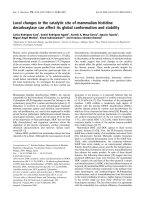
Alveolar enlargement induced by cigarette smoke
exposure is irreversible
The histological lung sections of the smoke-exposed
mice showed an increased a ir space enlargement and
destruction (Fig. 1B) compared with the air-exposed
mice (Fig. 1A). The alveolar enlargement is still present
after a smoking cessation period of 8 weeks (Fig. 1C).
The m ean linear intercept, a quantification method for
alveolar size, was used to quantify the presence and
severity of emphysema [37]. Significant airs pace enlarge-
ment was observed in mice after 20 weeks exposure to
cigarette smoke (Fig. 1D). Furthermore, airspace enlar-
gement induced by cigarette smoke exposure was not
reversible, since the increase in Lm was not significant ly
reduced after a period of 8 weeks without exposure to
cigarette smoke (Fig. 1D).
Right ventricle heart hypertrophy related to cigarette
smoke exposure is irreversible
Twenty weeks cigarette smoke exposure caused right
ventricular heart hypertrophy (Fig. 2). The right ventri-
cular mass was proportionally greater than the rest of
the lower heart (left ventricle and septum) i n smoke-
exposed mice compar ed to air-expose d mice. Moreover,
right ventricle heart hypertrophy was not reversible after
a p eriod of 8 weeks without cigarette smoke exposure,
because the heart hypertrophy ratio (RV/LV +S) was not
significantly decreased in the smoking cessation group
compared to smoke-exposed group.
Lung volume increase after cigarette smoke exposure is
irreversible after smoking cessation
It has been demonstrated that chronic inflammation in
the airways ultimately leads to alveolar enlargement,
increased pulmonary compliance as well as enhanced
lung volumes [40]. We measured the lung volumes in
the murine lung emphysema model and the lung
volume was significantly increased in mice exposed to
cigarette smoke for 20 weeks compared to the control
Braber et al. Respiratory Research 2010, 11:99
/>Page 3 of 11
mice (Fig. 3). After a period of 8 weeks without cigarette
smoke e xposure, the lung volume was still significantly
enhanced compared to the control group.
Smoking cessation reduces the inflammatory cell influx in
bronchoalveolar lavage fluid
Progression of COPD is associated with the accumula-
tion and activation of inflammatory cells in the BALF.
In the present lung emhysema model, the total number
of inflammatory cells was 5-fold increased in the BALF
after 20 weeks of cigarette smoke exposure (Table 1).
Differential cell counts demonstrated that most of the
cells in the BALF of the air-exposed mice were macro-
phages, with a few neutrophils and lymphocy tes. The
number of all these inflammatory cells in the BALF was
significantly increased after cigarette smoke exposure,
especially the neutrophils. Cigarette smoke exposure
also affected the BALF cell composition, since there was
a shift observed from mainly macrophages in the control
Control
Smoke
Smoke cessation
40
45
50
55
**
**
Lm ( m)
D
B
A
C
Figure 1 Cigarette smoke-induced alveolar enlargement is irreversible. Representative photomicrographs of hematoxylin and eosin stained
lung tissue of air-exposed mice (A), smoke-exposed mice (B), smoke-exposed mice 8 weeks after smoking cessation (C). Magnification, ×100.
Mean linear intercept (Lm) values of mice exposed to air (white bar), mice exposed to cigarette smoke for 20 weeks (black bar) and mice
exposed to cigarette smoke for 20 weeks plus a smoking cessation period of 8 weeks (grey bar) (D). n = 4-5 animals per group. Values are
expressed as mean +/- S.E.M. **P ≤ 0.01; significantly different from the control group.
Control Smoke Smoke ce ssation
0.10
0.15
0.20
0.25
0.30
***
***
RV/LV+S
Figure 2 Cigarette smoke-induced right ventricle heart
hypertrophy is irreversible. Right ventricle (RV) and left ventricle
(LV) + septum (S) were dissected after 20 weeks air exposure (white
bar), after 20 weeks smoke exposure (black bar) and after 20 weeks
smoke exposure plus a smoking cessation period of 8 weeks (grey
bar) to determine their weight ratio (RV(LV+S)). n = 6-7 animals per
group. Values are expressed as mean +/- S.E.M. ***P ≤ 0.001;
significantly different from the control group.
Braber et al. Respiratory Research 2010, 11:99
/>Page 4 of 11
animals towards neutrophils in the BAL F of smoke-
exposed mice. After smoking cessation of 8 weeks, we
found a significant decline in inflammatory cells in the
BALF, although the total cell number was still signifi-
cant different compared to the control group (Table 1).
First, the amount of neutrophils was strongly reduced
after smoking cessation, but these cell numbers were
still significantly increased compared to the control
mice. The macrophages w ere also decreased compared
to the smoke-exposed mice, however these numbers
were not returned to basal levels. Finally, the cigarette
smoke-induced increase of lymphocytes was not chan-
ged after cessation of cigarette smoke exposure. These
results indicate that smoking cessat ion leads to a reduc-
tion in inflammatory cell types and a change in cell
composition in the BALF, mainly caused by a decline in
neutrophils.
Lung inflammation is still present in lung tissue after
smoking cessation
Histologi cal lung s ectio ns demonstrated that pulmonary
inflammation with peribronchial and perivascular
inflammatory cell infiltrates was present in the airways
of smoke-exposed mice (Fig. 4B). The air-exposed ani-
mals had no detectable lung inflammation (Fig. 4A).
The smoking cessation group showed that the peribron-
chial and perivascular airway inflammation was still pre-
sent after a smoke-free period of 8 weeks (Fig. 4C),
since there was no notable difference in the leukocyte
aggregates compared to those found in smoke-exposed
lungs. The scores of peribronchial, perivascular and total
lung inflammation were significantly increased after
20 weeks cigarette smoke exposure compared to air-
exposed mice and these scores were still significantly
enhanced after a smoking c essation period of 8 weeks
(Fig. 4D).
Moreover, there was an accumulation of brown-
pigmented macrophages in lung tissue of smoke-
exposed mice (Fig. 5B) compared to the lung tissue of
the control mice (Fig. 5A). These pigmented macro-
phages were still present after a smoking cessation per-
iod of 8 weeks (Fig. 5C).
The effect of smoking cessation on smoke-induced
changes in cytokine and chemokine levels in BALF
The levels of different cytokines and chemokines (IL-1a,
IL-10, IL-12, TNF-a, CCL2, CCL3 and VEGF) were
measured in the BALF of control mice and in smoke-
exposed mice before and after smoking cessation. Differ-
ences between the cytoki ne/chemokine profiles in t he
BALF before and after smoking cessation were observed.
The concentrations of the pro-inflammatory cytokines
IL-1a and TNF-a were significantly elevated in the
BALF of the cigarette smoke-exposed mice compared to
the air-exposed mice (IL-1a: control: 0 pg/ml BALF ver-
sus smoke: 73.7 ± 8.7 pg/ml BALF, P < 0.001; TNF-a:
control: 17.1 ± 0.3 pg/ml BALF versus smoke: 33.1 ± 2.6
pg/ml BALF, P < 0.01). Both IL-1a and TNF-a returned
completely to basal levels after smoking cessation. The
cigarette smoke-enhanced IL-12 levels in the BALF did
not completely return to its basal level after smoking
cessation (Fig. 6A). In contrast to the pro-inflammatory
cytokines, the levels of the regulatory cytokine IL-10
were significantly decreased in the BALF after cigarette
smoke exposure. Although IL-10 levels were rising after
smoking cessation, the smoke-induced reduction was
Control Smoke Smoke cessation
0.0
0.5
1.0
1.5
2.0
2.5
*
*
Relative lung volume (ml)
Figure 3 Lung volume increase after cigarette smoke exposure
is not reversible after smoking cessation. The relative lung
volume was measured by fluid displacement. The relative lung
volumes were determined after 20 weeks air exposure (white bar),
after 20 weeks smoke exposure (black bar) and after 20 weeks
smoke exposure plus a smoking cessation period of 8 weeks (grey
bar). n = 4-5 animals per group. Values are expressed as mean +/-
S.E.M. *P ≤ 0.05; significantly different from the control group.
Table 1 Immune cells in BALF recovered from air-exposed mice, smoke-exposed mice and smoke-exposed mice 8
weeks after smoking cessation
Control Smoke Smoke cessation
Total cell count, × 10
4
30.0 ± 3.2 140.4 ± 2.6 *** 52.8 ± 5.0 ** ^^^
Differential cell count, × 10
4
Macrophages 29.2 ± 3.1 56.1 ± 1.1 *** 42.4 ± 4.2 * ^
Neutrophils 0.27 ± 0.1 79.9 ± 3.5 *** 6.1 ± 0.5 *** ^^^
Lymphocytes 0.51 ± 0.1 4.4 ± 1.0 * 4.4 ± 1.0 *
n = 4-5 animals per group. Values are expressed as mean +/- S.E.M. *P ≤ 0.05, **P ≤ 0.01, ***P ≤ 0.001; significantly different from the control group. ^P ≤ 0.05,
^^^ P ≤ 0.001; significantly different from the smoke group.
Braber et al. Respiratory Research 2010, 11:99
/>Page 5 of 11
AB
C
Control
Smoke
Smoke cessation
0
1
2
3
***
***
***
***
***
***
Periv ascular
Peribronchial
Total
Lung inflammation score
D
Figure 4 Lung inflammation is still present in lung tissue after smoking cessation. Representati ve photomicrographs of hematoxyli n and
eosin stained lung tissue of air-exposed mice (A), smoke-exposed mice (B), smoke-exposed mice 8 weeks after smoking cessation (C).
Magnification, ×100. The histological sections were scored for the presence of peribronchial and perivascular inflammation (D). Total lung
inflammation was defined as the average of the peribronchial and perivascular inflammation scores. n = 4-5 animals per group. Values are
expressed as mean +/- S.E.M. ***P ≤ 0.001; significantly different from the control group.
AB
C
Figure 5 Pigmented macrophage accumulation in the lung tissue before and after smoking cessation. Representative photomicrographs
of hematoxylin and eosin stained lung tissue of air-exposed mice (A), smoke-exposed mice (B), smoke-exposed mice 8 weeks after smoking
cessation (C). n = 4-5 animals per group. Magnification, ×400.
Braber et al. Respiratory Research 2010, 11:99
/>Page 6 of 11
still significantly different from the control group
(Fig. 6B). Furthermore, the chemokine levels CCL2 and
CCL3 were increased in the BALF of cigarette smoke-
exposed mice as compared to the control mice (CCL2:
control: 17.8 ± 0.2 pg/ml BALF versus smoke: 298.8 ±
47.7 pg/ml BALF, P < 0.01; CCL3: control: 12.1 ± 3.7
pg/ml BALF versus smoke: 133.6 ± 26.8 pg/ml BALF, P
< 0.01), while these chemokines returned completely
towards basal levels after smoking cessation. The VEGF
levels were enhanced in the BALF after chronic cigarette
smoke exposure and were still significantly eleva ted
compared to the air-exposed mice after 8 weeks smok-
ing cessation (Fig.6C).
Since no CXCL2 levels were detected in the BALF of
the smoke-exposed mice, CXCL2 levels were also exam-
ined in the lung homogenates of these animals. A signif-
icant increase of the CXCL2 concentration was observed
in the lung homogenates of the smoke-exposed mice
(4820.7 ± 820.1 pg/ml/mg protein, P < 0.05) compared
to the control animals (1108.1 ± 727.2 pg/ml/mg pro-
tein). After smoking cessation the smoke-induced
increase of CXCL2 levels was still evident (4175.6 ±
1338.6 pg/ml/mg protein).
Discussion
This study investigated the effects of smoking cessation
on airway remodeling and pulmonary inflammation.
First, airspace enlargement in the animal model for lung
emphysema was evident after 20 weeks c igarette smoke
exposure. This enlar gement was not significant redu ced
after smoking cessation, suggesting that induction of
lung emphysema by alveolar wall destruction is not
reversible. These findings are in agreement with the in
vivo data of Wright and Sun [41] and March et al. [42],
who demonstrated that emphysema was still present in
guinea pigs and mice a fter smoke exposure followed by
a smoking cessation period. Vernooy et al. [43] also
found that long-term LPS exposure results in irreversi-
ble alveolar enlargement in mice. The effect of cigarette
smoke is believed to be strain dependent. A/J mice were
used in the present COPD model, since this strain is
characterized as moderately susceptible to the develop-
ment of lung emphysema and to the lung inflammatory
response after acute cigarette smoke exposure [44,45].
The persistent emphysema observed in the present mur-
ine model is also similar to findings in people who have
stopped smoking. The alveolar enlargement and destruc-
tion seen in lung emphysema is generally thought to be
irreversible [46-48]. Besides the determination of lung
emphysema, we were interested in the lung volume. In
the current study, cigarette smoke-exposed mice showed
a significantly increased relative lung volume compared
to the air-exposed mic e, which is a characteristic feature
of lung emphysema [40]. This lung volume was stil l sig-
nificantly enhanced after smoking cessation, which sup-
ported the irreversible alveolar changes after cigarette
smoke exposure.
Furthermore, right ventricle heart hypertrophy was
found in m ice exposed to cigarette smoke, indicating
changes in the structure of the heart. Other authors also
demonstrated right ventricle heart hypertrophy as well
in animal models for lung emphysema as in COPD
patients [6,7,38,39,49]. A possible explanation for the
development of right ventricle heart hypertrophy could
be pulmonary hypertension, caused by hypoxic pulmon-
ary vasoconstriction or remodeling of the pulmonary
vessels, two important complications of COPD [6,50,51].
VEGF is identified as an endothelial cell specif ic growth
Figure 6 The effect of smoking cessation on smoke-in duced changes in cytokine and chemo kine levels in BALF. Levels of the pro-
inflammatory cytokine IL-12 (A), the regulatory cytokine IL-10 (B) and the growth factor VEGF (C) in the BALF of air-exposed mice (white bars),
smoke-exposed mice (black bars), smoke-exposed mice 8 weeks after smoking cessation (grey bars). n = 4-5 animals per group. Values are
expressed as mean +/- S.E.M. *P ≤ 0.05, **P ≤ 0.01, ***P ≤ 0.001; significantly different from the control group. ^P ≤ 0.05, ^^P ≤ 0.01;
significantly different from the smoke group.
Braber et al. Respiratory Research 2010, 11:99
/>Page 7 of 11
factor that contributes to angiogenesis and vascular per-
meabili ty [52]. In the current study the increased VEGF
levels observ ed in the BALF of the smoke-expose d mice
could be involved in the pulmon ary vascular remodeling
as a result of pulmonary hypertension, ultimately leading
to right ventricle heart hypertrophy. An enhanced
expression of VEGF was also observe d in the pulmonary
vessels and arteries of COPD patients, suggestin g an
important role for VEGF in the development of pulmon-
ary hypertension [53,54]. However, other studies suggest
that VEGF may have a protective role in the develop-
ment of pulmonary hypertension [55-57]. Like alveolar
enlargement, the right ventricle heart hypertrophy and
the increased VEGF in the BALF were irreversible after
smoking cessation. It is possible that the pulmonary
hypertension continued after the recovery period due to
the sustained lung damage and elevated VEGF levels,
which could lead to the ongoing heart hypertrophy. It
remains to be determined whether right ventricle heart
hypertrophy is directly related to lung emphysema or
whether other factors can play a role in the development
and maintaining of heart hypertrophy in COPD patients.
Airway inflammation was present in the airways of
mice exposed to cigarette smoke as shown by an
increase in total cell number in the BALF and by
inflammatory cell infiltration in the lung tissue. Analysis
of differential cell count s in BALF revealed a significant
increase in the number of macrophages, neu trophils and
lymphocytes in the smoke-exposed mice compared to
air-exposed mice, which is described in several in vivo
studies [58-61]. The histological lung sections and l ung
inflammation scores of t he smoke-exposed mice con-
firmed pulmonary inflammation with perivascular and
peribronchial cellular infiltrates, which has also been
demonstrated in other in vivo studies [62,63]. After
smoking c essation, the reduced numbers of inflamma-
tory cells in the BALF did not correlate with the sus-
tained inflammatory cell infiltration observed in lung
tissue. These results support the studies by Seagrave et
al. [64] and March et al. [42,64], who also observed air-
way inflammation and lower levels of inflammatory cells
in the BALF after smoking cessation. It should be noted
that it is very difficult to compare the numerous studies,
since the smoking cessation period, the duration of
smoking and the experimental set-up varied between
the stud ies, which could lead to discrepancies. Addition-
ally, several studies in COPD patients found a normal-
ized cell count in the BALF and sputum after smoking
cessation [24,25]. In contr ast, other studies indicate that
there is an ongoing airway inflammation in COPD
patients who had stopped smoking [29-32]. These find-
ings indica te that inflammatory changes in the airways
of smoke-exposed mice are at least partially reversed
after smoking cessation. The persistent airway
inflammation (especially macrophages and lym phocytes)
could be rela ted to the irreversible tissue damage in the
lungs, or to an ongoing microbial stimulus in the “ sensi-
tive” airways of smokers [65-67] as discussed by Will-
emse et al. [31]. Another explanation could be that
COPD may have an autoimmune component that regu-
lates the sustained airway inflammation after smoking
cessation [68,69].
Little is known about cytokine and chemokine levels
in the BALF after smoking cessation. To the best of our
knowledge, this is the first reported in vivo study in
which cytokine profiles were determined after cessation
of ciga rette smoke exposure. Increased levels of the pro-
inflammatory cytokines IL-1a,IL-12andTNF-a were
observed in the BALF of ci garette smoke-exposed mice.
IL-1a and TNF-a le vels returned to basal levels a fter
smoking cessation, while IL-12 was not normalized. The
cytokines IL-1a, IL-12 and TNF-a are mainly produced
by macrophages [70]. The alterations in these cytokine
levels are in line with the accumulated macrophage
levels before and reduced levels after smoking cessatio n.
As IL-12 is a potent Th1 skewing cytokine, we suggest a
Th1 polarization a fter cigarette smoke exposure. The
decreased IL-10 levels after smoke exp osure will amplify
this polarization towards Th1, since IL-10 down-regu-
lates the expressi on of Th1 cytokine s [71]. Other
authors also describe a possible association between
COPD and a Th1-driven immune response [72,73].
Moreover, after smoking cessation the IL-10 levels were
still significantly reduced compared to the air-exposed
animals. IL-10 could also play a role in function and dif-
ferentiation of the regulatory T cell, which is likely to be
associated with the control of immune responses in
COPD [74,75]. A significant increase of the CXCL2 con-
centration was observed in the lung homogenates of the
smoke-exposed mice compared to the control animals.
The CXCL2 increase is most probably important for the
neutrophil recruitment to the lungs following cigarette
smoke exposure, which is also indicated by Thatcher et
al. [63] . The chemokines CCL2 and CCL3 were also ele-
vated during COPD progression. This is in accordance
with the accumulated macrophage, n eutrophil and lym-
phocyte levels in the BALF of the smo ke-exposed mice,
since CCL2 is a monocyte chemoattractant and is pro-
duced by multiple cell types, including monocytes,
macrophages, endothelial cells and epithelial cells [76].
CCL3 is mainly released by monocytes/macrophages
and is involved in the recruitment and activation of pro-
inflammatory cells, such as T-cells, monocytes/macro-
phages and neutrophils [77,78]. Like IL-12, the synthesis
of CCL3 is typically associated with a Th1 milieu [79].
The C CL3 receptor, CCR1 is upregulated on Th1 cells
by IL-12 [80,81], while CCR5, is prim arily expressed on
Th1 cell s and promotes Th1 skewing [82,83]. Th1 cells
Braber et al. Respiratory Research 2010, 11:99
/>Page 8 of 11
secrete IL-2, IFN-у and TNF- a, which activate CD8+
T-cells. Since CCL3 attracts CD8+ lymphocytes, the ele-
vated CCL3 in the smoke-exposed mice could be related
to the increase in CD8+ T-cells seen in tissues of COPD
patients [84]. These Th1-related cytokines and chemo-
kines were markedly reduced after s moking cessation,
suggesting that the Th1 skewing will diminish after
smoking cessation.
Despite of the decrease in cell numbers and the reduc-
tion in cytokine and chemokin e levels in the BALF after
smoking cessation, the current study demonstrated that
smoking cessation does not result in a profound reduc-
tion of airway inflammation, which is associated with
the sustained emphysema. First, the neutrophils in the
BALF were strongly reduced after smoking cessation to
almost basal levels, but were still significantly increased
compared to the control group. The macrophages in the
alveolar cavity were also not completely restored toward
basal levels after smoking cessation. Furthermore, the
cigarette smoke-induced increase of lymphocytes was
not changed after cessation of cigarette smoke exposure.
Finally, the histological l ung sections showed that the
inflammatory cells and the brown-pigmented macro-
phages were still present in the lung tissue after smok-
ing cessation of 8 weeks, confirming the results
described by Seagrave et al. [64]. The pigmented macro-
phage has been a consistently reported inflammatory
cell type in COPD and contains characteristic brown-
pigmented cytoplasmic inclusions believed to be by-pro-
ducts of cigarette smoke [85-87]. It could be that these
brown-pigmented macrophages together with the ele-
vated lymphocytes in the BALF are responsible for the
sustained airway inflammation observed in the lung tis-
sue after smoking cessation. Future research is needed
to investigate whether this ongoing inflammation is per-
manent after smoking cessation.
In conclusion, cigarette smoke exposur e leads to irre-
versible lung damage and heart hypertrophy. The
inflammatory changes in the airways caused by cigarette
smoke exposure were only partially reversed after smok-
ing cessation. Although smoking cessation should be the
first step in reducing the progression of lung emphy-
sema, additional medication could be provided to tackle
the sustained airway inflammation.
Acknowledgements
The authors would like to thank Kim Verheijden en Marije Kleinjan for their
excellent technical assistance. This study was performed within the
framework of the Dutch Top Institute Pharma Project T1-103.
Authors’ contributions
SB performed the experimental studies and was involved in acquisition and
interpretation of data and drafted the manuscript. FP-N helped on the draft
of the manuscript. PAJ-H, AD-K and GF supervised the study and
contributed to the writing of the final paper. All authors read and approved
the final manuscript.
Competing interests
The authors declare that they have no competing interests.
Received: 1 April 2010 Accepted: 22 July 2010 Published: 22 July 2010
References
1. World Health Organization (WHO): Global Tobacco Treaty Enters into
Force with 57 Countries Committed. World Health Organization Geneva,
World Health Organization Framework Convention 2005, Press Release.
2. Brunnemann KD, Hoffmann D: Analytical studies on tobacco-specific N-
nitrosamines in tobacco and tobacco smoke. Crit Rev Toxicol 1991,
21(4):235-240.
3. Saetta M: Airway inflammation in chronic obstructive pulmonary disease.
Am J Respir Crit Care Med 1999, 160(5 Pt 2):S17-20.
4. Hogg JC: Pathophysiology of airflow limitation in chronic obstructive
pulmonary disease. Lancet 2004, 364(9435):709-721.
5. Mannino DM: Chronic obstructive pulmonary disease: definition and
epidemiology. Respir Care 2003, 48(12):1185-1191, discussion 1191-1183.
6. Naeije R: Pulmonary hypertension and right heart failure in chronic
obstructive pulmonary disease. Proc Am Thorac Soc 2005, 2(1):20-22.
7. Vonk-Noordegraaf A, Marcus JT, Holverda S, Roseboom B, Postmus PE: Early
changes of cardiac structure and function in COPD patients with mild
hypoxemia. Chest 2005, 127(6):1898-1903.
8. Jeffery PK: Structural and inflammatory changes in COPD: a comparison
with asthma. Thorax 1998, 53(2):129-136.
9. Keatings VM, Collins PD, Scott DM, Barnes PJ: Differences in interleukin-8
and tumor necrosis factor-alpha in induced sputum from patients with
chronic obstructive pulmonary disease or asthma. Am J Respir Crit Care
Med 1996, 153(2):530-534.
10. Pesci A, Balbi B, Majori M, Cacciani G, Bertacco S, Alciato P, Donner CF:
Inflammatory cells and mediators in bronchial lavage of patients with
chronic obstructive pulmonary disease. Eur Respir J 1998, 12(2):380-386.
11. Retamales I, Elliott WM, Meshi B, Coxson HO, Pare PD, Sciurba FC,
Rogers RM, Hayashi S, Hogg JC: Amplification of inflammation in
emphysema and its association with latent adenoviral infection. Am J
Respir Crit Care Med 2001, 164(3):469-473.
12. Saetta M, Di Stefano A, Turato G, Facchini FM, Corbino L, Mapp CE,
Maestrelli P, Ciaccia A, Fabbri LM: CD8+ T-lymphocytes in peripheral
airways of smokers with chronic obstructive pulmonary disease. Am J
Respir Crit Care Med 1998, 157(3 Pt 1):822-826.
13. Di Stefano A, Capelli A, Lusuardi M, Balbo P, Vecchio C, Maestrelli P,
Mapp CE, Fabbri LM, Donner CF, Saetta M: Severity of airflow limitation is
associated with severity of airway inflammation in smokers. Am J Respir
Crit Care Med 1998, 158(4):1277-1285.
14. Maeno T, Houghton AM, Quintero PA, Grumelli S, Owen CA, Shapiro SD:
CD8+ T Cells are required for inflammation and destruction in cigarette
smoke-induced emphysema in mice. J Immunol 2007, 178(12)
:8090-8096.
15. Thurlbeck WM, Muller NL: Emphysema: definition, imaging, and
quantification. AJR Am J Roentgenol 1994, 163(5):1017-1025.
16. Tuder RM, McGrath S, Neptune E: The pathobiological mechanisms of
emphysema models: what do they have in common? Pulm Pharmacol
Ther 2003, 16(2):67-78.
17. Churg A, Dai J, Tai H, Xie C, Wright JL: Tumor necrosis factor-alpha is
central to acute cigarette smoke-induced inflammation and connective
tissue breakdown. Am J Respir Crit Care Med 2002, 166(6):849-854.
18. Fuke S, Betsuyaku T, Nasuhara Y, Morikawa T, Katoh H, Nishimura M:
Chemokines in bronchiolar epithelium in the development of chronic
obstructive pulmonary disease. Am J Respir Cell Mol Biol 2004,
31(4):405-412.
19. de Boer WI: Potential new drugs for therapy of chronic obstructive
pulmonary disease. Expert Opin Investig Drugs 2003, 12(7):1067-1086.
20. Di Stefano A, Capelli A, Donner CF: Role of interleukin-8 in the
pathogenesis and treatment of COPD. Chest 2004, 126(3):676-678.
21. Traves SL, Culpitt SV, Russell RE, Barnes PJ, Donnelly LE: Increased levels of
the chemokines GROalpha and MCP-1 in sputum samples from patients
with COPD. Thorax 2002, 57(7):590-595.
22. Belvisi MG, Hele DJ, Birrell MA: New anti-inflammatory therapies and
targets for asthma and chronic obstructive pulmonary disease. Expert
Opin Ther Targets 2004, 8(4):265-285.
23. Rennard SI, Daughton DM: Smoking cessation. Chest 2000, 117(5 Suppl
2):360S-364S.
Braber et al. Respiratory Research 2010, 11:99
/>Page 9 of 11
24. Skold CM, Hed J, Eklund A: Smoking cessation rapidly reduces cell
recovery in bronchoalveolar lavage fluid, while alveolar macrophage
fluorescence remains high. Chest 1992, 101(4):989-995.
25. Swan GE, Hodgkin JE, Roby T, Mittman C, Jacobo N, Peters J: Reversibility
of airways injury over a 12-month period following smoking cessation.
Chest 1992, 101(3):607-612.
26. Scanlon PD, Connett JE, Waller LA, Altose MD, Bailey WC, Buist AS: Smoking
cessation and lung function in mild-to-moderate chronic obstructive
pulmonary disease. The Lung Health Study. Am J Respir Crit Care Med
2000, 161(2 Pt 1):381-390.
27. Pelkonen M, Notkola IL, Tukiainen H, Tervahauta M, Tuomilehto J,
Nissinen A: Smoking cessation, decline in pulmonary function and total
mortality: a 30 year follow up study among the Finnish cohorts of the
Seven Countries Study. Thorax 2001, 56(9):703-707.
28. Godtfredsen NS, Lam TH, Hansel TT, Leon ME, Gray N, Dresler C, Burns DM,
Prescott E, Vestbo J: COPD-related morbidity and mortality after smoking
cessation: status of the evidence. Eur Respir J 2008, 32(4):844-853.
29. Rutgers SR, Postma DS, ten Hacken NH, Kauffman HF, van Der Mark TW,
Koeter GH, Timens W: Ongoing airway inflammation in patients with
COPD who Do not currently smoke. Chest 2000, 117(5 Suppl 1):262S.
30. Gamble E, Grootendorst DC, Hattotuwa K, O’Shaughnessy T, Ram FS, Qiu Y,
Zhu J, Vignola AM, Kroegel C, Morell F, et al: Airway mucosal inflammation
in COPD is similar in smokers and ex-smokers: a pooled analysis. Eur
Respir J 2007, 30(3):467-471.
31. Willemse BW, ten Hacken NH, Rutgers B, Lesman-Leegte IG, Postma DS,
Timens W: Effect of 1-year smoking cessation on airway inflammation in
COPD and asymptomatic smokers. Eur Respir J 2005, 26(5):835-845.
32. Turato G, Di Stefano A, Maestrelli P, Mapp CE, Ruggieri MP, Roggeri A,
Fabbri LM, Saetta M: Effect of smoking cessation on airway inflammation
in chronic bronchitis. Am J Respir Crit Care Med 1995, 152(4 Pt
1):1262-1267.
33. Kawakami M, Paul JL, Thurlbeck WM: The effect of age on lung structure
in male BALB/cNNia inbred mice. Am J Anat 1984, 170(1):1-21.
34. Amy RW, Bowes D, Burri PH, Haines J, Thurlbeck WM: Postnatal growth of
the mouse lung. J Anat 1977, 124(Pt 1):131-151.
35. Tournoy KG, Kips JC, Schou C, Pauwels RA: Airway eosinophilia is not a
requirement for allergen-induced airway hyperresponsiveness. Clin Exp
Allergy 2000, 30(1):79-85.
36. Kwak YG, Song CH, Yi HK, Hwang PH, Kim JS, Lee KS, Lee YC: Involvement
of PTEN in airway hyperresponsiveness and inflammation in bronchial
asthma. J Clin Invest
2003, 111(7):1083-1092.
37. Thurlbeck WM: Measurement of pulmonary emphysema. Am Rev Respir
Dis 1967, 95(5):752-764.
38. Weathington NM, van Houwelingen AH, Noerager BD, Jackson PL,
Kraneveld AD, Galin FS, Folkerts G, Nijkamp FP, Blalock JE: A novel peptide
CXCR ligand derived from extracellular matrix degradation during
airway inflammation. Nat Med 2006, 12(3):317-323.
39. van Houwelingen AH, Weathington NM, Verweij V, Blalock JE, Nijkamp FP,
Folkerts G: Induction of lung emphysema is prevented by L-arginine-
threonine-arginine. Faseb J 2008, 22(9):3403-3408.
40. Senior RM, Shapiro SD: Chronic obstructive pulmonary disease:
epidemiology, pathophysiology, and pathogenesis. Fishman’s Pulmonary
Diseases and Disorders McGraw-Hill Inc., New YorkFishman AP, Elias JA,
Fishman JA, Grippi MA, Kaiser LR, Senior RM 1998, 1:659-681.
41. Wright JL, Sun JP: Effect of smoking cessation on pulmonary and
cardiovascular function and structure: analysis of guinea pig model. J
Appl Physiol 1994, 76(5):2163-2168.
42. March TH, Wilder JA, Esparza DC, Cossey PY, Blair LF, Herrera LK,
McDonald JD, Campen MJ, Mauderly JL, Seagrave J: Modulators of
cigarette smoke-induced pulmonary emphysema in A/J mice. Toxicol Sci
2006, 92(2):545-559.
43. Vernooy JH, Dentener MA, van Suylen RJ, Buurman WA, Wouters EF: Long-
term intratracheal lipopolysaccharide exposure in mice results in chronic
lung inflammation and persistent pathology. Am J Respir Cell Mol Biol
2002, 26(1):152-159.
44. Yao H, Edirisinghe I, Rajendrasozhan S, Yang SR, Caito S, Adenuga D,
Rahman I: Cigarette smoke-mediated inflammatory and oxidative
responses are strain-dependent in mice. Am J Physiol Lung Cell Mol Physiol
2008, 294(6):L1174-1186.
45. Guerassimov A, Hoshino Y, Takubo Y, Turcotte A, Yamamoto M, Ghezzo H,
Triantafillopoulos A, Whittaker K, Hoidal JR, Cosio MG: The development of
emphysema in cigarette smoke-exposed mice is strain dependent. Am J
Respir Crit Care Med 2004, 170(9):974-980.
46. Sharafkhaneh A, Hanania NA, Kim V: Pathogenesis of emphysema: from
the bench to the bedside. Proc Am Thorac Soc 2008, 5(4):475-477.
47. Jeffery PK: Remodeling in asthma and chronic obstructive lung disease.
Am J Respir Crit Care Med 2001, 164(10 Pt 2):S28-38.
48. Saetta M, Turato G, Maestrelli P, Mapp CE, Fabbri LM: Cellular and
structural bases of chronic obstructive pulmonary disease. Am J Respir
Crit Care Med 2001, 163(6):1304-1309.
49. Millard J, Reid L: Right ventricular hypertrophy and its relationship to
chronic bronchitis and emphysema. Br J Dis Chest 1974, 68(2):103-110.
50. Barbera JA, Peinado VI, Santos S: Pulmonary hypertension in chronic
obstructive pulmonary disease. Eur Respir J 2003, 21(5):892-905.
51. Naeije R, Barbera JA: Pulmonary hypertension associated with COPD. Crit
Care 2001, 5(6):286-289.
52. Roy H, Bhardwaj S, Yla-Herttuala S: Biology of vascular endothelial growth
factors. FEBS Lett 2006, 580(12):2879-2887.
53. Kranenburg AR, de Boer WI, Alagappan VK, Sterk PJ, Sharma HS: Enhanced
bronchial expression of vascular endothelial growth factor and receptors
(Flk-1 and Flt-1) in patients with chronic obstructive pulmonary disease.
Thorax 2005, 60(2):106-113.
54. Santos S, Peinado VI, Ramirez J, Morales-Blanhir J, Bastos R, Roca J,
Rodriguez-Roisin R, Barbera JA: Enhanced expression of vascular
endothelial growth factor in pulmonary arteries of smokers and patients
with moderate chronic obstructive pulmonary disease. Am J Respir Crit
Care Med 2003, 167(9):1250-1256.
55. Taraseviciene-Stewart L, Kasahara Y, Alger L, Hirth P, Mc Mahon G,
Waltenberger J, Voelkel NF, Tuder RM: Inhibition of the VEGF receptor 2
combined with chronic hypoxia causes cell death-dependent pulmonary
endothelial cell proliferation and severe pulmonary hypertension. Faseb
J 2001, 15(2):427-438.
56. Campbell AI, Zhao Y, Sandhu R, Stewart DJ: Cell-based gene transfer of
vascular endothelial growth factor attenuates monocrotaline-induced
pulmonary hypertension. Circulation 2001, 104(18):2242-2248.
57. Partovian C, Adnot S, Raffestin B, Louzier V, Levame M, Mavier IM,
Lemarchand P, Eddahibi S: Adenovirus-mediated lung vascular
endothelial growth factor overexpression protects against hypoxic
pulmonary hypertension in rats. Am J Respir Cell Mol Biol 2000,
23(6):762-771.
58. D’Hulst AI, Vermaelen KY, Brusselle GG, Joos GF, Pauwels RA: Time course
of cigarette smoke-induced pulmonary inflammation in mice. Eur Respir J
2005, 26(2):204-213.
59. Martorana PA, Beume R, Lucattelli M, Wollin L, Lungarella G: Roflumilast
fully prevents emphysema in mice chronically exposed to cigarette
smoke. Am J Respir Crit Care Med 2005, 172(7):848-853.
60. Bracke KR, D’Hulst AI, Maes T, Demedts IK, Moerloose KB, Kuziel WA,
Joos GF, Brusselle GG: Cigarette smoke-induced pulmonary inflammation,
but not airway remodelling, is attenuated in chemokine receptor 5-
deficient mice. Clin Exp Allergy 2007, 37(10):1467-1479.
61. Rang asamy T, Misra V, Zhen L, Tankersley CG, Tuder RM, Biswal S:
Cigarette smoke-induced emphysema in A/J mice is ass ociated with
pulmonary oxidative stress, apoptosis of lung cells, and global
alterations in gene expression. Am J Physiol Lung Cell Mol Physiol 2009,
296(6):L888-900.
62. Bracke KR, D’Hulst AI, Maes T, Moerloose KB, Demedts IK, Lebecque S,
Joos GF, Brusselle GG: Cigarette smoke-induced pulmonary inflammation
and emphysema are attenuated in CCR6-deficient mice. J Immunol 2006,
177(7):4350-4359.
63. Thatcher TH, McHugh NA, Egan RW, Chapman RW, Hey JA, Turner CK,
Redonnet MR, Seweryniak KE, Sime PJ, Phipps RP: Role of CXCR2 in
cigarette smoke-induced lung inflammation. Am J Physiol Lung Cell Mol
Physiol 2005, 289(2):L322-328.
64. Seagrave J, Barr EB, March TH, Nikula KJ: Effects of cigarette smoke
exposure and cessation on inflammatory cells and matrix
metalloproteinase activity in mice. Exp Lung Res 2004, 30(1):1-15.
65. Zalacain R, Sobradillo V, Amilibia J, Barron J, Achotegui V, Pijoan JI,
Llorente JL: Predisposing factors to bacterial colonization in chronic
obstructive pulmonary disease. Eur Respir J 1999, 13(2):343-348.
66. Soler N, Ewig S, Torres A, Filella X, Gonzalez J, Zaubet A: Airway
inflammation and bronchial microbial patterns in patients with stable
chronic obstructive pulmonary disease. Eur Respir J 1999, 14(5):1015-1022.
Braber et al. Respiratory Research 2010, 11:99
/>Page 10 of 11
67. Hill A, Gompertz S, Stockley R: Factors influencing airway inflammation in
chronic obstructive pulmonary disease. Thorax 2000, 55(11):970-977.
68. Agusti A, MacNee W, Donaldson K, Cosio M: Hypothesis: does COPD have
an autoimmune component? Thorax 2003, 58(10):832-834.
69. Cosio MG: Autoimmunity, T-cells and STAT-4 in the pathogenesis of
chronic obstructive pulmonary disease. Eur Respir J 2004, 24(1):3-5.
70. Chung KF: Cytokines in chronic obstructive pulmonary disease. Eur Respir
J Suppl 2001, 34:50s-59s.
71. Romagnani S: The Th1/Th2 paradigm. Immunol Today 1997, 18(6):263-266.
72. Brozyna S, Ahern J, Hodge G, Nairn J, Holmes M, Reynolds PN, Hodge S:
Chemotactic mediators of Th1 T-cell trafficking in smokers and COPD
patients. Copd 2009, 6(1):4-16.
73. Hodge G, Nairn J, Holmes M, Reynolds PN, Hodge S: Increased intracellular
T helper 1 proinflammatory cytokine production in peripheral blood,
bronchoalveolar lavage and intraepithelial T cells of COPD subjects. Clin
Exp Immunol 2007, 150(1):22-29.
74. Plumb J, Smyth LJ, Adams HR, Vestbo J, Bentley A, Singh SD: Increased T-
regulatory cells within lymphocyte follicles in moderate COPD. Eur Respir
J 2009, 34(1):89-94.
75. Barcelo B, Pons J, Ferrer JM, Sauleda J, Fuster A, Agusti AG: Phenotypic
characterisation of T-lymphocytes in COPD: abnormal CD4+CD25+
regulatory T-lymphocyte response to tobacco smoking. Eur Respir J 2008,
31(3):555-562.
76. Deshmane SL, Kremlev S, Amini S, Sawaya BE: Monocyte chemoattractant
protein-1 (MCP-1): an overview. J Interferon Cytokine Res 2009,
29(6):313-326.
77. Menten P, Wuyts A, Van Damme J: Macrophage inflammatory protein-1.
Cytokine Growth Factor Rev 2002, 13(6):455-481.
78. Maurer M, von Stebut E: Macrophage inflammatory protein-1. Int J
Biochem Cell Biol 2004, 36(10):1882-1886.
79. Schrum S, Probst P, Fleischer B, Zipfel PF: Synthesis of the CC-chemokines
MIP-1alpha, MIP-1beta, and RANTES is associated with a type 1 immune
response. J Immunol 1996, 157(8):3598-3604.
80. Colantonio L, Iellem A, Clissi B, Pardi R, Rogge L, Sinigaglia F, D’Ambrosio D:
Upregulation of integrin alpha6/beta1 and chemokine receptor CCR1 by
interleukin-12 promotes the migration of human type 1 helper T cells.
Blood 1999, 94(9):2981-2989.
81. D’Ambrosio D, Panina-Bordignon P, Sinigaglia F:
Chemokine receptors in
inflammation: an overview. J Immunol Methods 2003, 273(1-2):3-13.
82. Qin S, Rottman JB, Myers P, Kassam N, Weinblatt M, Loetscher M, Koch AE,
Moser B, Mackay CR: The chemokine receptors CXCR3 and CCR5 mark
subsets of T cells associated with certain inflammatory reactions. J Clin
Invest 1998, 101(4):746-754.
83. Loetscher P, Uguccioni M, Bordoli L, Baggiolini M, Moser B, Chizzolini C,
Dayer JM: CCR5 is characteristic of Th1 lymphocytes. Nature 1998,
391(6665):344-345.
84. Bisset LR, Schmid-Grendelmeier P: Chemokines and their receptors in the
pathogenesis of allergic asthma: progress and perspective. Curr Opin
Pulm Med 2005, 11(1):35-42.
85. Niewoehner DE, Kleinerman J, Rice DB: Pathologic changes in the
peripheral airways of young cigarette smokers. N Engl J Med 1974,
291(15):755-758.
86. Brody AR, Craighead JE: Cytoplasmic inclusions in pulmonary
macrophages of cigarette smokers. Lab Invest 1975, 32(2):125-132.
87. van der Strate BW, Postma DS, Brandsma CA, Melgert BN, Luinge MA,
Geerlings M, Hylkema MN, van den Berg A, Timens W, Kerstjens HA:
Cigarette smoke-induced emphysema: A role for the B cell? Am J Respir
Crit Care Med 2006, 173(7):751-758.
doi:10.1186/1465-9921-11-99
Cite this article as: Braber et al.: Inflammatory changes in the airways of
mice caused by cigarette smoke exposure are only partially reversed
after smoking cessation. Respiratory Research 2010 11:99.
Submit your next manuscript to BioMed Central
and take full advantage of:
• Convenient online submission
• Thorough peer review
• No space constraints or color figure charges
• Immediate publication on acceptance
• Inclusion in PubMed, CAS, Scopus and Google Scholar
• Research which is freely available for redistribution
Submit your manuscript at
www.biomedcentral.com/submit
Braber et al. Respiratory Research 2010, 11:99
/>Page 11 of 11