Chấn thương vai và khuỷu tay trong các vận động viên pptx
Bạn đang xem bản rút gọn của tài liệu. Xem và tải ngay bản đầy đủ của tài liệu tại đây (7.76 MB, 14 trang )
Shoulder and Elbow Injuries in the
Skeletally Immature Athlete
Frank S. Chen, MD, Veronica A. Diaz, MD, Mark Loebenberg, MD, and Jeffrey E. Rosen, MD
Abstract
Shoulder and elbow injuries in the skel-
etally immature are becoming more
frequent as more children and ado-
lescents participate in recreational and
competitive athletics requiring repet-
itive overhead motion. Although most
of these injuries result from chronic
overuse, traumatic injuries to the shoul-
der and elbow also occur. The injury
patterns in these patients are distinct
because their developing physes are
relatively weak. Whereas injuries in
adults tend to involve ligamentous and
soft-tissue structures, injuries in the
skeletally immature commonly involve
the physes as well.
Shoulder and Elbow
Anatomy
To manage these injuries effectively,
clinicians should understand func-
tional shoulder and elbow anatomy
in children and adolescents, as well
as the normal developmental se-
quence of the primary and secondary
ossification centers, which represent
potential sites of injury.
Shoulder
The proximal humeral physis,
which has formidable growth and re-
modeling potential,
1,2
contributes ap-
proximately 80% of the longitudinal
growth of the upper extremity. It is
composed of three primary ossifica-
tion centers—the humeral head, the
greater tuberosity, and the lesser tu-
berosity—that coalesce between the
ages of 5 and 7 years to form a sin-
gle proximal humeral epiphysis. Sub-
sequently, the proximal humeral phy-
sis fuses approximately between the
ages of 14 and 17 years in females and
16 and 18 years in males.
1,3
The capsuloligamentous and mus-
cular structures of the shoulder pro-
vide static and dynamic stability of
the glenohumeral joint.
4-6
The static
stabilizers function primarily at the
extremes of the range of motion
(ROM) as they reciprocally tighten
and loosen to limit humeral head
translation. The dynamic stabilizers
provide stability during the midrange
of motion, when the static stabilizers
are lax. The dynamic stabilizers con-
tract in a coordinated pattern to pro-
vide concavity-compression of the
humeral head within the glenoid cav-
ity, limiting abnormal translation.
4-6
Different portions of the joint cap-
sule, glenohumeral ligaments, and gle-
noid labrum provide static gleno-
humeral stability, depending on the
position of the arm.
4,6
The anterosu-
perior capsule, coupled with the struc-
tures of the rotator interval, limit in-
ferior and posterior translation of the
Dr. Chen is Attending Physician, Sports Medi-
cine Department, Palo Alto Medical Foundation,
Palo Alto, CA. Dr. Diaz is Resident, University
of Miami/Jackson Memorial Hospital, Miami, FL.
Dr. Loebenberg is Consultant Surgeon, Assaf
Harofeh Medical Center, Tel Aviv University
School of Medicine, Tzrifin, Israel. Dr. Rosen is
Assistant Professor, Orthopaedic Surgery, and Di-
rector, Child &Adolescent Sports Medicine, Sports
Medicine/Arthroscopic Surgery, NYU–Hospital
for Joint Diseases, New York, NY.
None of the following authors or the departments
with which they ar e affiliated has received anything
of value from or owns stock in a commercial com-
pany or institution related directly or indirectly
to the subject of this article: Dr. Chen, Dr. Diaz,
Dr. Loebenberg, and Dr. Rosen.
Reprint requests: Dr. Rosen, NYU–Hospital for
Joint Diseases, Suite 2, 305 Second Avenue, New
York, NY 10003.
Copyright 2005 by the American Academy of
Orthopaedic Surgeons.
The intensity of training and competition among young athletes can place them at
increased risk of acute and chronic injuries, which occur in patterns unique to the
skeletally immature athlete. Prompt recognition and treatment of these injuries are
critical to prevent long-term functional disability and deformity. Children and ad-
olescents participating in recreational and organized sports are particularly suscep-
tible to a broad spectrum of shoulder and elbow injuries involving both osseous and
soft-tissue structures. Understanding the relevant functional anatomy, biomechan-
ics of throwing, and pathophysiology of injury can help the clinician manage com-
mon acute traumatic injuries, some of which may result in chronic problems. Over-
use injuries occur more frequently than do acute, traumatic injuries, and early
recognition, coupled with appropriate treatment or prevention, can help restore and
maintain normal shoulder and elbow function.
J Am Acad Orthop Surg 2005;13:172-185
172 Journal of the American Academy of Orthopaedic Surgeons
humeral head in the adducted arm.
4,7
The middle glenohumeral ligament
functions to limit anteroposterior (AP)
translation in the abducted arm at the
midrange of external rotation. The in-
ferior glenohumeral ligament, a com-
plex structure possessing anterior and
posterior bands with an interposing
axillary pouch, serves as the prima-
ry restraint to AP as well as to infe-
rior translation in the abducted and
maximally externally rotated arm.
4,7
The posterior capsule, which does not
have any direct posterior ligamentous
reinforcements, is important in lim-
iting posterior humeral translation in
the adducted, internally rotated, and
forward-flexed arm.
4,5
Overall static
stability is further enhanced by the la-
brum, which deepens the concavity
of the glenoid socket in both the AP
and superior-inferior dimensions. The
labrum also acts as an anchor point
for the capsuloligamentous structures
and the long head of the biceps ten-
don. Labral injury or detachment is
commonly associated with injury to
the associated capsuloligamentous
structures.
4-6
The dynamic stabilizers are the ro-
tator cuff, the long head of the biceps,
the deltoid, and the scapulothoracic
muscles. The posterior cuff muscles
provide dynamic posterior gleno-
humeral stability, with the supraspina-
tus functioning as a powerful abduc-
tor of the arm in the scapular plane
and the infraspinatus and teres mi-
nor combining to externally rotate and
flex the humerus.
5,6,8
The subscapu-
laris, in addition to functioning as an
internal rotator of the humerus, is an
important dynamic anterior stabiliz-
er, given its confluence with the an-
terior capsule.
5,6,8
The long head of the
biceps is important in preventing an-
terior and superior humeral head
translation.
9
Finally, the deltoid and
scapulothoracic muscles—including
the trapezius, levator scapulae, ser-
ratus anterior, and both rhomboid
muscles—function to position the
scapula to provide maximum stabil-
ity at the glenohumeral articulation.
4-6
Elbow
Skeletal maturation of the elbow
occurs at primary and secondary os-
sification centers within the distal hu-
merus, radius, and ulna. Six second-
ary ossification centers represent
potential sites of injury. (1) The capi-
tellum has a variable ossification pat-
tern but usually appears by age 2
years in males. (2) Ossification of the
radial head occurs next, between age
4 and 5 years, followed by (3) the me-
dial epicondyle between age 6 and 7
years and (4) the trochlea between age
9 and 10 years. (5) The olecranon ap-
pears next, usually by age 11, fol-
lowed by (6) the lateral epicondyle
during adolescence.
10,11
Ossification
rates are highly variable and also dif-
fer by sex; rates in females usually
precede those of males by 6 to 12
months. Therefore, clinicians should
obtain radiographs of the contralat-
eral side to evaluate elbow injuries in
the skeletally immature patient.
The osseous anatomy of the el-
bow allows flexion-extension and
pronation-supination through the
ulnohumeral and proximal radioul-
nar articulations, respectively. In full
extension, the elbow possesses a
normal valgus-carrying angle of 11°
to 16°. This bony configuration pro-
vides approximately 50% of the
overall stability of the elbow, primar-
ily against varus stress in the ex-
tended elbow. The anterior joint cap-
sule, the ulnar collateral ligament
(UCL) complex, and the lateral col-
lateral ligament complex provide
the remainder of elbow stability.
12,13
The UCL complex consists of three
main portions: the anterior bundle,
posterior bundle, and oblique bundle
(transverse ligament). The anterior
bundle is functionally the most im-
portant in providing stability against
valgus stress and is further subdivid-
ed into distinct anterior and posteri-
or bands, possessing reciprocal
functions.
12-14
The anterior band is the
primary restraint to valgus stress at
lesser degrees of flexion and is more
susceptible to injury in the extended
elbow.
13,14
The posterior band, because
of its primary stabilizing role at high-
er degrees of elbow flexion, is func-
tionally more important than the an-
terior band in the overhead throwing
athlete.
Originating from the medial epi-
condyle, the flexor-pronator muscu-
lature provides dynamic valgus sta-
bility of the elbow.
15
From proximal
to distal, this muscle mass includes
the pronator teres, flexor carpi radi-
alis, palmaris longus, flexor digi-
torum superficialis, and flexor carpi
ulnaris. Electromyographic (EMG)
and biomechanical studies have
shown the pronator teres, flexor dig-
itorum superficialis, and flexor carpi
ulnaris muscles, which form muscu-
lotendinous units overlying the UCL
complex, to be primarily responsible
for maintaining dynamic valgus sta-
bility.
13,15
The lateral collateral ligament com-
plex is less well understood than the
medial ligamentous structures. It is
composed of three distinct portions:
the radial collateral ligament, the lat-
eral UCL, and the accessory lateral col-
lateral ligament.
12,14
The lateral UCL
has been shown to be the primary re-
straint against rotatory subluxation of
the ulnohumeral joint; injury to this
structure allows posterolateral rota-
tory instability to develop.
14
The ra-
dial collateral ligament is reportedly
an important secondary restraint of
the lateral elbow, along with the ex-
tensor muscles, including the exten-
sor digitorum communis, brachiora-
dialis, and extensor carpi radialis
longus and brevis.
12,14
These muscles
impart dynamic stability to the later-
al elbow. EMG studies have shown
that they exhibit complex, interdepen-
dent firing patterns throughout the
throwing motion; thus, they may be
vulnerable to overuse injuries.
8,15
Biomechanics of Throwing
The overall motion and kinematics of
throwing in adolescents are similar to
Frank S. Chen, MD, et al
Vol 13, No 3, May/June 2005 173
those in adults, with stresses that are
similar but of lesser absolute magni-
tude.
16
Proper throwing mechanics
can and should be taught at a young
age, along with strengthening of the
upper extremity, to reduce injury
rates.
Although specific techniques of
overhead throwing vary with differ-
ent sports, the basic motion is simi-
lar. The baseball pitch has been the
most studied and can be divided into
five main stages
8,17
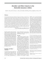
(Fig. 1). In stage
1, or windup, the elbow is flexed and
the shoulder is in slight internal ro-
tation; muscular activity is minimal.
Stage 2, or early cocking, begins
when the ball leaves the nondomi-
nant gloved hand and ends when the
forward foot contacts the gr ound. The
shoulder begins to abduct and rotate
externally. This stage entails early
activation of the deltoid followed by
activation of the supraspinatus, in-
fraspinatus, and teres minor mus-
cles.
5,6,8,17
Stage 3, or late cocking, is charac-
terized by further shoulder abduction
and maximal external r otation as well
as increasing elbow flexion and fore-
arm pronation. Activity levels of the
supraspinatus, infraspinatus, and te-
res minor reach their peak during the
midportion of this phase, and sub-
scapularis and periscapular muscu-
lar activity increase.
6,8,17
Tremendous
shear forces are generated across the
anterior shoulder, predominantly by
the rotator cuff muscles.
16,17
The long
head of the biceps and the subscap-
ularis also contribute to dynamic an-
terior shoulder stability during late
cocking.
8,9
In stage 4, or acceleration, the
shoulder musculature generates a
large forward force on the extremity,
resulting in internal rotation and ad-
duction of the humerus coupled with
rapid elbow extension.
5,6,17,18
Working
in concert with the periscapular mus-
cles, the subscapularis exhibits high
activity during this stage.
5,6,17,18
Stage
4 ends with ball release as tremen-
dous valgus stresses are generated
about the medial elbow struc-
tures.
13,18
The anterior bundle of the
UCL bears most of these forces. Sec-
ondary supporting structur es, such as
the flexor-pronator musculature, fa-
cilitate transmission of these signif-
icant stresses. Most elbow injuries oc-
cur during this stage because these
forces are concentrated on the medi-
al elbow structures. Ball release also
generates tremendous compression
and rotatory stresses laterally in the
radiocapitellar articulation, and pow-
erful triceps contraction imparts ten-
sile forces in the posterior compart-
ment.
13,19
In stage 5, or follow-through, all
excess kinetic energy is dissipated as
the upper extremity decelerates rap-
idly. The stage ends when all motion
is complete. Forceful deceleration of
the upper extremity occurs as the el-
bow reaches full extension and the
shoulder is maximally internally ro-
tated.
8,13
The biceps and brachialis ex-
hibit high activity levels during this
phase, as does the posterior cuff mus-
culature, which contracts eccentrical-
ly to stabilize the glenohumeral
joint.
8,9
The deltoid, latissimus dorsi,
and subscapularis muscles contribute
to shoulder stability and prevent hu-
meral head subluxation. Tremendous
torque is generated across the gleno-
humeral joint as the arm rapidly de-
celerates.
5,18
Acute Injuries of the
Shoulder and Elbow
Traumatic osseous and soft-tissue in-
juries of the shoulder and elbow in
the skeletally immature athlete span
a wide range of injury patterns, some
of which may lead to chronic insta-
bility. The most commonly observed
acute injury patterns in this popula-
tion are glenohumeral dislocations and
acute medial epicondylar fractures.
Traumatic Glenohumeral
Dislocations
Although relatively uncommon,
traumatic shoulder dislocations do oc-
cur, primarily during collision sports.
The incidence is as high as 7% in
young athletes participating in ice
hockey.
20
In addition, up to 40% of all
primary shoulder dislocations occur
in patients younger than 22 years.
20
AP, axillary, and lateral views of the
shoulder always should be obtained
because associated fractures of the gle-
Figure 1 Phases of the throwing motion in baseball. (Adapted with permission from
DiGiovine NM, Jobe FW, Pink M, Perry J: An electromyographic analysis of the upper ex-
tremity in pitching. J Shoulder Elbow Surg 1992;1:15-25.)
Shoulder and Elbow Injuries in the Skeletally Immature Athlete
174 Journal of the American Academy of Orthopaedic Surgeons
noid rim or Hill-Sachs lesions may oc-
cur. Magnetic resonance imaging
(MRI), MR arthrography, or comput-
ed tomography arthrography may
demonstrate a Bankart lesion or la-
bral detachment (Fig. 2). Intra-articular
contrast medium can be used to out-
line and properly visualize the labrum;
without contrast medium, these struc-
tures cannot be fully evaluated. La-
bral injury or detachment usually de-
notes concomitant injury of the
associated capsuloligamentous struc-
tures, which can result in distinct in-
stability patterns, depending on the
region of capsulolabral injury.
4-6,17
Recurrent instability after a trau-
matic injury in the skeletally imma-
ture patient is common; rates range
from 25% to 90% in adolescents and
up to 100% in patients with open phy-
ses.
21,22
Surgical intervention may be
indicated when symptoms of insta-
bility persist despite 4 to 6 months of
nonsurgical management—a brief pe-
riod of immobilization followed by dy-
namic shoulder stabilization with del-
toid, rotator cuf f, and scapular muscle
strengthening. Because recurrence
rates are high in this population and
because arthroscopic stabilization tech-
niques have advanced, early stabili-
zation increasingly is being recom-
mended for athletes with traumatic
instability with labral detachments or
bony Bankart lesions.
Arthroscopic techniques now ap-
pear to produce functional results
comparable to those of open Bankart
or anterior capsulolabral reconstruc-
tion procedures.
23-29
Arthroscopy has
the potential advantages of better vi-
sualization of the capsulolabral com-
plex and other intra-articular struc-
tures, less surgical dissection (which
decreases scarring), less damage to
surrounding tissues (which decreas-
es morbidity), and earlier and more
rapid rehabilitation with improved
ROM, especially in external rota-
tion.
25,26,29
Arthroscopic sutur e anchors
can be used for labral repair, and cap-
sular pathology can be addressed con-
comitantly with suture capsulorrha-
phy to maximize functional outcome
and minimize the risk of recurrence
(Fig. 3).
Postoperative shoulder immobili-
zation is generally maintained for the
first 10 to 14 days, followed by a pro-
gressive ROM and strengthening pro-
gram. In patients with anterior insta-
bility, shoulder abduction and external
rotation in the 90°–90° position over-
head should be avoided in the early
postoperative period. Therapy is di-
rected at strengthening the rotator cuff,
deltoid, and scapulothoracic muscles
to provide dynamic stabilization of the
shoulder. These techniques and pro-
tocols can achieve results compara-
ble to those of traditional open sta-
bilization techniques, so that the
patient may be allowed to return to
athletic activity at 3 to 6 months.
23-29
Medial Epicondylar Fractures
Avulsion fractures of the medial
epicondyle result from extreme val-
gus loads or violent muscle contrac-
tions during the throwing motion and
commonly occur in adolescents as the
medial epicondyle begins to fuse.
11,21
Patients may report feeling a “pop”
or “giving way” of the elbow, followed
by acute pain; they also may describe
locking or catching of the elbow. Ex-
amination reveals tenderness and
swelling over the medial epicondyle
with decreased ROM and valgus in-
stability.
11
Plain radiography shows
avulsion of the medial epicondylar
apophysis with varying degrees of dis-
placement, depending on the force of
the trauma.
10,11
Type 1 fractures (a large
fragment that may involve the entire
epicondyle) occur in younger children,
and type 2 fractures (small fragments)
in adolescents older than 15 years of
age with fused physes.
10,11
Treatment is guided by the extent
of fracture displacement. Minimally
displaced fractures are treated with
immobilization for 2 to 3 weeks, fol-
lowed by a rehabilitation protocol, in-
cluding protected active and active-
assisted ROM exercises.
10,11
Nonunions
have been reported as a result of in-
adequate immobilization and activ-
ity modification because repetitive
traction from resumed throwing leads
to residual motion and stress at the
fracture site, inhibiting physeal fusion.
Late surgical excision may be indicat-
ed for pain. For patients with fractures
displaced >5 mm, valgus stability
should be tested clinically and, if nec-
essary, should include valgus stress
radiography. Amedial joint line open-
ing >2 to 3 mm is considered abnor-
mal. In the presence of instability or
marked rotation or displacement of
the medial epicondyle fragment, sur-
gical reattachment by open reduction
and internal fixation (with smooth Kir-
schner wires) is indicated to restore
valgus stability. Anatomic reduction
may prevent late sequelae, such as
radiocapitellar degenerative chang-
es.
10,11,21
Chronic Overuse Injuries
Although skeletally immature ath-
letes sustain a variety of acute shoul-
der and elbow injuries, most of these
are chronic overuse injuries second-
Figure 2 Anterior labral detachment. Axial
T2-weighted MRI scan demonstrating avul-
sion of the anterior labrum (curved arrow)
and the fluid between the glenoid (G) and the
displaced labrum. S = subscapularis tendon.
(Reproduced with permission from Kingston
S: Diagnostic imaging of the upper extrem-
ity, in Jobe FW [ed]: Operative Techniques in Up-
per Extremity Sports Injuries. St. Louis, MO:
Mosby, 1996, p 46.)
Frank S. Chen, MD, et al
Vol 13, No 3, May/June 2005 175
ary to cumulative stresses from repet-
itive overhead throwing motion.
Chronic injuries occur predominant-
ly in baseball players, but participants
in other sports involving similar over-
head activity, such as football, tennis,
swimming, or volleyball, also are sus-
ceptible. These injuries occur in spe-
cific patterns depending on the nature
of the repetitive stresses and the de-
velopmental anatomy of the athlete.
Injuries of the Shoulder
Shoulder and elbow injuries increase
in frequency during the mid to late
teenage years. As the athlete matures
and gains strength, the shoulder is
subjected to greater stresses during
the throwing motion. The most com-
mon overuse injuries include Little
League shoulder, rotator cuff tendini-
tis, and glenohumeral instability—
anterior, posterior, and multidirec-
tional.
Little League Shoulder
Little League shoulder is epiphysi-
olysis of the proximal humerus sec-
ondary to repetitive microtrauma fr om
overhead activity. Patients present
with diffuse shoulder pain that is
worse with throwing. Arecent increase
in the throwing regimen often pre-
cedes the onset of symptoms.
21,30,31
Findings include tenderness and
swelling over the anterolateral shoul-
der, with weakness on resisted abduc-
tion and internal rotation. External ro-
tation contractures with decreased
internal rotation also may develop. Ra-
diographs usually reveal proximal
physeal widening, best appreciated on
an AP view taken with the shoulder
in external rotation. Depending on the
severity of the condition, radiographs
also may demonstrate metaphyseal
demineralization and fragmentation
coupled with physeal irregularity and
periosteal reaction.
21,30,31
Treatment involves an initial period
of 2 to 3 months of rest and activity
modification, followed by a progres-
sive throwing program. The protocol
calls for a light tossing schedule and
gradually progresses with increasing
distance and velocity. This protocol
has shown excellent results, with up
to 91% of patients remaining asymp-
tomatic.
21
Because of the great remod-
eling potential of the proximal hu-
merus, long-term consequences are
rare. However, problems can occur and
may include premature physeal clo-
sure with resultant humeral length dis-
crepancy or angular deformity, as well
as subsequent Salter-Harris fractures
of the proximal humeral epiphysis.
Factors that contribute to the de-
velopment of Little League shoulder
include excessive throwing, poor
technique, and muscle-tendon imbal-
ance. Coaches, trainers, and parents
should be aware of the American
Academy of Orthopaedic Surgeons
(AAOS) guidelines for pitching (Ta-
ble 1). Developing proper throwing
mechanics and limiting the number
of pitches and innings thrown ar e c ru-
cial for preventing Little League
shoulder. Control, not speed, should
be emphasized in training regimens.
In addition, educating coaches and
players about appropriate stretching,
strengthening, and conditioning and
proper throwing mechanics is vital.
Rotator Cuff Tendinitis and
Impingement
Adolescent overhead athletes—
especially those involved in baseball,
swimming, and tennis—often sustain
tendinitis or strains of the rotator cuff
as a result of outlet impingement,
cumulative tensile overload, and
instability associated with internal
impingement
32-35
(Fig. 4). Patients with
rotator cuff damage usually present
Figure 3 Arthroscopic repair of a Bankart lesion with suture anchors. The patient is in the lateral decubitus position. A, Elevator used to
free the labrum, which has healed medially on the glenoid neck. B, Mobilization of the dissected labrum onto the glenoid rim. C, Fixation
of the labrum to the glenoid with a suture anchor after arthroscopic knot tying.
Shoulder and Elbow Injuries in the Skeletally Immature Athlete
176 Journal of the American Academy of Orthopaedic Surgeons
with anterolateral shoulder pain that
worsens with continued activity. In ad-
dition, they may report mild stiffness
and weakness in the involved extrem-
ity. Physical examination should in-
clude provocative impingement ma-
neuvers and testing of ROM because
active internal rotation may be present
secondary to a tight posterior cap-
sule.
32,33
The Neer impingement sign
is pain elicited by forcing the arm into
a position of maximal forward eleva-
tion. The Hawkins impingement sign
is pain elicited by forcible internal ro-
tation with the arm forward elevat-
ed to 90°, which produces pain when
the supraspinatus tendon impinges on
the coracoacromial ligament or ante-
rior acromion. Pain also may be
present with resisted supraspinatus
testing, although significant weakness
is not typically noted unless an un-
derlying tear is present.
Examination for concomitant gle-
nohumeral instability is important be-
cause treatment must be geared to-
ward all etiologic factors. Standard
radiographic studies, including AP,
outlet, and axillary views, typically
do not show any marked osseous ab-
normalities. MRI is the imaging study
of choice for evaluation of rotator cuff
damage. Increased signal in the ten-
don and inflammation in the sub-
acromial space may be noted within
the insertion of the supraspinatus ten-
don, in cases of tendinitis. MRI also
may show evidence of partial or full-
thickness tears (Fig. 5), although these
are not commonly observed in ado-
lescents.
2
Initial treatment of rotator cuff in-
jury is nonsurgical, consisting of rest,
ice, nonsteroidal anti-inflammatory
drugs (NSAIDs), and physical ther-
apy. The physical therapy program
focuses on ROM and strengthening
of the shoulder muscles to correct un-
derlying muscular imbalance and to
provide dynamic glenohumeral sta-
bility. Proper rehabilitation is crucial
not only to relieve pain and expedite
return to play but also to prevent pro-
gression to partial or full-thickness
tears that might require surgical in-
tervention. Stretching is important to
establish and maintain full ROM, es-
pecially in patients with tight poste-
rior capsules with limited internal ro-
tation. A strengthening program is
instituted to increase strength in the
rotator cuff as well as in the scapular
stabilizers. During the acute phase of
tendinitis, exercises should be per-
formed below shoulder level to avoid
rotator cuf f outlet impingement, with
gradual progr ession as symptoms de-
Table 1
Pitching Recommendations for the Young Baseball Player
Age Maximum Pitches per Game Maximum Games per Week
8-10 52 ± 15 2 ± 0.6
11-12 68 ± 18 2 ± 0.6
13-14 76 ± 16 2 ± 0.4
15-16 91 ± 16 2 ± 0.6
17-18 106 ± 16 2 ± 0.6
Reproduced with permission from Pasque CB, McGinnis DW, Griffin LY: Shoulder, in
Sullivan JA, Anderson ST (eds): Care of the Young Athlete. Rosemont, IL: American
Academy of Orthopaedic Surgeons, and Elk Grove Village, IL: American Academy of
Pediatrics, 2000, p 347.
Figure 4 Swimmers subject their shoulders to excessive forces during both (A) the front crawl-stroke (cuff impingement, arrows) and (B)
the backstroke (anterior capsular tension). In panel B, the arrows indicate the pull of the rotator cuff on the proximal humerus. (Adapted
with permission from Wilkens KE: Shoulder injuries: Epidemiology, in Stanitski CL, DeLee JC, Drez D Jr [eds]: Pediatric and Adolescent Sports
Medicine. Philadelphia, PA: WB Saunders, 1994, p 181.)
Frank S. Chen, MD, et al
Vol 13, No 3, May/June 2005 177
crease. Usually, nonsurgical treatment
allows gradual r eturn to competition.
For patients who do not respond to
an initial 6- to 12-week period of mod-
ified activity and physical therapy, an
MRI should be considered to evalu-
ate for partial or full-thickness tears
and other intra-articular damage.
Arthroscopic treatment of rotator
cuff injury is reserved for injuries that
do not respond to nonsurgical man-
agement; results have been mixed in
young athletes with regard to pain re-
lief and r eturn to sports. Surgical suc-
cess depends on the nature of the un-
derlying rotator cuff damage as well
as any associated problems, such as
instability or labral tears.
2,35
It is im-
portant to distinguish between im-
pingement and concomitant under-
lying instability because failure to
address these subtle instability pat-
terns may compromise functional re-
sults. True outlet impingement is ex-
tremely rare in adolescents, who
typically present with secondary or
internal impingement as a result of
subtle instability patterns. These pat-
terns may be related to rotator cuff fa-
tigue, to superior labral anterior-
posterior (SLAP) lesions involving the
superior biceps–labral anchor complex,
or to true instability and are asciated
with articular-sided partial-thickness
rotator cuff tears. Arthroscopic acro-
mioplasty is rarely performed alone
in this population; rather, subacromial
bursectomy and débridement are usu-
ally accompanied by procedures that
address the associated damage (ie,
débridement of partial-thickness ro-
tator cuff tears and repair of SLAP/
labral lesions).
36
Progr essive ROM and
strengthening exercises may be initi-
ated early after surgery. As a general
rule, however, arthroscopy should be
a last resort in the treatment of rota-
tor cuff injuries in the adolescent ath-
lete and undertaken only when spe-
cific, clearly defined damage can be
addressed.
Anterior Glenohumeral
Instability
Anterior instability usually results
from chronic overload injuries in the
athlete engaged in overhead sports.
Excessive, repetitive external rotation
during the overhead motion places
tremendous stress on the anterior cap-
sular and ligamentous structures,
causing microtrauma that leads to lig-
amentous laxity. Initially, the rotator
cuff and periscapular muscles com-
pensate. However, these dynamic
stabilizers fatigue with repeated ac-
tivity, and anterior glenohumeral
translation ensues, with subsequent
development of instability. Secondary
impingement of the rotator cuff an-
terosuperiorly against the coracoac-
romial arch during forward flexion
may occur, causing tendinitis or even
undersurface tears.
31
Furthermore, as
the humeral head translates anteriorly
with shoulder abduction and exter-
nal rotation, internal impingement of
the rotator cuff also may occur
33
(Fig.
6). Normally, with the shoulder in the
apprehension position, the distance
between the rotator cuff and the pos-
terosuperior glenoid rim is small. As
the static stabilizers become lax and
the dynamic stabilizers fatigue, in-
creased anterior glenohumeral trans-
lation with the arm in the apprehen-
sion position pinches the cuff against
the posterosuperior glenoid rim, pro-
ducing internal impingement. Con-
comitant posterior capsular contrac-
tures caused by repetitive stress
may further exacerbate the impinge-
ment.
5,24
Athletes typically pr esent with de-
creased throwing effectiveness and
pain, especially during late cocking
and early acceleration. They also may
report a “dead arm.” On examination,
load-and-shift (Fig. 7) and fulcrum
tests may not demonstrate anterior
laxity. Mild anterior apprehension
with a positive relocation test may be
present, indicating internal impinge-
ment. Loss of internal rotation also
may be present, secondary to a tight
posterior capsule.
5,6,17
Athletes with
associated rotator cuff damage may
have appropriate findings. Usually, i n
the absence of a traumatic injury,
plain radiography shows no signif-
icant abnormalities. MRI may show
increased signal within the posterior
cuff, consistent with fraying in cases
of internal impingement, and if MRI
is performed with contrast medium,
it may show redundancy in the an-
terior capsule. Usually, labral damage
is not present unless there has been
a traumatic episode. Routine use of
MRI for instability secondary to over-
use is not needed unless the clinician
suspects associated damage.
Treatment of anterior instability
begins with an initial period of rest
followed by physical therapy and a
home exercise program that empha-
sizes strengthening and conditioning
of the rotator cuff, deltoid, and scap-
ular muscles. Both concentric and ec-
centric exercises are included as well
as stretching of the posterior capsule
if tightness is present. Improper
throwing mechanics also must be cor-
rected. Athletes are allowed to return
gradually to throwing once stability,
strength, and endurance have im-
Figure 5 Oblique coronal MRI scan of the
shoulder after intra-articularinjection of gad-
olinium, demonstrating partial-thickness un-
dersurface rotator cuff tear (arrow). (Repro-
duced with permission from Kingston S:
Diagnostic imaging of the upper extremity,
in Jobe FW [ed]: Operative Techniques in Up-
per Extremity Sports Injuries. St. Louis, MO:
Mosby, 1996, p 35.)
Shoulder and Elbow Injuries in the Skeletally Immature Athlete
178 Journal of the American Academy of Orthopaedic Surgeons
proved (usually within 3 months).
With well-supervised physical ther-
apy, most will be able to r eturn to their
prior level of activity in 6 months.
2,37
If symptoms persist despite 4 to 6
months of well-supervised nonsurgi-
cal management, surgery may be in-
dicated. Arthroscopy may reveal
stretching of the inferior glenohumer -
al ligament and anterior capsule, la-
bral fraying, or undersurface cuff
tears.
38,39
Débridement alone of the ro-
tator cuff and poster osuperior glenoid
is inadequate to address the under-
lying pathology; either open or ar-
throscopic anterior capsuloligamen-
tous reconstruction is recommended
for the best functional outcomes. An
arthroscopic anteroinferior suture
capsulorrhaphy is often sufficient to
address the underlying damage, and
current arthroscopic techniques have
results comparable to those of open
stabilization.
29,36
Arthroscopy allows
excellent visualization of the capsu-
lolabral complex with minimal inva-
siveness, which can decrease morbid-
ity and, more important, minimize
external rotation loss postoperative-
ly, which is critical in the overhead
athlete. The lax inferior glenohumeral
ligamentous complex and anteroin-
ferior capsule are imbricated arthro-
scopically and tightened using mul-
tiple nonabsorbable sutures. Suture
anchors may be placed along the gle-
noid rim to repair a labral detachment
as well as to perform capsular
plication
24-27
(Fig. 3, C).
Postoperative isometric strength-
ening exercises are started early. Sling
immobilization may be discontinued
after 10 to 14 days, followed by pro-
gressive ROM and strengthening ex-
ercises. The deltoid, rotator cuff, and
scapular muscles are targeted to pro-
vide dynamic stability and restor e nor-
mal glenohumeral and scapulothoracic
rhythm. Return to full, unrestricted
Figure 6 Progression of injury in internal impingement. A, The normal position of the hu-
meral head in the glenoid during abduction to 90° in the scapular plane and maximal ex-
ternal rotation. B, Anterior translation (curved arrow) leads to subluxation of the humeral
head and hyperangulation. C, This in turn leads to skeletal, labral, and tendinous lesions.
Inset: The posterosuperior region of the glenoid (broken line) is where impingement occurs.
(Reproduced with permission from Jobe CM, Pink MM, Jobe FW, Shaffer B: Anterior shoul-
der instability, impingement, and rotator cuff tear: Theories and concepts, in Jobe FW [ed]:
Operative Techniques in Upper Extremity Sports Injuries. St. Louis, MO: Mosby, 1996, p 175.)
Figure 7 Load-and-shift test for anterior in-
stability of the shoulder. With the patient seat-
ed, the examiner stabilizes the scapula with
one hand and then applies a compressive
force to the glenohumeral joint (arrows) and
measures anteroposterior excursion (dotted
lines). (Adapted with permission from Mc-
Farland EG, Shaf fer B, Glousman RE,Conway
JE, Jobe FW: Anterior shoulder instability, im-
pingement, and rotator cuff tear: Clinical and
diagnostic evaluation, in Jobe FW [ed]: Op-
erative Techniques in Upper Extremity Sports In-
juries. St. Louis, MO: Mosby, 1996, p 185.)
Frank S. Chen, MD, et al
Vol 13, No 3, May/June 2005 179
activity may take up to 6 to 12 months
in the throwing athlete.
Posterior Glenohumeral
Instability
Although not as common as ante-
rior pathology, posterior instability is
increasing in incidence as a result of
chronic microtrauma to the posterior
structures from repetitive overhead
activity. Less commonly, a single trau-
matic episode may result in posteri-
or capsular injury and subluxation,
which may be missed if lateral and
axillary radiographs are not ob-
tained.
20
Repetitive eccentric contrac-
tion during the deceleration and
follow-through stages of throwing
stretches the posterior capsule and
produces microtears within the pos-
terior cuff. Together, these factors can
contribute to development of poste-
rior instability.
37,40
Typically, athletes
present with pain during the decel-
eration phase of throwing, and pain
may be elicited on examination with
the arm in flexion, adduction, and in-
ternal rotation as the shoulder is pos-
teriorly subluxated. Usually, in the ab-
sence of a posterior labral tear, neither
plain radiography nor MRI shows
any damage.
Initial treatment is nonsurgical and
includes physical therapy to strength-
en the posterior rotator cuff and scapu-
lar muscles, especially the infraspina-
tus, teres minor, and posterior deltoid.
Proper throwing mechanics are em-
phasized along with leg and trunk
strengthening to transfer some of the
throwing stresses to the lower extrem-
ities. Usually, athletes are able to re-
turn to throwing after 4 to 6 months
of rehabilitation. Recurrent or recal-
citrant symptoms may require surgi-
cal intervention, with either open or
arthroscopic posterior capsulorrhaphy
to imbricate the redundant posterior
capsule.
41,42
Multidirectional Shoulder
Instability
Multidirectional instability (MDI)
is characterized by symptoms of sub-
luxation in more than one direction
(anterior, posterior, or inferior) in the
absence of a major traumatic event.
Commonly, MDI affects athletes par-
ticipating in sports that involve repet-
itive shoulder abduction and exter-
nal rotation. Competitive swimmers,
especially those swimming the but-
terfly stroke, and gymnasts often
exhibit symptoms of MDI.
2,31,37
Affect-
ed athletes typically possess under-
lying physiologic glenohumeral lax-
ity that is exacerbated by repetitive
microtrauma or by a traumatic insult,
resulting in inability to maintain dy-
namic stability. Athletes may report
a dead arm as well as a sensation of
the shoulder dislocating and sponta-
neously reducing. Symptoms may be
vague but usually correlate with the
direction of instability. Athletes with
anterior instability describe pain with
the arm in the overhead, abducted,
and externally rotated position. Those
with posterior instability typically re-
port pain with the arm in the forward-
elevated and internally rotated posi-
tion, such as when pushing open
heavy doors. Patients with inferior in-
stability may r eport discomfort when
they carry heavy objects with the arm
at the side. Occasionally, secondary
rotator cuff symptoms also may be re-
ported in conjunction with instabil-
ity.
31
On physical examination, general-
ized ligamentous laxity may be
present, with findings such as elbow
and metacarpophalangeal joint hy-
perextension. The affected shoulder
demonstrates increased glenohumer-
al translation in multiple directions.
Comparing the affected shoulder
with the contralateral shoulder is
mandatory, and there may be multi-
ple positive findings on load-and-
shift, relocation, and fulcrum tests
and apprehension maneuvers. Typ-
ically, a sulcus sign significant for in-
ferior laxity is also present. It is im-
portant to determine the direction or
directions of increased glenohumer-
al translation that actually replicate
the patient’s symptoms because lax-
ity does not necessarily indicate in-
stability. Imaging studies are often un-
remarkable; plain radiographs usually
show no osseous abnormalities unless
the patient has had an actual dislo-
cation, in which case a humeral head
or glenoid defect may be observed.
MRI arthrography with intra-articular
contrast medium may show a redun-
dant or patulous capsule with in-
creased capsular volume, usually with
no evidence of labral damage.
Initial treatment consists of rest
and wet heat before, and ice after, ac-
tivity. Most importantly, the patient
should begin rehabilitation that em-
phasizes strengthening of the rotator
cuff, deltoid, and scapulothoracic
musculature to provide dynamic sta-
bility. Sur gery is indicated when ther e
are residual symptoms after a min-
imum of 6 months of therapy. Usu-
ally, the loose redundant capsule is
reconstructed and imbricated in the
direction or directions of predomi-
nant instability (anterior, inferior, or
posterior, or combinations of these).
Because current arthroscopic capsu-
lorrhaphy techniques can achieve re-
sults similar to those of open inferior
capsular shifts, these are typically pre-
ferred in overhead athletes.
43,44
Ther-
mal energy to “shrink” the redundant
capsule is not indicated, given the
failure rates for MDI;
45-47
rather, su-
ture capsulorrhaphy techniques to
eliminate redundancy by imbricating
the capsule and reducing its overall
volume are preferable. Occasionally,
these techniques ar e augmented with
suture anchors along the glenoid rim
for additional fixation.
43,44
Suture cap-
sulorrhaphy may be accomplished for
the anterior and posterior capsule as
well as the rotator interval, depend-
ing on the nature of the injury.
Patients should be counseled re-
garding tr eatment goals, including ini-
tial shoulder “tightening,” during
which the shoulder is immobilized for
a period of 2 to 4 weeks while gentle
isometric exercises are performed. This
is followed by gradual increase in
ROM and strengthening over an ex-
Shoulder and Elbow Injuries in the Skeletally Immature Athlete
180 Journal of the American Academy of Orthopaedic Surgeons
tended period, with return to unre-
stricted activity by 6 months.
Injuries of the Elbow
Elbow injuries occur more frequent-
ly than shoulder injuries, with 50%
to 75% of adolescent baseball players
reporting elbow pain.
2
Most of these
injuries result from chronic repetitive
stresses; they can be limited by de-
creasing the frequency and duration
of throwing and by improving pitch-
ing mechanics. Although these inju-
ries are most common in pitchers,
they also occur frequently in other
overhead athletes.
2,21
Little League Elbow
Initially described as an avulsion
fracture of the medial epicondyle, Lit-
tle League elbow is a general term r e-
lating to several abnormalities in the
elbow of the young over head athlete,
including medial epicondylar avul-
sion, medial epicondylar apophysitis,
and accelerated apophyseal growth
with delayed closure of the epicondy-
lar growth plate.
2,10,11,21
Little League
elbow results from repetitive valgus
stresses and tension overload of the
medial structures. Repetitive contrac-
tion of the flexor-pronator muscula-
ture stresses the chondro-osseous or-
igin, leading to inflammation and
subsequent apophysitis. Af fected ath-
letes are usually younger than age 10
years and typically report a triad of
medial elbow pain, decreased throw-
ing effectiveness, and decreased
throwing distance.
2,10,11,21
Patients
may exhibit medial swelling, focal
tenderness over the medial epi-
condyle, and occasional flexion con-
tractures.Although results of plain ra-
diography are sometimes normal,
radiographic changes include irreg-
ular ossification of the medial epi-
condylar apophysis early in the dis-
ease process, followed by accelerated
growth, marked by apophyseal en-
largement, separation, and eventual-
ly fragmentation.
2,10,11,21
Generally, treatment consists of 2
to 4 weeks of rest and NSAIDs, fol-
lowed by stretching and strengthen-
ing exercises of the elbow, with grad-
ual return to throwing at 6 weeks if
the athlete is symptom free.
10
Occa-
sionally, symptoms may persist for
extended periods, typically because
of inadequate rest or activity modi-
fication. In these instances, brief splint
or cast immobilization may be nec-
essary, and the patient should not re-
sume throwing until the following
season.
10
Other factors contributing to
exacerbation of symptoms include a
high number of pitches thrown and
innings pitched as well as improper
throwing mechanics, all of which
should be addressed and monitored
closely in young overhead athletes.
Ulnar Collateral Ligament
Injuries and Valgus Instability
UCL injuries are uncommon in
skeletally immature athletes. Pa-
tients with this injury report medial
elbow pain that is exacerbated dur-
ing the late cocking and acceleration
stages of throwing. Examination for
valgus stability is performed with
the elbow flexed 25° to 30° to unlock
the olecranon from its fossa as a val-
gus stress is applied; this maneuver
tests the anterior band of the ante-
rior bundle of the UCL. The poste-
rior band is tested by the milking
maneuver (Fig. 8), performed by
pulling the patient’s thumb with the
forearm supinated, shoulder ex-
tended, and elbow flexed more than
90°.
47
Usually, results of plain radi-
ography are normal unless late
changes associated with chronic lax-
ity and valgus extension overload
have developed. Valgus stress views
also may be obtained to assess sta-
bility; a medial joint opening >2 mm
wide indicates instability (Fig. 9).
However, MRI is more useful and
provides good visualization of the
UCL as well as of the surrounding
structures
19
(Fig. 10). Recently, com-
puted tomography arthrography
also has been used adjunctively to
evaluate undersurface tears of the
UCL as well as other intra-articular
structures.
Initial treatment of UCLinjuries in-
cludes a short period of immobiliza-
tion coupled with ice and NSAIDs to
control pain. Once the acute inflam-
mation subsides, a supervised ther-
Figure 8 Elbow examination for medial instability. A, The examination for valgus stability
is done with the elbow flexed 25° to 30° (to unlock the olecranon) testing the anterior band
of the anterior bundle of the ulnar collateral ligament. The examiner firmly grasps the pa-
tient’s elbow and forearm applying varus-valgus stress while palpating the UCL. B, The milk-
ing maneuver tests the posterior band of the anterior bundle of the ulnar collateral ligament.
The maneuver is performed by applying downward and valgus stress with the forearm su-
pinated, and elbow flexed more than 90°. (Adapted with permission from Kvitne RS, Jobe
FW: Ligamentous and posterior compartment injuries of the elbow, in Jobe FW [ed]: Oper-
ative Techniques in Upper Extremity Sports Injuries. St. Louis, MO: Mosby, 1996, p 415.)
Frank S. Chen, MD, et al
Vol 13, No 3, May/June 2005 181
apy program aimed at restoring flex-
ibility, muscle tone, strength, and
endurance is begun to provide dy-
namic elbow stability and strength-
ening. A hinged elbow brace may be
worn for the first 6 weeks to protect
against valgus stress. The flexor-
pronator muscles should be targeted
with specific therapy because they are
important secondary dynamic stabi-
lizers of valgus stress.
15
In addition,
a thorough evaluation of the athlete’s
throwing motion is essential to iden-
tify improper mechanics that must be
corrected to prevent further ligamen-
tous injury.
Surgery is reserved primarily for
the older athlete with valgus instabil-
ity despite at least 6 months of non-
surgical management. Direct repair is
indicated only in cases of epicondy-
lar avulsions with good ligamentous
tissue quality; otherwise, open graft
reconstruction of the anterior bundle
of the UCL is necessary to restore val-
gus stability. The technique has been
well described in adult athletes.Apal-
maris longus autograft or a similar
graft is used in a figure-of-8 construct
through osseous tunnels in the me-
dial epicondyle and proximal ulna.
This technique has allowed most
overhead athletes to return to previ-
ous levels of function.
47,48
Osteochondritis Dissecans
Usually, osteochondritis disse-
cans (OCD) affects adolescents older
than age 13 years. It typically involves
the lateral compartment, specifically
the capitellum, and less commonly the
radial head. The etiology is unknown
but may involve microtraumatic vas-
cular insufficiency from chronic com-
pressive and rotatory forces as a re-
sult of repetitive throwing.
11
OCD
must be differ entiated from Panner’s
disease, a self-limiting osteochondro-
sis that occurs in younger patients.
10,11
Unlike the pain in OCD, pain in Pan-
ner’s disease occurs acutely with frag-
mentation of the entire capitellar os-
sific nucleus. In addition, normal
capitellar growth resumes after this
initial fragmentation, with no resid-
ual deformity or late sequelae.
10
Patients with OCD usually de-
scribe an insidious onset of symptoms
characterized by dull, poorly local-
ized pain that is worse with activity
and relieved with rest. Elbow swell-
ing and flexion contractures may be
Figure 9 A, Valgus stress radiograph of the elbow. The elbow is flexed 25° while a valgus
force is applied. B, Anteroposterior radiograph of the elbow, without valgus stress, in a 17-
year-old male pitcher with ulnar collateral ligament instability. C, Anteroposterior radiograph
of the elbow of the same patient, with valgus stress applied. Note the widening of the me-
dial aspect of the elbow joint (arrow). (Panel A adapted with permission from Kvitne RS,
Jobe FW: Ligamentous and posterior compartment injuries of the elbow, in Jobe FW [ed]:
Operative Techniques in Upper Extremity Sports Injuries. St. Louis, MO: Mosby, 1996, p 416.)
Figure 10 Proton-density coronal MRI scan
of acute ulnar collateral ligament rupture
demonstrates a midligament rupture (aster-
isk) and the proximal and distal ends of the
torn ligament (arrows). (Reproduced with
permission from Kingston S: Diagnostic im-
aging of the upper extremity, in Jobe FW [ed]:
Operative Techniques in Upper Extremity Sports
Injuries. St. Louis, MO: Mosby, 1996, p 75.)
Shoulder and Elbow Injuries in the Skeletally Immature Athlete
182 Journal of the American Academy of Orthopaedic Surgeons
present, as well as locking and catch-
ing of the elbow as loose bodies de-
velop. Initial results of plain radiog-
raphy may be normal but usually
show rarefaction and irregular ossi-
fication of the involved region.
10,11
With disease pr ogr ession, a demarcat-
ed island of subchondral bone may
be observed, along with radial head
enlargement. MRI is extremely use-
ful and important in determining the
size of OCD lesions as well as the
presence of fragment separation or
displacement.
49
Treatment depends on the stage
and size of the lesion, and results are
better in younger patients. Nondis-
placed stage 1 lesions without chon-
dral separation are treated with ac-
tivity restriction, although brief
immobilization may be required in
selected cases. Protected-elbow ROM
is maintained until radiographic
follow-up demonstrates healing and
revascularization. Most patients are
able to resume throwing in 6 to 12
months.
10
Management of stage 2 le-
sions with chondral fissuring or par-
tial detachment is more controversial;
recommendations range from nonsur-
gical management to fragment fixa-
tion and bone grafting.
26,31
However,
even with successful reattachment,
subsequent collapse and degeneration
may occur. Consequently, fragment ex-
cision with débridement and subchon-
dral drilling to promote a reparative
response has been advocated.
50,51
Stage
3 lesions with complete detachment
and displacement are treated similar-
ly. Removal of loose bodies coupled
with drilling or curettage appear to
have the best results in returning pa-
tients to activity.
26,49-51
Appropriate
postoperative therapy is important,
but some patients still have residual
loss of motion and pain with activ-
ity; others eventually may undergo late
development of radiocapitellar degen-
erative changes.
51,52
Posterior Compartment Injuries
Injuries of the posterior compart-
ment are uncommon and are second-
ary to extension overload fr om repet-
itive triceps contraction during
deceleration and follow-through.
Childhood injuries usually involve
olecranon apophysitis and osteo-
chondrosis with irregular ossifica-
tion.
11
In older adolescents, injuries
tend to progress to avulsion or stress
fractures of the olecranon apophysis
that result in physeal widening, de-
layed fusion, or fragmentation seen
on plain radiography.
10
As the athlete
nears skeletal maturity, valgus exten-
sion overload from repetitive throw-
ing leads to posteromedial impinge-
ment, with subsequent osteophyte
formation on the olecranon that can
potentially fragment and become
loose bodies.
19,47,53
Traction spurs also
can develop on the olecranon tip,
along with scar tissue within the pos-
terior compartment. Symptoms of
pain, locking, or catching within the
elbow may result, especially during
the acceleration and follow-though
stages of throwing.
Physical examination usually shows
pain on terminal extension secondary
to posteromedial impingement. Val-
gus stability should be assessed to rule
out associated UCL laxity that may
predispose to valgus extension over-
load. AP and lateral radiographs usu-
ally demonstrate osteophytes along
the posteromedial olecranon. MRI is
useful for visualizing the associated
soft-tissue structures, especially the
UCL in cases of valgus laxity. MRI also
is helpful in demonstrating the pres-
ence of intra-articular loose bodies.
19,47,53
Treatment is individualized, based
on the nature of the injury and the pa-
tient’s age. Younger patients with olec-
ranon stress fractures and osteochon-
drosis may be treated with a period
of rest and activity modification (4 to
6 weeks) followed by ROM and
strengthening exercises. Patients with
avulsion fractures displaced <2 mm
usually respond to a period of splint
or cast immobilization followed by
progressive ROM and functional ex-
ercises.
11
Small apophyseal fragments
that do not compromise the extensor
mechanism may be surgically excised
in patients with recalcitrant symptoms.
Large fragments displaced >2 to 4 mm
usually require surgical reattachment
to restore the extensor mechanism and
to optimize functional results.
11
In older adolescents with postero-
medial impingement, initial treatment
is nonsurgical, with a period of activ-
ity modification, ice, and NSAIDs fol-
lowed by a physical therapy program
aimed at stretching and strengthen-
ing the elbow, coupled with en-
durance training. Therapies such as
ultrasound, moist heat, and phono-
phoresis also may be used. Athletes
with persistent symptoms despite non-
surgical management may be candi-
dates for arthroscopic débridement
with removal of osteophytes and loose
bodies to relieve symptoms and in-
crease motion.
19,47,53
Aprogressive ROM
and strengthening program is insti-
tuted early after surgery, with the goal
of initiating a progressive throwing
program at appr oximately 6 to 8 weeks.
Lateral
Epicondylitis/Apophysitis
Athletes playing racquet sports are
prone to lateral epicondylitis as a re-
sult of repetitive wrist extension, al-
though other overhead athletes may
have similar symptoms secondary to
repetitive, eccentric contraction of the
wrist extensors, especially during the
follow-through phase of throwing.
31,54
Repetitive microtrauma to the later-
al epicondylar apophysis and exten-
sor tendon origin leads to apophysi-
tis in younger persons and to extensor
tendinitis in older athletes. Typical-
ly, patients report pain at the lateral
epicondyle and extensor origin that
is exacerbated with activity. Physical
examination usually reveals focal ten-
derness over the lateral epicondyle and
extensor origin, in addition to pain on
resisted wrist and finger extension.
Typically, plain radiography does not
reveal significant abnormalities, al-
though widening or fragmentation of
the apophysis may be seen in some
patients.
Frank S. Chen, MD, et al
Vol 13, No 3, May/June 2005 183
In adolescents, treatment of later-
al epicondylitis is primarily nonsur-
gical. An initial period of rest and ac-
tivity modification with cessation of
the offending activity is usually sup-
plemented with ice and NSAIDs.
Physical therapy aimed at stretching
and strengthening the wrist extensors
and forearm musculature is also rec-
ommended, coupled with correction
of improper throwing or stroke me-
chanics and adjustment of equipment
size (eg, racquet grip size). Counter-
force bracing also may be added to
decrease stresses on the extensor or-
igin. In severe cases, a prolonged pe-
riod of rest and activity modification
may be necessary, as well as the ju-
dicious use of corticosteroid injections
for recalcitrant symptoms in older
athletes approaching skeletal matu-
rity. Surgery for lateral epicondylar
and extensor tendon débridement is
rarely necessary in the adolescent ath-
lete because most patients respond to
nonsurgical management.
Summary
Injuries to the shoulder and elbow are
becoming more common as increas-
ing numbers of young athletes par-
ticipate in highly competitive athlet-
ics. Although mechanisms of injury
in the adolescent and adult popula-
tions are similar, anatomic differenc-
es result in distinct injury patterns
unique to the skeletally immature
athlete. Evaluation of the athlete
should begin with the patient’s age,
handedness, sport, and position
played. For overhead athletes, it is im-
portant to note the phase of the over-
head motion that produces the symp-
toms, the number of pitches per game
or number of competitions per week,
and any recent changes in training
and performance technique. The cli-
nician should understand the func-
tional anatomy of the shoulder and
elbow as well as the biomechanics of
the overhead throwing motion or
stress unique to the sport (eg, base-
ball, swimming, racquet sports).
A variety of chronic overuse inju-
ries can develop in the young athlete
as a result of cumulative stresses
from repetitive athletic activity.
Acute shoulder dislocations and
avulsion fractures of the medial epi-
condyle can lead to chronic instabil-
ity. In general, symptomatic shoul-
der and elbow conditions respond
well to nonsurgical treatment insti-
tuted at an early stage. To achieve
the best outcomes, these injuries
must be recognized, diagnosed, and
managed promptly. Instruction in
proper throwing mechanics coupled
with careful attention to the number
of pitches and innings thrown are
important in preventing injury re-
currence. The AAOS recommends
limiting the number of pitches per
game to 60 to 100, with no more than
30 to 40 in a single practice session.
The AAOS further suggests that in-
nings pitched be limited to 4 to 10
per week. Furthermore, sidearm
throwing should be strongly dis-
couraged because athletes who
throw with a sidearm motion are
three times more prone to injury
than are those who use a more over-
head technique.
37
Advancements in rehabilitation
protocols and in surgical treatment of
refractory symptoms have improved
the restoration of function in throw-
ing athletes. With further insight into
the relevant anatomy, biomechanics,
and pathophysiology, advancements
can continue to be made in the non-
surgical and surgical management of
these unique athletic injuries.
References
1. Curtis RJ: Shoulder injuries: Anatomy,
biomechanics and physiology, in Stan-
itski CL, DeLee JC, Drez D Jr (eds): Pe-
diatric and Adolescent Sports Medicine.
Philadelphia, PA: WB Saunders, 1994,
pp 183-190.
2. Ireland ML, Hutchinson MR: Upper ex-
tremity injuries in young athletes. Clin
Sports Med 1995;14:533-569.
3. Webb LX, Mooney JF: Fractures and
dislocations about the shoulder, in
Green NE, Swiontkowski MF (eds):
Skeletal Trauma in Children. Philadel-
phia, PA: WB Saunders, 1998.
4. Cole BJ, Warner JJP: Anatomy, biome-
chanics, and pathophysiology of gleno-
humeral instability, in Iannotti JP, Wil-
liams GR (eds): Disorders of the Shoulder:
Diagnosis and Management. New York,
NY: Lippincott, Williams & Wilkins,
1999, pp 207-232.
5. Tibone JE, McMahon PJ: Biomechanics
and pathologic lesions in the overhead
athlete, in Iannotti JP, Williams GR
(eds): Disorders of the Shoulder: Diagno-
sis and Management. New York, NY: Lip-
pincott, Williams & Wilkins, 1999, pp
233-250.
6. Jobe FW, Tibone JE, Pink MM, et al: The
shoulder in sports, in Rockwood CA,
Matsen FA III (eds): The Shoulder,ed2.
Philadelphia, PA: WB Saunders, 1998,
pp 1214-1238.
7. O’Brien SJ, Neves MC, Arnoczky SP, et
al: The anatomy and histology of the in-
ferior glenohumeral ligament complex
of the shoulder. Am J Sports Med 1990;
18:449-456.
8. DiGiovine NM, Jobe FW, Pink M, Perry
J: An electromyographic analysis of the
upper extremity in pitching. J Shoulder
Elbow Surg 1992;1:15-25.
9. Itoi E, Kuechle DK, Newman SR, Mor-
rey BJ,An KN: Stabilising function of the
biceps in stable and unstable shoulders.
J Bone Joint Surg Br 1993;75:546-550.
10. Difelice GS, Meunier MJ, Paletta GA:
Elbow injury in the adolescent athlete,
in Altchek DW, Andrews JR (eds): The
Athlete’s Elbow. New York, NY: Lippin-
cott, Williams & Wilkins, 2001, pp 231-
248.
11. Bradley JP: Upper extremity: Elbow in-
juries in children and adolescents, in
Stanitski CL, DeLee JC, Drez D Jr (eds):
Pediatric and Adolescent Sports Medicine.
Philadelphia, PA: WB Saunders, 1994,
pp 242-261.
12. Morrey BF, An KN: Functional anato-
my of the ligaments of the elbow. Clin
Orthop 1985;201:84-90.
13. Callaway GH, Field LD, Deng XH, et al:
Biomechanical evaluation of the medial
Shoulder and Elbow Injuries in the Skeletally Immature Athlete
184 Journal of the American Academy of Orthopaedic Surgeons
collateral ligament of the elbow. J Bone
Joint Surg Am 1997;79:1223-1231.
14. Regan WD, Korinek SL, Morrey BF, An
KN: Biomechanical study of ligaments
around the elbow joint. Clin Orthop
1991;271:170-179.
15. Davidson PA, Pink M, Perry J, Jobe FW:
Functional anatomy of the flexor pro-
nator muscle group in relation to the
medial collateral ligament of the elbow.
Am J Sports Med 1995;23:245-250.
16. Fleisig GS, Barrentine SW, Zheng N, Es-
camilla RF, Andrews JR: Kinematic and
kinetic comparison of baseball pitching
among various levels of development.
J Biomech 1999;32:1371-1375.
17. Meister K: Injuries to the shoulder in
the throwing athlete: I. Biomechanics/
pathophysiology/classification of inju-
ry. Am J Sports Med 2000;28:265-275.
18. Pappas AM, Zawacki RM, Sullivan TJ:
Biomechanics of baseball pitching: A
preliminary report. Am J Sports Med
1985;13:216-222.
19. Miller CD, Savoie FH III: Valgus exten-
sion injuries of the elbow in the throw-
ing athlete. J Am Acad Orthop Surg 1994;
2:261-269.
20. Cleeman E, Flatow EL: Shoulder dislo-
cations in the young patient. Orthop
Clin North Am 2000;31:217-229.
21. Kocher MS, Waters PM, Micheli LJ: Up-
per extremity injuries in the pediatric
athlete. Sports Med 2000;30:117-135.
22. Marans HJ, Angel KR, Schemitsch EH,
Wedge JH: The fate of traumatic ante-
rior dislocation of the shoulder in chil-
dren. J Bone Joint Surg Am 1992;74:1242-
1244.
23. Stein DA, Jazrawi L, Bartolozzi AR: Ar-
throscopic stabilization of anterior
shoulder instability: A review of the lit-
erature. Arthroscopy 2002;18:912-924.
24. Abrams JS, Savoie FH III, Tauro JC,
Bradley JP: Recent advances in the eval-
uation and treatment of shoulder insta-
bility: Anterior, posterior, and multidi-
rectional. Arthroscopy 2002;18(9 suppl
2):1-13.
25. Weiss KS, Savoie FH III: Recent advanc-
es in arthroscopic repair of traumatic
anterior glenohumeral instability. Clin
Orthop 2002;400:117-122.
26. Kim SH, Ha KI, Kim SH: Bankart repair
in traumatic anterior shoulder instabil-
ity: Open versus arthroscopic tech-
nique. Arthroscopy 2002;18:755-763.
27. Cole BJ, Romeo AA: Arthroscopic
shoulder stabilization with suture an-
chors: Technique, technology, and pit-
falls. Clin Orthop 2001;390:17-30.
28. Mishra DK, Fanton GS: Two-year out-
come of arthroscopic bankart repair
and electrothermal-assisted capsulor-
rhaphy for recurrent traumatic anterior
shoulder instability. Arthroscopy 2001;
17:844-849.
29. Nelson BJ, Arciero RA: Arthroscopic
management of glenohumeral instabil-
ity. Am J Sports Med 2000;28:602-614.
30. Carson WG Jr, Gasser SI: Little Lea-
guer’s shoulder: A report of 23 cases.
Am J Sports Med 1998;26:575-580.
31. Ireland ML, Andrews JR: Shoulder and
elbow injuries in the young athlete. Clin
Sports Med 1988;7:473-494.
32. Hawkins RJ, Kennedy JC: Impinge-
ment syndrome in athletes. Am J Sports
Med 1980;8:151-158.
33. Jobe FW, Kvitne RS, Giangarra CE:
Shoulder pain in the overhand or
throwing athlete: The relationship of
anterior instability and rotator cuff im-
pingement. Orthop Rev 1989;18:963-975.
34. Paley KJ, Jobe FW, Pink MM, Kvitne
RS, ElAttrache NS: Arthroscopic find-
ings in the overhand throwing athlete:
Evidence for posterior internal im-
pingement of the rotator cuff. Arthros-
copy 2000;16:35-40.
35. Blevins FT: Rotator cuff pathology in ath-
letes. Clin Sports Med 1996;15:673-700.
36. Payne LZ, Altchek DW, Craig EV, War-
ren RF: Arthroscopic treatment of par-
tial rotator cuff tears in young athletes:
A preliminary report. Am J Sports Med
1997;25:299-305.
37. Pasque CB, McGinnis DW,Yurko-Griffin
L: Shoulder, in Sullivan JA,Anderson SJ
(eds): Car e o f the Young Athlete. Rosemont,
IL: American Academy of Orthopaedic
Surgeons and Elk Grove Village, IL:
American Academy of Pediatrics, 2000,
pp 323-348.
38. Craig EV: Shoulder arthroscopy in the
throwing athlete. Clin Sports Med 1996;
15:673-700.
39. Hurley JA, Anderson TE: Shoulder ar-
throscopy: Its role in evaluating shoul-
der disorders in the athlete. Am J Sports
Med 1990;18:480-483.
40. Fronek J, Warren RF, Bowen M: Poste-
rior subluxation of the glenohumeraljoint.
J Bone Joint Surg Am 1989;71:205-216.
41. Misamore GW, Facibene WA: Posterior
capsulorrhaphy for the treatment of
traumatic recurrent posterior sublux-
ations of the shoulder in athletes.
J Shoulder Elbow Surg 2000;9:403-408.
42. Wolf EM, Eakin CL: Arthroscopic cap-
sular plication for posterior shoulder
instability. Arthroscopy 1998;14:153-163.
43. McIntyre LF, Caspari RB, Savoie FH III:
The arthroscopic treatment of multidi-
rectional shoulder instability: Two-year
results of a multiple suture technique.
Arthroscopy 1997;13:418-425.
44. Treacy SH, Savoie FH III, Field LD: Ar-
throscopic treatment of multidirection-
al instability. J Shoulder Elbow Surg 1999;
8:345-350.
45. Anderson K, Warren RF, Altchek DW,
Craig EV, O’Brien SJ: Risk factors for
early failure after thermal capsulorrha-
phy. Am J Sports Med 2002;30:103-107.
46. Wong KL, Williams GR: Complications
of thermal capsulorrhaphy of the
shoulder. J Bone Joint Surg Am 2001;
83(suppl 2 pt 2):151-155.
47. Kvitne RS, Jobe FW: Ligamentous and
posterior compartment injuries, in Jobe
FW (ed): Operative Techniques in Upper
Extremity Sports Injuries. St. Louis, MO:
Mosby-Year Book, 1996, pp 411-430.
48. Conway JE, Jobe FW, Glousman RE,
Pink M: Medial instability of the elbow
in throwing athletes: Surgical treatment
by ulnar collateral ligament repair or
reconstruction. J Bone Joint Surg Am
1992;74:67-83.
49. Janarv PM, Hesser U, Hirsch G: Osteo-
chondral lesions in the radiocapitellar
joint in the skeletally immature: Radio-
graphic, MRI, and arthroscopic find-
ings in 13 consecutive cases. J Pediatr
Orthop 1997;17:311-314.
50. Baumgarten TE, Andrews JR, Satter-
white YE: The arthroscopic classifica-
tion and treatment of osteochondritis
dissecans of the capitellum. Am J Sports
Med 1998;26:520-523.
51. Ruch DS, Cory JW, Poehling GG: The
arthroscopic management of osteo-
chondritis dissecans of the adolescent
elbow. Arthroscopy 1998;14:797-803.
52. Takahara M, Ogino T, Fukushima S, et
al: Nonoperative treatment of osteo-
chondritis dissecans of the humeral
capitellum. Am J Sports Med 1999;27:
728-732.
53. Woods GW, Tullos HS: Elbow instabil-
ity and medial epicondyle fractures.
Am J Sports Med 1977;5:23-30.
54. Gill TJ, Micheli LT: The immature ath-
lete: Common injuries and overuse syn-
dromes of the elbow and wrist. Clin
Sports Med 1996;15:401-423.
Frank S. Chen, MD, et al
Vol 13, No 3, May/June 2005 185