Báo cáo y học: "Bovine herpesvirus 4 based vector as a potential oncolytic-virus for treatment of glioma" pps
Bạn đang xem bản rút gọn của tài liệu. Xem và tải ngay bản đầy đủ của tài liệu tại đây (4.2 MB, 6 trang )
SHOR T REPOR T Open Access
Bovine herpesvirus 4 based vector as a potential
oncolytic-virus for treatment of glioma
Marco Redaelli
1
, Carla Mucignat-Caretta
1
, Andrea Cavaggioni
1
, Antonio Caretta
3
, Domenico D’Avella
2
,
Luca Denaro
2
, Sandro Cavirani
4
, Gaetano Donofrio
4*
Abstract
The application of gene therapy for malignant gliomas is still under study and the use of specific vectors
represents an important contribution. Here, we investigated bovine herpesvirus 4 (BoHV-4), which is non-
pathogenic if injected into the rodent brain. We show that the vector can infect mouse, rat and human glioma cell
lines and primary cultures obtained from human glioblastoma in vitro. BoHV-4 was injected into a tumour grown
in rat brain. Although virus expression was scattered across the tumour mass, it was mainly located in the
peripheral area of larger gliomas. These data support BoHV-4 as a candidate vector for glioma treatment.
Findings
Gene therapy for the selective treatment of brain
tumours is intriguing, particularly given the limited effi-
cacy of currently available t herapeutic options. In thir-
teen studies that performed clinical trials with gene
therapy, results showed an increase in mean survival
time ranging from 8.9 months to 14.4 months [1]. The
optimization of potential vectors is essential for clinical
effectiveness of cancer gene therapy.
Bovine herpesvirus 4 (BoHV-4) belongs to the Herpes-
viridae family, gamma-herpesviridae subfamily [2]. T he
monocyte/macrophage lineage is one of the sites of per-
sistence of infection in cattle, a natural host, and in
experimental hosts the rabbit [3]. BoHV-4 is able to
replicate in a broad range of host species both in vivo
and in vitro [4]. BoHV-4 replicates and causes a cyto-
pathic effect (CPE) in a large number of immortalized
cell lines and primary cultures [3,5,6].
Although BoHV-4 is not considered a neurotropic
virus, it has been isolated in peripheral and central ner-
vous systems during persistent infection [7]. While
BoHV-4 induces apoptosis in some cancer cell lines [6],
the association between the virus and disease is at pre-
sent unclear. BoHV-4 does not replicate in mouse or rat
brain, but reporter gene expression has been shown in
ependymal cells and the rostral migratory stream (RMS)
area after the injection into the lateral ventricle of both
mouse and rat brain [8]. These data prompted us to
investigate the use of BoHV-4 as a vector for gene ther-
apy or oncolytic therapy of brain tumours.
As a first approach, the replicating competence of
BoHV-4 was initially tested in vitro using three different
cell lines, the GL261 mouse glioblastoma cell line, the
F98 rat glioma cell line and the GLI36 human glioma
cell line. Cells were maintained in monolayer using
complete growth medium (CGM) with 90% Dulbecco
Modified Eagle’s Medium (DMEM), 10% FBS, 100 I.U./
ml penicillin, 10 μg/ml streptomycin, 10 μg/ml tetracy-
cline, 25 μg/ml Plasmocin (InVivogen, Milan, Italy).
Cells were incubated at 37°C in a humidified environ-
mentwith95%airand5%CO
2
, for up to 80-90%
confluence (4-6 days).
Infection was performed with 1 TCID50/cell of a recom-
binant BoHV-4 expressing EGFP (BoHV-4EGFPΔTK) [3]
and its effects were observed after 24, 48, 72, 96, 144, 216
hours post infection with an epi-fluorescence microsco pe
(Leica). Indeed BoHV-4EGFPΔTK infected, replicated and
induced cytopathic effects (CPE) in all three cell lines
tested (Figure 1A, C and 1E). To quantify the newly pro-
duced progeny virus, the non-penetrated infectious viral
particles were inactivated by low-pH treatment after infec-
tion. Cultures were washed with medium and cultured
until CPE appeared, after which 1 ml of the medium was
removed from each well and centrifuged for 5 min at 3000
rpm in a bench top centrifuge to remove any cellular deb-
ris and TCID50 were determined (tittering was repeated
* Correspondence:
4
Department of Animal Health, University of Parma, Italy
Full list of author information is available at the end of the article
Redaelli et al. Virology Journal 2010, 7:298
/>© 2010 Redaelli et al; licensee BioMed Central Ltd. This is an Open A ccess article distrib uted under the terms of the Creative Commons
Attribu tion License (htt p://creativecommons.org/licenses/by/2.0), which permits unrestricted use, distribution, and reproduction in
any medium, provided the original work is properly cited.
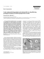
Figure 1 Representative pictures (10×) of BoHV-4-EFGPΔTK infected F98 (A), GLI36 (C) and GL261 (E) cells at 96 hours (hs) post
infection (P.I.), visualized by phase contrast (PC) fluorescence with a FITC filter for EGFP expression or with DAPI filter for nuclear
counterstaining (bar = 100 μm). The respective titers (expressed as log
10
of Tissue Cells Infectious Dose/50 [TCID
50
] per ml
-1
) of viral particles
released during the time at 24 and 96 hours (hs) post infection (P.I.) are shown in B, D and F. Values are the mean ± standard error of three
independent experiments. (G) GL261 mouse glioblastoma cell line (a, bar = 25 μm), F98 rat glioma cell line (b, bar = 25 μm) and GLI36 human
glioma cell line (c, bar = 10 μm) infected with BoHV-4EGFPΔTK for 72 hours. CPE induced by infection shows a prevalence of necrosis (ANOVA,
**p < 0.001, *p < 0.05).
Redaelli et al. Virology Journal 2010, 7:298
/>Page 2 of 6
three times for each cell line). All three cell lines sustained
productive infection (Figure 1B, D and 1F). In order to
analyze the CPE induced by BoHV-4EGFPΔTK, cells were
fixed with met hanol and stained with Wright’ sstain.A
total of 600 cells were counted from each slide, and the
percentage of apoptotic and necrotic cells was calculated.
At least 6 control and 6 treated slides were counted for
each treat ment. Mo novariate ANOVA was used to test
differences in the percentage of dead cells between control
and infected cells. The CPE induced in vitro by BoHV-
4EGFPΔTK infection was prevalently necrosis (Figure 1G)
rather than apoptosis. Similar result s were obtained with
Annexin V and Propidium Io dide staining (data not
shown). These results, together with the data previously
obtained in vivo where BoHV-4 did not replicate in the
mouse and rat brain, but reporter gene expression was
shown following injection into the mouse and rat lateral
ventricle, prompted us to investigate the use of BoHV-4 as
avectorforthegenetherapyorasanoncolyticvirusof
brain tumours. Thus, a rat glioma model was constructed.
Fifteen four-month-old, male Fisher rats were pre-anesthe-
tiz ed with i soflurane and subsequently anesthetized with
zolazepam tiletamine (20 mg/kg body weight) and xylazine
(75 mg/kg body weight). Eight × 10
6
F98 glioma cells were
suspended in 8 μl DMEM and injected 1 mm anterior and
1.5 mm lateral to the bregma, 3.7 mm below the pial sur-
face. Injection was carried out for 16 minutes and was per-
formed using a Hamilton syringe. Animals were
monitored daily for neurological signs and weight loss. At
the appearance of neurological signs, animals were re-
anesthetized as above and 6 μlof10
6
pfu of BoHV-
4EGFPΔTK were injected into the same position as the
previous injection. Animals were then monitored every 12
hours. Any animals showing severe worsening of neurolo-
gical conditions were humanely euthanize. Rat brains were
analyzed at different post-injection times: 48, 72, 86, 96,
120, 132, 144 and 216 hours. Briefly, anesthetized rats
were first perfused with PBS for 15 min and then with 4%
formalin in PBS for 30 min. Brains were carefully removed,
post-fixed for 2 hours in 4% formalin in PBS, equilibrated
for 24 h in 30% sucrose in PBS at 4°C and frozen at -80°C
unti l sectioning with a cryostat at 16 μm. Sections of the
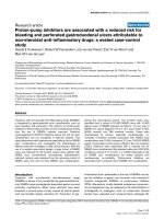
BoHV-4-injected, rat brain gliomas showed EGFP expres-
sion in the peripheral area of larger tumours (Figure 2a,
b), scattered across the mass of smaller t umours (Figure
2c), and in the solid peripheral area of cystic tumours (Fig-
ure 2d). These same sections, following observation of
EGFP expression, were then stained with hematoxylin-
eosin. In order to confirm co-localization of the tumour
area with EGFP-positive transduced cells, five four-month-
old male rats were inoculated with 8 × 10
6
F98 glioma
cells labelled with the red fluorescent cell linker PHK26,
according to manufacturer ’sinstructions(Sigma).Cells
maintain fluorescence for more than 100 mitotic divisions
[9]. When BoHV-4EGFPΔTK was injected into the rat
brains at the same position as the marked glioma cells, co-
localization between the red fluores cent-marked tumo ur
area and the EGFP positive cells was observed, without
detection of the EGFP signal within the brain parenchyma
(data not shown). In another experiment, primary cultures
from biopsies of 2 patients with glioblastoma (both males,
59 and 79 years of age respectively) were prepared. Speci-
mens were dissociate d not more t han 30 minutes after
surgery by shaking for 5 minutes in 0.25% Trypsin, 0.02%
EDTA (1 ml/mm
3
tissue). The suspension was inactivated
with CGM, and centrifuged at 37°C for 10 minutes at
1350 rpm. The supernatant was discharged and the pellet
resuspended in 10 ml of CGM, changed every 72 hour for
three weeks. Cells were then infected with BoHV-
4EGFPΔTK and analyzed 24, 48 and 72 hours post infec-
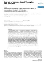
tion as described above. Indeed, these primary cultures of
human glioblastoma were susceptible to BoHV-4 infection
as shown by EGFP expression, and also in this case infec-
tion leaded to a mainly necrotic CPE (Figure 3).
We here report the capaci ty of BoHV- 4 to infec t and
replicate in glioma cell lines and glioblastoma primary
cultures in vitro and the ability of BoHV-4 to selecti vely
infect gliomas induced in the rat brain in vivo.
BoHV-4 is not oncogenic, unlike other g-herpesviruses
like KSHV, EBV and HVS [10]. In addition, the attenua-
tion by gene inactivation is not mandatory, due to the
mild pathogenicity of the virus in natural and experi-
mental hosts. Interestingly, previous studies demon-
stratedthatBoHV-4EGFPΔTK infection is not
permissive in the rat and mouse brain [8], unlike the
replication-competent behaviour of BoHV-4EGFPΔTK
in a different number of cell lines in vitro.
The data from clinical trials underline the need to
refine gene therapy protocols through combination with
other therapeutic strategies or by improving the effi-
ciency and selectivity of vectors [1]. A recent clinical
trial with combined cytokine/suicide gene therapy for
glioma supported the efficacy of the transduction of
therapeutic genes to the targeted tumour cells in human
patients [11]. These data suggest a possible application
in the long-term control of tumour growth.
The present study demonstrates the safety of the vector
in vivo and t he efficiency of the transduction of the
reporter gene both in vitro and in vivo. In vitro, the ability
of BoHV-4 to infect different glioma cell lines, as demon-
strated by the expression of the reporter gene, suggests
the suitability of this vector for gene therapy. The selec-
tivity of the virus for glioma cells in the nervous system
and its safety have been also tested in vivo. The evolution
of infection and the distribution of EGFP-positive cells
within the tumour area shows the selectivity of the virus
for the tumour cells and its oncolytic properties. More-
over the non-replicative behaviour of the virus in the
Redaelli et al. Virology Journal 2010, 7:298
/>Page 3 of 6
Figure 2 Frozen sections (horizontal) of the BoHV-4 injected rat brain gliomas. EGFP expression in the peripheral area of tumour 48 hours
post BoHV-4 injection (a, bar = 500 μm), hematoxylin eosin of the whole section (b) with magnification of the tumour area in the insert (c).
EGFP expression in the solid peripheral area of a cystic tumour 96 hours post BoHV-4 injection (d, bar = 500 μm), hematoxylin eosin of the
whole section (e) with magnification of the tumour area in the insert (f). EGFP expression in the whole mass of non necrotic tumours 132 hours
post BoHV-4 injection (g, bar = 150 μm), hematoxylin eosin of the whole section (h) with magnification of the tumour area in the insert (i).
Redaelli et al. Virology Journal 2010, 7:298
/>Page 4 of 6
brain parenchyma [8] is important for its safe use. These
data are suppo rted by the analysis of the infection in the
F98-PHK26red model in vivo,thatalsoconfirmthat
BoHV-4 infection is co nfined to the tumour area. Lastly,
the infection of human primary culture of brain tumour
extends our results in rat gliomas to human gliomas.
In conclusion, the capability to establish an infection
of glioma cells in vitro, of both immortalized cell lines
as well as primary cultures, the in vivo non-pathogeni-
city and the affinity for the glioma cells in vivo set
BoHV-4 up as a candidate for gene delivery and onco-
lyses to the glial tumours of the nervous system.
List of abbreviations
BoHV-4: Bovine herpesvirus 4; CPE: Cytopathic effect; CGM: Complete growth
medium; RMS: Rostral migratory stream; DMEM: Dulbecco Modified Eagle’s
Medium; EGFP: Enhanced green fluorescent protein; FBS: Fetal bovine serum.
Acknowledgements
We would like to tank Professor Laura Kramer for English language
correction and Italian Ministry of University and Scientific Research and the
Fondazione Cariparma (Cassa di Risparmio di Parma, Italy) for funding
contributions to the project.
Author details
1
Department of Human Anatomy and Physiology, University of Padova, Italy.
2
Department of Neuroscience, University of Padova, Italy.
3
Department of
Pharmaceutical Sciences, University of Parma, Italy.
4
Department of Animal
Health, University of Parma, Italy.
Authors’ contributions
RM: performed the experiments and wrote the paper. M-CC, CA and CA:
intellectually contributed. DD and DL: provided human glioma samples. GD:
Conceive the experiments, performed the experiments and wrote the paper.
Competing interests
The authors declare that they have no competing interests.
Received: 22 September 2010 Accepted: 3 November 2010
Published: 3 November 2010
References
1. Pulkkanen KJ, Yla-Herttuala S: Gene therapy for malignant glioma: current
clinical status. Mol Ther 2005, 12(4):585-598.
2. Zimmermann W, Broll H, Ehlers B, Buhk HJ, Rosenthal A, Goltz M: Genome
sequence of bovine herpesvirus 4, a bovine Rhadinovirus, and
Figure 3 Primary cultures from two human glioblastoma analyzed 24, 48 and 72 hours post BoHV-4EGFPΔTK infection. The cells were
visualized with a FITC filter for EGFP expression (a, b, c, d, e, f, bar = 50 μm) and by phase contrast (PC) (a
i
,b
i
,c
i
,d
i
,e
i
,f
i
). After 72 hours post
infection the cultures were completely infected. CPE induced by infection shows a prevalence of necrosis (g, h, t-test, ***p < 0.001, *p < 0.05).
Redaelli et al. Virology Journal 2010, 7:298
/>Page 5 of 6
identification of an origin of DNA replication. Journal of virology 2001,
75(3):1186-1194.
3. Donofrio G, Cavirani S, Simone T, van Santen VL: Potential of bovine
herpesvirus 4 as a gene delivery vector. J Virol Methods 2002, 101(1-
2):49-61.
4. Egyed L: Bovine herpesvirus type 4: a special herpesvirus (review article).
Acta veterinaria Hungarica 2000, 48(4):501-513.
5. Gillet L, Minner F, Detry B, Farnir F, Willems L, Lambot M, Thiry E,
Pastoret PP, Schynts F, Vanderplasschen A: Investigation of the
susceptibility of human cell lines to bovine herpesvirus 4 infection:
demonstration that human cells can support a nonpermissive persistent
infection which protects them against tumor necrosis factor alpha-
induced apoptosis. Journal of virology 2004, 78(5):2336-2347.
6. Gillet L, Dewals B, Farnir F, de Leval L, Vanderplasschen A: Bovine
herpesvirus 4 induces apoptosis of human carcinoma cell lines in vitro
and in vivo. Cancer research 2005, 65(20):9463-9472.
7. Yamamoto Y, Murakami K, Inoshima Y, Nakane T, Saika K, Sentsui H:
Characterization of a bovine herpesvirus type 4 isolated from the spinal
cord of a cow with astasia. Archives of virology 2000, 145(11):2363-2370.
8. Redaelli M, Cavaggioni A, Mucignat-Caretta C, Cavirani S, Caretta A,
Donofrio G: Transduction of the rat brain by Bovine Herpesvirus 4. Genet
Vaccines Ther 2008, 6:6.
9. Horan PK, Slezak SE: Stable cell membrane labelling. Nature 1989,
340(6229):167-168.
10. Jung JU, Choi JK, Ensser A, Biesinger B: Herpesvirus saimiri as a model for
gammaherpesvirus oncogenesis. Seminars in cancer biology 1999,
9(3):231-239.
11. Colombo F, Barzon L, Franchin E, Pacenti M, Pinna V, Danieli D, Zanusso M,
Palu G: Combined HSV-TK/IL-2 gene therapy in patients with recurrent
glioblastoma multiforme: biological and clinical results. Cancer gene
therapy 2005, 12(10):835-848.
doi:10.1186/1743-422X-7-298
Cite this article as: Redaelli et al.: Bovine herpesvirus 4 based vector as
a potential oncolytic-virus for treatment of glioma. Virology Journal 2010
7:298.
Submit your next manuscript to BioMed Central
and take full advantage of:
• Convenient online submission
• Thorough peer review
• No space constraints or color figure charges
• Immediate publication on acceptance
• Inclusion in PubMed, CAS, Scopus and Google Scholar
• Research which is freely available for redistribution
Submit your manuscript at
www.biomedcentral.com/submit
Redaelli et al. Virology Journal 2010, 7:298
/>Page 6 of 6