Phẫu thuật vai và khuỷu tay docx
Bạn đang xem bản rút gọn của tài liệu. Xem và tải ngay bản đầy đủ của tài liệu tại đây (4.24 MB, 8 trang )
Symptomatic Os Acromiale
Abstract
Os acromiale, the joining of the acromion to the scapular spine by
fibrocartilaginous tissue rather than bone, is an anatomic variant
that has been reported in approximately 8% of the population
worldwide. It is more common in blacks and males than in whites
and females. Although it is often an incidental finding, os
acromiale has been identified as a contributor to shoulder
impingement symptoms and rotator cuff tears. When nonsurgical
management of a symptomatic os acromiale fails to relieve
symptoms, surgical intervention is considered. Options include os
acromiale excision, open reduction and internal fixation, and
arthroscopic decompression. Excision usually is reserved for small
to midsized fragments (preacromion) or after failed open reduction
and internal fixation. Persistent deltoid dysfunction may result
from excision of a large os acromiale. Open reduction and internal
fixation preserves large fragments while maintaining deltoid
function. Cannulated screw fixation has been shown to result in
good union rates. Arthroscopic techniques have shown mixed
results when used for treating impingement secondary to an
unstable os acromiale. Associated rotator cuff tears may be
addressed arthroscopically or through an open transacromial
approach, followed by open reduction and internal fixation of the
os acromiale.
G
ruber,
1
in 1863, first reported
on separation of the acromion
in a study of 100 cadavers; 3 of the
100 specimens exhibited a fibrocar-
tilaginous union of the acromial os-
sification centers. Numerous other
anatomists have produced descrip-
tive studies of os acromiale.
2-4
The
reported incidence ranges from 1.3%
to 30%.
5-10
The relatively high 30%
rate was reported in an archeological
study of remains from an excavated
cemetery.
7
The rate is attributed to
familial ties of the persons buried in
that cemetery. Two separate studies
of the Hamann-Todd Osteological
Collection discovered an 8% inci-
dence of os acromiale (17 of 210
specimens), with roughly one third
having bilateral involvement.
9,10
In
addition, these studies revealed that
blacks and males were twice as like-
ly to have an os acromiale as whites
and females, respectively. Other re-
ports indicate bilateral involvement
in as many as 62% of patients.
6
Anatomy
An os acromiale represents a failure
of fusion of the anterior acromial
apophysis. The acromial apophysis
develops from four separate centers
of ossification: the basiacromion,
Christopher A. Kurtz, MD
Byron J. Humble, DO
Mark W. Rodosky, MD
Jon K. Sekiya, MD
Dr. Kurtz is Lieutenant Commander,
Medical Corps, United States Navy, and
Head, Division of Sports Medicine,
Bone and Joint/Sports Medicine
Institute, Department of Orthopaedic
Surgery, Naval Medical Center
Portsmouth, Portsmouth, VA. Dr.
Humble is Lieutenant, Medical Corps,
United States Navy, Bone and Joint/
Sports Medicine Institute, Naval Medical
Center Portsmouth. Dr. Rodosky is
Assistant Professor and Chief, Division
of Shoulder and Elbow Surgery, Center
for Sports Medicine, Department of
Orthopaedic Surgery, University of
Pittsburgh Medical Center, Pittsburgh,
PA. Dr. Sekiya is Assistant Professor,
Center for Sports Medicine, University
of Pittsburgh Medical Center.
None of the following authors or the
departments with which they are
affiliated has received anything of value
from or owns stock in a commercial
company or institution related directly or
indirectly to the subject of this article:
Dr. Kurtz, Dr. Humble, Dr. Rodosky, and
Dr. Sekiya.
The views expressed in this article are
those of the authors and do not reflect
the official policy or position of the
Department of the Navy, Department of
Defense, or the United States
Government.
Reprint requests: Dr. Sekiya, Center for
Sports Medicine, University of
Pittsburgh Medical Center, 3200 S
Water Street, Pittsburgh, PA 15203.
J Am Acad Orthop Surg 2006;14:
12-19
Copyright 2006 by the American
Academy of Orthopaedic Surgeons.
12 Journal of the American Academy of Orthopaedic Surgeons
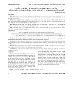
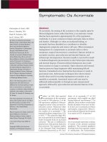
meta-acromion, mesoacromion, and
preacromion (Figure 1). The basi-
acromion fuses to the scapular spine
at approximately age 12 years. The
meta-acromion serves as the origin
of the posterior deltoid muscle, and
the mesoacromion anchors the mid-
dle tendinous portion of the deltoid.
The preacromion is the attachment
site for both the anterior deltoid fi-
bers and the coracoacromial liga-
ment. The three anterior acromial
ossification centers develop from
several ossification nuclei, but by
between ages 15 and 18 years, they
coalesce into the meta-acromion,
mesoacromion, and preacromion.
Complete union of all centers may
occur as late as age 25 years;
11
there-
fore, caution is warranted when di-
agnosing an unfused os acromiale
before that age. Some authors dis-
pute the concept of four discrete os-
sification centers and contend that
the acromion ossifies from one con-
tinuous cartilaginous anlage.
8
The types of os acromiale are de-
fined by the unfused segment imme-
diately anterior to the site of non-
union. For example, failed fusion
between the meta-acromial and
mesoacromial ossification centers is
called a mesoacromiale. The great
majority of ossa acromiale are
mesoacromial.
8-11
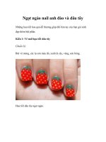
Preacromial frag-
ments occur much less frequently,
and a meta-acromiale is rare (Figure
2). Mudge et al
12
reported on the ex-
tremely rare variant of a preacromial
and mesoacromial double fragment.
Pathophysiology
Os acromiale is often an incidental
radiographic finding discovered
while examining a patient with
shoulder pain. The os acromiale may
be completely unrelated to the true
source of the patient’s discomfort.
13
A complete evaluation for all sourc-
es of potential pain must be under-
taken before attributing symptoma-
tology to the os acromiale.
In patients in whom the os acro-
miale is believed to be pathologic,
the pain-generating potential from
an unstable os likely stems from two
main sources. First, the nonunion
site may be inherently painful, with
pain directly at the nonunion site.
Physical findings include tenderness
at the nonunion site or localized pain
with manipulation of the unstable
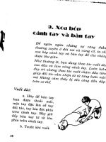
fragment. Furthermore, magnetic
resonance imaging (MRI)
14
(Figure 3)
and bone scan
15,16
may demonstrate
evidence of inflammatory reaction at
the site of nonunion. Second, an un-
stable os acromiale may produce a
dynamic type of outlet-based im-
pingement syndrome.
15,17,18
Both
flexion of the anterior fragment with
deltoid contraction and elevation of
the arm can decrease the size of the
supraspinatus outlet, thereby pro-
ducing the symptoms of classic ex-
ternal impingement.
15,18
Figure 1
The acromial ossification centers
comprising the acromial apophysis.
BA = basiacromion, MS = meso-
acromion, MT = meta-acromion,
PA = preacromion
Figure 2
Axial T2-weighted MRI scan
demonstrating a meta-acromiale of the
right shoulder. The site of nonunion
is indicated by the arrow.
Figure 3
A, T2-weighted axial MRI scan of the right shoulder demonstrating reactive edema
at the nonunion site (arrow). B, T2-weighted coronal oblique image of the same
patient demonstrating superior osteophyte formation (arrow).
Christopher A. Kurtz, MD, et al
Volume 14, Number 1, January 2006 13
Patient Assessment
In patients with symptomatic os
acromiale, complaints are frequently
those of classic outlet impingement
syndrome.
16-22
Patients relate diffi-
culty with overhead activities and
with sleeping. They may report lim-
ited range of motion or clicking in
the shoulder.
12,17
Patients also de-
scribe pain located directly over the
superior acromion, especially when
the fragment becomes more unsta-
ble.
15,22,23
Finally, patients may notice
weakness caused by associated rota-
tor cuff dysfunction.
12,24
A history of
trauma is less common; if present, its
role in the development of os acromi-
ale is usually minor.
A standard physical examination
reveals many findings of classic im-
pingement, including pain with im-
pingement signs, painful arc of mo-
tion, and difficulty with forward
elevation, even in the presence of an
intact cuff.
15
Rotator cuff weakness is
often present.
In addition to the typical impinge-
ment findings, the physical examina-
tion may reveal abnormalities
unique to an unstable os acromiale.
The patient may experience tender-
ness directly at the nonunion site;
further, gross motion of the anterior
acromion may be present. A diagnos-
tic subacromial injection (impinge-
ment test) may give a mixed
response, with alleviation of im-
pingement signs but with variable re-
lief of the localized tenderness. In the
presence of uncertainty regarding the
source of localized tenderness, a di-
agnostic injection into the nonunion
site itself may be beneficial.
Radiographic
Assessment
Three-view tangential radiographs
are essential for assessing any pa-
tient with shoulder problems. With
os acromiale, the axillary lateral
view is essential. An os acromiale is
easily missed with anteroposterior
or y-view scapular radiographs. Most
authors stress that the axillary later-
al view is critical
8,12,15-17,19-21,23,25
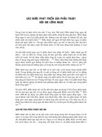
(Fig-
ure 4). The axillary lateral view re-
veals the size and shape of the
acromial fragment as well as any de-
generative change at the site.
In addition to the standard axil-
lary lateral view, the acromial profile
view described by Andrews et al
26
(Figure 5) provides another means of
detecting an os acromiale that is not
readily apparent on more conven-
tional views. Plain radiographs of
the contralateral shoulder may be
helpful, especially when evaluating
a patient who is not skeletally ma-
ture. With contralateral views, how-
ever, the incidence of bilateral in-
volvement may be as high as 62%.
6
MRI is a helpful and frequently
Figure 4
Anteroposterior (A), outlet (B), and axillary lateral (C) radiographic views of the right shoulder in the same patient. The os
acromiale is most readily apparent on the axillary lateral projection (black arrow in panel C).
Symptomatic Os Acromiale
14 Journal of the American Academy of Orthopaedic Surgeons
used adjunct in radiographic evalua-
tion of the shoulder. Axial cuts
through the acromion reliably detect
an os acromiale. When the axial pro-
jection is either incomplete (ie, not
taken superior enough to include the
acromion) or absent, other orienta-
tions may offer more subtle clues.
The sagittal and oblique cuts are eas-
ily misinterpreted. For instance, the
os acromiale may be mistaken for
the acromioclavicular joint. The
presence of a double acromioclavic-
ular joint on a single image (Figure 6)
should raise the suspicion of an os
acromiale; however, this finding is
often not present.
14
In most patients,
the os acromiale defect appears as a
vertical band of low signal intensity
in a position posterior to a line bi-
secting the humeral head on oblique
sagittal images.
14,27
This is in con-
trast with the acromioclavicular
joint, which lies anterior. MRI also
may detect hypertrophic osteophyte
formation, edema, or widening at
the site of nonunion, indicating in-
stability of the os acromiale.
27
Final-
ly, MRI is useful for confirming the
presence of other associated pathol-
ogy, such as rotator cuff tears.
Other imaging modalities also
may be helpful in evaluating an os
acromiale. Computed tomography
(CT) readily delineates an unfused
acromion on the axillary projec-
tion.
20,22
Three-dimensional CT re-
constructions clearly show the os
acromiale.
20
Bone scanning, when
positive, is useful in confirming the
os acromiale as a contributing factor
in a painful shoulder,
15,16
especially
when evaluating a patient on the
cusp of skeletal maturity.
Nonsurgical
Management
Initial management of the symp-
tomatic os acromiale should be
nonsurgical. Nonsteroidal anti-
inflammatory drugs should be pre-
scribed, as well as physical therapy
with an impingement protocol. Sub-
acromial corticosteroid injection
also may be used. Local corti-
costeroid injection at the nonunion
site may provide sufficient relief of
symptoms to avoid surgery.
16
Gen-
erally, nonsurgical management
should be tried for at least 6 months.
However, the incidence of a full-
thickness rotator cuff tear may be as
high as 50%;
15,18
such a tear may be
grounds for early surgical manage-
ment.
28
Surgical Management
Surgical management is warranted
when nonsurgical treatment fails. A
number of surgical approaches have
been advocated, including fragment
excision, open reduction and inter-
nal fixation (ORIF), and arthroscop-
ic subacromial decompression. Var-
ious techniques are reported for each
approach, and each procedure has
benefits and drawbacks.
Open Fragment Excision
Open fragment excision has had
mixed results. Mudge et al
12
treated
six patients with excision in con-
junction with rotator cuff repair.
Four patients had excellent results;
the remaining two were poor. De-
spite their results, Mudge et al
12
ad-
vocated ORIF and bone grafting for
larger fragments. Edelson et al
8
re-
ported an anatomically based tech-
nique of excision and deltoid ad-
vancement in five patients; four of
Figure 6
T2-weighted coronal oblique MRI scan
of the left shoulder. The acromioclav-
icular joint is anterior (narrow arrow),
and the acromial defect is posterior
(wide arrow).
Figure 5
Acromial radiographic profile view of the right shoulder in a patient with a meso-
acromiale. The arrow indicates the site of nonunion. A = acromion, C = clavicle,
H = humeral head, M = mesoacromiale
Christopher A. Kurtz, MD, et al
Volume 14, Number 1, January 2006 15
five patients were satisfied. The au-
thors attributed the one failure to an
irreparable rotator cuff tear and con-
comitant distal clavicle resection re-
sulting in superior humeral head mi-
gration and loss of forward flexion.
As a result, they recommended
ORIF in the presence of an irrepara-
ble rotator cuff tear.
Warner et al
15
performed frag-
ment excision on three patients; two
had poor results. Both poor results
involved mesoacromial fragment ex-
cision with resultant pain and weak-
ness. The one satisfactory result in-
volved resection of a preacromiale.
In general, patients who undergo
open resection of the anterior acro-
mion are at high risk for deltoid dys-
function;
29
thus, open fragment exci-
sion should be reserved for very
small fragments or as a salvage pro-
cedure for patients with failed at-
tempted ORIF.
15,17
Open Reduction and
Internal Fixation
Numerous case reports
19,22,24
and
case series
8,15,18,25,30
have been pub-
lished regarding ORIF of an unstable
os acromiale. Nearly all techniques
involve some sort of internal fixa-
tion with bone grafting. Edelson et
al
8
treated two patients with ORIF
consisting of malleolar screw fixa-
tion and local bone grafting. Both
achieved union, and both required
hardware removal. The indication
for fusion rather than excision was
primary pain at the nonunion site
with absence of impingement symp-
toms. Warner et al
15
performed ORIF
on 11 patients (12 shoulders) with
two techniques, both of which in-
volved débridement of the nonunion
site and bone grafting perpendicular
to the nonunion via a bone trough.
Five of 12 shoulders were fixed with
pins and tension band wiring; 4 of 5
failed to unite. In contrast, only one
failed fusion was reported in seven
shoulders fixed with cannulated
screws and a tension band construct.
Average time to union was 9 weeks.
Nine of 12 patients required subse-
quent hardware removal, including
five of seven with successful fusions.
Hertel et al
30
performed ORIF for
15 unstable acromial fragments with
takedown of the nonunion and ten-
sion band wiring without bone graft-
ing. Two distinct surgical approach-
es were employed. Seven patients
were operated on with an anterior
deltoid-off approach, and eight pa-
tients with a transacromial approach
with preservation of the deltoid ori-
gin. Union was achieved in three of
the seven deltoid-off patients and in
seven of the eight transacromial
deltoid-preserving patients. The au-
thors attributed the increased union
rate with deltoid preservation to
maintenance of the acromial blood
supply via the acromial branch of
the thoracoacromial artery.
Satterlee
18
reported successful fu-
sion in six of six patients with an un-
stable os acromiale. The procedure
involved dorsal wedge osteotomy
and nonunion takedown, elevation
of the anterior fragment, fixation
with two 4.5-mm Herbert screws,
and local bone graft held in place
with a figure-of-8 suture passed
through the cannulated screws. One
patient underwent hardware remov-
al but was asymptomatic. Ryu et
al
25
used two parallel 3.5-mm cannu-
lated screws and greater tuberosity
bone grafting in four patients. Fusion
was achieved in all four, with a time
to union of 10 to 16 weeks.
ORIF of an unstable os acromiale
is indicated for larger fragments.
Success is predictable with any of a
variety of techniques. Factors associ-
ated with successful union include
use of a rigid construct
15,18,25
and
preservation of the acromial vascu-
larity.
30
Even with successful union,
hardware removal is not uncom-
mon. Pain is the most common rea-
son for hardware removal.
Arthroscopic Subacromial
Decompression
Arthroscopic subacromial decom-
pression has been advocated as a
means to avoid the complications
associated with ORIF (eg, risk of
nonunion, revision for hardware re-
moval). Early experience with ar-
throscopic treatment was not very
successful because many patients
were treated with simple decom-
pression. Although the deltoid inser-
tion was preserved, standard arthro-
scopic decompression failed to
eliminate the painful nonunion.
Hutchinson and Veenstra
23
re-
ported on three patients who under-
went arthroscopic subacromial de-
compression for impingement
syndrome associated with an unsta-
ble os acromiale. The authors per-
formed decompression of the entire
acromial fragment back to the junc-
tion with the intact acromion. Two
patients had recurrence of symp-
toms after a 6- to 8-month period of
relief. The third patient was im-
proved but not pain free and required
a change in employment to avoid
overhead activities. In all patients,
the presence of the os acromiale was
not discovered until the time of sur-
gery, despite preoperative radio-
graphs revealing its presence. Based
on this small series, the authors con-
cluded that standard techniques for
arthroscopic subacromial decom-
pression cannot be recommended for
impingement secondary to an unsta-
ble os acromiale.
Jerosch et al
31
performed 122 ar-
throscopic subacromial decompres-
sions for impingement syndrome, of
which 12 had os acromiale. No pa-
tient had a rotator cuff tear. Patients
with an os acromiale had a trend to-
ward less favorable results, but the
difference did not reach statistical
significance. Even with the slightly
worse outcomes, the authors recom-
mended arthroscopic subacromial
decompression as a reasonable op-
tion for managing impingement syn-
drome with an os acromiale.
In an effort to improve results
with standard arthroscopic tech-
niques, Wright et al
21
employed a
more aggressive arthroscopic ap-
proach for treating os acromiale–as-
sociated impingement. They treated
Symptomatic Os Acromiale
16 Journal of the American Academy of Orthopaedic Surgeons
13 shoulders in 12 patients who had
failed nonsurgical management; all
patients had complete pain relief
with preoperative subacromial injec-
tions. None of the patients was di-
rectly tender at the nonunion site.
The authors used a more aggressive
bone resection, especially of almost
the entire mobile anterior tip, leav-
ing only a thin superior cortical
shell. Ten of 12 patients achieved
satisfactory postoperative Universi-
ty of California, Los Angeles (UCLA)
scores, and 11 of the 12 patients
themselves rated the outcome as sat-
isfactory. No complications were re-
ported. The authors concluded that
arthroscopic subacromial decom-
pression with resection is a reason-
able alternative and can achieve
good results, provided that bone re-
section is adequate.
In addition to addressing the os
acromiale and associated impinge-
ment syndrome, many patients re-
quire concurrent treatment of a rota-
tor cuff tear. A complete tear or
significant partial tear should be ad-
dressed at the same time as the os
acromiale, regardless of the surgical
approach selected. With excision,
the cuff repair can be achieved
through standard open, mini-open,
or arthroscopic means, depending on
the technique. With ORIF, an
acromion-splitting approach is a
good option;
24
open repair of the cuff
is done through the acromial defect
before bone fixation. With arthro-
scopic decompression, the cuff may
be addressed by arthroscopic repair,
débridement, or a mini-open ap-
proach.
Each surgical technique has ad-
vantages and disadvantages. Al-
though open fragment excision may
be warranted for the preacromial os,
it can result in significant deltoid
dysfunction for larger segments.
ORIF preserves deltoid function and
addresses the os acromiale as a pri-
mary pain generator; however, risk
of nonunion is a concern, and revi-
sion for hardware removal is com-
mon. Arthroscopic decompression
has minimal risk, but results may be
mixed, and pain at the nonunion site
may persist. The clinical scenario
and surgeon experience are evaluat-
ed to determine the technique that
will most benefit the patient.
Management
Techniques
The initial step in management is
determining whether the os acromi-
ale is incidental or symptomatic.
Tenderness at the nonunion site,
pain with motion of the mobile seg-
ment, and imaging studies showing
reactive changes are all indications
that the os acromiale is not an inci-
dental finding. For patients in whom
the os acromiale is determined to be
coincidental, management of the
other shoulder pathology is indicat-
ed. In some instances, impingement
may exist in the presence of an os
acromiale with a stable fibrous
union. A standard arthroscopic sub-
acromial decompression without re-
section of the os may be indicated in
patients who fail nonsurgical treat-
ment.
For the symptomatic os acromi-
ale, a nonsurgical approach is fol-
lowed, consisting of nonsteroidal
anti-inflammatory drugs, physical
therapy, and judicious use of sub-
acromial corticosteroid injections.
As mentioned, this nonsurgical ap-
proach generally is given a 6-month
trial unless some other consider-
ation (eg, full-thickness rotator cuff
tear) warrants abandonment. For pa-
tients who require surgery, the ap-
proach is tailored to the individual
clinical situation.
A symptomatic preacromial frag-
ment or a small mesoacromial frag-
ment anterior to the posterior aspect
of the acromioclavicular joint can be
treated with excision using an ar-
throscopic technique of fragment ex-
cision with decompression of the
remaining mesoacromion. The frag-
ment is excised to the superior cor-
tical plate, leaving the deltoid intact.
The anterior edge of the remaining
acromion is smoothed over to the
deltoid attachment. An arthroscopic
approach preserves the deltoid fascia
and allows for treatment of all asso-
ciated pathology.
A symptomatic large mesoacro-
mial fragment is by far the most
common presentation. The non-
union is located at or behind the lev-
el of the posterior acromioclavicular
joint. Arthroscopic examination of
the glenohumeral joint always
should be performed. Rotator cuff
integrity is assessed, and any other
associated pathology (eg, superior la-
bral injury) is addressed. Subacromi-
al arthroscopy (Figure 7) should de-
termine both segment motion and
rotator cuff disease. Rotator cuff re-
pair may be done arthroscopically if
the tear is amenable. When the os is
stable, a standard arthroscopic sub-
acromial decompression is per-
formed. In the presence of an unsta-
ble os in a patient with low-demand
shoulder function, the os is arthro-
scopically resected to a cortical
plate. When it is unstable and the pa-
tient requires higher-demand upper
extremity function, ORIF should be
performed.
ORIF is undertaken via a trans-
acromial approach, as described by
Hertel et al.
30
Superior osteophytes
Figure 7
Arthroscopic view of the subacromial
space from the posterior viewing portal.
A spinal needle was inserted through
the acromial defect. Note the
downgoing hook on the anterior
fragment (dashed line). A = anterior,
P = posterior
Christopher A. Kurtz, MD, et al
Volume 14, Number 1, January 2006 17
are removed, and the nonunion is
taken down until bleeding bone is
seen on each opposing fragment face.
The débridement creates a dorsally
based open wedge that allows for el-
evation of the anterior fragment be-
fore fixation. The fragment is elevat-
ed and temporarily fixed with
Kirschner wires. The wires can be
drilled posterior-to-anterior in the
anterior fragment, then advanced
retrograde after the fragment is re-
duced. A large tenaculum is used to
provide compression of the reduced
fragments. Screws may be placed
either anterior-to-posterior or
posterior-to-anterior. Placing screws
posterior-to-anterior avoids compro-
mising the more important anterior
deltoid (Figure 8). Compression tech-
niques are used. Standard-head
screws may be used, but intramedul-
lary screws will reduce the need for
further surgery. Demineralized bone
matrix may be added to increase
union rates. Substantial bone defects
may be grafted with autogenous
bone obtained from either the iliac
crest or anterior tibia (Gerdy’s tuber-
cle). Gerdy’s tubercle bone graft is
generally less painful than iliac crest
graft, and surgical access is easier
when the patient is in the beach-
chair position. A nonabsorbable su-
ture placed through the screws and
looped superiorly in a figure-of-8
configuration will aid not only in se-
curing any bone graft but also in lo-
calizing the screws in the event
hardware removal is required. An
acromion-splitting approach (Figure
9) followed by ORIF is used when an
open rotator cuff repair is needed.
Subacromial arthroscopy can be per-
formed after ORIF to evaluate for
unwanted prominence or a residual
acromial hook requiring decompres-
sion. The deltoid fascia is closed, and
the patient is placed in a shoulder
immobilizer.
The patient is kept in an immobi-
lizer for a minimum of 6 weeks post-
operatively. Passive motion only is
allowed for the initial 6 weeks. Gen-
tle active-assisted and active motion
are begun at 6 weeks. Radiographs
are obtained at 6 weeks and period-
ically thereafter until union. Time to
union is variable, with an average of
8 to 12 weeks;
15,18,25
however, it may
take 16 to 20 weeks to achieve
union.
15,25
Strengthening and activi-
ty progression are withheld until
union is achieved.
Summary
Os acromiale is not an uncommon
finding during the workup of a pa-
tient with a painful shoulder. An ax-
illary lateral radiograph is critical in
identifying an os acromiale. The
finding may be incidental or symp-
tomatic. Unstable os fragments gen-
erally exhibit high signal or widen-
ing on MRI. For the symptomatic os
acromiale, management is initially
nonsurgical. Surgery is indicated
only for patients who fail nonsurgi-
cal treatment. Surgical options in-
clude arthroscopic sub−total exci-
sion, arthroscopic subacromial
decompression of stable fragments,
and ORIF of unstable fragments. Re-
sults are variable, and the surgical
approach should be tailored to fit the
patient’s specific clinical scenario.
References
1. Gruber W: Über die Arten der Adro-
mialknochen und accidentellen Ak-
romialgelenke. Arch Anat Physiol
und Wissen Med 1863:373-393.
2. Poirer P: Os acromial. Bull Soc
Anatomy 1887;62:881-882.
Figure 8
Immediate postoperative anteroposterior (A) and axillary (B) radiographs of the
right shoulder following ORIF with 4.5-mm Herbert screws.
Figure 9
Acromion-splitting approach for rotator
cuff repair of the right shoulder. The
deltoid attachment is preserved for
each fragment. A = anterior fragment,
P = posterior fragment
Symptomatic Os Acromiale
18 Journal of the American Academy of Orthopaedic Surgeons
3. Struthers J:On separate acromion pro-
cess simulating fracture. Edinburgh
Med J 1895;41:900-908.
4. Gray DJ: Variations in human scapu-
lae. Am J Phys Anthropol 1942;29:
57-72.
5. Liberson F: Os acromiale–A contested
anomaly. J Bone Joint Surg Am 1937;
19:683-689.
6. Liberson F: The value and limitation
of the oblique view as compared with
the ordinary anteroposterior exposure
of the shoulder: A report of the use of
the oblique view in 1,800 cases. AJR
Am J Roentgenol 1937;37:498-509.
7. Angel JL, Kelly JO, Parrington M,
Pinter S: Life stresses of the free black
community as represented by the
First African Baptist Church, Phila-
delphia, 1823-1841. Am J Phys
Anthropol 1987;74:213-229.
8. Edelson JG, Zuckerman J, Hershko-
vitz I: Os acromiale: Anatomy and
surgical implications. J Bone Joint
Surg Br 1993;75:551-555.
9. Nicholson GP, Goodman DA, Flatow
EL, Bigliani LU: The acromion: Mor-
phologic condition and age-related
changes. A study of 420 scapulas.
J Shoulder Elbow Surg 1996;5:1-11.
10. Sammarco VJ: Os acromiale: Frequen-
cy, anatomy, and clinical implica-
tions. J Bone Joint Surg Am 2000;82:
394-400.
11. McClure JG, Raney RB: Anomalies of
the scapula. Clin Orthop 1975;110:
22-31.
12. Mudge MK, Wood VE, Frykman GK:
Rotator cuff tears associated with os
acromiale. J Bone Joint Surg Am
1984;66:427-429.
13. Burkhart SS: Os acromiale in a profes-
sional tennis player. Am J Sports
Med 1992;20:483-484.
14. Uri DS, Kneeland JB, Herzog R: Os
acromiale: Evaluation of markers for
identification on sagittal and coronal
oblique MR images. Skeletal Radiol
1997;26:31-34.
15. Warner JJ, Beim GM, Higgins L: The
treatment of symptomatic os acromi-
ale. J Bone Joint Surg Am 1998;80:
1320-1326.
16. Jerosch J, Hepp R, Castro WH: An un-
fused acromial epiphysis: A reason for
chronic shoulder pain. Acta Orthop
Belg 1991;57:309-312.
17. Swain RA, Wilson FD, Harsha DM:
The os acromiale: Another cause of
impingement. Med Sci Sports Exerc
1996;28:1459-1462.
18. Satterlee CC:Successful osteosynthe-
sis of an unstable mesoacromion in 6
shoulders: A new technique.
J Shoulder Elbow Surg 1999;8:125-
129.
19. Sterling JC, Meyers MC, Chesshir W,
Calvo RD: Os acromiale in a baseball
catcher. Med Sci Sports Exerc 1995;
27:795-799.
20. Granieri GF, Bacarini L: A little-
known cause of painful shoulder: Os
acromiale. Eur Radiol 1998;8:130-
133.
21. Wright RW, Heller MA, Quick DC,
Buss DD: Arthroscopic decompres-
sion for impingement syndrome sec-
ondary to an unstable os acromiale.
Arthroscopy 2000;16:595-599.
22. Carlson DW, Kim DH: Os acromiale.
Am J Orthop 2002;31:458.
23. Hutchinson MR, Veenstra MA: Ar-
throscopic decompression of shoulder
impingement secondary to os acromi-
ale. Arthroscopy 1993;9:28-32.
24. Richman N, Curtis A, Hayman M:
Acromion-splitting approach through
an os acromiale for repair of a massive
rotator cuff tear. Arthroscopy 1997;
13:652-655.
25. Ryu RK, Fan RS, Dunbar WH V: The
treatment of symptomatic os acromi-
ale. Orthopedics 1999;22:325-328.
26. Andrews JR, Byrd JW, Kupferman SP,
Angelo RL: The profile view of the
acromion. Clin Orthop 1991;263:
142-146.
27. Park JG, Lee JK, Phelps CT: Os acro-
miale associated with rotator cuff im-
pingement: MR imaging of the shoul-
der. Radiology 1994;193:255-257.
28. Ortiguera CJ, Buss DD: Surgical man-
agement of the symptomatic os acro-
miale. J Shoulder Elbow Surg 2002;
11:521-528.
29. Neer CS II, Marberry TA: On the dis-
advantages of radical acromionecto-
my. J Bone Joint Surg Am 1981;63:
416-419.
30. Hertel R, Windisch W, Schuster A,
Ballmer FT: Transacromial approach
to obtain fusion of unstable os acromi-
ale. J Shoulder Elbow Surg 1998;7:
606-609.
31. Jerosch J, Steinbeck J, Strauss JM,
Schneider T: Arthroscopic subacromial
decompression—indications in os ac-
romiale? [German]. Unfallchirurg
1994;97:69-73.
Christopher A. Kurtz, MD, et al
Volume 14, Number 1, January 2006 19