Surgical Atlas of pediatric otolaryngology - part 2 pptx
Bạn đang xem bản rút gọn của tài liệu. Xem và tải ngay bản đầy đủ của tài liệu tại đây (937.27 KB, 69 trang )
72 Surgical Atlas of Pediatric Otolaryngology
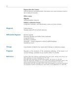
• The tympanomeatal flap is replaced. A tympanostomy tube is inserted
into the tympanic membrane if eustachian tube function is still poor in
order to prevent middle-ear effusion or another portion of the tympan-
ic membrane from retracting (Figure 3–48).
• A layer of Gelfoam is placed over the tympanic membrane and graft.
Two strips of Adaptic gauze impregnated with antibiotic ointment are
inserted into the external canal.
Postoperative Care
• The postoperative care is the same as that described in Chapter 2 under
Postauricular Approach.
Figure 3–48 A tympanostomy
tube is inserted into the tympan-
ic membrane.
Myringoplasty and Tympanoplasty 73
REFERENCES
1. Saito H, Kazama Y, Yazawa Y. Simple maneuver for closing traumatic eardrum perforation by
micropore strip tape patching. Am J Otol 1990;11:427–30.
2. Paparella MM. Otologic surgery in children. Otolaryngol Clin North Am 1977;10:145–51.
3. Sheehy JL, Anderson RG. Myringoplasty: a review of 472 cases. Ann Otol Rhinol Laryngol
1980;89:331–4.
4. Koch WM, Friedman EM, McGill TJI, et al. Tympanoplasty in children. The Boston Children’s
Hospital Experience. Arch Otolaryngol Head Neck Surg 1990;116:35–40.
5. Smyth GD. Tympanic reconstruction. Otolaryngol Clin North Am 1972;5:111–25.
6. Shih L, de Tar T, Crabtree JA. Myringoplasty in children. Otolaryngol Head Neck Surg
1991;105:74–7.
7. Tos M, Orntoft S, Stangerup SE. Results of tympanoplasty in children after 15 to 27 years. Ann
Otol Rhinol Laryngol 2000;109:17–23.
8. Vrabec JT, Deskin RW, Grady JJ. Meta-analysis of pediatric tympanoplasty. Arch Otolaryngol
Head Neck Surg 1999;125:530–4.
9. Bluestone CD, Klein JO. Otitis media in infants and children. WB Saunders; 2001. p. 313–7.
10. Potsic WP, Winawer MR, Marsh RR. Tympanoplasty for the anterior-superior perforation in
children. Amer J Otol 1996;17:115–8.
11. Lempert J. Endaural, antauricular surgical approach to the temporal bone: principles involved
in this new approach. Summary report of 1,780 cases. Arch Otolaryngol Head Neck Surg
1937;27:555–87.
12. Blaney SPA, Tierney P, Bowder DA. The surgical management of the pars tensa retraction pock-
et in the child—results following simple excision and ventilation tube insertion. Int J Pediatr
Otorhinolaryngol 1999;50:133–7.
13. Palva T, Johnsson L-G, Ramsey H. Attic aeration in temporal bones from children with recur-
ring otitis media: tympanostomy tubes did not cure disease in Prussak’s Space. Am J Otol
2000;21:485–93.
14. Hasebe S, Takahashi H, Honjo I, Sudo M. Organic change of effusion in the mastoid in otitis
media with effusion and its relation to attic retraction. Int J Pediatr Otorhinolaryngol
2000;53:17–24.
15. Gerber MJ, Mason JC, Lambert PR. Hearing results after primary cartilage tympanoplasty.
Laryngoscope 2000;110:1994–9.
16. Bluestone CD. Definitions, terminology, and classification. In: Bluestone CD, Rosenfeld RM,
editors. Evidence-based otitis media. Hamilton, Ontario: B C Decker Inc; 1999. p. 94–6.
17. Khanna SM, Tonndorf J. Tympanic membrane vibration in cats studied by time-averaged
holography. J Acoust Soc Am 1972;51:1904–20.
18. Chan KC, Sculerati N, Casselbrant ML, et al. Comparison of eustachian tube function tests
between children with cholesteatoma/retraction pocket and those with chronic otitis media
with effusion. In: Tos M, Thomsen J, Peitersen E, editors. Cholesteatoma and Mastoid
Surgery; 1989; Amsterdam: Kugler & Ghedini; 1989. p. 485–7.
OSSICULOPLASTY
James S. Batti, MD
Charles D. Bluestone, MD
OSSICULAR RECONSTRUCTION
Etiology of Ossicular Abnormalities
Ossicular-related causes of conductive hearing loss can be congenital or
acquired, and are mainly due to discontinuity or fixation:
•
Ossicular discontinuity occurs in the following scenarios presented in
order of decreasing frequency: eroded incudostapedial joint, absent
incus, absent incus and stapes superstructure, and absent incus and
stapes including the footplate.
1
Austin
2
defined four groups in the
absence of an intact incus: (1) malleus handle present, stapes super-
structure present, (2) malleus handle present, stapes superstructure
absent, (3) malleus handle absent, stapes superstructure present, and (4)
malleus handle absent, stapes superstructure absent.
• Ossicular fixation most commonly occurs when the malleus head is anky-
losed to the attic wall or when tympanosclerosis of the attic is present.
Kartush
3
modified Austin’s classification of ossicular defects by adding
two other groups related to ossicular fixation: (1) ossicle head fixation
with all ossicles present, and (2) stapes fixation with all ossicles present.
Moretz
4
added still another category, nonclassifiable, to describe unusual sit-
uations requiring ossiculoplasty that are not easily included in the other
categories. These include lateralized tympanic membrane and some con-
genital abnormalities.
This chapter reviews methods for reconstructing the ossicular chain from tympanic mem-
brane to oval window, with emphasis on specific techniques for children with ossicular fix-
ation or discontinuity. Information is also provided regarding outcomes and prognostic fac-
tors, with the caveat that most published data relate to adults. Lastly, the major reasons for
failure are discussed and the current knowledge of ossiculoplasty in children is summarized.
76 Surgical Atlas of Pediatric Otolaryngology
Options for Ossicular Reconstruction
The many options for ossicular chain reconstruction can be classified into
three groups:
1. Autograft prostheses include tissue harvested from the patient and used for
reconstructing the ossicular chain. Examples include the patient’s own
ossicles or cartilage.
2. Homograft prostheses are derived from human donor tissue, screened and
treated to avoid transmission of disease, and preserved for later use.
Examples include tympanic membrane, ossicles, and cartilage.
3. Allograft prostheses are synthetic and biocompatible. Examples include
high density polyethylene sponge (Plasti-Pore), aluminum oxide, ceram-
ic, and hydroxyapatite.
5
Recommended methods of ossicular chain reconstruction are listed in
Tables 4–1 to 4–3. Many of the preferred methods attempt to utilize the
patient’s own tissue; however, when this is not possible, prosthetic devices
can be used depending on the remaining ossicle(s). Prosthetic devices are
classified according to the desired reconstruction:
• Incus prostheses are used when the malleus and stapes are present.
•
Incus-stapes prostheses are used when the stapes footplate is present along
with an intact malleus.
•
Partial ossicular replacement prostheses (PORPs) are used when the stapes
superstructure is intact.
•
Total ossicular replacement prostheses (TORPs) are used when only the
stapes footplate is available.
78 Surgical Atlas of Pediatric Otolaryngology
ADVANCEMENT FLAP
Indications
• Lateralized tympanic membrane following any method of tympanoplas-
ty, but more often following the lateral graft technique (see Chapter 3
under
Tympanoplasty)
Anesthetic Considerations
• In children, the procedure is performed under general anesthesia.
• Local anesthetic (1% lidocaine with 1:100,000 epinephrine) is infiltrat-
ed into all four quadrants of the ear canal (6, 9, 12, and 3 o’clock) just
lateral to the bony-cartilaginous junction for hemostasis and to enhance
the anesthesia.
Procedure
• Coronal view demonstrating the lateralized tympanic membrane (Figure
4–1).
• A transcanal incision is made just medial to the bony-cartilaginous junc-
tion (Figure 4–2
A).
• The wide tympanomeatal flap is elevated (Figure 4–2
B).
• The middle ear is entered by elevating the annulus (Figure 4–3
A).
• The tympanomeatal flap and lateralized tympanic membrane are elevat-
ed to expose the entire middle ear space; the flap is attached only to the
anterior canal wall (Figure 4–3
B).
Figure 4–1 Advancement flap
for lateralized tympanic mem-
brane. Coronal view showing that
the grafted tympanic membrane
does not connect to the malleus,
which usually results in mild to
moderate conductive hearing loss.
80 Surgical Atlas of Pediatric Otolaryngology
• The tympanomeatal flap is advanced medially against the malleus, leav-
ing bare bone in the ear canal medial to the bony-cartilaginous junction
(Figure 4–4).
• Gelfoam is placed lateral to the flap and two strips of Adaptic (with
antibiotic ointment) are inserted into the medial and lateral canal as
packing (Figure 4–5).
• An addition to the method described above is to incise part of the tympa-
nomeatal flap and insert the handle of the malleus through the incision.
This holds the flap against the malleus, but the incision is generally unnec-
essary if the packing in the external canal rests firmly against the flap.
Postoperative Care
• The packs are removed in 1 week, and the child is re-examined in about
1 month.
Figure 4–4 Tympanomeatal flap
is advanced medially against the
malleus, which leaves exposed
bone in the canal wall medial to
the bony-cartilaginous junction.
Figure 4–5 Coronal view show-
ing tympanomeatal flap advanced
onto the tympanic membrane;
Gelfoam is placed lateral to the
flap and two strips of Adaptic
(with antibiotic ointment) are
inserted into the medial and
lateral ear canal.
Ossiculoplasty 81
INCUS INTERPOSITION
Indications
• The most commonly encountered abnormality with the ossicular chain
involves the incus. The incus interposition procedure can be utilized
when there is either discontinuity or fixation involving the incudomal-
leal or incudostapedial joint.
Anesthetic Considerations
• The anesthesia is the same as that described for the advancement flap.
Procedure
• A transcanal incision is made just medial to the bony-cartilaginous junc-
tion (see Figure 4–2
A).
• The wide tympanomeatal flap is elevated (see Figure 4–2
B).
• The middle ear is entered by elevating the annulus (see Figure 4–3
A).
• Utilizing a right angle or curved needle, the incus is disarticulated from
any remaining attachments in the attic.
• The incus is removed and sculpted (Figure 4–6
A). A groove for the
malleus handle is created in the articulating surface of the incus body.
The facet for the stapes is then created in the body of the incus near its
junction with the long process.
• The incus is inserted between the malleus and stapes superstructure,
completing the interposition (Figure 4–6
B).
• Gelfoam is placed lateral to the flap and the ear canal is filled with
antibiotic ointment.
Postoperative Care
• After an initial postoperative visit, the child is followed up in 1 month.
Figure 4–6 Incus interposition. A, The incus is removed and sculpted. B, The sculpted incus is inserted between the malleus
and head of the stapes.
A
B
82 Surgical Atlas of Pediatric Otolaryngology
PARTIAL OSSICULAR REPLACEMENT PROSTHESIS
(PORP)
Indications
• Ossicular chain abnormality in which an intact stapes superstructure is
bridged with a synthetic biocompatible prosthesis to the tympanic
membrane, graft, or malleus
Anesthetic Considerations
• The anesthesia is the same as that described for the advancement flap.
Procedure
• A transcanal incision is made just medial to the bony-cartilaginous junc-
tion (see Figure 4–2
A).
• The wide tympanomeatal flap is elevated (see Figure 4–2
B).
• The middle ear is entered by elevating the annulus (see Figure 4–3
A).
• The PORP is inserted on the stapes (Figure 4–7). A notch can be made
in the prosthesis to secure the PORP and accommodate the stapedial
tendon.
• A cartilage graft can be placed lateral to the prosthesis to aid in preven-
tion of extrusion of the prosthesis.
• Gelfoam is placed lateral to the flap and the ear canal is filled with
antibiotic ointment.
Postoperative Care
• After an initial postoperative visit, the child is followed up in 1 month.
• A postoperative audiogram is obtained in 2-3 months.
B
A
Figure 4–7 Placement of a partial ossicular replacement prosthesis (PORP). A, Surgeon’s view of PORP in place. B, Lateral
view of the PORP positioned on the stapes head.
Ossiculoplasty 83
TOTAL OSSICULAR REPLACEMENT PROSTHESIS (TORP)
Indications
• Ossicular chain abnormality in which an intact stapes footplate is
bridged with a synthetic biocompatible prosthesis to the tympanic
membrane, graft, or malleus
Anesthetic Considerations
• The anesthesia is the same as that described for the advancement flap.
Procedure
• A transcanal incision is made just medial to the bony-cartilaginous junc-
tion (see Figure 4–2
A).
• The wide tympanomeatal flap is elevated (see Figure 4–2
B).
• The middle ear is entered by elevating the annulus (see Figure 4–3
A).
• The TORP is inserted on the stapes footplate (Figure 4–8).
A
Figure 4–8 Placement of a total
ossicular replacement prosthesis
(TORP).
A, Surgeon’s view of
TORP in place.
B, Lateral view
of the TORP positioned on the
stapes footplate.
B
84 Surgical Atlas of Pediatric Otolaryngology
• A cartilage graft is placed between the TORP and tympanic membrane
to reduce the chance of extrusion (Figure 4–9).
• Gelfoam is placed lateral to the flap and the ear canal is filled with
antibiotic ointment.
Postoperative Care
• After an initial postoperative visit, the child is followed up in 1 month.
• A postoperative audiogram is obtained in 2-3 months.
Figure 4–9 Cartilage graft
between the TORP and
tympanic membrane.
Ossiculoplasty 85
OUTCOMES AND PROGNOSTIC FACTORS
Table 4–4 presents a summary of the published data on hearing level and
extrusion rate outcomes for various methods of ossicular reconstruc-
tion.
2,5–21
Several trends are apparent. Successful closure of the air-bone gap
to less than 20 dB hearing level is achieved by less than 80% of authors,
with TORP results being generally poorer than those for PORP or incus
interposition. Furthermore, hearing results tend to worsen with time in
nearly all studies that reported serial outcome data. This observation, com-
bined with the nontrivial extrusion rates in some studies, suggests a need
for long-term follow-up of all patients after ossiculoplasty.
Several prognostic factors for ossiculoplasty success have been reported.
Bellucci
22
noted a relationship between outcomes and middle-ear status
(never infected, intermittent discharge, unremitting discharge, and cleft
palate or nasopharyngeal deformity) and Austin
2
emphasized the availabil-
ity of the malleus handle and stapes superstructure. Black
23
proposed a
combined system using the acronym SPITE for preoperative predictive fac-
tors of poor outcome:
(S) Surgical – complexity of surgery; necessity of scutum and drum repair
(P) Prosthetic – absence of malleus or stapes; presentation of a 50 dB air-
bone gap
(I) Infection – chronic otorrhea; myringitis
(T) Tissue – poor general condition of tissue, referring to extremes of
youth (under 5 years) or advanced age (over 70 years); meatoplasty
required; poor mucosa of the middle ear
(E) Eustachian tube dysfunction–eustachian tube dysfunction / middle-
ear effusion present; severely collapsed tympanic membrane
Factors that failed to show statistically significant adverse effects in audio-
logic results included any prior failed surgery, scutum defect repair without
tympanic membrane repair, myringoplasty, and staged surgery.
Loss of the stapes superstructure was found by both Mills
24
and Smyth
and Patterson
25
to be associated with a poorer outcome in ossiculoplasty. In
order to achieve success in ossiculoplasty, Smyth and Patterson
25
concluded
that the average postoperative air conduction over the speech frequencies
(0.5, 1.0, 2.0, and 4.0 kHz) must be < 30 dB, or the interaural difference
must be reduced to < 15 dB. Fifteen dB corresponds to the cross-attenua-
tion effect of the skull.
26
If these criteria are not met, the patient will likely
be unaware of any audiometric improvement.
Reasons for Ossiculoplasty Failure
Ossiculoplasty failure may occur because of problems with the prosthesis,
middle ear, or eustachian tube. A common cause of ossiculoplasty failure is
inadequate contact between the prosthesis and the graft, which may be caused
by sliding or reabsorption of the cartilage. Additional causes of functional fail-
ure include: (1) improperly sized prosthesis (too short), (2) sliding of the pros-
thesis, (3) fracture of the stapes crura, and (4) contraction and movement of
the healing tympanic membrane. Each of these results in poor contact
between the footplate and the graft.
27
Ossiculoplasty 87
Middle-ear disease may also cause ossiculoplasty failure. There are many
uncertainties in the hostile biological environment associated with surgery
for chronic ear disease—mucosal disease, middle-ear adhesions, and
eustachian tube dysfunction—that contribute to failure of the surgery.
These abnormalities promote middle-ear effusion, retraction of the tym-
panic membrane, atelectasis of the middle ear, and extrusion of the graft or
prosthesis. Perforation of the tympanic membrane, with or without extru-
sion of the prosthesis, may also occur.
Eustachian tube dysfunction is also a common cause of tympanic mem-
brane perforation and prosthesis extrusion, because of graft retraction and
increased tension against the prosthesis. Sustained tension may break the
prosthesis, or result in partial or complete extrusion. One proposed method
to decrease failure is to cut the tensor tympani tendon during ossicular
reconstruction. This may flatten and slightly lateralize the tympanic mem-
brane, thereby facilitating placement of the prosthesis and decreasing the
tendency of the tympanic membrane to medialize in patients with
eustachian tube dysfunction.
16
RECOMMENDATIONS FOR OSSICULOPLASTY IN
CHILDREN
Few studies of ossicular reconstruction in children have been reported. Sil-
verstein et al
28
reported 18 cases using Plasti-Pore PORPs and TORPs, but
obtained poor results with a 44% failure rate and 17% extrusion rate. Con-
versely, Sheehy
29
and Kessler et al
30
reported using PORPs and TORPs in
children with success rates similar to those in adults. In Kessler’s study, for
example, the mean patient age was 9.8 years and hearing results of an air-
bone gap < 20 dB were noted in 54% of cases with an extrusion rate of
13%. Tos and Lau
31
evaluated autografts and homografts in children and
found 58% had hearing results of an air-bone gap < 20 dB which remained
stable. Due to the lack of long-term use of middle-ear prostheses in chil-
dren, autograft materials are primarily used to reconstruct the ossicular
chain whenever possible.
32
The most effective method of managing ossicular chain abnormalities is
disease prevention, ie, tympanic membrane retraction treated with place-
ment of a ventilation tube, cartilage graft, or both (see Chapters 1 and 3).
The hesitancy to perform ossiculoplasty in children is primarily related to
eustachian tube dysfunction with difficulty in controlling middle-ear disease
and cholesteatoma. With some reported failure rates higher in children than
in adults, many argue that ossicular reconstruction should be be post-
poned.
10
The principles of successful tympanoplasty, however, are similar for
adults and children. Once the child’s ear is made safe and stable, ossicular
reconstruction is the next goal and completes the restoration of normal mid-
dle-ear function. Some claim that children differ only in that they may be
more likely to require postsurgical tympanostomy tube insertion to main-
tain a stable ear.
31,33
Despite a paucity of studies that have evaluated short- and long-term
outcomes of ossiculoplasty in children, the surgeon must have some guide-
lines for procedure timing. A useful rule of thumb is that eustachian tube
function may be considered adequate for ossiculoplasty when there has
88 Surgical Atlas of Pediatric Otolaryngology
been no otitis media (in an ear with an intact tympanic membrane) for at
least four consecutive seasons (12 months). This should minimize the inci-
dence of postoperative atelectasis or middle-ear effusion, which can result
in failure or extrusion. Similarly, ossicular reconstruction in children who
have had a cholesteatoma removed from the middle ear is usually withheld
until the middle ear is found to be free of disease (eg, at the time of “sec-
ond look” tympanotomy), because residual or recurrent cholesteatoma at
the site of the reconstruction will usually result in failure of the graft or
prosthesis. Nonetheless, the timing and treatment option chosen should be
individualized for each child.
REFERENCES
1. Hough J. Incudostapedial joint separation: etiology, treatment and significances. Laryngoscope
1959;69:644–53.
2. Austin DF. Ossicular reconstruction. Otolaryngol Clin North Am 1972;5:145–60.
3. Kartush JM. Ossicular chain reconstruction: capitulum to malleus. Otolaryngol Clin North Am
1994;27:689–715.
4. Moretz WH Jr. Ossiculoplasty with an intact stapes: superstructure versus footplate prosthesis
placement. Laryngoscope 1998;108:1–12.
5. Chole RA, Skarada DJ. Middle ear reconstructive techniques. Otolaryngol Clin North Am
1999;32:489–503.
6. Nikolaou A, Bourikas Z, Maltas V, Aidonis A. Ossiculoplasty with the use of autografts and syn-
thetic prosthetic materials : a comparison of results in 165 cases. J Laryngol Otol 1992;106:
692–4.
7. Jackson CG, Glasscock ME III, Nissen AJ, et al. Ossicular chain reconstruction: the TORP and
PORP in chronic ear disease. Laryngoscope 1983;93:981–8.
8. Grote J. Reconstruction of the middle ear with hydroxyapatite implants: long-term results. Ann
Otol Rhinol Laryngol 1990;144 Suppl:12–6.
9. Wehrs RE. Incus interposition and ossiculoplasty with hydroxyapatite prostheses. Otolaryngol
Clin NA 1994;27:677–88.
10. Schwetschenau EL, Isaacson G. Ossiculoplasty in young children with the Applebaum incud-
ostapedial joint prosthesis. Laryngoscope 1999;109:1621–5.
11. Colletti V, Fiorino FG, Sittoni, V. Minisculptured ossicle grafts versus implants: long-term
results. Am J Otol 1987;8:553–9.
12. Bayazit Y, Goksu N, Beder L. Functional results of Plasti-Pore prostheses for middle-ear ossicu-
lar chain reconstruction. Laryngoscope 1999;109:709–11.
13. Goldenberg RA. Hydroxylapatite ossicular replacement prostheses: preliminary results. Laryn-
goscope 1990;100:693–700.
14. Brackmann DE, Sheehy JL, Luxford WM. TORPs and PORPs in tympanoplasty: a review of
1042 operations. Otolaryngol Head Neck Surg 1984;92:32–7.
15. Smyth GD. Five year report on partial ossicular replacement prostheses and total ossicular
replacement prostheses. Otolaryngol Head Neck Surg 1982;90:343–6.
16. Slater PW, Rizer FM, Schuring AG, Lippy WH. Practical use of total and partial ossicular
replacement prostheses in ossiculoplasty. Laryngoscope 1997;107:1193–8.
Ossiculoplasty 89
17. Daniels RL, Rizer FM, Schuring AG, Lippy WL. Partial ossicular reconstruction in children: a
review of 62 operations. Laryngoscope 1998;108:1674–81.
18. Macias JD, Glasscock ME III, Widick MH, et al. Ossiculoplasty using the Black hydroxyap-
atite hybrid ossicular replacement prostheses. Am J Otol 1995;16:718–21.
19. Black B. Design and development of a contoured ossicular replacement prosthesis: clinical trials
of 125 cases. Am J Otol 1990;11:85–9.
20. Brackmann DE, Sheehy JL. Tympanoplasty with TORPs and PORPs. Laryngoscope
1979;89:108–14.
21. Colletti V, Fiorino FG. Malleus to footplate prosthetic interposition: experience with 265 patients.
Otolaryngology Head Neck Surg 1999;120:437–44.
22. Bellucci RJ. Dual classification of tympanoplasty. Laryngoscope 1973;83:1754–8.
23. Black B. Ossiculoplasty prognosis: the SPITE method of assessment. Am J Otol
1992;13:544–51.
24. Mills RP. The influence of pathological and technical variables on hearing results in ossiculo-
plasty. Clin Otolaryngol Allied Sciences 1993;18:202–5.
25. Smyth GD, Patterson CG. Results of middle ear reconstruction: do patients and surgeons agree?
Am J Otol 1985;6:276–9.
26. Browning G. Clinical Otology and Audiology. London, England: Butterworths; 1986.
27. Sellari-Franceschini S, Piragine F, Bruschini P, Berrettini S. TORPS and PORPS: causes of fail-
ure. Am J Otol 1987;8:551–2.
28. Silverstein H, McDaniel AB, Lichtenstein R. A comparison of PORP, TORP, and incus homo-
graft for ossicular reconstruction in chronic ear surgery. Laryngoscope 1986;96:159–65.
29. Sheehy JL. Cholesteatoma surgery in children. Am J Otol 1985;6:170–2.
30. Kessler A, Potsic WP, Marsh RR. Total and partial ossicular replacement prostheses in children.
Otolaryngol Head Neck Surg 1994;110:302–3.
31. Tos M, Lau T. Stability of tympanoplasty in children. Otolaryngol Clin N Am 1989;22:15–28.
32. Bluestone CD, Stool SE, Kenna M. Pediatric Otolaryngology. 3
rd
ed. Philadelphia: WB Saun-
ders; 1996.
33. Chandrasekhar SS, House JW, Devgan U. Pediatric tympanoplasty. A 10 year experience. Arch
Otolaryngol Head Neck Surg 1995;121:873–8.
MASTOIDECTOMY AND
CHOLESTEATOMA
Charles D. Bluestone, MD
MASTOIDECTOMY
Many procedures include a mastoidectomy, but the most common indica-
tions in infants and children are mastoiditis (acute and chronic),
cholesteatoma, or coexistence of these diseases.
There are three traditional procedures:
1. Simple (cortical, complete) mastoidectomy
2. Modified radical mastoidectomy
3. Radical mastoidectomy
A fourth procedure,
tympanomastoidectomy, combines the simple mas-
toidectomy with a middle-ear procedure, maintaining the posterior and
superior canal walls.
The basic steps in performing the three standard mastoidectomy proce-
dures are described below. The approach in all cases is postauricular (see
Chapter 2), and intraoperative monitoring of facial nerve function is used
routinely.
1
SIMPLE (CORTICAL, COMPLETE) MASTOIDECTOMY
A simple or complete mastoidectomy, which is more appropriately called a
cortical mastoidectomy, is indicated for acute mastoid osteitis.
2,3
An impor-
tant distinction is acute mastoiditis
without osteitis (with or without
periosteitis), which generally does not require surgical management. When
surgery is needed, the term acute “coalescent” mastoiditis is commonly
In the first section of this chapter, I describe my indications and surgical technique for
mastoidectomy. In the next section, I describe specific surgical procedures for
cholesteatoma (depending upon the site and extent of the disease), which may or may not
include a mastoidectomy.
92 Surgical Atlas of Pediatric Otolaryngology
used, but a more consistent term related to the underlying pathology is
acute mastoid osteitis. The term acute “surgical” mastoiditis is also used, but
again does not appropriately describe the pathology.
Another indication for cortical mastoidectomy, which is more common
in the antibiotic era than acute mastoid osteitis, is in conjunction with
surgery for middle-ear disease. When performed in this manner, the proce-
dure becomes a “canal wall–up” tympanomastoidectomy (see Tympanomas-
toidectomy
later in this chapter).
Indications
• Acute mastoid osteitis, with or without subperiosteal abscess (or other
extensions into the temporal bone and deep neck)
• Chronic suppurative otitis media (and mastoiditis), when nonsurgical
management fails
• Cholesteatoma (with or without chronic suppurative otitis media),
when the cholesteatoma extends into the mastoid gas cells (see
Cholesteatoma later in this chapter)
• Cochlear implant, in which a posterior tympanotomy is part of the pro-
cedure (see Chapter 9)
• Other reasons, such as facial nerve decompression, translabyrinthine
labyrinthectomy, neoplasm, and mastoid trauma, which are relatively
uncommon indications in infants and children
Anesthetic Considerations
• The anesthesia and the preparation for this procedure are described in
Chapter 2.
Procedure
• A postauricular approach and a drill are used to uncover the mastoid
antrum (Figure 5–1).
• The mastoid antrum is identified (Figure 5–2).
94 Surgical Atlas of Pediatric Otolaryngology
• A curette removes the thinned bone over the incus (Figure 5–3); drilling
at this stage could injure the incus and result in conductive, sen-
sorineural (due to acoustic trauma), or mixed hearing loss.
• Dissection is complete when the anterior epitympanum, zygomatic cells,
body of the incus, and head of the malleus are identified (Figure 5–4),
and there is free flow of the irrigant from the mastoid into the middle ear.
• Specimens for culture and antibiotic susceptibility are taken from the
mastoid mucosa and bone, and also from the middle ear and mastoid
purulent material.
• A tympanostomy tube (with or without the addition of a wide-field
myringotomy) is placed when acute mastoid osteitis is an indication for
surgery (Figure 5–5).
• The postauricular wound is closed with an absorbable suture. The need
for drainage, if any, relates to the primary indication for surgery:
♦
For acute mastoid osteitis, a plastic drain with holes cut into the por-
tion that lies within the mastoid cavity, is placed in the mastoid cav-
ity (Figure 5–6).
♦
For chronic suppurative otitis media, a rubber band or Penrose drain is
used.
♦
For cholesteatoma, without acute or chronic infection, placement of a
drain is optional.
• When the procedure is performed for acute mastoid osteitis, no packing
is inserted into the external canal.
Postoperative Care
• The child is maintained on intravenous antimicrobial therapy, which
can be adjusted after the results of the culture and susceptibility studies
are available.
• The drain is removed when there is no further drainage from the wound.
• The child can be discharged on a culture-directed, oral antimicrobial
agent when afebrile and when there is no further discharge from the
middle ear or mastoid wound.
96 Surgical Atlas of Pediatric Otolaryngology
MODIFIED RADICAL MASTOIDECTOMY
A modified radical mastoidectomy is most commonly performed for con-
genital or acquired cholesteatoma, chronic suppurative otitis media with
mastoiditis, or both. The mastoid cavity, the epitympanum, and the exter-
nal canal are exteriorized into a common cavity, but the tympanic mem-
brane is either maintained or grafted. In a study of 232 Pittsburgh children
with cholesteatoma, there were 244 surgical procedures, of which 24%
were modified radical mastoidectomies.
4
A Bondy modified radical mas-
toidectomy was performed in selected cases (eg, small, constricted mastoid)
in which cholesteatoma was localized to the epitympanum and lateral to
the ossicles. Today, however, a canal wall–up tympanomastoidectomy, if
possible, is preferred over a modified radical mastoidectomy for
cholesteatoma (see
Cholesteatoma later in this chapter).
When chronic suppurative otitis media and mastoiditis fail to improve
following intensive medical management, a tympanomastoidectomy is usu-
ally the next step in management (see
Tympanomastoidectomy later in this
chapter).
5
If, during surgery, there appears to be a persistent obstruction
between the middle ear and the mastoid cavity when the simple mas-
toidectomy is completed (ie, irrigation fluid fails to flow freely between the
two areas), then the canal wall may have to be removed and the operation
converted into a modified radical mastoidectomy. An alternative would be
a posterior tympanotomy approach to the facial recess, but this technique
is not as effective in controlling and preventing the infection as removing
the canal wall. An alternative to removing the posterior canal wall in a child
would be to remove the incus.
Neither removal of the posterior canal wall nor the incus is desirable in
a child, thus the surgeon should make every effort to be conservative by
removing as much disease (eg, granulation tissue) as possible from the facial
recess and attic, to promote adequate drainage from the aditus ad antrum
and mastoid into the middle ear.
Indications
• Cholesteatoma: When the disease extends to the mastoid air cells and
cannot be effectively managed using the more preferred method of an
intact canal wall–up tympanomastoidectomy (see
Cholesteatoma later in
this chapter)
•
Chronic suppurative otitis media and mastoiditis: When nonsurgical
methods fail and a simple mastoidectomy will most likely be, or has
been, unsuccessful in providing adequate aeration between the middle
ear and the mastoid cavity
Anesthetic Considerations and Preparation
• The anesthesia and the preparation for this procedure have been
described in Chapter 2 under
Postauricular Approach.
• When chronic suppurative disease (with or without cholesteatoma) is
present, perioperative antimicrobial therapy is usually administered; an
agent effective against
Pseudomonas aeruginosa is usually recommended,
because it is the most commonly isolated organism.
Mastoidectomy and Cholesteatoma 97
Procedure
• A simple mastoidectomy is usually performed first (Figure 5–7).
• The posterior canal wall is taken down to the facial ridge (Figure 5–8).
• The tympanic membrane is replaced (Figure 5–9); the epitympanum
and the mastoid cavity are exteriorized.
Figure 5–7 A complete “simple” mastoidectomy is usually
performed first.
Figure 5–8 The posterior canal wall is taken down to the
facial ridge.
Figure 5–9 The tympanic mem-
brane is replaced.
98 Surgical Atlas of Pediatric Otolaryngology
• In children, the mastoid cavity is usually not grafted or obliterated
because residual disease may be obscured, and the cavity frequently
becomes smaller with advancing age.
• A layer of Gelfoam is placed on the tympanic membrane/graft, and two
strips of Adaptic gauze (Johnson & Johnson Medical, Inc, Arlington,
TX) are lightly packed into the external canal; the mastoid cavity may or
may not require packing depending upon the degree of bleeding
encountered when performing the mastoidectomy.
• A drain in the postauricular wound is usually unnecessary, since the mas-
toid (and the wound) is in continuity with the external canal.
Postoperative Care
• The postoperative care is similar to that described for the Postauricular
Approach
discussed in Chapter 2.
• When the indication is chronic suppurative otitis media and mastoidi-
tis, perioperative and postoperative intravenous antimicrobial therapy is
usually administered.
• Cavity care is more difficult in the infant and young child, and the pro-
cedure may have to be performed in the operating room with the patient
under general anesthesia, especially when residual cholesteatoma is pre-
sent. Thus, one of the goals of cholesteatoma surgery at this age should
be to make every effort to avoid a cavity by preserving the canal wall (see
Cholesteatoma later in this chapter).
RADICAL MASTOIDECTOMY
Radical mastoidectomy creates a common cavity that consists of the mid-
dle ear, epitympanum, mastoid cavity, and external canal. The operation is
not performed as frequently today as it was in the preantibiotic era; how-
ever, it is performed when extensive cholesteatoma, which cannot be con-
trolled with a less radical procedure, is present. In children, an extensive
rapidly growing cholesteatoma is not uncommon, and radical mastoidec-
tomy is still performed in selected cases. In our series of 244 surgical pro-
cedures for cholesteatoma, 26% were radical mastoidectomies.
4
In the past, radical mastoidectomy was advocated when a suppurative
intracranial complication developed in a patient who had acute or chronic
otitis media and mastoiditis, but today, a lesser procedure is usually safe and
effective in individualized patients, especially when cholesteatoma is
absent. Even when cholesteatoma is present, the availability of the telescope
frequently allows a canal wall–up tympanomastoidectomy, which is a more
desirable procedure in children than a radical mastoidectomy (see
Cholesteatoma later in this chapter).
Closure of the eustachian tube at the bony (protympanic) portion can
prevent troublesome postoperative recurrent or chronic otorrhea caused by
reflux of nasopharyngeal secretions (see Chapter 6). This author does not
routinely perform this part of the procedure, because all patients do not
have postoperative drainage. Moreover, future reconstruction of an aerated
middle-ear space may not be possible unless a tympanostomy tube is insert-
ed or a perforation is present in the reconstructed tympanic membrane.
Mastoidectomy and Cholesteatoma 99
Indications
• Extensive congenital or acquired cholesteatoma, when a less radical pro-
cedure is not possible.
• For suppurative intracranial complications of cholesteatoma, on an indi-
vidualized basis, or for selected children who have chronic suppurative oti-
tis media (and mastoiditis), when a less radical procedure (eg, canal wall–up
tympanomastoidectomy) is not likely to control the disease process.
Anesthetic Considerations and Preparation
• When suppurative disease is present within the middle-ear cleft, intra-
venous antimicrobial therapy is frequently administered perioperatively
(and postoperatively) and should be given when there is a suppurative
complication of middle-ear mastoid disease.
• The anesthesia and preparation for this procedure are the same as
described earlier in this chapter.
• If an intracranial procedure is to be performed in conjunction with the
mastoidectomy, the patient should also be prepared for that procedure.
Procedure
• The posterior external auditory canal is taken down and a facial ridge is
created as in a modified radical mastoidectomy (Figure 5–10).
• The tympanic is removed. Removal of the malleus and incus is includ-
ed in the classic operation, but depends upon the extent of the disease
(Figure 5–11).
• A meatoplasty, in which soft tissue and a portion of conchal cartilage are
removed through the postauricular wound, is usually performed.
Postoperative Care
• The postoperative care is similar to that described above following a
modified radical mastoidectomy.
Figure 5–10 The posterior ear canal is taken down, and a
facial ridge is created.
Figure 5–11 The tympanic membrane, malleus, and incus
are removed.
100 Surgical Atlas of Pediatric Otolaryngology
TYMPANOMASTOIDECTOMY
A tympanomastoidectomy combines a simple mastoidectomy with a mid-
dle-ear surgical procedure, which frequently includes a tympanoplasty, ie,
tympanomastoidectomy with tympanoplasty. The goals of this procedure, in
addition to eradication of the disease, are to maintain an intact canal wall,
and to maintain, or reconstruct, the tympanic membrane and ossicular
chain. For children, this procedure should be the goal of the operation, since
it is much more desirable than a radical modified radical mastoidectomy.
Tympanomastoidectomy is used when chronic suppurative otitis media
(and mastoiditis), or cholesteatoma, or both, are present (see
Cholesteatoma
later in this chapter). When chronic suppurative otitis media (without
cholesteatoma) is unresponsive to medical management, including intra-
venous antimicrobial therapy, a tympanomastoidectomy is indicated, which
includes a simple mastoidectomy (see
Simple Mastoidectomy above).
5
The
middle ear is entered as described in Chapter 2 under
Postauricular Approach.
CHOLESTEATOMA
Classification and Etiology
Aural cholesteatoma can be congenital or acquired. Congenital
cholesteatoma
is caused by a congenital rest of epithelial tissue within the
middle ear (including intratympanic), or in other portions of the temporal
bone, which may appear as a white cyst-like structure or as sheets of tissue.
The tympanic membrane is usually intact, and the cholesteatoma is appar-
ently not a sequela of otitis media or eustachian tube dysfunction; howev-
er, Tos
6
recently proposed that a congenital cholesteatoma may be acquired
and may be a sequela of otitis media.
The most common site of congenital cholesteatoma, in the early phase,
is within the middle ear in the anterosuperior quadrant of the mesotympa-
num. Disease frequently extends into the anterior attic, or into the pos-
terosuperior portion of the mesotympanum, and can also invade the facial
recess, sinus tympani, and the attic. Also, the site can be in the posterosu-
perior portion of the mesotympanum. More advanced congenital middle-
ear cholesteatoma can extend into the aditus ad antrum, mastoid, petrous
apex, labyrinth, and can even spread outside the temporal bone, such as
into the intracranial cavity. The tympanic membrane may not be intact if
the disease is extensive.
Acquired cholesteatoma can be a sequela of middle-ear disease or may arise
from implantation of epithelium, caused by trauma or surgery (ie, iatro-
genic) of the middle ear (including the tympanic membrane), ear canal, or
mastoid. Acquired cholesteatoma can be present anywhere in the middle-
ear cleft, can extend to any portion of the temporal bone, and can spread
outside the temporal bone. Often the cause of the cholesteatoma, either
congenital or acquired, is uncertain, especially when the disease is far
advanced and the tympanic membrane is not intact.
Of 232 children operated on at the Children’s Hospital of Pittsburgh
between 1973 and 1990, 43 (18%) had a congenital cholesteatoma
(excluding intratympanic), 83 (36%) had an acquired cholesteatoma, and
in 106 (46%) children, the cholesteatoma could not be distinguished as