Báo cáo y học: "Unusual trivial trauma may end with extrusion of a well-functioning penile prosthesis: a case report" pps
Bạn đang xem bản rút gọn của tài liệu. Xem và tải ngay bản đầy đủ của tài liệu tại đây (333.11 KB, 3 trang )
BioMed Central
Page 1 of 3
(page number not for citation purposes)
Journal of Medical Case Reports
Open Access
Case report
Unusual trivial trauma may end with extrusion of a well-functioning
penile prosthesis: a case report
Nader Salama*
1,2
, Tomoteru Kishimoto
2
, Hiro-Omi Kanayama
2
and
Susumu Kagawa
2
Address:
1
Departments of Urology, Alexandria School of Medicine, Alexandria, Egypt and
2
Tokushima School of Medicine, Tokushima City, Japan
Email: Nader Salama* - ; Tomoteru Kishimoto - ; Hiro-
Omi Kanayama - ; Susumu Kagawa -
* Corresponding author
Abstract
Background: Diabetes mellitus (DM) is the most common indication for insertion of a penile
prosthesis and is a risk factor for infection of such prostheses.
Case presentation: Two patients presented with infected prostheses following unusual trivial
penile trauma. Both patients underwent exploration and removal of the prostheses with uneventful
recovery.
Conclusion: Appropriate sizing of the prosthesis should be taken into account to ensure good
concealment and avoid easy exposure of the penis to unexpected trauma. Use of the newly
designed antibiotic-coated prostheses appears preferable. As soon as signs of prosthesis infection
appeared, extrusion of the device should be expedited.
Background
Penile prostheses continue to be required even in the era
of newly available oral medications. These prostheses can
be either semirigid or hydraulic. Implantation of a semi-
rigid prosthesis is relatively straightforward with a low
complication rate and offers effective treatment of erectile
dysfunction that has been unresponsive to pharmaco-
therapy. Significant benefits to quality of life have been
reported for both patients and their partners [1]. DM is
the most common indication for prosthesis implantation
and also represents a risk factor for prosthesis infection
[1].
This report describes the cases of two patients who experi-
enced unusual trivial penile trauma resulting in infection
and ultimately extrusion of a successfully inserted and
well functioning penile prosthesis.
Case presentation
Case 1
A 57-year-old man was admitted to our clinic with a his-
tory of fever and pain, erythema and swelling of the penis.
He had undergone placement of a Mentor Acuform penile
prosthesis (13 mm) 18 months earlier. He claimed the
prosthesis had been functioning well, giving him and his
two wives, as he had a polygamous marriage, an excellent
degrees of satisfaction [1]. He had a 20-years history of
DM (type II) but the disease was under control. He also
reported having bumped his penis into the suitcase of the
preceding passenger while boarding an airplane five days
prior to presentation.
Published: 27 June 2007
Journal of Medical Case Reports 2007, 1:34 doi:10.1186/1752-1947-1-34
Received: 28 March 2007
Accepted: 27 June 2007
This article is available from: />© 2007 Salama et al; licensee BioMed Central Ltd.
This is an Open Access article distributed under the terms of the Creative Commons Attribution License ( />),
which permits unrestricted use, distribution, and reproduction in any medium, provided the original work is properly cited.
Journal of Medical Case Reports 2007, 1:34 />Page 2 of 3
(page number not for citation purposes)
Case 2
A 64-year-old man was admitted to our clinic with similar
complaints. He had undergone placement of the same
type of penile prosthesis three years earlier. He reported
the prosthesis had been functioning well, providing a
high degree of satisfaction for him and his wife [1]. He
had a 17-year history of DM (type II) with good control.
He also described having trapped his penis against a toilet
seat while sitting down to defecate four days earlier.
At presentation, both patients displayed fever (38.6°C
and 39°C, respectively), and reported receiving broad
spectrum antibiotics from general practitioners in their
home towns. They denied any previous similar episodes
since prostheses implantation. Physical examination in
both cases revealed an erythematous, edematous and
indurated penis with mildly macerated skin. The first
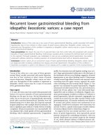
patient also had ischemic spots over the penile shaft and
localized soft swelling (3 × 2.5 cm) on the left side of the
peno-scrotal junction (Fig. 1). Penile and perineal palpa-
tion indicated intact devices in place, and this was further
confirmed by radiography of the pelvis. However, the
appearance of the patients' organs looked abnormal with
poor concealment of the devices. The patients' white
blood counts were elevated (13.200/mL and 14,100/mL,
respectively). Urine analyses and cultures yielded negative
results. Diabetes was well controlled in both patients as
evidenced by normal levels of fasting and postprandial
blood sugars and glycosylated hemoglobin levels. Ultra-
sound examination of the genitalia was performed to
identify any possible hematoma but yielded no relevant
results other than edema at the peno-scrotal junction of
the first patient. Blood examination for bleeding, coagula-
tion, prothrombin and partial thromboplastin times
yielded normal results.
After discussion in each case, we decided to perform an
exploration and extrusion of each penile prosthesis. The
operations were performed under spinal anesthesia. The
tunica was opened and a significant volume of whitish-
yellow purulent material was noted around both cylinders
of the device in both patients. Cultures of this material in
both patients yielded positive results for Staphylococcus epi-
dermidis (S. epidermidis). The localized swelling seen at the
peno-scrotal junction of the first patient was confirmed to
represent soft tissue edema but not hematoma. Removal
of the prostheses followed by continuous irrigation and
suction drainage resulted in rapid and complete resolu-
tion of the local inflammatory process and infection-asso-
ciated symptoms within three to four days and recovery
was uneventful in both cases.
Discussion
Penile prosthesis infection has been reported in many
studies with an incidence of about 8.9 %, mostly occur-
ring in the first year postoperatively [2]. DM is prominent
in the etiology of erectile dysfunction and has also been a
feature of most cases of penile prosthesis infection [1,2].
Problems in neurovascular, immune and micro-circula-
tory systems are well-known to be associated with DM [3],
and may contribute to the higher rate of prosthesis infec-
tion in diabetic patients.
In the present report, S. epidermidis was isolated on culture
taken from the explored wounds of both patients. This
supports the findings of several studies showing S. epider-
midis as the most common organism found at removal of
penile prostheses due to infection [4]. S. epidermidis is
present in all portions of the body living within the super-
ficial layers of the epidermis surrounded by the biofilm; a
protective coating [5]. Given the symbiotic nature of this
bacteria living outside the immunological system of the
body it incites little immunological response when it is
the cause of infection related to a prosthesis. Patients
infected with this organism may remain asymptomatic for
long periods. These bacteria probably arrived in the pros-
thesis as surgical contaminants during the initial surgery
[6].
Both patients, in the current report, had penile prostheses
with 13 mm diameter. These prostheses are somewhat
bulky and cannot be satisfactorily crammed into relatively
small organs nor allow for complete concealment. This
lack of appropriate concealment might facilitate easier
exposure of patient organs to unexpected trauma, as evi-
denced by the soft tissue edema in the first patient.
Although the trauma reported in these cases appeared triv-
ial it may have been sufficient to break up the biofilm gen-
erated by the offending organism; at least partially, with
Appearance of the penis on initial examination in Case 1Figure 1
Appearance of the penis on initial examination in Case 1. The
arrow shows edema on the peno-scrotal junction.
Journal of Medical Case Reports 2007, 1:34 />Page 3 of 3
(page number not for citation purposes)
detachment and dispersal of the organism in a planktonic
fashion leading to rapid progression of the infection proc-
ess. This is consistent with the findings of Costerton et al
[7] who showed that trauma could represent a potential
triggering events for disengagement of bacterial biofilms.
In support of our explanation, two lines of evidence were
present. First, the isolated organism was S. epidermidis. The
biofilm made by this bacterium is formed of multiple cell
layers resting on the biomaterial surface and protected by
an amorphous slimy material [8]. This slime is not a true
capsule, but is loosely bound to the staphylococcal cells.
This slime may be less resistant to shearing force during
washing or during trauma causing its break up [9]. Sec-
ond, both patients were diabetic and several in-vitro stud-
ies have showed that glucose and its analogues, although
inducing the formation of S. epidermidis biofilm, also dis-
tinctly inhibit its strong attachment to biomaterials [10].
However, this trivial trauma induced only minimal bio-
film detachment and the antibiotics, therefore, received
by our patients were thus ineffective to stop the impend-
ing infection. This suggestion agrees with several previous
studies proposing that if the biofilm is not sufficiently
damaged, antibiotic diffusion into the periprosthetic area
will be hindered making the antibiotic concentration sig-
nificantly lower compared to the level in serum [11]. Sev-
eral episodes of such trivial trauma might have affected
each of our patients since they underwent implantation
surgery. Nevertheless, they passed un-noticed as they were
not associated with the signs of significant inflammation
related to the reported trauma that made these occasions
memorable.
On the other hand, the chronic pressure exerted by the cyl-
inders of these 13 mm prostheses with subsequent tissue
ischemia, and in presence of DM with its well known
microcirculatory and immuno-compromising problems
[3], could provide a good environment for prosthesis
infection to occur. While most reported penile prosthesis
infections occur in the early post-operative period [2], late
infections have been also documented. A review of the lit-
erature revealed late prosthesis infection due to hematog-
enous seeding from significant remote entry sites, in the
absence of trauma, including patients with active Crohn's
disease, skin ulcers and dental abscesses. However, these
reports involved only a small number of patients and so
they appeared to occur infrequently although the infec-
tion was obvious when it occurred [12]. Our two cases
appear to represent the first reported instances of late-
onset prosthesis infection precipitated by trivial acciden-
tal trauma in the absence of any demonstrable source of
infection.
Conclusion
When the implantation of a malleable penile prosthesis is
considered, appropriate sizing should be taken into
account to ensure good concealment and to allow the
patient to avoid easy exposure of the penis to unexpected
trauma. Patients with such prostheses should also be care-
fully instructed about the importance of concealing the
device. New antibiotic-coated prostheses should be con-
sidered for insertion particularly in patients with condi-
tions such as diabetes to decrease the subsequent risk of
device infection. Device extrusion should be expedited as
soon as signs of prosthesis infection appear, since antibi-
otic use alone is likely to be of little value.
Competing interests
The author(s) declare that they have no competing inter-
ests.
Authors' contributions
The contributing authors made a critical review of this
manuscript.
Acknowledgements
Both patients have provided written informed consent for the publication
of this case report. Funding support for this research is not available.
References
1. Salama N: Satisfaction with the malleable penile prosthesis
among couples from the Middle East: is it different from that
reported elsewhere? Int J Impotence Res 2004, 16(2):175-180.
2. Cacan M, Demirel F, Karabacak O, Yalcınkaya F, Altug U: Risk fac-
tors for penile prosthetic infection. Int Urol Nephrol 2003,
35:209-213.
3. Powers AC: Diabetes mellitus. In Harrison Principles of Internal Med-
icine Edited by: Kasper DL, Braunwald E, Fauci AS, Hauser SL, Longo
DL, Jameson JL. McGraw-Hill: New York; 2005:2152-2180.
4. Montague DK: Periprosthetic infections. J Urol 1987, 138:68-69.
5. Voung C, Otto M: Staphylococcus epidermidis infections.
Microb Infect 2002, 4:481-489.
6. Cheng KJ, Irvin RT, Costerton JW: Autochthonous and patho-
genic colonization of animal tissues by bacteria. Canad J Micro-
biol 1981, 27(5):461-490.
7. Costerton JW, Stewart PS, Greenberg EP: Bacterial biofilms a
common cause of persistent infections. Science 1999,
284:1318-1322.
8. Marrie TJ, Costerton JW: Scanning and transmission electron
microscopy of in situ bacterial colonization of intravenous
and intraarterial catheters. J Clin Micrbiol 1984, 19(5):687-693.
9. Gotz F: Staphylococcus and biofilms. Mol Microbiol 2002,
43:1367-1378.
10. Telgman U, Horn H, Morgenroth E: Influence of growth history
onsloughing and erosion from biofilms. Water Res 2004,
38:3671-3684.
11. Jarow JP: Risk factors for penile prosthetic infection. J Urol
1996, 156:402-404.
12. Carson CC, Robertson CN: Late hematogenous infection of
penile prosthesis. J Urol 1988, 139:112-118.