Cochlear Implants: Fundamentals and Application - part 3 pdf
Bạn đang xem bản rút gọn của tài liệu. Xem và tải ngay bản đầy đủ của tài liệu tại đây (722.31 KB, 87 trang )
136 3. Surgical Pathology
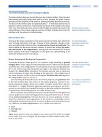
F
IGURE
3.15. A photomicrograph of the basal turn of the cat cochlea where the electrode
entry through the round window membrane was grafted with fascia, and an otitis media
was induced with Streptococcus pneumoniae. This has formed a fibrous tissue sheath or
type 1 seal. ET—electrode track.
mococcal otitis media, but there was a reduced incidence of infection when the
entry point was grafted.
Therefore, for safety it is essential to place a graft around the electrode where
it enters the cochlea. Although there was no statistically significant difference
between fascia and Gelfoam, it is recommended that fascia and not Gelfoam be
used. Gelfoam was used in the animal models to produce otitis media as described
previously. If bacteria are introduced at surgery with Gelfoam around the elec-
trode entry point, it could act as a home (nidus) for infection (Clark and Shepherd
1997). These experimental results apply to the Nucleus free-fitting array only. It
must be stressed that a two-element array with members close to each other should
not pass from the middle to the inner ear. A space between them is a conduit for
infection, a home to allow pathogens to multiply, as well as a site to increase the
pathogenicity of the organisms and reduce the ingress of antibodies and anti-
biotics. This is especially important considering the above studies showing the
invasiveness of S. pneumoniae.
Host Factors and Foreign Bodies
Implanted foreign bodies, as discussed above (see Biocompatibility of Materials),
are not totally inert, and should be evaluated for tissue toxicity. Foreign bodies
markedly increase the pathogenic potential of organisms of low virulence, for
Infection 137
example, Staphylococcus epidermidis (Lowy and Hammer 1983). Many studies
have shown that a bacterial inoculum that is normally “subinfective” will lead to
a severe infection in the presence of implanted material such as devitalized and
crushed muscle and gelatin (Vaudaux et al 1994). Finally on the basis of the
above evidence it is apparent that any dead space between the two members of a
dual element array would not only be a pathway for infection to enter the inner
ear and a home for the pathogens to multiply, but also would allow them to
become more virulent.
The effect of a dead space either within a foreign body or between two bodies
has been investigated by Zimmerli et al (1982) using Teflon perforated cylinders
(tissue cages). With this and other implants producing a dead space (Bergan 1981;
Marchant et al 1986), an inflammatory exudate accumulated within the cages
within 2 to 4 weeks. If these tissue cages were infected with an organism at levels
much below those normally causing infection, there would be a virulent inflam-
mation with the ingress of polymorphonucleocytes and the formation of pus. This
demonstrated that a dead space could make organisms more virulent. Further-
more, the tissue cage model also showed that parenteral antibiotics were ineffec-
tive against the organisms in the cage if administered more than 12 hours after
the inoculation. This inefficacy of antibiotic therapy is commonly observed in the
clinical context of foreign body infections (Vaudaux et al 1994).
Furthermore, it was shown that the phagocytic activity of neutrophils in the
cages was markedly deficient and lower than observed with neutrophils from
acute and chronic peritoneal exudates or blood. This suggested the neutrophils
could be damaged through contact with the surface of foreign bodies, and this
would reduce their antibacterial activity (Zimmerli et al 1982). Or alternatively
it was associated with a reduced level of opsonins and complement in the tissue
cages (Zimmerli et al 1982). In a later phase of infection, complement-mediated
opsonic activity was reduced, and this too limited the ability of body to handle
infection. Thus any dead space created within and across the inner ear is not only
likely to be a path or home for infection, but also will increase the virulence of
the organism and reduce the body’s ability to deal with the infection either through
phagocytic action or complement-mediated responses. It has also been shown
with dead space that the access for antibiotics is significantly reduced. In addition,
the studies with the infected tissue cages showed there was no associated bacter-
emia or spread by the bloodstream.
The penetration of antibiotics to infected locations almost always depends on
passive diffusion. The rate is proportional to the concentrations of a drug in the
plasma or extracellular fluid. Drugs that are extensively bound to protein may not
penetrate to the same extent as those with lesser links. Drugs that are highly
protein bound may have reduced activity because there is a smaller fraction to
react with its target. For example, the drugs cefotaxime and ceftriaxone, both
third-generation cephalosporins and the treatment of choice for H. influenzae and
S. pneumoniae infections, have different degrees of binding. Ceftriaxone is used
for adults and Cefotaxime in children. Ceftriaxone, however, is 90% to 95%
protein bound, and that greatly reduces its efficacy. On the other hand, cefotaxime
138 3. Surgical Pathology
is only 36%. Vancomycin should be added to the therapy if the minimum inhib-
itory concentration (MIC) for these antibiotics is greater than 0.12 mg/L. Thus if
there is a dead space as seen with a two-element array, the penetration of the
antibiotics could be considerably reduced. In addition, in preventing infection
spreading to the meninges many antibiotics that are polar and at a physiological
pH do not cross the blood–brain barrier at all well. Some such as penicillin G
are even actively transported out of the CSF by active transport mechanisms in
the choroid plexus. The concentrations of penicillin and cephalosporins in the
CSF are usually only 0.5% to 5% of the steady-state level in the plasma (Quag-
liarello et al 1986). The integrity of the blood–brain barrier, however, is dimin-
ished during bacterial infection.
With infections from S. pneumoniae and other pathogens, there is also the
added problem of their developing a biofilm, a slime on the surface of the foreign
material, and this will allow them to resist antibiotics and antibodies. Bacteria
that adhere to implant materials by encasing themselves in a hydrated matrix of
polysaccharide protein form a slimy layer known as a biofilm (Stewart and Cos-
terton 2001). Bacteria in the biofilm are resistant to antibiotics. For example, a
b-lactimase negative strain of Klebsiella pneumoniae had a MIC of 2 lg/mL of
ampicillin in aqueous suspension, but when grown as a biofilm the organism was
scarcely affected by 4 hours’ treatment with 5000 lg/mL of ampicillin, a dose
that would eradicate free-floating bacteria. The antibiotic resistance that normally
occurs due to efflux pumps, modifying enzymes, and target mutations does not
seem to apply to this mechanism of drug resistance with biofilms.
Furthermore, because active and inactive microbes are closely situated and
because surviving bacteria can use dead ones as nutrients, the new cells remaining
after antibiotic therapy can restore the biofilm to its original state in a matter of
hours.
Single-Component Array and the Natural Defenses Against Infection
A single component array that is surrounded with a fibrous tissue sheath can, as
described above, effectively work with the body’s three defense mechanisms to
prevent the ingress of infection from an otitis media to the cochlea and thence
the meninges. The above studies demonstrated that the sheath around the single
component array enabled three lines of defense to be used against the spread of
infection. The first line of defense is the surface activity of mucus-secreting cells,
and their extension around the electrode. The second line of defense is the mo-
bilization of phagocytes in and around the sheath. The third line of defense is the
mobilization of type B lymphocytes, and type T lymphocytes to the sheath and
between the sheath and the electrode.
With the first line of defense against the spread of infection from otitis media,
the surface cells around the electrode entry changed into mucus-secreting cells
and extended around and along the electrode array. They produced mucus that is
bacteriostatic, and the hairs of the mucous cells beat to and fro to sweep the
bacteria away. Their growth around the electrode is illustrated in Figure 3.8.
Infection 139
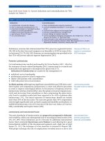
F
IGURE
3.16. Phagocytosis of bacteria—the second line of defense. Photomicrograph
shows granulocytes and debris in apposition to mucous lining cells.
The second line of defense operates when the bacteria release toxins into the
sheath. The blood vessels dilate and bring the phagocytes to the site so they can
digest the bacteria. This is illustrated in Figure 3.16.
The third line of defense is the production of type B and T lymphocytes, in
response to the bacterial surface antigen. The B lymphocytes produce antibodies
and the T lymphocytes are killer cells that pierce cells. Note that in Figure 3.11
the lymphocytes not only lie in the connective tissue around the sheath, but also
enter between the sheath and the array.
Clinical Protocol
The results obtained from animal studies indicate that there is a risk of otitis
media extending into the inner ear after implantation during the first few weeks,
a period of increased vulnerability due to the increased permeability of the tissues
and the need for the seal to form. To minimize the risk of infection spreading into
the inner ear during or after implantation, it is recommended that surgery should
be carried out under strict aseptic conditions, preferably using a laminar flow of
filtered sterile air. Systemic antibiotics should be administered at the beginning
and conclusion of the operation to eradicate organisms introduced during the
procedure that could invade the inner ear during the period of increased vulner-
ability when the electrode seal is being established. As a further safeguard the
operative wound should be irrigated with an antibiotic solution of ampicillin and
140 3. Surgical Pathology
cloxacillin. Although not the first-line antibiotics for the treatment of S. pneu-
moniae infections, they have a broad spectrum of activity. In the U.S., of the
children who had meningitis, one child with a ventriculoperitoneal shunt devel-
oped the infection within a day or two of having the implant, and two with normal
cochleae developed it within 24 hours. It is likely that the causal S. pneumoniae
could have been introduced into the perilymph and thence the CSF. For this reason
irrigation seems warranted. This is especially desirable as unpublished studies by
Black and Clark showed that antibiotic concentrations were very low in the peri-
lymph of the cat unless the cochlea was infected; furthermore the blood–brain
barrier does not allow antibiotics to easily enter the CSF in the uninflammed
condition. In children with the Mondini syndrome, special care should be taken
as there can be a wide dehiscence between the scala tympani or the scala vestibuli
and internal auditory canal. The data in the experimental animal presented in the
sections above also demonstrate the necessity of a fascial autograft, which should
be placed around the electrode in the cochleostomy. I have experimental unpub-
lished data to suggest that if there are gaps between strips of fascia, they could
be a passage for pathogens to enter the cochlea. If there is a “perilymph gusher”
at surgery, then the fascia will need to be compressed quite firmly. The fascial
autograft can be taken from the temporalis fascia. It is not desirable to use crushed
muscle, as it can become necrotic and a home for infection. Bone pate´ provides
spicules of bone that are not absorbed and may also be a nidus for infection, as
may Gelfoam. Furthermore, as stated above, there are serious risks associated
with the use of a two-element electrode array.
After the tissue around a cochleostomy or the implanted round window has
healed, the response to infection appears similar to that of a nonimplanted cochlea.
However, certain microorganisms could have a detrimental impact as seen with
S. pneumoniae or P. aeruginosa. Improving the seal at the entry point still requires
further research with other biocompatible materials and techniques.
Deafness and the Central Auditory Pathways
Spiral Ganglion
With the loss of hair cells there is a rapid and extensive reduction of the unmy-
elinated peripheral processes in the organ of Corti (Terayama et al 1977), and a
more gradual degeneration of the myelinated portion of the peripheral processes
within the spiral lamina as well as the spiral ganglion cells (Webster and Webster
1981; Spoendlin 1984; Leake and Hradek 1988; Shepherd and Javel 1997). Some
surviving cells and processes may be demyelinated. These changes as discussed
above are due to the loss of trophic factors from the hair cells, and vary according
to the type of lesion and animal species. In the human there is better preservation
of the spiral ganglion cells over longer periods of time than in other animals, for
example, the guinea pig. Otte et al (1978) found 45% of cochleae from profoundly
deaf people had at least a third or more of the number of ganglion cells found in
Deafness and the Central Auditory Pathways 141
the normal population. In 93 cochleae from profoundly deaf people Nadol et al
(1989) found the main spiral ganglion population was half the normal. The loss
was greater in older subjects, for longer durations of hearing loss, and in the basal
turn. Etiology had the greatest impact and the depletion was most extensive in
people with viral labyrinthitis, congenital or genetic deafness, or bacterial men-
ingitis. The least extensive loss occurred after aminoglycosides and sudden id-
iopathic deafness (Nadol et al 1989; Nadol 1997). The physiological effects of
these pathological changes and their impact on electrical stimulation with a co-
chlear implant are discussed in Chapter 5.
Cochlear Nucleus
Pathological changes in the central auditory pathways, as well as in the spiral
ganglion, can follow loss of cochlear function. As distinct from spiral ganglion
cell loss, occurring at any stage of life, transneuronal degeneration of higher order
neurons only develops with the loss of cochlear function at a critical period early
in life. Ablation of the cochlea in the experimental animal during a narrow time
window near the onset of hearing is the only period when significant cell death
is demonstrated in the anteroventral cochlear nucleus (AVCN) (Tierney et al 1997;
Mostafapour et al 2000). With cochlear destruction in 6-day-old mice, the co-
chlear nucleus (CN) population was reduced to 34% of normal (Trune 1982). The
changes were not due solely to ablation of the cochlea, but also to the loss of
activity in the auditory nerve. Born and Rubel (1988) found transneuronal cell
death and reduction in soma size also occurred when a sodium channel blocker
was applied (Pasic and Rubel 1989). These changes could be prevented by elec-
trical stimulation of the auditory nerve, but not by direct excitation of the neurons
in the CN (Hyson and Rubel 1989; Zirpel and Rubel 1996). They were the result
of presynaptic release of the transmitter shown to be glutamate (Zirpel and Rubel
1996). The effects were associated with reduced protein synthesis (Sie and Rubel
1992), and increased intracellular Ca
2ם
(Zirpel et al 1995). Mostafapour et al
(2000) found evidence that suggested neuronal death was due to the inactivation
of an antiapoptotic (anti–cell death) gene bcl-2. Early loss of hearing also led to
a significant decrease in the expression of messenger RNA (mRNA)-encoded
receptors to glutamate (Marianowski et al 2000). In addition, there was an increase
in the expression of receptors to c-aminobutyric acid, a major inhibitor (Mari-
anowski et al 2000), as well as a long-term deficiency in glycinergic synaptic
inhibition. In mammals the changes were most marked in the CN, but higher
order effects could be observed. The significance of these events is not clear, but
they presumably affect both place and temporal frequency codes, as discussed in
Chapters 5 and 6. It is also unclear when and whether these changes occur in
humans. They do, however, suggest the importance of early electrical stimulation
of the auditory nerve.
If animals are deafened after the onset of hearing, there is no transneuronal
degeneration, but a shrinkage in the soma size associated with downregulation of
142 3. Surgical Pathology
its metabolism, and a reduction in the neuropil (a complex mesh of terminal axons,
dendrites, and neuroglial processes). The reduction in soma size was first dem-
onstrated by Powell and Erulkar (1962), who destroyed the cochlea in mature
cats, and reported neuronal shrinkage in the CN and superior olivary complex
(SOC). In another study, a reduction in soma size by a third occurred within 1
week of the hearing loss (Pasic and Rubel 1989). The deafening had a marked
effect on the metabolic activity (Wong-Riley et al 1978; Durham et al 1993).
There was also a loss of the neuropil or axon terminals innervating the ventral
cochlear nucleus (VCN) (Powell and Erulkar 1962; Trune 1982). This may have
been due to a loss of spiral ganglion cells, and a reduction in the number of their
axons converging on the AVCN cells. It resulted, too, in an increase in the packing
density in the AVCN.
Cochlear ablation in adult experimental animals also led to a loss of synapses
in the AVCN. This too could be related to the loss of auditory neurons converging
on the AVCN cells. This was followed by the generation of synapses over a long
period from the remaining afferent input (Benson et al 1997). The loss of hearing
also affected the terminal boutons. For example, the end bulbs terminating on the
globular bushy cells were smaller as were the end bulbs of Held terminating on
spherical bushy cells (Ryugo et al 1997; Redd et al 2000). This effect could have
been the result of a downregulation in the metabolism of the remaining spiral
ganglion cells. The above changes were accompanied by a temporary reduction
in the expression of mRNA receptors to glutamate (Sato et al 2000), the main
excitatory neurotransmitter in the auditory pathway. There was also a deficiency
in glycinergic synaptic inhibition (Willott et al 1997).
As the sensorineural hearing loss led to a loss of the terminal axons and syn-
apses on the cells in the AVCN as well as soma size, this would limit the pro-
cessing of temporal and place information as discussed in Chapter 5. As these
effects were secondary to the loss of spiral ganglion cells it makes it essential to
stimulate these cells electrically as soon as possible after deafening to preserve
the input to the AVCN. The connections could thus be preserved for improved
strategies that may be developed later to provide fine temporal spatial patterns of
excitation for the temporal coding of frequency.
Chouard et al (1983) found the soma size of octopus cells in the VCN of the
guinea pig was preserved with electrical stimulation. In a study by Xu et al (1990),
kittens were deafened 37 to 40 days after birth with ototoxic drugs. The animals
were stimulated 80 to 90 days after birth on one side. The mean soma areas in
the AVCN were significantly greater than the unstimulated control side. There
was a weaker trend for the cells to be larger in the posteroventral cochlear nucleus
on the stimulated side.
Pons and Midbrain
A bilateral sensorineural hearing loss at the onset of hearing resulted in a signifi-
cant reduction in synaptic density in the central nucleus of the inferior colliculus
Deafness and the Central Auditory Pathways 143
(ICC) (Hardie et al 1998). In view of the loss of neurons in the AVCN discussed
above, this would lead to a loss of input and synaptic connections at the IC. A
unilateral loss, however, did not lead to a loss in density. This was associated
with an increase in the proportion of neurons projecting from the ipsilateral side
(Nordeen et al 1983; Moore and Kitzes 1985). This suggests that the relative level
of neural activity in the pathway from each VCN determines the success of each
side in forming or retaining synapses in the auditory midbrain (Moore 1990).
A sensorineural loss after the onset of hearing also affected the higher brain
centers in the pons and midbrain. There was a reduction in the soma area of
neurons in the trapezoid body (Pasic et al 1994), the SOC and nucleus of the
lateral lemniscus (Powell and Erulkar 1962), and ICC (Nishiyama et al 2000).
Human Brainstem
There are few studies on the human central auditory pathways following a pro-
found hearing loss. A reduction in the soma area was found in the CN by Clark,
Shepherd et al. (1988) Seldon and Clark (1991), and Moore et al (1997), but also
in the medial superior olive (MSO) and IC (Moore et al 1997). There was also a
reduction in the volume of the CN, especially the VCN, in the studies by Clark,
Shepherd et al (1988) and Seldon and Clark (1991). These findings, also discussed
in Chapter 4, were essentially consistent with those from experimental animals.
The brainstem and temporal bone of the first University of Melbourne/Bionic
Ear Institute patient to have a bilateral cochlear implant were also studied (Yu-
kawa et al 2001a,b). The sections were compared with those from a second person
who had a cochlear implant on one side. The bilateral patient died at the age of
59 years. He went profoundly deaf in the left ear at 31 years due to a head injury,
and became profoundly deaf in the right ear at 36 years. At 46 years he had a
right cochlear implant and a left cochlear implant at 51 years. Thus the right ear
was implanted for 13 years and the left for 8 years. He had only fair speech
discrimination with the right implant and satisfactory results with the left. Bin-
aural psychophysical studies showed there was a marked reduction in the inter-
aural temporal discrimination difference limens for electrical stimulation. It was
well below that for normal hearing, as discussed in Chapter 6. The unilateral
subject died at the age of 62 years. She suffered a hearing loss due to mumps and
then had a 27-year history of a slow progressive loss and had a profound hearing
loss for 5 years prior to implantation. She had the implant for 1.5 years in the left
ear. The brainstems were sectioned and the MSO analyzed, as it is considered an
important nucleus for coding interaural time differences (see Chapter 5). The
trigeminal nucleus was also examined as a control for tissue fixation and pro-
cessing artifacts. The cell density and volume were determined for each nucleus.
Cell numbers and volume were determined by a technique in which a criterion
was established to ensure that the cells were not counted twice.
The results are shown in the Table 3.1 for cell density and volume, and statis-
tical significance was determined with the Mann-Whitney U test. There was a
144 3. Surgical Pathology
T
ABLE
3.1. The cell density and volume measures for the right and left medial superior
olive from one patient with a bilateral and another with a unilateral cochlear implant.
Side Cell density (ן10
מ5
/lm
3
) Mean
Bilateral Right 1.23 1.17*
Left 1.1
Unilateral Right 2.2 1.96
Left 1.71
*p Ͻ .05, Mann-Whitney U test.
Side Cell volume (lm
3
) Mean
Bilateral Right 2241 2005**
Left 1693*
Unilateral Right 2688 2577
Left 2511*
*p Ͻ .05, **p Ͻ .0001, Mann-Whitney U test.
significant difference between the bilateral and the unilateral subjects. For the
combined right and left sides there was a lower cell density and volume for the
bilateral compared with the unilateral subject. This suggests that the MSO was
affected by the hearing loss occurring well after the onset of hearing, and this is
the reason the patient did not have satisfactory interaural temporal difference
limens. It is also of interest that for both patients the cell volume was lower on
the left side. This is consistent with the fact that the first patient received more
help from the left implant, and the second unilateral patient had a left implant. In
both cases there would have been more contralateral stimulation to the right, thus
helping to preserve its function. This is consistent with the experimental animal
studies showing that electrical stimulation maintains cell viability and size (Miller
and Altschuler 1995).
Prenatal (Congenital) and Postnatal Hearing Loss
Deafness may occur before or during birth (prenatal and perinatal, respectively)
when it is referred to as congenital. It can also occur after birth (postnatal). Con-
genital deafness may arise from genetic causes, chromosomal abnormalities, or
diseases affecting the mother during pregnancy. In about two thirds of children
with prelinguistic severe or profound sensorineural deafness without syndromes
(before language develops), the cause is thought to be genetic (Morton 1991).
Postnatal deafness is mostly from disease or injury, but may also be the result of
delayed genetic effects.
Genetic and Chromosomal
Body cells contain 46 chromosomes, and the genes are located at different points
along the chromosomes. In the male the body cell divides into two germ cells;
PreNatal (Congenital) and Postnatal Hearing Loss 145
the sperms each contain 23 chromosomes. The same occurs in the female for the
ova. When the two germ cells containing 23 chromosomes unite, they form a new
cell with 46 chromosomes. Two chromosomes determine the sex of the individual.
In the male, one of the two sex chromosomes is small (Y chromosome) and
inherited from the father, and the other, the X chromosome, is inherited from the
mother. The female has two X chromosomes, one being inherited from the father
and one from the mother. The other 22 chromosomes are referred to as autosomes.
If a parent passes on a dominant gene causing deafness, it only requires one
chromosome of the pair to have the deafness gene for the child to be affected. If
it is a recessive gene, the child needs to have one on each chromosome pair. A
sex-linked inheritance may occur in the male when the X gene is affected, and
thus without protective effects from the Y or male chromosome. Genetic deafness
may be classified thus as dominant or recessive. Most genetic deafness presenting
congenitally is transmitted as a recessive, and about half those with recessive
deafness have no accompanying abnormalities.
Congenital, Genetic Deafness
Nonsyndromic
As stated, genetic deafness frequently occurs alone without other abnormalities
(nonsyndromic). In about 80% of children with nonsyndromic deafness, the in-
heritance is autosomal recessive (Dahl et al 2001). Using DNA markers, genetic
linkage studies have shown over 20 genes for nonsyndromic deafness (Van Camp
and Smith 2002). A mutation of the connexin 26 gene has been found to account
for up to 50% of cases of nonsyndromic deafness in children of European descent
(Maw et al 1995; Denoyelle et al 1997). In addition 50% to 90% of chromosomes
on which a connexin 26 mutation has been determined have the same specific
mutation (deletion of a guanine nucleotide at position 35, i.e., 35delG) (Denoyelle
et al 1997). A similar incidence to the European data was found for a group of
Australian deaf children (Dahl et al 2001). Furthermore, over 40 connexin 26
mutations have been reported (Denoyelle et al 1999). On the other hand, the
incidence of connexin mutations is very low in Asian-American and African-
American populations (Morell et al 1998).
Connexin 26 belongs to a family of proteins that mediate the exchange of
molecules between adjacent cells. The number refers to the size of the protein in
thousands of daltons. Connexin is highly expressed in the cells lining the cochlear
duct and the stria vascularis. It is thought that it is important for the recycling of
(K
ם
) ions from sensory hair cells into the endolymph in the process of transduc-
tion of sound to electrical signals. The slope of the hearing loss (over 2000 to
8000 Hz) was greater than in children without connexin 26 mutant alleles (Wilcox
et al 2000). It is not known to what extent cochlear implants benefit children with
connexin 26 and other genetic disorders.
Nonsyndromic deafness has variable anatomical and histological features. First,
there may be total lack of development of the inner ear, and the x-ray will show
complete absence. It can be difficult to distinguish this condition from bony laby-
146 3. Surgical Pathology
rinthitis. This condition is called the Michel syndrome, and it is inherited as
autosomal dominant. It will not be possible to implant children with this condi-
tion, but fortunately it only accounts for a small proportion of genetic deafness.
Second, only 1
1
⁄
2
turns of the cochlea may develop, rather than the normal 2
1
⁄
2
turns. This condition is often associated with underdevelopment of the vestibular
structures, and is called the Mondini syndrome. Endolymphatic hydrops (disten-
tion of the endolymphatic system) is often present, and there may be some residual
hearing. It is inherited as autosomal dominant and is characterized pathologically
by an absence of the septum (interscalar) between the apical and the middle turns,
thus creating a common cavity. In a child with the Mondini syndrome who died
from infection in the nonimplanted ear as discussed above, the temporal bones
showed a wide dehiscence between the scala tympani and the internal auditory
canal that could have accounted for the CSF leak at surgery (C. Suzuki et al
1998). The histology also showed there was a wide vestibular aqueduct, and
expansive communication between the cochlea and vestibule that could lead to a
misplaced electrode. There was a hypoplastic modiolus, and the spiral ganglion
cell population was 10,826. In the unimplanted ear there was inflammatory ne-
crosis of the round window membrane, and many polymorphonuclear leukocytes
in the adjacent scala tympani, indicating the route for the spread to the cochlea.
The extension to the meninges probably occurred through the abnormally patent
modiolus. This 6-year-old child was developing speech and language, and this
indicates the importance of providing an implant. However, because of meningitis
it stresses the need to ensure there is an adequate seal around the electrode entry
into the inner ear, and the aggressive treatment of any middle ear infection.
In other cases the modiolus may be better developed, and this is apparent on
the computed tomography (CT) scan. A perimodiolar electrode array could be
used, as the spiral ganglion cells lie centrally. On the other hand, the modiolus
may be deficient, and the cochlear nerve fibers lie peripherally. When this happens
it is preferable to use the straight but flexible Nucleus array. This array produces
less trauma, and lies closer to the nerve fibers. Schmidt (1985) examined eight
bones and found a significantly reduced population in those where there had been
a severe hearing loss.
The Mondini dysplasia may be associated with a wide cochlear aqueduct (peri-
lymph gusher) (Nadol 1984). This is seen on the CT scan, and indicates that a
large outflow of CSF (perilymph gusher) may occur when an opening is made
into the cochlea for the insertion of the electrode array. So in summary, satisfac-
tory to good results have been reported for cochlear implants with the Monidini
dysplasia (Silverstein et al 1988; Turrini et al 1997; M. Suzuki et al 1998).
A related condition is the large vestibular aqueduct syndrome (LVAS) first
described by Valvassori and Clemis (1978) on radiological findings. An autoso-
mal-recessive or X-linked inheritance was suggested by Griffith et al (1996). A
profound hearing loss was reported in 39% of patients (Jackler and De La Cruz
1989).
Children with the Mondini dysplasia have a higher risk of meningitis whether
they have an implant or not. Phelps et al (1994) report an incidence of four of 20
PreNatal (Congenital) and Postnatal Hearing Loss 147
children with congenital dysplasia (unimplanted) developed meningitis. In an
analysis of the 19 Nucleus patients who developed meningitis out of 16,500
implantees in North America (see Chapter 10), at least 9 had a deformity of the
cochlea. It is unclear if any were device-related, but it again serves to emphasize
the importance of sealing the round window entry point together with extreme
care in the antibacterial management.
Finally, if the development of the osseous cochlea is complete, but the sensory
elements have failed to develop, they may be represented only by mounds of
undifferentiated cells. This is referred to as the Scheibe syndrome. It is the com-
monest of all inherited congenital deafness, and is autosomal recessive.
Syndromic
In a number of children deafness is associated with other abnormalities, and
hearing loss may be the first symptom. With Waardenburg’s syndrome, the fea-
tures other than deafness are a lateral displacement of the inner canthus of the
eye, heterochromia of the iris, and a white forelock. It is inherited as autosomal
dominant. Pathologically there is atrophy of the organ of Corti and stria vascularis,
and a reduction in the number of ganglion cells. In albinism, where there is loss
of pigmentation resulting in fair skin and poor vision, the deafness is bilateral
and severe. It is inherited as an autosomal-dominant or -recessive or sex-linked
trait. With onchodystrophy there is sensorineural deafness and nail dystrophy.
Pendred’s syndrome may account for 10% of recessive deafness. In this syndrome
there is abnormal iodine metabolism. It is often associated with a Mondini de-
formity of the cochlea. In Jervell’s syndrome there is a bilateral severe hearing
loss and cardiac abnormality (prolonged Q-T interval) that can lead to sudden
death (Stokes-Adams attacks). It is inherited as autosomal recessive. Usher’s dis-
ease is a congenital condition in which there is combined sensorineural hearing
loss and retinitis pigmentosa. It is inherited as sex linked or autosomal dominant,
and there is a recessive form. So it is in fact a collection of conditions. There are
a number of other syndromes that have associated deafness, and more details can
be obtained from standard texts.
Deafness may also occur due to chromosome abnormalities. Normally the 22
pairs of autosomal chromosomes are grouped according to similar morphologies
from A to G. Trisomy 13 to 15 (D) have an extra chromosome located in the
group D, and trisomy 18 (E) in group E. These conditions are often associated
with other ear or body defects, and the children die early.
Delayed
Delayed sensorineural deafness coming on sometime after the baby is born can
also be genetic, and deafness is commonly the only abnormality. It is inherited
as an autosomal-dominant condition, and there is a progressive sensorineural
hearing loss. A delayed sensorineural loss may also be associated with other
abnormalities and there are a number of these conditions (see Chapter 9).
148 3. Surgical Pathology
Acquired
Prenatal and Perinatal
There are a number of nongenetic causes of congenital deafness. These are in-
fective agents; trauma, in particular drugs; and metabolic conditions. The most
common infective agents are toxoplasmosis, rubella, cytomegalovirus (CMV),
and herpes simplex, together referred to as TORCH. O’Sullivan et al (1997)
showed that the most common viral causes of a hearing loss in the Melbourne
Cochlear Implant Clinic were CMV and rubella. Rubella and other viruses cross
the placental barrier to infect the fetus, and this impairs the development of the
cochlea and other organs. With rubella the hearing loss is more severe if the
infection affects the mother in the first 3 months of pregnancy (first trimester),
but it may occur following infections in the second or third trimesters. It is nec-
essary to make the diagnosis by detecting the virus in the pharynx, urine, or CSF,
and the presence of a specific immunoglobulin M (IgM) antibody in the chord
blood or body serum. There are also persistent elevated levels of rubella IgG in
the serum. In a review of 300 children with congenital rubella, 50% of the mothers
had no clinical evidence of the disease, so there was a high incidence of subliminal
infections. Maternal rubella infection during pregnancy must be confirmed by
viral isolation or serological tests. Prospective studies based on laboratory diag-
nosis show the incidence of deafness to be from 50% to 70%. Rubella can also
be associated with cardiac defects or mental retardation. The hearing loss is pre-
dominantly bilateral, but may be asymmetrical. In a small proportion the deafness
became more severe with time. This was probably due to persistent infection,
indicated by the continued shedding of the rubella virus after birth. The central
auditory pathways may also be affected, and this could account for the lack of
language development. This could also apply to results with cochlear implanta-
tion. It has been shown, too, that a child is more likely to develop deafness from
rubella when there is a genetic predisposition shown by a positive family history.
CMV results in deafness that is often severe to profound, and like rubella can
progress. It too affects the central nervous system, with impaired vision, cerebral
palsy, epilepsy, and intellectual disability. CMV infections are highly prevalent
and can be detected in 0.5% to 2.4% of all live births (Pass, Stagno et al. 1980).
Rasmussen (1990) estimated that 10% of infected newborns are at risk from
hearing loss, impaired vision, or neuromuscular abnormalities. Although 90% of
patients are without symptoms, there may be swollen lymph nodes and enlarge-
ment of the liver and spleen. In children presenting with a severe-to-profound
hearing loss, it is considered important to undertake serological tests on both the
mother and child, as well as viral cultures from the saliva and urine up to the age
of approximately 4 years (S. Locarnini, personal communication). This helps in
deciding whether the child has had a CMV infection, and whether it was of
congenital origin when the effects are more severe. The children with CMV at
the University of Melbourne’s Cochlear Implant Clinic have not had as good
results as other children, and this may be due to the involvement of central au-
ditory pathways.
PreNatal (Congenital) and Postnatal Hearing Loss 149
Herpes simplex encephalitis is a viral infection usually from genital herpes. It
may present neonatally as a localized mucocutaneous or disseminated infection.
When it is disseminated there is a 30% risk of meningoencephalitis that is likely
to occur in the second or third week of life. Herpes simplex infections leading to
sensorineural hearing loss may also involve the central auditory pathways.
The pre- and perinatal viral infections infect the fetus and neonate in the same
way as a postnatal invasion. Lindsay (1973) has shown that the spread of the
virus by the bloodstream to the endolymph produces a different pathological
picture from the one where the spread is from the meninges or lining of the brain
via the cochlear aqueduct to the perilymph in the scala tympani of the basal turn.
With an endolymphatic involvement there is often a normal spiral ganglion or
cochlear nerve population. With spread to the perilymph there is more often
degeneration of spiral ganglion cells and nerve fibers and variable changes in the
cochlear duct including hydrops. Malformations such as a rudimentary organ of
Corti and underdeveloped stria vascularis and tectorial membrane are rare. In
most cases the lesions are due to small hemorrhages that are probably the result
of increased coagulability produced by the viruses. The vestibular system is only
affected in a small number of cases.
The only specific bacterial cause of deafness is syphilis. This organism cannot
cause malformations of the cochlea, as the treponema is not able to pass through
the placental blood barrier before the fifth month. Its effects are either through
inflammation of the meninges and nerve or due to labyrinthitis. The latter is more
common and the hearing loss increases in a steplike fashion. Loss of the spiral
ganglion cells is more likely to occur with this condition.
Parasitic protozoa are also agents that can lead to a severe sensorineural hearing
loss in the fetus. They are single-celled motile organisms. In particular Toxo-
plasma gondii infections in the mother (toxoplasmosis) can pass through the
placenta after 6 weeks. It is acquired either by contact with oocyte-shedding
kittens or by eating cyst-ridden undercooked meat. It is a common condition, and
some 87% of the population over 30 years of age have serologically positive tests.
The infection of the mother is generally not apparent. The diagnosis is made from
serological tests, the Savin’s lysis, and complement fixation tests. The significance
of the serological test depends on the age of the child. A positive result at 3 to
12 months would indicate congenital toxoplasmosis. The deafness is associated
with chorioretinitis and the characteristic deterioration of the fundus of the eye,
hydrocephalus or microcephaly, with calcification of the brain seen on x-ray. In
the cochlea there is calcification of the stria vascularis and spiral ligament, and
inflammation of the whole vestibule. Physical trauma in the form of misapplied
forceps during delivery may lead to loss of hearing through fractures of the skull
base. Chemical agents during pregnancy, such as ototoxic antibiotics, can lead to
profound hearing loss. Poor blood supply to the fetus (anoxia), through a hem-
orrhage behind the placenta (antepartem hemorrhage) or the placental cord around
the neck during delivery, is a factor. Other anoxic and metabolic causes are hy-
pertension, toxemia, diabetes, renal disease, and Rh blood incompatibility. It is
especially important to assess the condition of the baby after birth and to calculate
150 3. Surgical Pathology
an Apgar score, which is based on the color, reflex responses, respiratory effort,
heart rate, and muscle tone.
Child and Adulthood
The viral causes of a severe postnatal hearing loss are mumps, measles, influenza,
and chicken pox. These produce a viral labyrinthitis that affects the endolymphatic
duct. Pathological changes are more pronounced in the basal cochlea and include
degeneration of the organ of Corti, atrophy of the stria vascularis, displacement
and distortion of tectorial membrane, and distortion and degeneration of the sac-
cule. The utricle and semicircular canals are seldom involved.
The most common bacterial cause of a hearing loss in the newborn (neonate)
and in later childhood is labyrinthitis following meningitis. A study by Goodhill
(1950) on 904 deaf children showed 10% had meningitis as the cause of their
hearing loss. When deafness occurs it is mostly a very severe or a total loss, and
is usually due to infection of the inner ear (labyrinthitis). The incidence in men-
ingitis normally varies from 5% to 30%, depending on the causal organism. An-
tibiotics have now reduced the incidence.
With meningitis the infection is transmitted to the perilymph either through the
internal auditory canal or via the cochlear aqueduct. When through the internal
auditory canal the spread is via the perineural and perivascular spaces. In the
cochlea the pathological changes are the formation of serofibrinous exudate, in-
filtration with pus cells (polymorphonuclear leukocytes), and then the formation
of granulation tissue followed by healing characterized by fibrosis and ossifica-
tion. Ossification is usually more marked near the round window, where the peri-
lymphatic spread to the basal turn occurs.
Personal studies in the cat show that osteoid tissue commences within 2 weeks.
Therefore, in the human once the infection is controlled and the hearing loss
established, surgery should be considered to ensure that the electrode array can
be inserted an adequate distance. From the study of Blamey et al (1992) it has
been shown that up to 21 channels of stimulation are important (see Chapter 7).
Furthermore, as discussed above, if electrical stimulation is commenced early,
spiral ganglion cells will be preserved. Sometimes there is only fibrous tissue
rather than bone in the scala tympani, and for this reason magnetic resonance
imaging (MRI) should be carried out before operating on a patient with a history
of meningitis.
A head injury can produce fractures at the base of the skull. Sensorineural
hearing loss is more likely with a transverse than a longitudinal fracture of the
temporal bone. However, the fracture lines are not easily categorized as transverse
and longitudinal. With a transverse fracture of the temporal bone the cochlear
nerve may have been sectioned in which case the results will be unsatisfactory,
and this may be seen with a CT scan and the status of the cochlear nerve observed
with MRI. Ototoxic drugs such as neomycin, kanamycin, polymyxin, and chlor-
amphenicol, as well as loop diuretics, cause a hearing loss both in children and
in adults. The antibiotics usually have their effects on the outer hair cells. With
References 151
antibiotics ototoxicity may occur suddenly after a few injections and can continue
after the withdrawal of the drug. It may continue for many months after treatment.
The effect of ototoxic drugs on spiral ganglion cell numbers varies with species,
and there is a marked loss in the guinea pig within weeks (Webster and Webster
1978). However, in the human, as discussed above, there is greater spiral ganglion
cell survival than would be predicted from experimental animal data (Ylikoski et
al 1981). The results of implantation in these patients can be expected to be good.
References
Benitez, J. T. 1977. Stapedectomy and fatal meningitis. ORL 39: 94–100.
Benson, C. G., J. S. Gross, S. K. Suneja and S. J. Potashner. 1997. Synaptophysin im-
munoreactivity in the cochlear nucleus after unilateral cochlear or ossicular removal.
Synapse 25: 243–257.
Bergan, T. 1981. Pharmacokinetics of tissue penetration of antibiotics. Reviews of Infec-
tious Diseases 3: 45–66.
Berkowitz, R. G., B. K H. G. Franz, R. K. Shepherd, G. M. Clark and D. Bloom. 1984.
Cochlear implant and otitis media. A pilot study to assess the feasibility of pseudomonas
aeruginosa and streptococcus pneumoniae infection in the cat. Journal of the Oto-
Laryngological Society of Australia 5: 297–299.
Berkowitz, R. G., B. K H. G. Franz, R. K. Shepherd, G. M. Clark and D. Bloom. 1987.
Pneumococcal middle ear infection and cochlear implantation. Annals of Otology, Rhi-
nology and Laryngology 96(suppl 128): 55–56.
Black, R. C., G. M. Clark, R. K. Shepherd, S. J. O’Leary and C. W. Walters. 1983.
Intracochlear electrical stimulation of normal and deaf cats investigated using brainstem
response audiometry. Acta Oto-Laryngologica-supplement 399: 5–17.
Blamey, P. J., B. C. Pyman, M. Gordon, et al. 1992. Factors predicting postoperative
sentence scores in postlinguistically deaf adult cochlear implant patients. Annals of
Otology, Rhinology and Laryngology 101: 342–348.
Block, S.L. 1997. Causative pathogens, antibiotic resistance and therapeutic considerations
in acute otitis media. Pediatric Infectious Disease Journal 16: 449–456.
Bluestone, C.D., J.S. Stephenson, L.M. Martin. 1992. Ten-year review of otitis media
pathogens. Pediatric Infectious Disease Journal 11(suppl): S7–S11.
Born, D. E. and E. W. Rubel. 1988. Afferent influences on brain stem auditory nuclei of
the chicken: presynaptic action potentials regulate protein synthesis in nucleus mag-
nocellularis neurons. Journal of Neuroscience 8: 901–919.
Brennan, W. J. and G. M. Clark. 1985. An animal model of acute otitis media and the
histopathological assessment of a cochlear implant in the cat. Journal of Laryngology
and Otology 99: 851–856.
Chouard, C. H., B. Meyer, P. Josset and J. F. Buche. 1983. The effect of the acoustic nerve
chronic electric stimulation upon the guinea pig cochlear nucleus development. Acta
Otolaryngologica 95: 639–645.
Clark, G. M. 1987. The University of Melbourne–Nucleus multi-electrode cochlear im-
plant. Advances in Oto-Rhino-Laryngology. Vol. 38. Basel, Karger.
Clark, G. M., H. G. Kranz and H. Minas. 1973. Behavioral thresholds in the cat to fre-
quency modulated sound and electrical stimulation of the auditory nerve. Experimental
Neurology 41: 190–200.
152 3. Surgical Pathology
Clark, G. M., B. C. Pyman and R. E. Pavillard. 1980. A protocol for the prevention of
infection in cochlear implant surgery. Journal of Laryngology and Otology 94(12):
1377–1386.
Clark, G. M., B. C. Pyman, R. L. Webb, Q. E. Bailey and R. K. Shepherd. 1984. Surgery
for an improved multiple-channel cochlear implant. Annals of Otology, Rhinology and
Laryngology 93(3 pt 1): 204–207.
Clark, G. M. and R. K. Shepherd. 1997. Biological safety. In: Clark, G. M., R. S. C. Cowan
and R. C. Dowell, eds. Cochlear implantation for infants and children: advances. San
Diego, Singular: 29–46.
Clark, G. M., R. K. Shepherd, B. K H. G. Franz and D. Bloom. 1984. Intracochlear
electrode implantation. Round window membrane sealing procedures and permeability
studies. Acta Oto-Laryngologica-supplement 410: 5–15.
Clark, G. M., R. K. Shepherd, B. K H. G. Franz, et al. 1988. The histopathology of the
human temporal bone and auditory central nervous system following cochlear implan-
tation in a patient. Correlation with psychophysics and speech perception results. Acta
Oto-Laryngologica-supplement 448: 1–65.
Clark, G. M., R. K. Shepherd, J. F. Patrick, R. C. Black and Y. C. Tong. 1983. Design and
fabrication of the banded electrode array. Annals of the New York Academy of Sciences
405: 191–201.
Coleman, D. L., R. N. King and J.D. Andrade. 1974. The foreign body reaction: a chronic
inflammatory response. Journal of Biomedical Materials Research 8: 199–211.
Cranswick, N. E. 1984. Studies in the cochlear round window. B. Med. Sci thesis, Uni-
versity of Melbourne.
Cranswick, N. E., B. K H. G. Franz, G. M. Clark and R. K. Shepherd. 1987. Middle ear
infection postimplantation: response of the round window membrane to Streptococcus
pyogenes. Annals of Otology, Rhinology and Laryngology 96(suppl 128): 53–54.
Dahl, H. H. M., K. Saunders, T. M. Kelly, et al. 2001. Prevalence and nature of connexin
26 mutations in children with non-syndromic deafness. Medical Journal of Australia
175: 191–194.
Dahm, M., G. M. Clark, B. K H. Franz, R. K. Shepherd, M. J. Burton and R. Robins-
Browne. 1994. Cochlear implantation in children: labyrinthitis following pneumococcal
otitis media in unimplanted and implanted cat cochleas. Acta Oto-Laryngologica 114:
620–625.
Dahm, M., R. L. Webb, G. M. Clark, et al. 1995. Cochlear implants in children: the value
of cochleostomy seals in the prevention of labyrinthitis following pneumococcal otits
media. Australian Journal of Oto-Laryngology 2: 90–92.
Dahm, M., J. Xu, M. Tykocinski, R. K. Shepherd and G. M. Clark. 2000. Intracochlear
damage following insertion of the Nucleus 22 standard electrode array: a post mortem
study of 14 implant patients. CI 2000, the 6th International Cochlear Implant Confer-
ence, Miami Beach, Florida: 149.
Denoyelle, F., S. Marlin, D. Weil, et al. 1999. Clinical features of the prevalent form of
childhood deafness, DFNB1, due to a connexin-26 gene defect: implications for genetic
counselling. Lancet 353: 1298–1303.
Denoyelle, F., D. Weil, M. A. Maw, et al. 1997. Prelingual deafness; high prevalence of a
30delG mutation in the connexin 26 gene. Human Molecular Genetics 6: 2173–2177.
Durham, D., F. M. Matschinsky and E. W. Rubel. 1993. Altered malate dehydrogenase
activity in nucleus magnocellularis of the chicken following cochlear removal. Hearing
Research 70: 151–159.
Franz, B. K H. G. and G. M. Clark. 1987. Refined surgical technique for insertion of
References 153
banded electrode array. Annals of Otology, Rhinology and Laryngology 96(suppl 128):
15–16.
Franz, B. K H. G., G. M. Clark and D. Bloom. 1984. Permeability of the implanted round
window membrane in the cat—an investigation using horseradish peroxidase. Acta Oto-
Laryngologica-supplement 410: 17–23.
Franz, B. K H. G., G. M. Clark and D. Bloom. 1987. Effect of experimentally induced
otitis media on cochlear implants. Annals of Otology, Rhinology and Laryngology 96:
174–177.
Friedmann, I. 1974. Pathology of the ear. Oxford, Blackwell Scientific.
Giebink, G. S., I. K. Berzins and P. G. Quie. 1980. Animal models for studying pneu-
mococcal otitis media and pneumococcal vaccine efficacy. Annals of Otology Rhinology
and Laryngology 89: 339–343.
Goodhill, V. 1950. The nerve-deaf child: significance of RH, maternal rubella and other
etiologic factors. Annals of Otology and Rhinology 59: 1123–1147.
Goycoolea, M. V., M. M. Paparella, G. Goldberg and A. M. Carpenter. 1980. Permeability
of the round window membrane in otitis media. Archives of Otolaryngology 106: 430–
433.
Griffith, A. J., A. Arts, C. Downs, et al. 1996. Familial large vestibular aqueduct syndrome.
Laryngoscope 106: 960–965.
Hardie, N. A., A. Martsi-McClintock, L. M. Aitkin and R. K. Shepherd. 1998. Neonatal
sensorineural hearing loss affects synaptic density in the auditory midbrain. Neuro-
Report 9: 2019–2022.
Hodges, K. B., J. E. Penny, D. Brown and C. Herley. 1984. Scanning electron microscopy
of the cochlea in rats with Streptococcus pneumoniae otitis media. Archives of Otolar-
yngology 110: 429–436.
Homsy, C. A. 1970. Biocompatibility in selection of materials for implantation. Journal
of Biomedical Materials Research 4: 341–356.
House, W. F., W. M. Luxford and B. Courtney. 1985. Otitis media in children following
the cochlear implant. Ear and Hearing 6(3 suppl): 24S–26S.
Hyson, R. L. and E. W. Rubel. 1989. Transneuronal regulation of protein synthesis in the
brain-stem auditory system of the chick requires synaptic activation. Journal of Neu-
roscience 9: 2835–2845.
Igarashi, M., R. Saito, B. R. Alford, M. V. Filippone and J. A. Smith 1974. Temporal bone
findings in pneumococcal meningitis. Archives of Otolaryngology 99: 79–83.
Jackler, R. K. and A. De La Cruz. 1989. The large vestibular aqueduct syndrome. Laryn-
goscope 99: 1238–1243.
Johnson, C. E., S. A. Carlin, D. M. Super, et al. 1991. Cefixime compared with amoxicillin
for treatment of acute otitis media. Journal of Pediatrics 119: 117–122.
Kawano, A., H. L. Seldon and G. M. Clark. 1996. Computer-aided three-dimensional
reconstruction in human cochlear maps: measurement of the lengths of organ of corti,
outer wall, inner wall, and Rosenthal’s canal. Annals of Otology, Rhinology and Lar-
yngology 105: 701–709.
Kawano, A. S., H. L. Seldon, G. M. Clark, R. Madsen and C. Raine. 1995. Intracochlear
factors contributing to psychophysical percepts following cochlear implantation: a case
study. Annals of Otology, Rhinology and Laryngology 104: 54–57.
Kawano, A., H. L. Seldon, G. M. Clark and R. Ramsden. 1998. Intracochlear factors
contributing to psychophysical percepts following cochlear implantation. Acta Oto-
Laryngologica 118: 313–326.
154 3. Surgical Pathology
Kerr, A. and H. F. Schuknecht. 1968. The spiral ganglion in profound deafness. Acta Oto-
Laryngologica 65: 568–598.
Leake, P. A. and G. T. Hradek. 1988. Cochlear pathology of long term neomycin induced
deafness in cats. Hearing Research 33: 11–34.
Leake-Jones, P. A. and S. J. Rebscher. 1983. Cochlear pathology with chronically im-
planted scala tympani electrodes. Annals of the New York Academy of Sciences 405:
203–223.
Lehnhardt, E. 1993. Intracochlear placement of cochlear implant electrodes in soft surgery
technique. HNO 41(7): 356–359.
Lewis, D. M., J. L. Schram, S. J. Meadema and D. J. Lim. 1980. Experimental otitis media
in chinchillas. Annals of Otology Rhinology and Laryngology 68: 344–350.
Lindsay, J. R. 1973. Histopathology of deafness due to postnatal viral disease. Archives
of Otolaryngology 98: 258–264.
Loeb, G. E., A. E. Walker, S. Uematsu and B. W. Konigsmark. 1977. Histological reaction
to various conductive and dielectric films chronically implanted in the subdural space.
Biomedical Materials Research 11: 195–210.
Lowy, F. D. and S. M. Hammer. 1983. Staphylococcus epidermitis infections. Annals of
Internal Medicine 99: 834–839.
Luotonen, J., E. Herva, P. Karma, M. Timonen, M. Leinonen and P.H. Makela. 1981. The
bacteriology of acute otitis media in children with special reference to Streptococcus
pneumoniae as studied by bacteriological and antigen detection methods. Scandinavian
Journal of Infectious Disease 13: 177–183.
Luxford, W. M. and W. F. House 1985. Cochlear implants in children: medical and surgical
considerations. Ear and Hearing 6(3 suppl): 20S–23S.
Majno, G. and I. Joris 1996. Cells, tissues and disease: principles of general pathology.
Cambridge, MA, Blackwell Science: 229–246.
Marchant, R. E., J. M. Anderson, E. Castillo and A. Hiltner. 1986. The effects of an
enhanced inflammatory reaction on the surface properties of cast Biomer. Journal of
Biomedical Materials Research 20: 153–168.
Marianowski, R., W H. Liao, T. Van Den Abbeele, et al. 2000. Expression of NMDA,
AMPA and GABA A receptor subunit mRNAs in the rat auditory brainstem. I. Influence
of early auditory deprivation. Hearing Research 150: 1–11.
Matz, G. J., H. B. Lockhart and J. R. Lindsay. 1968. Meningitis following stapedectomy.
Laryngoscope 78: 56–63.
Maw, M. A., D. R. Allen-Powell, R. J. Goodey, et al. 1995. The contribution of the DFNB
1 locus to neurosensory deafness in a Caucasian population. American Journal of Human
Genetics 57: 629–635.
Meyerhoff, W. L., D. A. Shea and G. S. Giebink. 1980. Experimental pneumococcal otitis
media: a histopathologic study. Otolaryngology-Head and Neck Surgery 88: 606–612.
Miller, J. M. and R. A. Altschuler. 1995. Effectiveness of different electrical stimulation
conditions in preservation of spiral ganglion cells following deafness. Annals of Otol-
ogy, Rhinology and Laryngology 104(suppl 166): 57–60.
Moore, D. R. 1990. Auditory brainstem of the ferret: early cessation of developmental
sensitivity of neurons in the cochlear nucleus to removal of the cochlea. Journal of
Comparative Neurology 302: 810–823.
Moore, D. R. and L. M. Kitzes. 1985. Projections from the cochlear nucleus to the inferior
colliculus in normal and neonatally cochlear-ablated gerbils. Journal of Comparative
Neurology 240: 180–195.
Moore, J. K., J. K. Niparko, L. M. Perazzo, M. R. Miller and F. H. Linthicum. 1997. Effect
References 155
of adult-onset deafness on the human central auditory system. Annals of Otology Rhi-
nology and Laryngology 106: 385–390.
Morell, R. J., H. J. Kim, L. J. Hood, et al. 1998. Mutations in the connexin 26 gene (GJB2)
among Ashkenazi Jews with nonsyndromic recessive deafness. New England Journal
of Medicine 339: 1500–1505.
Morton, N. E. 1991. Genetic epidemiology of hearing impairment. Annals of the New
York Academy of Science 630: 16–31.
Mostafapour, S. P., S. L. Cochran, N. M. Del Puerto and E. W. Rubel. 2000. Patterns of
cell death in mouse anteroventral cochlear nucleus neurons after unilateral cochlea re-
moval. Journal of Comparative Neurology 426: 561–571.
Mundy, G. R. 1995. Local control of bone formation by osteoblasts. Clinical Orthopaedics
and Related Research 313: 19–26.
Nadol, J. B. 1984. Histological considerations in implant patients. Archives of Otolaryn-
gology 110: 160–163.
Nadol, J. B. 1997. Patterns of neural degeneration in the human cochlea and auditory
nerve: implications for cochlear implantation. Otolaryngology Head and Neck Surgery
117: 220–228.
Nadol, J. B., Y S. Young and R. B. Glynn. 1989. Survival of spiral ganglion cells in
profound sensorineural hearing loss: implications for cochlear implantation. Annals of
Otology, Rhinology and Laryngology 98: 411–416.
Nishiyama, N., N. A. Hardie and R. K. Shepherd. 2000. Neonatal sensorineural hearing
loss affects neurone size in cat auditory midbrain. Hearing Research 140: 18–22.
Nordeen, K. W., H. P. Killackey and L. M. Kitzes. 1983. Ascending projections to the
inferior colliculus following unilateral cochlear ablation in the neonatal gerbil, Meriones
unguiculatus. Journal of Comparative Neurology 214: 144–153.
O’Sullivan, P., S. Ellul, R. C. Dowell, B. C. Pyman and G. M. Clark. 1997. The relationship
between aetiology of hearing loss and outcome following cochlear implantation in a
paediatric population. In: Clark, G. M., ed. Cochlear implants. XVI World Congress of
Otorhinolaryngology Head and Neck Surgery. Bologna, Monduzzi. 169–172.
Otte, J., H. Schuknecht and A. Kerr. 1978. Ganglion cell populations in normal and patho-
logical human cochleae. Implications for cochlear implantation. Laryngoscope 88:
1231–1246.
Palva, T., A. Palva and J. Karja. 1972. Fatal meningitis in a case of otosclerosis operated
upon bilaterally. Archives of Otolaryngology 96: 130–137.
Paparella, M. M., M. V. Goycoolea and W. L. Meyerhoff. 1980. Inner ear pathology and
otitis media: a review. Annals of Otology, Rhinology and Laryngology 89 (suppl 68):
249–253.
Pasic, T. R., D. R. Moore and E. W. Rubel. 1994. Effect of altered neuronal activity on
cell size in the medial nucleus of the trapezoid body and ventral cochlear nucleus of the
gerbil. Journal of Comparative Neurology 348: 111–120.
Pasic, T. R. and E. W. Rubel. 1989. Rapid changes in cochlear nucleus cell size following
blockade of auditory nerve electrical activity in gerbils. Journal of Comparative Neu-
rology 283: 474–480.
Pass, R. F., S. Stagno, G. J. Myers and C. A. Alford. 1980. Outcome of symptomatic
congenital cytomegalovirus infection: results of long-term congenital follow-up. Pedi-
atrics 66: 758–762.
Phelps, P.D., A. King, L. Michaels, F.R.C. Path. 1994. Cochlear dysplasia and meningitis.
American Journal of Otology 15: 551–557.
156 3. Surgical Pathology
Powell, T. P. and S. D. Erulkar. 1962. Transneuronal cell degeneration in the auditory relay
nuclei of the cat. Journal of Anatomy 96: 249–268.
Purser, S., R. K. Shepherd and G. M. Clark. 1991. Evaluation of a sealing device for the
intracochlear electrode entry point. Journal of the Oto-Laryngological Society of Aus-
tralia 6: 472–480.
Quagliarello, V. J., W. J. Long and W. M. Scheld. 1986. Morphologic alterations of the
blood-brain barrier with experimental meningitis in the rat. Temporal sequence and role
of encapsulation. Journal of Clinical Investigation 77: 1084–1095.
Rasmussen, L. 1990. Immune responses to human cytomegalorvirus infection. In: Mc-
Dougall J., ed. Cytomegalovirus. Berlin, Springer-Verlag: 221–254.
Redd, E. E., T. Pongstaporn and D. K. Ryugo. 2000. The effects of congenital deafness
on auditory nerve synapses and globular bushy cells in cats. Hearing Research 147:
160–174.
Roos, K. L. and K. L. Tyler. 2001. Bacterial meningitis and other suppurative infections.
In: Braunwald, E., A. S. Fuaci, D. L. Kasper, et al., eds. Harrison’s Principles of Internal
Medicine. 15th ed. New York, McGraw-Hill: 2462–2471.
Rutledge, L. J., M. L. Lewis and F. Sanabria. 1963. Fatal meningitis related to stapes
operation. Archives of Otolaryngology 78: 637–641.
Ryugo, D. K., T. Pongstaporn, D. M. Huchton and J. K. Niparko. 1997. Ultrastructural
analysis of primary endings in deaf white cats: morphological alterations in endbulbs
of Held. Journal of Comparative Neurology 385: 230–244.
Sato, K., S. Shiraishi, H. Nakagawa, H. Kuriyama and R. A. Altschuler. 2000. Diversity
and plasticity in amino acid receptor subunits in the rat auditory brainstem. Hearing
Research 147: 137–144.
Schachern, P. A., M. M. Paparella, M. Goycoolea, B. Goldberg and P. Schlievert. 1981.
The round window membrane following application of staphylococcal exotoxin: an
electromicroscopic study. Laryngoscope 91: 2007–2017.
Schecterson, L. C. and M. Bothwell. 1994. Neurotrophin and neurotrophin receptor mRNA
expression in developing inner ear. Hearing Research 73: 92–100.
Schmidt, J. M. 1985. Cochlear neuronal populations in developmental defects of the inner
ear. Implications for cochlear implantation. Acta Oto-Laryngologica 99: 14–20.
Schuknecht, H. F. 1974. Pathology of the ear. Cambridge, MA, Harvard University Press.
Schuknecht, H. F. and J. Gulya. 1986. Anatomy of the temporal bone with surgical im-
plications. Philadelphia, Lea and Febiger.
Seldon, H. L. and G. M. Clark. 1991. Human cochlear nucleus: comparison of Nissl-
stained neurons from deaf and hearing patients. Brain Research 551: 185–194.
Shepherd, R. K., G. M. Clark and R. C. Black. 1983a. Chronic electrical stimulation of
the auditory nerve in cats. Physiological and histopathological results. Acta Oto-Lar-
yngologica-supplement 399: 19–31.
Shepherd, R. K., G. M. Clark, R. C. Black and J. F. Patrick. 1983b. The histopathological
effects of chronic electrical stimulation of the cat cochlea. Journal of Laryngology and
Otology 97: 333–341.
Shepherd, R. K., B. K H. G. Franz and G. M. Clark. 1990. The biocompatibility and
safety of cochlear prostheses. In: Clark, G. M., Y. C. Tong and J. F. Patrick, eds. Cochlear
prostheses. London, Churchill Livingstone: 69–98.
Shepherd, R. K. and E. Javel. 1997. Electrical stimulation of the auditory nerve: I. Cor-
relation of physiological responses with cochlear status. Hearing Research 108: 112–
144.
Shepherd, R. K., R. L. Webb, G. M. Clark, et al. 1984. Implanted material tolerance studies
References 157
for a multiple-channel cochlear prosthesis. Acta Oto-Laryngologica-supplement 411:
71–81.
Sie, K. C. Y. and E. W. Rubel. 1992. Rapid changes in protein synthesis and cell size in
the cochlear nucleus following eighth nerve activity blockade or cochlear ablation. Jour-
nal of Comparative Neurology 320: 501–508.
Silverstein, H., E. Smouha and N. Morgan. 1988. Multichannel cochlear implantation in
a patient with bilateral Mondini deformities. American Journal of Otology 9(6): 451–
455.
Simmons, F. B. 1967. Permanent intracochlear electrodes in cats. Tissue tolerance and
cochlear microphonics. Laryngoscope 77: 171–186.
Snyderman, R. and R. J. Uhuig. 1992. Chemoattractant stimulus-response coupling. In:
Gallin, J. I., ed. Inflammation: basic principles and clinical correlates. New York, Raven
Press: 421–441.
Spoendlin, H. 1975. Neuroanatomical basis of cochlear coding mechanisms. Audiology
14: 383–407.
Spoendlin, H. 1984. Factors inducing retrograde degeneration of the cochlear nerve. An-
nals of Otology Rhinology and Laryngology 112: 76–82.
Stewart, P. S. and J. W. Costerton. 2001. Antibiotic resistance of bacteria in biofilms.
Lancet 358: 135–138.
Suzuki, C., I. Sando, J. J. Fagan, D. B. Kamerer and A. S. Knisely. 1998. Histopathological
features of a cochlear implant and otogenic meningitis in Mondini dysplasia. Archives
of Otolaryngology–Head and Neck Surgery 124: 462–466.
Suzuki, M., K. C. Cheng, M. S. Krug and T. J. Yoo. 1998. Successful prevention of
retrocochlear hearing loss in murine experimental allergic encephalomyelitis with T cell
receptor Vbeta8-specific antibody. Annals of Otology, Rhinology and Laryngology 107:
917–927.
Swartz, M. N. and P. R. Dodge. 1965. Bacterial meningitis—a review of selected aspects.
New England Journal of Medicine 272: 725–731, 779–787, 842–848, 898–902, 1003–
1010.
Tanaka, K. and S. Motomura. 1981. Permeability of the labyrinthine windows in guinea
pigs. Archives of Otolaryngologica 233: 67–73.
Terayama, Y., K. Kaneko and K. Tanaka. 1979. Ultrastructural changes of the nerve ele-
ments following disruption of the organ of corti. II. Nerve elements outside the organ
of corti. Acta Oto-Laryngologica 88: 27–36.
Terayama, Y., Y. Kaneko, K. Kawamoto and N. Sakai. 1977. Ultrastructural changes of
the nerve elements following disruption of the organ of Corti. I. Nerve elements in the
organ of Corti. Acta Otolaryngologica 83: 291–302.
Terrahe, K. and U. Westphal. 1968. Elektronenmikroskopische resorptionsstudien an der
gesunden und entzundlichen mittelohrschleimhaut. Archiv fur Klinische und Experi-
mentelle Ohren, Nasen und Kehlkopfheilkunde 191: 458.
Tierney, T. S., F. A. Russel and D. R. Moore. 1997. Susceptibility of developing cochlear
nucleus neurons to deafferentation-induced death abruptly ends just before the onset of
hearing. Journal of Comparative Neurology 378: 295–306.
Trune, D. R. 1982. Influence of neonatal cochlear removal on the development of mouse
cochlear nucleus: I. Number, size and density of its neurons. Journal of Comparative
Neurology 209: 409–424.
Turrini, M., E. Orzan, M. Gabana, E. Genovese, E. Arslan and U. Fisch. 1997. Cochlear
implantation in a bilateral Mondini dysplasia. Scandinavian Audiology Supplementum
46: 78–81.
158 3. Surgical Pathology
Valvassori, G. E. and J. D. Clemis. 1978. The large vestibular aqueduct and associated
anomalies of the inner ear. Laryngoscope 88: 723–728.
Van Camp, G. and R. J. Smith. 2002. Hereditary hearing loss home page. http://
www.uia.ac.be/dnalab/hhh (June 5, 2002).
Vaudaux, P. E., D. P. Lew and F. A. Waldvogel. 1994. Host factors predisposing to and
influencing therapy of forming body infections. In: Bisno, A. L. and F. A. Waldvogel,
eds. Infections associated with indwelling medical devices. 2nd ed. Washington, DC,
ASM Press.
Waring, G. W. and L. Weinstein. 1948. Treatment of pneumococcal meningitis. American
Journal of Medicine 5: 402–418.
Webster, D. B. and M. Webster. 1978. Cochlear nerve projections following organ of corti
destruction. Otolaryngology 86: 342–353.
Webster, M. and D. B. Webster. 1981. Spiral ganglion neuron loss following organ of Corti
loss: a quantitative study. Brain Research 212: 17–30.
Wilcox, S. A., K. Saunders, A. H. Osborn, et al. 2000. High frequency hearing loss cor-
related with mutations in the GJB2 gene. Human Genetics 106: 399–405.
Willott, J., J. Milbrandt, L. Bross and D. Caspary. 1997. Glycine immunoreactivity and
receptor binding in the cochlear nucleus of C57BL16J mouse. Hearing Research 141:
12–18.
Wolff, D. 1964. Untoward sequelae eleven months following stapedectomy. Annals of
Otology, Rhinology and Laryngology 73: 297–304.
Wong-Riley, M. T. T., M. M. Merzenich and P. A. Leake-Jones. 1978. Changes in endog-
enous enzymatic reactivity to DAB induced by neuronal inactivity. Brain Research 141:
185–192.
Wood, N. K., E. J. Kaminski and R. J. Oglesby. 1970. The significance of implant shape
in experimental testing of biological materials: disc vs rod. Journal of Biomedical Ma-
terials Research 4: 1–12.
Xu, J., R. K. Shepherd, R. E. Millard and G. M. Clark. 1997. Chronic electrical stimulation
of the auditory nerve at high stimulus rates: a physiological and histopathological study.
Hearing Research 105: 1–29.
Xu, S. A., R. K. Shepherd and G. M. Clark. 1990. Rapid and permanent hearing loss in
cats following co-administration of kanamycin and ethacrynic acid. Proceedings of the
Australian Physiological and Pharmacological Society 21: 44P.
Ylikoski, J., A. Belal and W. F. House. 1981. Morphology of human cochlear nerve after
labyrinthectomy. Acta Oto-Laryngologica 91: 161–166.
Ylikoski, J., U. Pirvola, M. Moshnyakov, J. Palgi, U. Arumae and M. Saarma. 1993.
Expression patterns of neurotrophin and their receptor mRNAs in the rat inner ear.
Hearing Research 65: 69–78.
Yukawa, K., S. O’Leary, M. Clarke and G. M. Clark. 2001a. Histopathology of the brain-
stem of a binaural cochlear implant subject. Program and abstracts of the Third Inter-
national Congress of Asia Pacific Symposium on Cochlear Implant and Related Sci-
ences, Osaka, Japan: 48.
Yukawa, K., S. J. O’Leary, M. Clarke and G. M. Clark. 2001b. Histopathology of the
binaural cochlear implant patient. Abstract for the Australian Society for Otolaryngology
Head and Neck Surgery Annual Scientific Meeting.
Zimmerli, W., F. A. Waldvogel, P. Vaudaux and U. E. Nydegger. 1982. Pathogenesis of
foreign body infection: description and characteristics of an animal model. Journal of
Infectious Diseases 146: 487–497.
Zirpel, L., E. A. Lachica and W. R. Lippe. 1995. Deafferentation increases the intracellular
References 159
calcium of cochlear nucleus neurons in the embryonic chick. Journal of Neurophysi-
ology 74: 1355–1357.
Zirpel, L. and E. W. Rubel. 1996. Eighth nerve activity regulates intracellular calcium
concentration of avian cochlear nucleus neurons via a metabotropic glutamate receptor.
Journal of Neurophysiology 76: 4127–4139.
160
4
Neurobiology
Overview
Electrical stimulation as well as the toxicity of materials, trauma, and infection
may cause spiral ganglion cell and auditory nerve loss. Electrical stimulation
within physiological limits can also help preserve the auditory nerve and spiral
ganglion cells and prevent degeneration. This chapter discusses the effects of
acute and chronic intracochlear electrical stimulation as well as the effects of
stimulating the cochlear nucleus. The possible neural damage mechanisms asso-
ciated with electrical stimulation at high stimulus intensities and rates are ap-
praised. Electrical stimulation may result in adverse effects through the direct
effect of electrical charge on the biochemistry of the neuron. Electrochemical
reactions associated with the electrical stimulus, such as the release of platinum
ions into the biological environment, can also be the cause of the above responses
(Agnew et al 1977). In addition, scanning electron microscopy of the electrode
array, together with chronically recorded electrode impedance data, provide an
important insight into the long-term performance of the stimulating electrodes
and the tissue response. The clinical implications of neural and other tissue dam-
age with electrical stimulation are reviewed. Finally, the results from several
studies on the histopathology of human temporal bones following cochlear im-
plantation are described.
Definition of Terms
Current and Charge
Electrical current is the passage of electrons in a conducting material. Current, I,
is defined as the rate at which the number of electrons or charge, Q, measured
in coulombs, passes a given point. Consequently I ס dQ/dt, expressed in cou-
lombs/s or amperes. Furthermore, if the current is steady it is referred to as DC
(direct current), or if it alternates, AC (alternating current).