Advanced therapy in thoracic surgery - part 3 docx
Bạn đang xem bản rút gọn của tài liệu. Xem và tải ngay bản đầy đủ của tài liệu tại đây (1.79 MB, 45 trang )
term survivor is more likely to have a second primary
malignancy than a relapse of the small cell lung cancer,
and many of these new tumors arise in the lung. In the
University of Toronto series, eight patients underwent
surgical resection at the time of “relapse” following a long
disease-free interval after initial treatment for small cell
lung cancer. Two were found to have nonsmall cell
tumors, and both achieved long-term survival after
surgery. It is recommended, therefore, that a biopsy
should be undertaken for long-term survivors of small
cell lung cancer who develop a new lung lesion. If nons-
mall cell pathology is documented, the patient should be
staged completely, and surgery should be considered if
the standard medical and surgical criteria for resection
that would be applied to all patients with nonsmall cell
tumors are met.
Summary
Combined modality therapy with surgery and
chemotherapy is feasible; the toxicity is manageable and
postoperative morbidity and mortality rates acceptable.
Patient selection is important, and the results of the
LCSG trial indicate that surgical resection does not bene-
fit the majority of patients with limited small cell lung
cancer. The chances of long-term survival and cure are
strongly correlated with pathologic TNM subgroups, and
consideration of surgery for patients with small cell lung
cancer should be limited to those with stage I and
perhaps stage II cancer. Therefore, before surgery is
undertaken, patients should undergo full staging of the
mediastinum, including mediastinoscopy.
Surgery may be considered for patients with T1–2N0
small cell tumors, and whether it is offered as the initial
treatment or after induction chemotherapy does not
seem to be important, as has been shown by Wada and
colleagues and the University of Toronto Group.
28,34
If a
small cell tumor is identified unexpectedly at the time of
thoracotomy, complete resection and mediastinal lymph
node resection should be undertaken if possible.
Chemotherapy is recommended postoperatively for all
patients, even those with pathologic stage I tumors.
Surgery likely has very little role to play for most
patients with stage II tumors and virtually no role for
those with stage III tumors. Even though chemotherapy
can result in dramatic shrinkage of bulky mediastinal
tumors, the addition of surgical resection does not
contribute significantly to long-term survival for the
majority of patients, as has been shown conclusively by
the LCSG trial.
The final group of patients who may benefit from
surgical resection are those with combined small cell and
nonsmall cell tumors. If a mixed histology cancer is iden-
tified at diagnosis, the initial treatment should be
chemotherapy to control the small cell component of the
disease, and surgery should be considered for the non-
small cell component. For patients who demonstrate an
unexpectedly poor response to chemotherapy, and for
those who experience localized late relapse after treatment
for pure small cell tumors, a repeat biopsy should be
performed. Surgery may be considered if nonsmall cell
pathology is confirmed.
References
1. Hansen HH, Dombernowsky P, Hirsch FR. Staging proce-
dures and prognostic features in small cell anaplastic bron-
chogenic carcinoma. Semin Oncol 1978;5:280–7.
2. Martini N, Wittes RE, Hilaris BS, et al. Oat cell carcinoma
of the lung. Clin Bull 1975;5:144–8.
3. Mountain C. Clinical biology of small cell carcinoma: rela-
tionship to surgical therapy. Semin Oncol 1978;5:272–9.
4. Working Party on the Evaluation of Different Methods of
Therapy in Carcinoma of the Bronchus. Comparative trial
of surgery and radiotherapy for the primary treatment of
small celled, or oat-celled carcinoma of the bronchus.
Lancet 1966;2:979–86.
5. Fox W, Scadding JG. Medical research council comparative
trial of surgery and radiotherapy for primary treatment of
small celled or oat-celled carcinoma of the bronchus. Ten-
year follow-up. Lancet 1973;2:63–5.
6. Bates M, Levison V, Hurt R, Sutton M. Treatment of oat-cell
carcinoma of bronchus by pre-operative radiotherapy and
surgery. Lancet 1975;1:1134–5.
7. Levison V. Pre-operative radiotherapy and surgery in the
treatment of oat-cell carcinoma of the bronchus. Clin
Radiol 1980;31:345–8.
8. Sherman DM, Neptune W, Weichselbaum R, et al. An
aggressive approach to marginally resectable lung cancer.
Cancer 1978;41:2040–5.
9. Bergsagel DE, Jenkin RDT, Pringle JF. Lung cancer: clinical
trial of radiotherapy alone versus radiotherapy plus
cyclophosphamide. Cancer 1972;30:321.
10. Medical Research Council Lung Cancer Working Party.
Radiotherapy alone or with chemotherapy in the treatment
of small cell carcinoma of the lung. Br J Cancer
1979;40:1–10.
11. Shields TW, Humphrey EW, Eastridge CE, Keehn RJ.
Adjuvant cancer chemotherapy after resection of carci-
noma of the lung. Cancer 1977;40:2057–62.
12. Shields TW, Higgins GA, Matthews MG, Keehn RJ. Surgical
resection in the management of small cell carcinoma of the
lung. J Thorac Cardiovasc Surg 1982;84:481–8.
13. Shore DF, Paneth M. Survival after resection of small cell
carcinoma of the bronchus. Thorax 1987;35:819–22.
110
/ Advanced Therapy in Thoracic Surgery
14. Lennox SC, Flavell G, Pollock DJ, et al. Results of resection
for oat-cell carcinoma of the lung. Lancet 1968;2:925–7.
15. Higgins GS, Shields TW, Keehn RJ. The solidary pulmonary
nodule. Ten-year follow-up of Veterans
Administration–Armed Forces Co-operative study. Arch
Surg 1975;110:570–5.
16. Shepherd FA. The role of chemotherapy in the treatment of
small cell lung cancer. Chest Surg Clin N Am 1997;7:113–33.
17. Elliott JA, Osterlind K, Hirsch FR, Hansen HH. Metastatic
patterns in small cell lung cancer: correlation of autopsy
findings with clinical parameters in 537 patients. J Clin
Oncol 1987;5:246–54.
18. Warde P, Payne D. Does thoracic irradiation improve survival
and local control in limited-stage small cell carcinoma of the
lung? A meta-analysis. J Clin Oncol 1992;10:890–5.
19. Pingnon J-P, Arriagada R, Ihde D, et al. A meta-analysis of
thoracic radiotherapy for small cell lung cancer. N Engl J
Med 1992;327:1618–24.
20. Murray N, Coy P, Pater J, et al. Importance of timing for
thoracic irradiation in the combined modality treatment of
limited-stage small cell lung cancer. J Clin Oncol
1993;11:336–44.
21. Shepherd FA, Ginsberg RJ, Evans WK, et al. Reduction in
local recurrence and improved survival in surgically treated
patients with small cell lung cancer. J Thorac Cardiovasc
Surg 1983;86:498–504.
22. Comis R, Meyer J, Ginsberg S, et al. The impact of TNM
stage on results with chemotherapy and adjuvant surgery in
small cell lung cancer [abstract C-844]. Proc Am Soc Clin
Oncol 1984;3:226.
23. Hirsch FR, Osterlind K, Hansen H. The prognostic signifi-
cance of histopathologic subtyping of small cell carcinoma
of the lung according to the classification of the World
Health Organization. Cancer 1983;52:2144–50.
24. Magnum MD, Greco FA, Hainsworth JD, et al. Combined
small cell and non-small cell lung cancer. J Clin Oncol
1989;7:607–12.
25. Shepherd FA, Ginsberg RJ, Feld R, et al. Surgical treatment
for limited small cell lung cancer. J Thorac Cardiovasc Surg
1991;101:385–93.
26. Heyne KH, Lippman SM, Lee JJ, et al. The incidence of
second primary tumors in long-term survivors of small cell
lung cancer. J Clin Oncol 1992;10:1519–24.
27. Tucker MA, Murray N, Shaw EG, et al. Second cancers
related to smoking and treatment for small cell lung cancer.
J Natl Cancer Inst 1997;89:1782–8.
28. Sagman U, Lishner M, Maki E, et al. Second primary malig-
nancies following diagnosis of small cell lung cancer. J Clin
Oncol 1992;10:1525–33.
29. Ihde DC, Tucker MA. Second primary malignancies in
small cell lung cancer: a major consequence of modest
success. J Clin Oncol 1992;10:1511–3.
30. Osterlind K, Hansen HH, Hansen M, et al. Mortality and
morbidity in long-term surviving patients treated with
chemotherapy with or without irradiation for small cell
lung cancer. J Clin Oncol 1986;4:1044–52.
31. Hayata Y, Funatsu H, Suemasu K, et al. Surgical indications
for small cell carcinoma of the lung. Jpn J Clin Oncol
1978;8:93–100.
32. Meyer J, Comis RL, Ginsberg SJ, et al. The prospect of
disease control by surgery combined with chemotherapy in
stage I and stage II small cell carcinoma of the lung. Ann
Thorac Surg 1983;36:37–43.
33. Meyer JA, Gullo JJ, Ikins PM, et al. Adverse prognostic
effect of N2 disease in treated small cell carcinoma of the
lung. J Thorac Cardiovasc Surg 1984;88:495–501.
34. Wada H, Yokomise H, Tanaka F, et al. Surgical treatment of
small cell carcinoma of the lung: advantage of preoperative
chemotherapy. Lung Cancer 1995;13:45–56.
35. Osterlind K, Hansen M, Hansen HH, Dombernowsky P.
Influence of surgical resection prior to chemotherapy on the
long-term results in small cell lung cancer. A study of 150
operable patients. Eur J Cancer Clin Oncol 1986;22:589–93.
36. Maassen W, Greschuchna D. Small cell carcinoma of the
lung—to operate or not? Surgical experience and results.
Thorac Cardiovasc Surg 1986;34:71–6.
37. Shepherd FA, Evans WK, Feld R, et al. Adjuvant chemother-
apy following surgical resection for small cell carcinoma of
the lung. J Clin Oncol 1988;6:832–8.
38. Karrer K, Denck H, Karnicka-Mlodkowska H, et al. The
importance of surgery as the first step in multi-modality
treatment of small cell bronchial carcinoma. Int J Clin
Pharmacol Res 1990;10:257–63.
39. Ulsperger E, Karrer K, Denck H. ISC-lung cancer study
group. Multi-modality treatment for small cell bronchial
carcinoma. Eur J Cardiothorac Surg 1991;5:306–10.
40. Macchiarini P, Hardin M, Basolo F, et al. Surgery plus adju-
vant chemotherapy for T1–3N0M0 small cell lung cancer.
Am J Clin Oncol 1991;14:218–24.
41. Hara N, Ichinose Y, Kuda T, et al. Long-term survivors in
resected and non-resected small cell lung cancer. Oncology
1991;48:441–7.
42. Davis S, Crino L, Tonato M, et al. A prospective analysis of
chemotherapy following surgical resection of clinical stage
I–II small cell lung cancer. Am J Clin Oncol 1993;16:93–5.
43. Friess GG, McCracken JD, Troxell ML, et al. Effects of initial
resection of small cell carcinoma of the lung: a review of
Southwest Oncology Group study 7628. J Clin Oncol
1985;3:964–8.
44. Littlewood TH, Smith AP, Bentley DP. Treatment of small
cell lung cancer by pneumonectomy and single course high
dose chemotherapy. Thorax 1987;42:315–6.
45. Shah SS, Thompson J, Goldstraw P. Results of operation
without adjuvant therapy in the treatment of small cell
lung cancer. Ann Thorac Surg 1992;54:498–501.
Surgical Management of Small Cell Lung Cancer
/
111
46. Prager RL, Foster JM, Hainsworth JD, et al. The feasibility
of adjuvant surgery in limited-stage small cell carcinoma:
a prospective evaluation. Ann Thorac Surg 1984;38:622–7.
47. Williams CJ, McMillan I, Lea R, et al. Surgery after initial
chemotherapy for localized small cell carcinoma of the
lung. J Clin Oncol 1987;5:1579–88.
48. Johnson DH, Einhorn LH, Mandelbaum I, et al. Post
chemotherapy resection of residual tumor in limited stage
small cell lung cancer. Chest 1987;92:241–6.
49. Baker RR, Ettinger DS, Ruckdeschel JD, et al. The role of
surgery in the management of selected patients with small
cell carcinoma of the lung. J Clin Oncol 1987;5:697–702.
50. Shepherd FA, Ginsberg RJ, Patterson GA, et al. A prospec-
tive study of adjuvant surgical resection after chemotherapy
for limited small cell lung cancer. J Thorac Cardiovasc Surg
1989;97:177–86.
51. Benfield GFA, Matthews HR, Watson DCT, et al.
Chemotherapy plus adjuvant surgery for local small cell
lung cancer. Eur J Surg Oncol 1989;15:341–4.
52. Zatopek N, Holoye P, Ellerbroek NA, et al. Resectability of
small cell lung cancer following induction chemotherapy in
patients with limited disease (stage II–IIIb). Am J Clin
Oncol 1991;14:427–32.
53. Hara N, Ohta M, Ichinose Y, et al. Influence of surgical
resection before and after chemotherapy on survival in
small cell lung cancer. J Surg Oncol 1991;47:53–61.
54. Eberhardt W, Wilke H, Stamatis G, et al. Preliminary results
of a stage oriented multimodality treatment including
surgery for selected subgroups of limited disease small cell
lung cancer [abstract 235]. Lung Cancer 1997;18 Suppl 1:61.
55. Yamada K, Saijo N, Kojima A, et al. A retrospective analysis
of patients receiving surgery after chemotherapy for small
cell lung cancer. Jpn J Clin Oncol 1991;21:39–45.
56. Muller LC, Salzer GM, Huber H, et al. Multi modal therapy
of small cell lung cancer in TNM stages I–IIIa. Ann Thorac
Surg 1992;54:493–7.
57. Shepherd FA. Induction chemotherapy for locally advanced
non-small cell lung cancer. Ann Thorac Surg
1993;55:1585–92.
58. Lad T, Piantadosi S, Thomas P, et al. A prospective random-
ized trial to determine the benefit of surgical resection of
residual disease following response of small cell lung cancer
to combination chemotherapy. Chest 1994;106(6
Suppl):3205–35.
59. Shepherd FA, Ginsberg RJ, Haddad R, et al. Importance of
clinical staging in limited small cell lung cancer: a valuable
system to separate prognostic subgroups. J Clin Oncol
1993;8:1592–7.
60. Shepherd FA, Ginsberg RJ, Patterson GA, et al. Is there ever
a role for salvage operations in limited small cell lung
cancer? J Thorac Cardiovasc Surg 1991;101:196–200.
61. Mangum MD, Greco FA, Hainsworth JD, et al. Combined
small cell and non-small cell lung cancer. J Clin Oncol
1989;7:607–12.
112
/ Advanced Therapy in Thoracic Surgery
113
CHAPTER
9
GENE THERAPY AND
THORACIC S
URGERY
ROBERT I. G
ARVER JR,
MD
Gene therapy can be broadly defined as the administra-
tion of nucleic acids that direct the production of a new
protein within targeted cells. By this definition, oligonu-
cleotide therapeutics are not considered gene therapy
since these short deoxyribonucleic acid (DNA) sequences
do not, themselves, direct new protein production. In
addition, lytic viruses that only contain viral genes and
function as cytolytic therapy are not considered gene
therapy for the discussion here.
Specific proteins have been identified that play a criti-
cal role in the initiation or regulation of many clinical
entities affecting the thorax. Since gene therapy has the
potential for modifying proteins critical to a given disease
state, the rationale for developing this modality is intu-
itively obvious. As technologies developed in the past 10
to 15 years enabled a relatively large number of basic
“proof of concept” gene therapy studies in preclinical
models, many proponents of gene therapy made over-
reaching predictions of success in the clinical utility of
present-day gene therapy. It is now widely appreciated
that gene therapy has not yet fulfilled these prophecies of
rapid success, owing largely to the limitations of current
gene-delivery technologies.
This chapter endeavors to achieve two specific objec-
tives: (1) to provide an understanding of the most
common gene therapy technologies, with some apprecia-
tion for the limitations that need to be overcome for
improved efficacy, and (2) to review specific gene therapy
approaches directed toward clinical problems facing the
thoracic surgeon, including lung cancer, mesothelioma,
and lung transplantation.
Gene Therapy Basics
Choice and Engineering of the Therapeutic Gene
Although messenger ribonucleic acid (RNA) has been
used as the nucleic acid in a handful of preclinical gene
therapy studies, the vast majority of gene therapy strate-
gies employ the administration of DNA. The primary
DNA components required for the production of the
new protein within the host cells include the transcrip-
tion regulatory sequence (also known as the promoter)
and the contiguous coding sequence, a combination vari-
ably designated as the transgene, expression cassette, or
therapeutic gene (Figure 9-1). Several commonly used
transcription regulatory sequences continuously direct
relatively high levels of coding sequence transcription
Transgene
FIGURE 9-1. Transgene components. The typical transgene employed
for gene therapy is a deoxyribonucleic acid containing a promoter
(transcription regulatory sequence) that directs the production of tran-
scripts from the contiguous coding sequence. Variations in types of
promoters and coding sequence options are indicated in the figure
text.
and are commonly referred to as constitutive promoters.It
is also possible, depending on the host cell target, to
select a transcription regulatory sequence that will only
be active in specific tissues such as hepatocytes or
prostate cells. The selection of such tissue-specific regula-
tory sequences obviously can be exploited as a means of
targeting the new protein production to specific sites or
tissues, a strategy sometimes referred to as transcription
targeting.
Almost any coding sequence can be used in a gene
therapy context, but experience over the past decade has
defined several different thematic approaches (Table 9-1).
The initial gene therapy approaches focused primarily on
protein replacement/augmentation for heritable protein
deficiencies such as ␣
1
-antitrypsin deficiency or cystic
fibrosis. More recently, the administration of transgenes
encoding for angiogenic factors that stimulate new vascu-
lature (eg, VEGF) has received significant attention based
on potential efficacy observed in small numbers of
patients. Gene therapy approaches for neoplastic diseases
relevant to thoracic surgical practice have spawned several
other strategies that seek to destroy the tumor cells,
directly or indirectly. Direct antineoplastic genetic therapy
approaches include toxin therapies in which the cancer
cells are modified to produce a toxin (eg, diphtheria
toxin) or a protein that converts a prodrug into a toxin
(herpes simplex virus thymidine kinase [HSVTK]).
Another direct antineoplastic strategy is the production of
a protein that can dominantly suppress the effects of
mutated oncogenes/tumor suppressor genes that perpetu-
ate the neoplastic phenotype (eg, the introduction of
wild-type TP53 into neoplastic cells with mutated or
deleted TP53). Indirect antineoplastic strategies include
the introduction of genes that direct the production of
immunomodulatory agents with the aim of increasing the
immune response directed against the neoplastic cells (eg,
granulocyte-macrophage colony–stimulating factor).
Standard genetic engineering technologies have been
exploited to further improve the native coding sequences.
For example, fusion genes have been made so that the
protein produced in transduced cells may be bifunc-
tional, or targeted to a specific cellular compartment. The
portion of the gene coding for an enzyme active site can
be mutated to code for “superenzymes” that are more
efficient than the normal gene product.
Vectors
The fundamental requirement for any gene therapy strat-
egy is a delivery system, that is, a vector, that effectively
delivers the therapeutic nucleic acid. The vectors can be
used to facilitate gene delivery in two contexts. The first
approach, ex vivo gene therapy, refers to a process
wherein the target tissue is removed from the individual,
exposed to the gene therapy vector in a tissue culture
context, and at some point thereafter reintroduced into
the host. Ex vivo gene therapy can be accomplished by
every gene therapy vector system and has been shown to
be quite safe. This gene therapy paradigm is obviously
limited to those clinical situations in which the geneti-
cally modified cells are replaced within the host so as to
direct an immune response or produce a deficient
protein. The second gene delivery approach is in vivo
gene therapy, which refers to the administration of the
gene therapy vector directly into the host/patient. As is
intuitively obvious, the vector requirements here are
more stringent than in ex vivo gene therapy, because
vector toxicity, host cell inactivation of the vector, and
achievement of therapeutically meaningful delivery of
the vector and its gene to the targeted tissue are all
important barriers to success.
The ideal gene therapy vector is one that efficiently
transfers the functional, therapeutic DNA into all cells of
a target tissue following intravenous administration, an
ideal that can be designated as a targetable-injectable
vector.This targetable-injectable vector system is also
nontoxic and easily manufactured and stored. In the very
brief review of currently available vector systems, the
reader will appreciate that the ideal vector has not yet
been developed, and the significant limitations of avail-
able vector systems is the greatest impediment to clinical
efficacy attained by gene therapy.
viral vector systems
A variety of viruses have been modified for gene therapy
applications (Figure 9-2). In most cases portions of the
viral genome have been deleted so as to render the virus
replication defective as well as provide space for the addi-
tion of the therapeutic transgene. At the time of this
writing, three viral vector systems have played a domi-
nant role in both preclinical and in human clinical trials:
retroviruses, adenoviruses, and adeno-associated viruses
(Table 9-2).
114
/ Advanced Therapy in Thoracic Surgery
TABLE 9-1. Thematic Gene Therapy Approaches
Theme Common Targets Coding Sequence
Example
Protein augmentation Heritable protein ␣
1
-Antitrypsin
deficiencies
Direct toxins Neoplasia Diphtheria toxin A
Indirect toxins Neoplasia Herpes simplex virus
thymidine kinase (HSVTK)
Dominant suppression Neoplasia TP53
Immunopotentiation Neoplasia Granulocyte-macrophage
colony–stimulating factor
Angiogenesis Vascular disease, VEGF
neoplasia
Radiosensitizers Neoplasia Cytosine deaminase
article by Gao and colleagues for a recent, comprehensive
review of AAV vectors.
3
) In spite of these limitations,
AAVs have recently been shown to have potential utility
in hemophilia.
nonviral vector systems
A large number of nonviral vector systems have been
employed for basic studies of gene transfer (see
Figure 16–2), but the majority of these methods are too
toxic or impractical for clinical applications. The clinically
relevant methods use one or more compounds that
condense the DNA and facilitate target cell entry. (For a
comprehensive review of nonviral vectors, see Nishikawa
and Huang’s article.
4
) Cationic lipids have been the most
widely used class of nonviral vectors. These positively
charged lipids form a complex with the negatively
charged DNA to condense the DNA and serve to mask the
negative charges that otherwise repel the DNA from cell
membranes that are also slightly negative in charge. The
lipids interact with the cell membranes by a receptor-
independent mechanism, resulting in cytoplasmic entry
by endocytosis. Cationic lipids have been combined with
other condensing agents as well as targeting moieties as a
means of improving efficacy and attempting to target the
delivery to specific cell types. The advantages of cationic
lipids over the viral vectors include (1) the elimination of
many biohazard concerns associated with recombinant
virus systems, and (2) the potentially greater uniformity,
longer shelf-life, and easier storage than the viral agents.
However, the central limitation of the lipid-based vectors
is the markedly lower gene-transduction efficiency
compared with that of the viral systems, in part the
consequence of host cell destruction of the therapeutic
DNA that gains entry into the cytoplasm by receptor-
independent pathways. Some specific formulations have
been developed that can improve the efficiency, but a
large gap in efficiency remains between cationic lipids and
viral vectors.
practical implications of the limitations posed
by available gene therapy vector systems
In the context of pathophysiologic states addressed by
thoracic surgeons, the shortcomings of currently available
vectors both limit the utility of intrathoracic gene therapy
and dictate the means of administration. All intrathoracic
gene therapy strategies have required direct, local admin-
istration of the vector at the target. For example, the TP53
gene therapy for nonsmall cell lung cancer (NSCLC) has
used intratumoral injections containing the vectors with
the wild-type TP53 expression cassette. This means of
vector administration results in limited distribution of the
vector within the target tissue site, although efficacy can
be better than expected owing to “bystander effects.”
Bystander Effects
If one accepts the premise that gene therapy works by
modifying the genetic makeup of cells one cell at a time,
then the corollary of this expectation is that the majority
of cells within a target tissue must be transduced with the
therapeutic gene for efficacy. In a variety of contexts,
preclinical animal models have shown that some gene
therapy strategies overcome this apparent limitation of
gene therapy by a bystander effect. The bystander effect
simply refers to the transduced cells exerting therapeuti-
cally desirable effects on surrounding, nontransduced
cells that have not been genetically modified (Figure 9-3).
The existence of the bystander effect was first described
with the HSVTK system, in which cells modified to
express the viral thymidine kinase convert the antiviral
drug ganciclovir into its toxic form, which subsequently
kills the cell. In preclinical studies it was found that the
proportion of cells killed greatly exceeded the proportion
of cells actually modified to contain the HSVTK protein.
Mixing experiments, in which HSVTK-containing cells
were mixed with naive cells, confirmed that ganciclovir
sensitivity had apparently spread to neighboring cells that
had not been genetically modified. It was subsequently
shown that the HSVTK bystander effect was largely the
consequence of the modified ganciclovir produced within
the genetically modified cells disseminating to neighbor-
ing cells by intercellular junctional communications.
5
Other bystander effects for other transgenes have been
identified, although the mechanisms are not all as
completely understood.
116
/ Advanced Therapy in Thoracic Surgery
FIGURE 9-3. Bystander effect. Shown is a schematic representation of
nine cells, but only the cell in the center of this group has received the
intact viral transgene represented by the bar in the nucleus. However, all
of the neighboring cells are killed with the single genetically modified
cell as a consequence of the bystander effect. Several bystander effects
have been described that work by different mechanisms.
New Gene Reci
p
ien
t
Gene Therapy and Thoracic Surgery
/
117
Summary
The initial gene therapy experience has identified genetic
strategies that can be effective in modifying target tissues
in a clinically meaningful way, based on preclinical
animal models. These “proof of principle” studies have
helped identify gene products that play essential roles in
a variety of pathophysiologic states.
It should be clear from the previous sections that the
greatest barrier to broad, clinical use of gene therapy for
any clinical situation, including intrathoracic diseases, is
the development of better vector systems. Incremental
improvements have been made in the extant vector
systems, such as the development of some rudimentary
targeting methodologies that can direct the vector away
from some tissues that may sustain vector damage (eg,
hepatocytes) and toward the desired target. However, it
can be argued that the vector field has not produced any
vector systems that are not simply modifications of exist-
ing vectors in more than a decade. Once the vector
barrier is overcome, gene therapy will become a main-
stream therapeutic modality—but for the present, it
remains one with narrow applications that can be
addressed with the limited vector technology we have
today.
Intrathoracic Gene Therapy Relevant
to Thoracic Surgical Problems
The remainder of this chapter highlights selected gene
therapy strategies that have been most extensively studied
for intrathoracic conditions encountered by the thoracic
surgeon. Lung cancer and mesothelioma are the two
conditions relevant here that have been subjected to the
majority of gene therapy investigations, although a few
studies have addressed aspects of lung transplantation.
Preclinical Studies of Lung Cancer Gene Therapy
There are two overriding rationales for the interest in
exploring the feasibility and efficacy of gene therapy for
lung cancer. First, novel therapy development is highly
appropriate for this common neoplasm that has an unac-
ceptably low disease-free survival. Second, the past two
decades have led to the identification of common
somatic mutations that have been shown to play a key
role in the initiation and perpetuation of the trans-
formed respiratory epithelium that comprises lung
cancer. The identification of these mutations has
provided targets for gene therapy, and, in fact, some of
the gene therapy experiments have demonstrated the
critical role that some mutations play in perpetuation of
the transformed state.
Table 9-3 lists strategies and specific transgenes that
have been employed in preclinical gene therapy studies
for lung cancer. Given the propensity for small cell lung
cancer (SCLC) to undergo widespread metastasis and the
current lack of a targetable-injectable vector system, the
interest in pursuing gene therapy for this category of
lung cancer has been relatively limited. The efforts for
SCLC have been primarily restricted to using tissue-
specific promoters to direct HSVTK expression within
the neoplastic cells. There have been no clinical trials of
gene therapy for SCLC.
The preclinical studies of gene therapy listed in
Ta ble 9-3 illustrate that considerably more effort has been
expended in exploring gene therapy for NSCLC. Various
HSVTK strategies have been employed that use tissue-
specific promoters or fusion proteins, for example.
Although NSCLC has not been a favorite target of
immunotherapy studies, several studies have employed
cytokines, immunogenic proteins (MDA7), or cofactors
that promote immune responses. Radiosensitization and
antiangiogenesis strategies have also been exploited in a
limited number of studies. As Table 16-3 shows, the
largest effort has been directed toward the addition of
genes encoding proteins that counteract a variety of onco-
genes, antioncogenes, or other proteins that are essential
in maintaining the neoplastic phenotype, a process that is
broadly defined here as dominant suppression.In most of
these studies, the successful production of the transgene
protein leads to the death of the neoplastic cell.
TABLE 9-3. Examples of Preclinical Gene Therapy
Strategies for Lung Cancer
Cancer Gene Therapy Strategy Example
Small cell lung cancer Indirect toxin GRP promoter-directed
HSVTK
26
Neuron-specific,
enolase-directed
HSVTK
27
MYC-MAX-directed
HSVTK
28
Nonsmall cell lung cancer Indirect toxin Multiple HSVTK
29
HGPRT
30
Immunopotentiation IL2
31
IL1/IL3
32
CD4L
33
MDA7
34
Dominant suppression TP53
10
p16INK4a
35
RB2/p130
36
k-ras ribozyme
37
p27
38
cyclin D antisense
39
E1A
40
c-erb2 antisense
41
IGFB-3
42
Radiosensitizer Na
+
/I
+43
Angiogenesis flt–receptor decoy
44
GRP = ; MYC-MAX = .
Clinical Studies of Lung Cancer
The tumor suppressor (antioncogene) gene TP53 encodes
a protein that serves as a critical transcriptional regulator
of many other genes that modulate cell growth/division
and apoptosis (as reviewed in Malkin’s article
6
). One of
the most important TP53 functions relevant to cancer
therapy is its function as a genomic quality-control moni-
tor, whereby damaged DNA is detected early in the course
of the cell cycle. Normally functioning TP53 halts the
progression of the cell cycle in those cells with damaged
DNA and initiates either a process of DNA repair or the
onset of apoptosis. Since many chemotherapies, as well as
radiotherapy, act by inducing DNA damage in the malig-
nant cells, those cells with mutated or absent TP53
protein might be expected to demonstrate resistance to
the therapy as they continue to complete cell cycles in the
absence of the TP53 checkpoint. This expectation has
been confirmed experimentally and has led to a broad
interest in the development of therapies that can compen-
sate for the loss of TP53 function.
Not surprisingly, gene therapy has been investigated as
one means of correcting the somatic mutations of TP53
in neoplastic cells. In the context of intrathoracic disease,
NSCLC has been most extensively studied in both preclin-
ical and clinical studies of TP53 gene therapy. Since
neoplastic cells have multiple somatic mutations in addi-
tion to those associated with TP53, it was not intuitively
obvious that the correction of the TP53 protein alone by
the addition of a wild-type TP53 gene would be sufficient
to change the cell phenotype. However, extensive experi-
mentation with multiple neoplastic cell types, including
NSCLC, has firmly established that addition of wild-type
TP53 into neoplastic cells with defective/absent TP53
generally induces those cells to undergo apoptotic cell
death.
7
In other words, these experiments showed that it
was not necessary to address the multiple other mutations
in oncogenes and other growth regulatory genes that are
commonly present in concert with TP53 mutations for
TP53 gene therapy to trigger neoplastic cell death.
Since approximately 50% of NSCLCs contain a defec-
tive or absent p53 gene product, Dr. Jack Roth and
colleagues pioneered studies that examined the effects of
transducing wild-type TP53 genes into NSCLC with
defective or absent TP53.Initial studies established that
NSCLC cell lines with mutated or deleted TP53—but not
those with wild-type TP53—were killed by the addition
of the normal TP53 gene.
8
Other in vitro studies by
several groups have also shown that TP53 gene therapy
can also function to both chemosensitize and radiosensi-
tize NSCLC cells that are defective in TP53.
9
However,
many expected that TP53 gene therapy was little more
than an in vitro laboratory phenomenon, where condi-
tions allowed a large majority of the cells to receive the
wild-type TP53 gene. Since there was no apparent mech-
anism for a TP53 bystander effect, it was not clear that
TP53 gene therapy would be efficacious in vivo, where
only a minority of cells would receive the new gene
because of the current vector limitations. Importantly,
subsequent animal studies established that the intratu-
moral administration of wild-type TP53 into engrafted
NSCLC with mutant/absent TP53 by either retroviral or
adenoviral vectors resulted in a marked reduction in
tumor nodule growth.
10
The preclinical efficacy in the
tumor nodules could only be explained by some type of
bystander effect. There is some data suggesting that TP53
gene therapy may exert a bystander effect via antiangio-
genic effects, but this is not yet completely understood.
11,12
The success of the preclinical studies has been followed
by an initial phase I clinical study in which wild-type
TP53 carried within an adenoviral vector was adminis-
tered via intratumoral injection into inoperable NSCLC
with mutated/absent TP53.In this dose-
escalation trial that included 25 evaluable patients, 23 of
25 received multiple intratumoral injections with
minimal toxicity. Two of 25 patients had a partial
response (PR), 16 of 25 had stable disease over 2 to 14
months, and the remainder progressed.
13
Toxicity
associated with the gene therapy was minimal. A sub-
sequent phase I trial by the same group assessed the safety
of using adenoviral-mediated TP53 gene therapy in
conjunction with chemotherapy. In this trial 24 patients
received cisplatin, followed 3 days later by an intratumoral
administration of TP53.Two of 24 patients had a PR, and
17 of 24 had stable disease; again, toxicity was minimal.
14
A phase II trial by a different group also examined
adenoviral-mediated TP53 gene therapy in combination
with chemotherapy. In this trial 25 patients received an
intratumoral TP53 gene in combination with one of two
chemotherapy regimens (carboplatin plus paclitaxel, or
cisplatin plus vinorelbine) given to patients with
advanced-stage disease. Response rates and median
survivals in patients receiving either chemotherapy regi-
men with the TP53 gene therapy were not significantly
improved relative to the controls who received the
chemotherapy alone.
15
A second phase II trial has been
reported by the Swisher and colleagues in abstract form
that examined TP53 gene therapy in combination with
radiotherapy.
16
In this trial, subjects receiving radiother-
apy concomitantly received intratumoral injections of
Ad-p53 on days 1, 18, and 32. There were 13 evaluable
patients: 5 of 13 had a complete response and 2 of 13 a
PR, but 19% of the patients also experienced grade 3/4
toxicities.
It is worth noting here that one small, earlier phase I
study examined the safety of administering a recombi-
118
/ Advanced Therapy in Thoracic Surgery
nant adenovirus with a -galactosidase gene that func-
tions as a marker without any known therapeutic effect
into NSCLC.
17
In this trial of six patients, the virus
administration was well tolerated and, surprisingly, four
of six patients had PRs in the treated tumors. This result
raises a question about the mechanism responsible for
the responses seen in some of the adenovirus-p53 trials
that may involve effects related to the adenoviral vector
as well as the p53 gene product resulting from the
successful gene transfer in the treated lung cancers.
In summary, TP53 gene therapy has certainly substan-
tiated the importance of mutated TP53 in the mainte-
nance of the neoplastic phenotype. Since many
carcinomas contain a multitude of somatic mutations in
genes relevant to cell growth, it was particularly notewor-
thy that the preclinical studies of TP53 gene therapy have
identified mutant TP53 as a key target for future thera-
pies—whether they be gene therapy or other modalities.
The limited clinical studies have generally shown that the
adenoviral delivery of wild-type TP53 is well tolerated,
although the studies of concomitant radiotherapy and
Ad-p53 did reveal significant toxicity that might temper
further increases in the amount of gene therapy vector
administered. The efficacy suggested by these early studies
of very few patients has been quite modest, and certainly
the data do not yet support the widespread use of TP53
gene therapy in NSCLC. However, as pointed out in
earlier sections, it seems highly probable that the develop-
ment of better vector systems could dramatically improve
the efficacy of TP53 gene therapy for NSCLC, as well as
reducing toxicity associated with the adenoviral vector
system. It should also be noted that approximately 50% of
NSCLCs involve wild-type TP53 and are therefore not
expected to derive any benefit from TP53 gene therapy, no
matter how ideal a future vector system may be.
Mesothelioma
Malignant mesothelioma of the pleural space is a rela-
tively rare neoplasm that responds poorly to conven-
tional therapy. The team of Albelda and Kaiser has
pioneered efforts to develop a gene therapy approach for
this problematic neoplasm. Their efforts have focused on
a toxic gene therapy strategy employing an HSVTK-plus-
ganciclovir system.
18
As was briefly alluded to earlier, the
HSVTK gene encodes for the viral thymidine kinase that,
in and of itself, is not toxic to cells. However, cells
containing the HSVTK protein phosphorylate antiher-
petic drugs such as ganciclovir into a nucleotide analog
that kills the host cell. The HSVTK-plus-ganciclovir
system has been widely examined in preclinical and clini-
cal gene therapy investigations for three reasons: (1) the
protein encoded by the HSVTK gene is not, itself, toxic,
so nonspecific gene transfer (into untargeted cells) does
not lead to problems, (2) the drugs used in conjunction
with HSVTK are already approved and available for
human use, and (3) the toxicity is conferred only when
ganciclovir is present, and in the event of undesirable
toxicity, further problems could be greatly mitigated by
simply withholding further ganciclovir infusions.
In preclinical studies of HSVTK gene therapy for
mesothelioma, investigators employed an animal model
in which human mesothelioma was engrafted into the
peritoneal cavities of immunosuppressed mice or the
pleural space of rats.
19
Multiple intraperitoneal adminis-
trations of adenovirus with an HSVTK transgene
resulted in significant reductions in tumor burden and
survival advantage compared with those of controls.
These promising findings led to a phase I trial of
adenoviral-mediated HSVTK gene therapy that was
administered via thoracoscopic injection into the tumor
mass. The results of this phase I trial of 20 evaluable
patients revealed some transient side effects, but only 11
of 20 had demonstrable gene transfer in spite of the
direct thoracoscopic administration of the adenoviral
vector into the tumor masses, a result that underscores
the limitations of available vector systems.
20
In this initial
phase I report, investigators were unable to identify
tumor reduction in any of the patients, although a
minority of the patients appeared to have stable disease.
An extension of this trial is currently underway.
A novel alternative form of the HSVTK-plus-
ganciclovir approach has been developed by
Schwarzenberger and colleagues.
21
In their strategy an
ovarian carcinoma cell line designated PAI-STK is geneti-
cally modified to permanently express the HSVTK
protein. In preclinical animal studies of ovarian cancer as
well as mesothelioma, it was observed that these PAI-STK
cells preferentially adhere to the neoplastic cells within
the host by an unclear mechanism, and subsequently lead
to the killing of the neoplastic cells when ganciclovir is
administered, presumably by the bystander mechanism.
The results of the first phase I trial of this strategy for
mesothelioma have reported that the intrapleural infu-
sion of the PAI-STK cells were well tolerated up to the
maximal infused dose (3 ϫ 10
9
cells).
22
Some scinti-
graphic data suggested that the PAI-STK cells did home
to areas of mesothelioma within the pleural space. In this
study there was no report of efficacy.
In summary, the current status of gene therapy for
mesothelioma is similar to that for NSCLC, in that some
small clinical trials have shown that the gene therapy
approaches used have been relatively safe. However, the
currently published trials of gene therapy for mesothe-
lioma have not shown any significant clinical efficacy. The
formidable challenge for mesothelioma gene therapy is
the development of a gene-delivery system that will trans-
Gene Therapy and Thoracic Surgery
/
119
duce the therapeutic gene into more than that portion of
the tumor that resides at the edge of the pleural effusion
space into which the vectors have been administered.
Lung Transplantation
A small number of preclinical studies have started
addressing lung allografts as targets of gene therapy.
These proof of principle investigations have sought to
examine the feasibility of modifying allograft cells as a
means of mitigating acute rejection, although other
longer-term objectives may also be achieved by similar
means. One important aspect of lung allografts is the
opportunity to infuse the vasculature or the bronchial
tree in an isolated fashion for extended periods of time
without the concern of vector effects beyond the lung, as
would be the case in an intact host.
Rat lungs have been excised, and either naked plasmid
DNA or cationic lipid complexes of plasmid containing
marker genes have been instilled into the bronchial
tree.
23,24
In these studies successful gene transfer and
subsequent gene expression were documented. One study
infused Brown Norway rat lungs with an adenoviral
vector containing a transgene for CTLA-4Ig protein that
greatly mitigates acute rejection in the rat allograft lung
transplant model system.
25
The lungs were subsequently
engrafted into allogeneic Lewis rat recipients, and lungs
that had been treated with the vector had a significant
reduction in the histologic grade of rejection.
Certainly the handful of gene therapy studies directed
toward lung transplantation have not clearly defined the
optimal target genes and strategies necessary for a thera-
peutically meaningful intervention, but they are likely to
stimulate further studies.
The author thanks Dr. Paul Reynolds for his thought-
ful review of this chapter. This work was supported in
part by a VA Merit Review awarded to Robert I. Garver Jr.
References
1. Hu WS, Pathak VK. Design of retroviral vectors and helper
cells for gene therapy. Pharmacol Rev 2000;52:493–511.
2. Hitt MM, Graham FL. Adenovirus vectors for human gene
therapy [review]. Adv Virus Res 2000;55:479–505.
3. Gao GP, Wilson JM, Wivel NA. Production of recombinant
adeno-associated virus. Adv Virus Res 2000;55:529–43.
4. Nishikawa M, Huang L. Nonviral vectors in the new millen-
nium: delivery barriers in gene transfer. Hum Gene Ther
2001;12:861–70.
5. Mesnil M, Yamasaki H. Bystander effect in herpes simplex
virus–thymidine kinase/ganciclovir cancer gene therapy:
role of gap-junctional intercellular communication. Cancer
Res 2000;60:3989–99.
6. Malkin D. The role of p53 in human cancer [review]. J
Neurooncol 2001;51:231–43.
7. Baker SJ, Markowitz S, Fearon ER, et al. Suppression of
human colorectal carcinoma cell growth by wild-type p53.
Science 1990;249:912–5.
8. Fujiwara T, Grimm EA, Mukhopadhyay T, et al. A retroviral
wild-type p53 expression vector penetrates human lung
cancer spheroids and inhibits growth by inducing apopto-
sis. Cancer Res 1993;53:4129–33.
9. Fujiwara T, Grimm EA, Mukhopadhyay T, et al. Induction
of chemosensitivity in human lung cancer cells in vivo by
adenovirus-mediated transfer of the wild-type p53 gene.
Cancer Res 1994;54:2287–91.
10. Fujiwara T, Cai D, Georges RN, et al. Therapeutic effect of a
retroviral wild-type p53 expression vector in an orthotopic
lung cancer model. J Natl Cancer Inst 1994;86:1458–62.
11. Rizk NP, Chang MY, Kouri CE, et al. The evaluation of
adenoviral p53-mediated bystander effect in gene therapy
of cancer. Cancer Gene Ther 1999;6:291–301.
12. Nishiszaki M, Fujiwara T, Tanida T, et al. Recombinant
adenovirus expressing wild-type p53 is antiangiogenic: a
proposed mechanism for bystander effect. Clin Cancer Res
1999;5:1015–23.
13. Swisher SG, Roth JA, Nemunaitis J, et al. Adenovirus-
mediated p53 gene transfer in advanced non-small cell lung
cancer. J Natl Cancer Inst 1999;91:763–71.
14. Nemunaitis J, Swisher SG, Timmons T, et al. Adenovirus-
mediated p53 gene transfer in sequence with cisplatin to
tumors of patients with non-small cell lung cancer. J Clin
Oncol 2000;18:609–22.
15. Schuler M, Hermann R, DeGreve JL, et al. Adenovirus-
mediated wild-type p53 gene transfer in patients receiving
chemotherapy for advanced non-small cell lung cancer:
results of a multicenter phase II study. J Clin Oncol
2001;19:1750–8.
16. Swisher S, Roth JA, Komaki R, et al. A phase II trial of aden-
oviral mediated p53 gene transfer (IRPR/INGN 201) in
conjunction with radiation therapy in patients with local-
ized non-small cell lung cancer (NSCLC) [abstract]. Proc
Am Soc Clin Oncol 2000;19:461a.
17. Tursz T, Cesne AL, Baldeyrou P, et al. Phase I study of a
recombinant adenovirus-mediated gene transfer in lung
cancer patients. J Natl Cancer Inst 1996;88:1857–63.
18. Smythe WR, Hwang HC, Amin KM, et al. Use of recombi-
nant adenovirus to transfer the herpes simplex virus thymi-
dine kinase (HSVtk) gene to thoracic neoplasms: an
effective in vitro drug sensitization system. Cancer Res
1994;54:2055–9.
19. Smythe WR, Kaiser LR, Hwang HC, et al. Successful
adenovirus-mediated gene transfer in an in vivo model of
human malignant mesothelioma. Ann Thorac Surg
1994;57:1395–401.
120
/ Advanced Therapy in Thoracic Surgery
20. Sterman DH, Treat J, Litzky LA, et al. Adenovirus-mediated
herpes simplex virus thymidine kinase/ganciclovir gene
therapy in patients with localized malignancy: results of a
phase I clinical trial in malignant mesothelioma. Hum
Gene Ther 1998;9:1083–92.
21. Schwarzenberger P, Lei D, Freeman SM, et al. Antitumor
activity with the HSV-tk-gene-modified cell line PA-1-STK
in malignant mesothelioma. Am J Respir Cell Mol Biol
1998;19:333–7.
22. Harrison LHJ, Schwarzenberger PO, Byrne PS, et al. Gene
modified PA1-STK cells home to tumor sites in patients
with malignant pleural mesothelioma. Ann Thorac Surg
2000;70:407–11.
23. Nagahiro I, Mora BN, Boasquevisque CH, et al. Toxicity of
cationic liposome-DNA complex in lung isografts.
Tr ansplantation 2000;69:1802–5.
24. D’Ovidio F, Daddi N, Suda T, et al. Efficient naked plasmid
cotransfection of lung grafts by extended lung/plasmid
exposure time. Ann Thorac Surg 2001;71:1817–23.
25. Ugurlu MM, Griffin MD, O’Brien T, et al. The effects of
CTLA-4Ig on acute lung allorgraft rejection: a comparison
of intrabronchial gene therapy with systemic administra-
tion of protein. Transplantation 2001;71:1867–71.
26. Morimoto E, Inase N, Mlyake S, et al. Adenovirus-mediated
suicide gene transfer to small cell lung carcinoma using a
tumor-specific promoter. Anticancer Res 2001;21:329–31.
27. Tanaka M, Inase N, Miyake S, et al. Neuron specific enolase
promoter for suicide gene therapy in small cell lung carci-
noma. Anticancer Res 2001;21:291–4.
28. Nishino K, Osaki T, Kumagai T, et al. Adenovirus-mediated
gene therapy specific for small cell lung cancer cells using
Myc-Max binding motif. Int J Cancer 2001;91:851–6.
29. Smith MJ, Rousculp MD, Goldsmith KT, et al. Surfactant
protein A–directed toxin gene kills lung cancer cells in vitro.
Hum Gene Ther 1994;5:29–35.
30. Trudeau C, Yuan S, Galipeau J, et al. A novel parasite-derived
suicide gene for cancer gene therapy with specificity for lung
cancer cells. Hum Gene Ther 2001;12:1673–80.
31. Tan Y, Xu M, Wang W, et al. IL-2 gene therapy of advanced
lung cancer patients. Anticancer Res 1996;16:1993–8.
32. Esandi MC, van Someren GD, Bout A, et al. IL-1/IL-3 gene
therapy of non-small cell lung cancer (NSCLC) in rats using
“cracked” adenoproducer cells. Gene Ther 1998;5:778–88.
33. Noguchi M, Imaizumi K, Kawabe T, et al. Induction of antitu-
mor immunity by transduction of CD40 ligand gene and
interferon-gamma gene into lung cancer. Cancer Gene Ther
2001;8:421–9.
34. Saeki T, Mhashilkar A, Chada S, et al. Tumor-suppressive
effects by adenovirus-mediated mda-7 gene transfer in non-
small cell lung cancer cell in vitro. Gene Ther 2000;7:2051–7.
35. Lee JH, Lee CT, Yoo CG, et al. The inhibitory effect of
adenovirus-mediated P16INK4a gene transfer on the prolifer-
ation of lung cancer cell line. Anticancer Res 1998;18:3257–61.
36. Claudio PP, Howard CM, Pacilio C, et al. Mutation in the
retinoblastoma-related gene RB2/p130 in lung tumors and
suppression of tumor growth in vivo by retrovirus-mediated
gene transfer. Cancer Res 2000;60:372–82.
37. Zhang YA, Nemunaitis J, Scanlon KJ, et al. Anti-tumorigenic
effect of a K-ras ribozyme against human lung cancer cell
line heterotransplants in nude mice. Gene Ther
2000;7:2041–50.
38. Park KH, Seol JY, Yoo CG, et al. Adenovirus expressing
p27(Kip 1) induces growth arrest of lung cancer cell lines and
suppresses the growth of established lung cancer xenografts.
Lung Cancer 2001;31:149–55.
39. Schrump DS, Chen A, Consoli U. Inhibition of lung cancer
proliferation by antisense cyclin. Cancer Gene Ther
1996;3:131–5.
40. Chang JY, Xia W, Shao R, et al. Inhibition of intratracheal
lung cancer development by systemic delivery of E1A.
Oncogene 1996;13:1405–12.
41. Casalini P, Menard S, Malandrin SM, et al. Inhibition of
tumorigenicity in lung adenocarcinoma cells by c-erB-2 anti-
sense expression. Int J Cancer 1997;72:631–6.
42. Hochscheid R, Jaquest G, Wegmann B. Transfection of
human insulin-like growth factor–binding protein 3 gene
inhibits cell growth and tumorigenicity: a cell culture model
for lung cancer. J Endocrinol 2000;166:553–63.
43. Boland A, Ricard M, Opolon P, et al. Adenovirus-mediated
transfer of the thyroid sodium/iodide symporter gene into
tumors for a targeted radiotherapy. Cancer Res
2000;60:3484–92.
44. Kong HL, Hecht D, Song W, et al. Regional suppression of
tumor growth by in vivo transfer of cDNA encoding a
secreted form of the extracellular domain of the flt-1 vascular
endothelial growth factor receptor. Hum Gene Ther
1998;9:823–33.
Gene Therapy and Thoracic Surgery
/
121
122
CHAPTER 10
DATABASES AND CLINICAL
OUTCOMES:THE
GENERAL
THORACIC SURGERY
D
ATABASE
JOSEPH C. CLEVELAND
J
R, MD
JOHN D. MITCHELL, MD
FREDERICK L. GROVER, MD
Quality improvement in cardiac care during the past
three decades has made substantial progress. Multi-
institutional databases have been developed specifically
to monitor outcomes in cardiac surgery. The leaders in
this effort include the Department of Veterans Affairs
(VA) National Cardiac Database, the Society of Thoracic
Surgeons (STS) National Database, the Northern New
England (NNE) Database, and the New York State
Database. Historically, a primary focus of these databases
was to collect and track cardiac surgical outcomes, with
the specific aim that feedback to participating programs
could improve outcomes. Secondarily, these databases
also provide enormously powerful multi-institutional
data from which clinical questions can be effectively
answered.
1,2
Continued challenges include measuring
other outcomes and extending these databases to other
areas in cardiothoracic surgery including congenital
heart surgery and general thoracic surgery. These models
have been rigorously developed for cardiac surgery and
should be easily adaptable for other areas of surgery for
monitoring and improving quality of care. The purpose
of this chapter is to review the status of the General
Thoracic Surgery Database and to provide an overall
perspective of the existing models of cardiac surgical
databases.
Historical Perspective: The
Development of the VA and STS
Databases
Beginning in 1987 and 1989, respectively, the VA
Department and the STS developed national cardiac
surgical databases to risk adjust outcomes and to estab-
lish a process of quality improvement in cardiac surgery.
Although these two databases evolved from the same
paradigm, they differ substantially. The VA database
(Continuous Improvement in Cardiac Surgery Program)
involves mandatory reporting. The purpose of this data-
base is to screen quality of outcomes, provide quality
improvement, and determine the viability of cardiac
surgery programs. Conversely, the STS National Adult
Cardiac Surgery Database is a voluntary, surgeon-driven
process. This latter database is primarily aimed toward
providing internal assessments of quality of cardiac
surgical care and local, institutional guidance in quality
improvement.
In 1972 the VA established the Cardiac Surgery
Consultants Committee to monitor cardiac surgical
outcomes within the nationwide VA system. This
committee originally used unadjusted raw mortality
statistics to evaluate volume and death rates among VA
medical centers. The committee realized that raw death
statistics represented an unappealing and inadequate
method to determine the quality of cardiac care between
the participating centers. Thus, in 1987 the VA Cardiac
Surgery Consultants Committee implemented risk-
adjusted methodology to appropriately track cardiac
surgical outcomes. The obvious benefits of risk adjusting
data include that a more fair and accurate assessment of
quality of care can be achieved, and it prevents surgeons
from denying operations to patients deemed “high risk”
with the perception that one or two excess deaths in
high-risk patients could adversely affect raw mortality
results. To implement this risk-adjusted mortality, a data
Databases and Clinical Outcomes: The General Thoracic Surgery Database
/
123
form was developed that captures variables relative to
coronary artery bypass grafting (CABG), valvular
surgery, and great vessel surgery.
The primary end point for analysis is 30-day surgical
death. The definition of 30-day surgical death includes
any death from any cause within 30 days after surgery, or
death occurring after 30 days that is a direct result of a
perioperative complication. Multivariate logistic regres-
sion analysis was employed to identify significant risk
factors and to determine the odds ratios that were
initially predictive of death and complications for CABG
only and valve-CABG procedures. Semiannually, masked
confidential reports are distributed to each VA center
performing cardiac surgery for local quality improve-
ment. Thus, since 1990 risk-adjusted outcomes have been
used within the VA system to provide local self-
assessment and quality outcomes purposes.
The STS initiated the development of a voluntary,
national, adult cardiac surgery database in 1989. The
motivation for the development of this database included
the desire for surgeons to conscientiously review their
surgical results, and to allay the growing concerns of vari-
ous public (Health Care Financing Administration) and
private entities regarding the results of cardiac surgery.
Under the leadership of Fred Edwards, MD, chair of the
adult cardiac surgery database committee, the statistical
methodology and risk adjustment in the STS Database
used Bayes’ theorem. Subsequently, in 1995 this risk-
adjustment modeling was changed to multivariate analy-
sis. During the period from 1990 to 1997, the STS
National Cardiac Surgical Database was maintained with
Summit Medical Inc., which warehoused the data and
developed the software package for the database. In 1997
the STS executive leadership decided to license multiple
software vendors and to move the data storage and analy-
sis to the Duke Clinical Research Institute. This move has
enabled easier data analysis and queries of the database
for research purposes. A copy of the current STS software
with core STS data elements and definitions is available
at < Biannual reports are
generated for participants in the STS database, and these
reports graphically display the observed-to-expected
ratios of death and major complications for the partici-
pating center, its region, and the nation.
Both the STS and VA databases have been used for 10
to 12 years. Although many similarities exist with regard
to sharing common risk factors and similar odds ratios
for outcomes, distinct differences are also present. The
VA is composed of a 99% male population with a high
incidence of comorbidities, and many VA patients lack
insurance. Also, as noted previously, the VA database
involves mandatory reporting. Although the VA database
is used for oversight and the authority exists to close
programs with poor outcomes, this occurrence rarely
happens. The review of programs in the VA system
includes outside consultants who offer constructive
advice rather than providing a punitive construct. In
contrast, the STS database is voluntary, with over 450
centers currently participating. Both databases report
outcomes every 6 months, and both databases have
measured processes of care such as internal mammary
artery use and length of stay.
Challenges Facing Both the STS and
VA Databases
Recent changes in patient confidentiality and data
reporting will require creative solutions. Currently, the
STS database strips all patient identifiers and scrambles
the surgery and birth dates. However, it is unknown what
impact the laws of the new Health Insurance Portability
and Accountability Act (HIPAA) of 1996 will exert over
the collection and reporting of patient data. It is hoped
that reason will prevail and that these federal regulations
imposed upon the STS database will not be overly oner-
ous or difficult.
The second major challenge facing the STS database is
the cost of maintaining the database. In the past, hospi-
tals shared the cost of data managers (usually nurses), the
software, and data storage and analysis. As hospitals are
forced to justify costs and minimize expenses, they are
shifting more of the cost burden to the individual cardio-
thoracic surgical practices. The current cost of software
in the STS is approximately $9,000 (US), and the cost of
data storage is roughly $2,000 (US). This cost is not
insignificant, and it discourages universal participation in
the database. Clearly, at a time when increasing scrutiny
exists regarding surgical outcomes, cardiothoracic
surgeons need to offer a unified front in supporting the
efforts of the database. Software and data analysis are
provided in the VA system. Clearly, these issues will also
play a role in the implementation and development of
the General Thoracic Surgical Database.
Extrapolating from Current Databases
to the General Thoracic Surgery
Database
For the past several years, the STS has been developing a
General Thoracic Surgery Database. This is taking
considerable effort and is being led by David Harpole,
MD, and Bill Putnam, MD, in conjunction with the
General Thoracic Committee and the General Thoracic
Surgery Club. This database has been developed to be a
simple one with only two pages of data elements, includ-
ing a small administrative section; a demographic
124
/ Advanced Therapy in Thoracic Surgery
section; and data elements involving preoperative risk
factors, operative details, and postoperative events such
as pulmonary, cardiovascular, gastrointestinal, and other
organ system morbidities, infections, and bleeding. In
addition, air leak data is collected and, obviously, mortal-
ity data at 30 days.
Included are tracheal/bronchial, pulmonary, esopha-
gogastric, chest wall, diaphragm, mediastinum, neck and
pleural, pericardial, vascular, and cardiac procedures as
they pertain to general thoracic procedures. In addition,
in patients with carcinoma, the final pathology and TNM
status are captured.
In July 2002 the STS offered this database to interested
surgeons, the cost of which was quite reasonable, with
the software package included in the annual fees of $750
to $1,250 (US) depending on the number of active
surgeons performing these procedures per center or
group. Data on patients operated on during the calendar
year 2002 were sent to the Duke Clinical Research
Institute (the data warehousing and analysis center for
the general thoracic and adult cardiac databases) for
analysis and reports. The data forms and definitions are
available on the STS Web page (<>).
It is envisioned that this database will continue to be
expanded over time and will serve as a quality-improvement
tool for those performing general thoracic surgery in a simi-
lar fashion to the way that the adult cardiac surgery and
congenital databases are useful for those performing cardiac
surgery. It is also expected that, pending HIPAA regulations,
provisions will be made for long-term follow-up of these
patients.
The STS has also developed a minimal data set for
congenital heart procedures, with an expected data
harvest and analysis for the fall of 2002. This follows a
long period of planning under the leadership of
Constantine Mavroudis, MD, who is charged with devel-
oping the congenital heart database, including major
international collaboration on defining congenital
cardiac surgical procedures and diseases. In addition,
data from this basic congenital cardiac database will be
analyzed by the Duke Clinical Research Institute for the
STS, with reports being generated and distributed to the
congenital heart surgery members. There is also a more
complex, complete data set available for the large centers
desiring detailed information.
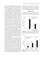
Remarkably, both the VA and STS databases during
their existence have demonstrated a very significant reduc-
tion in risk-adjusted operative mortality approaching 25 to
30% (Figures 10-1 and 10-2).
2
This has occurred in spite of
patient risk factors increasing throughout the 1990s.
Similar reductions in mortality following the implementa-
tion of analysis of outcome data and their distribution to
surgeons and their colleagues have occurred in northern
New England, where they noted a 24% reduction in deaths
following the institution of round-robin site visits for feed-
back of outcome data and training in quality improve-
ment.
3
New York State has also reported a similar
reduction in risk-adjusted operative mortality. Of great
interest is the fact that for three of these databases (those
of the STS, VA, and northern New England), there has
been no public reporting of data, just internal dissemina-
tion of these data to those providing the care.
References
1. Mavroudis C, Jacobs J. Congenital Heart Surgery
Nomenclature and Database Project: overview and mini-
mum dataset. Ann Thorac Surg 2000;69:1–387.
2. Grover FL, Cleveland JC Jr, Shroyer AL. Quality improvement
in cardiac care. Arch Surg 2002;137:28–36.
3. O’Connor GT, Plume SK, Olmstead EM, et al for the
Northern New England Cardiovascular Disease Study
Group. A regional intervention to improve the in-hospital
mortality associated with coronary bypass grafting surgery.
JAMA 1996;275:841–846.
FIGURE 10-1. Unadjusted and adjusted operative mortality rates
between April 1987 and September 1991.
FIGURE 10-2. Ratio of observed-to-expected mortalities between
1990 and 1999.
125
CHAPTER
11
PRIMARY AND SECONDARY
CHEST WALL T
UMORS
L. PENFIELD FABER
, MD
Chest wall tumors are neoplasms in the bones or soft
tissues of the thoracic cage. Primary tumors of the chest
wall develop in the bones or soft tissues of the thorax.
Bony tumors include chondroid, osseous, giant cell, and
marrow-derived tumors. Soft tissue tumors include those
of a fibrous, fibrohistiocytic, adipose, neurologic, and
muscular character. Secondary chest wall tumors include
tumors that arise from an adjacent organ that invade the
chest wall, and tumors that have metastasized to the
bones or soft tissue of the chest from a distant site.
Malignant tumors arising from the lung are the most
common of these, followed by breast cancer and malig-
nant tumors of the pleura.
Hedblom reviewed and reported on 213 tumors of the
chest wall collected from the literature between 1898 and
1921 and updated his collected series to 313 cases in
1933.
1
The majority of these cases, unlike findings in
other series, involved primary chest wall malignancies,
and Hedblom was among the first to pathologically cate-
gorize the various primary tumors of the thoracic wall.
Primary chest wall tumors are a heterogeneous group
of tumors of bone and soft tissue (Table 11-1).
Altogether, they comprise only 1 to 2% of all primary
tumors of the body.
2
Primary malignant chest wall tu-
mors account for approximately 4% of all new cancers
diagnosed annually. Malignant tumors of the soft tissues
are slightly more common than malignant tumors of
bone, and soft tissue sarcoma is the most common pri-
mary chest wall malignancy.
3
The most common benign tumors of chest wall bone,
in decreasing order of frequency, are osteochondroma
and fibrous dysplasia, chondroma, aneurysmal bone cyst,
and eosinophilic granuloma. Osteochondroma consti-
tutes approximately 30 to 50% of benign bony lesions.
The incidence of fibrous dysplasia is approximately the
same as that of osteochondroma, whereas chondroma
and bone cysts account for approximately 10 to 25% of
benign lesions of bone.
4
TABLE 11-1. Primary Tumors of the Chest Wall
Tissue Involvement Tumor
Benign tumors of bone
Bone Osteoid osteoma
Cartilage Enchondroma
Osteochondroma
Fibrous Fibrous dysplasia
Vascular Hemangioma
Marrow Eosinophilic granuloma
Osteoclast Giant cell tumor
Aneurysmal bone cyst
Benign tumors of soft tissue
Fibrous Fibroma
Adipose Lipoma
Nerve Schwann cell
Neurofibroma
Muscle Angioleiomyoma
Malignant tumors of bone
Bone Osteosarcoma
Cartilage Chondrosarcoma
Fibrous Malignant fibrous histiocytoma
Vascular Hemangiosarcoma
Marrow Plasmacytoma
Cellular Ewing’s sarcoma
Askin’s tumor (peripheral
neuroectodermal tumor)
Malignant tumors of soft tissue
Fibrous Desmoid
Fibrosarcoma
Fibrohistiocytic Malignant fibrous histiocytoma
Adipose Liposarcoma
Nerve Neurofibrosarcoma
Schwann cell sarcoma
Muscle Rhabdomyosarcoma
The most common malignant tumors of the bony
thorax, in decreasing order of frequency, include chon-
drosarcoma, Ewing’s sarcoma, osteosarcoma, and solitary
plasmacytoma. Chondrosarcoma is the single most
common malignant tumor of the chest wall. Myeloma of
the bony chest wall must be considered a systemic disease
and not a primary lesion. Solitary plasmacytoma, al-
though commonly associated with the development of
multimyeloma, is defined as a primary lesion and is less
frequently identified.
The most common benign lesions of thoracic soft tissue
are fibroma, hemangiomas, lipomas, and giant cell tumors.
Malignant fibrous histiocytomas, desmoid tumors,
liposarcomas, and fibrosarcomas are the most common
malignant soft tumors of the chest wall. Primary soft
tissue sarcomas of the thoracic wall are more common
than malignant tumors of the bony thorax, and when
considered as a group constitute the most common form
of chest wall malignancy.
Secondary chest wall tumors are most commonly lung
cancer, breast cancer, and metastatic disease.
Clinical Presentation
Approximately one-half of malignant tumors of the bony
chest wall occur in the ribs, with the remainder presenting
in the scapula, sternum, and clavicle. Malignant tumors of
the ribs are frequently found in the anterior aspect of the
upper seven ribs, but there is an equal distribution of
benign rib tumors throughout the thorax.
5
Specific
tumors are found in particular areas of the bony thorax;
the chondroma and endochondroma often arise anteri-
orly in the costal cartilages or sternum. The chondrosar-
coma most often occurs anteriorly at the costochondral
junction. Benign and soft tissue malignancies of the chest
wall occur in all locations with equal frequency. In Dahlin
and Unni’s series, 96% of sternal tumors were malignant,
with the most common types being chondrosarcoma,
plasmacytoma, and osteogenic sarcoma.
6
Chest wall tumors occur in all age groups. Ewing’s
sarcoma occurs in younger patients, and plasmacytoma
presents in the elderly. Chondrosarcoma commonly
occurs in adults. A male-to-female ratio of 2 to 1 occurs
for malignant chest wall tumors, whereas desmoid tumors
are more common in females. Burt reported that only 2%
of malignant chest wall tumors were asymptomatic.
3
Fifty
percent of patients presented with a painless mass,
whereas 33% had a painful mass and 15% had pain with-
out a mass being present. Pain is not necessarily a predic-
tor of malignancy; in Hedblom’s series, pain was present
in 40% of benign chondromas.
1
Benign osteoma presents
with severe pain relieved only by medication. Asym-
ptomatic lesions are more often benign, and most of these
are detected on chest radiographs. Soft tissue sarcomas
present as a painless mass, but tumors of bone and carti-
lage, both benign and malignant, present with pain.
Generalized symptoms of fever, malaise, and fatigue can
be presenting complaints associated with eosinophilic
granuloma and Ewing’s sarcoma. Rapidly growing tumors
are more likely to be malignant.
It is difficult to differentiate a benign from a malig-
nant chest wall tumor by physical examination. Bony
tumors are fixed to the chest wall; soft tissue tumors may
also be fixed, but others are quite mobile. Both malignant
and benign lesions can be tender to palpation.
Diagnosis
The chest radiograph is the initial study to be obtained.
Comparison with prior films is important to evaluate the
rate of growth. Computed tomography (CT) defines the
extent of pleural, mediastinal, and soft tissue involvement
(Figure 11-1). Metastatic disease to the lung is readily
identified, and the radiologic characteristics of the tumor
can assist in the clinical diagnosis. Magnetic resonance
imaging (MRI) has become extremely valuable in evaluat-
ing chest wall tumors (Figure 11-2). It precisely defines the
anatomic extent of the tumor, as well as showing adjacent
organ involvement. A significant advantage of MRI is that
multiplanar imaging with high-contrast resolution defines
anatomic planes for planned resection. Differential signal
intensity evaluates adjacent vascular structures and
becomes critical in the assessment of the upper chest with
involvement of the brachial plexus and subclavian vessels.
The MRI is supplemental to the CT, and both studies assist
in the planning of extensive and difficult chest wall resec-
tions. Positron emission tomography (PET) is useful to
define metastatic disease when a secondary chest wall
tumor is present. This particularly applies to lung and
breast cancer. PET is not particularly helpful in defining
the extent of a primary chest wall tumor.
126
/ Advanced Therapy in Thoracic Surgery
FIGURE 11-1. Computed tomography scan illustrating the involve-
ment of ribs, sternum, and mediastinum by a chondrosarcoma.
Benign Bony Tumors
Fibrous Dysplasia
Fibrous dysplasia is a common lesion of the chest wall,
accounting for 20 to 30% of rib tumors.
5,10
This lesion
occurs equally in males and females and is common in
the second and third decades of life. It usually presents as
a painless mass involving the bony chest wall; most
lesions are solitary, but multiple areas are seen occasion-
ally. Fibrous dysplasia usually is a painless mass identified
on a routine chest radiograph, but it can be painful in
association with a pathologic fracture or an expansion
and stretching of the periosteum. Patients with multiple
lesions show abnormal skin pigmentation and preco-
cious puberty (Albright’s syndrome).
The radiographic appearance is that of a lytic lesion
involving one or more ribs (Figure 11-3). There fre-
quently is an expansion of the rib with significant thin-
ning of the cortex, and the medullary aspect of the rib
has a homogeneous ground-glass appearance. Pathologic
fracture is a frequent cause of diagnosis precipitating
radiologic evaluation. The gross appearance of the
affected rib identifies a central fibrous area with a shell of
expanded cortex on the surface (Figure 11-4). Malignant
transformation into osteosarcoma or fibrous sarcoma is
exceedingly rare and is not an indication for excision of
this lesion.
11
Excision of fibrous dysplasia is indicated for
pain or uncertainty of the diagnosis. In cases in which
the diagnosis is questioned, incisional or excisional
biopsy is indicated. Recurrence after complete excision is
rare.
Osteochondroma
Osteochondroma is among the most common benign
neoplasms of the chest wall. The rib is the common loca-
tion for these tumors, which usually begin in childhood
and continue to grow until skeletal maturation is attained.
Patients with osteochondromas are usually < 30 years of
age, and males are more commonly affected than females.
A chest radiograph is usually diagnostic with the find-
ing of a pedunculated or sessile bony excrescence on the
surface of the rib, with the medulla of the bone continu-
ous with the medulla of the lesion. A thin cartilaginous
cap is present with areas of scattered calcification. Multi-
ple lesions suggest a diagnosis of familial osteochondro-
matosis.
Tr eatment for the osteochondroma is complete excision,
and recurrence is rare. Malignant transformation of these
lesions is suggested by pain and continued growth after
closure of the epiphyseal plate.
5
Malignancy of these lesions
is rare and is estimated to occur in < 1%. Malignant trans-
formation can occur in 20% of these lesions in cases of
familial osteochondromas or multiple exostoses. The
malignant tumor is well differentiated, and local recurrence
is unusual if complete resection is accomplished.
128
/ Advanced Therapy in Thoracic Surgery
FIGURE 11-3. A, Chest radiograph depicting a chest wall mass. B,
Computed tomography scan showing classic appearance of fibrous
dysplasia with cortical thinning.
FIGURE 11-4. Resected rib of fibrous dysplasia. Cortical thinning is
noted. Reprinted with permission from Faber LP, Somers J, Templeton
AC. Chest wall tumors. Curr Probl Surg 1995;32:661–756.
Chondromas
Chondromas constitute approximately 15 to 20% of all
benign tumors of the chest wall. They may occur in the
medulla as an enchondroma (Figure 11-5) or less
frequently on the periosteum as a periosteal chondroma,
but most commonly from costal cartilage. They usually
present anteriorly at the costal chondral junction and are
a slowly enlarging painless mass. Pain can also be present.
Chondromas occur in all age groups, most frequently
between the ages of 10 and 30 years. Males and females
are affected equally. On radiographs, the chondroma
appears as a small lytic area in the bone with sclerotic
margins. The lytic areas are typically round or oval, and a
rib lesion is expansile with thinning of the cortex.
There is no reliable imaging technique to distinguish a
low-grade chondrosarcoma from a chondroma. Histologic
differentiation between a low-grade chondrosarcoma and
a cellular chondroma can also be difficult. Because of this
problem, en bloc resection is recommended with wide
margins. All tumors arising from the costal cartilages
should be considered malignant and treated with wide
excision. Inadequate resection frequently results in local
recurrence of the malignant form.
Eosinophilic Granuloma
Eosinophilic granuloma is a part of the spectrum of
Langerhans cell histiocytosis, a diffuse infiltrative process
of antigen-presenting macrophages together with a vari-
able accompaniment of eosinophils, lymphocytes, and
plasma cells. Eosinophilic granuloma is limited to bone,
whereas other forms of the disease are systemic. In infants
it presents as an infiltration of lymph nodes, liver, spleen,
and bone marrow (Letterer-Siwe disease). Hand-Schuller-
Christian disease presents with bone involvement accom-
panied by diabetes insipidus. Isolated rib involvement
occurs in 10 to 20% of instances of eosinophilic granu-
loma, and there is a strong predilection in males.
If there is localized chest pain, chest radiography is
required to determine whether there is an expansile
lesion of the rib (Figure 11-6). Malignancy cannot be
Primary and Secondary Chest Wall Tumors
/
129
FIGURE 11-5. Anterior chest wall enchondroma (arrow).
FIGURE 11-6. A, Radiograph of the rib illustrates eosinophilic granu-
loma (arrow). B, Computed tomography scan depicts the lesion
(arrows). C, Resected rib specimen.
ruled out by the radiologic appearance, and excisional
biopsy is performed to establish the diagnosis and
provide a cure if the lesion is solitary.
5
Radiation therapy
can be effective for multiple lesions or tumors in areas
that are difficult to resect.
Aneurysmal Bone Cysts
Bone cysts account for < 1% of chest wall tumors
(Figure 11-7). They commonly occur in the ribs and
usually are associated with a pathologic fracture
(Figure 11-8). A chest radiograph can be diagnostic if it
shows a demarcated lytic lesion surrounded by a thin
shell of periosteum. Callus formation may be present if
there has been a previous fracture. The lesion is usually
asymptomatic unless fracture has occurred. Simple exci-
sion is indicated for associated pain or when there is a
question of metastatic cancer.
Osteoid Osteoma
Osteoid osteoma is a rare benign tumor of the rib or
vertebral body. The presentation is usually sharp pain
that requires radiography; films demonstrate a small,
radiolucent area surrounded by a marked area of sclero-
sis. A bone scan illustrates intense uptake in the center of
the lesion, with less dense activity surrounding the
central nidus.
Surgical resection is recommended for pain, and local
recurrence is rare after complete excision. Lesions of the
scapula and sternum can be treated with exposure of the
nidus by cortical shaving and curetting of the nidus.
Hemangioma
Hemangiomas present in the vertebral body or ribs and
are pain free. The tumor can arise in the soft tissue and
protrude into the thorax or develop in a rib with a
sclerotic-reticular pattern (Figure 11-9).
12
Increased
blood flow through the rib causes absorption, and adja-
cent slow flow causes deposition of new bone. The lesion
presents as linear areas of bone deposition and lucency.
Diagnosis is usually made by radiography and CT, and
excision is not required except in cases of fracture or
cosmetic deformity. Low-grade malignant hemangioen-
dothelioma presents with soft tissue involvement outside
the rib; in this instance, excision is recommended. Clear
margins must be obtained as there is a significant inci-
dence of local recurrence with this lesion.
13
Other benign bony lesions of the chest wall include
osteoblastoma, ossifying fibroma, nonossifying fibroma,
ossifying lipoma, and giant cell tumors. These lesions can
usually be diagnosed by precise radiologic interpretation
and do not require excision unless there is associated
pathologic fracture or a question of malignancy.
Malignant Bony Tumors
Chondrosarcoma
Chondrosarcoma is the most common primary malig-
nant neoplasm of the chest wall and commonly presents
anteriorly, arising from either the costochondral arches
or the sternum. It is also the most common primary
malignant tumor of the sternum. It most commonly
affects males and is found in people 20 to 40 years old.
McAfee and colleagues reported that 12.5% of patients
with chondrosarcomas reported a previous history of
chest wall trauma in the location of the tumor.
14
Chondrosarcomas also develop as a result of malignant
degeneration of a benign chondroma or osteochon-
droma and are categorized as secondary chondrosarco-
mas. These lesions are usually of low-grade malignancy,
appear at an earlier age, and have a better prognosis than
the standard primary chondrosarcoma.
The tumor usually presents as a slowly enlarging,
painful mass located on the anterior chest wall or sternum.
The mass is hard, slightly tender, and fixed firmly to the
chest wall (Figure 11-10). The radiographic appearance
defines a lobulated mass arising in the medullary portion
of the bone with radiolucency and stippled calcification.
130
/ Advanced Therapy in Thoracic Surgery
FIGURE 11-7. Resected bone cyst.
FIGURE 11-8. Bone cyst with rib fracture (arrow).
recurrence significantly decreases survival and also
increases the risk of distant metastasis. There is no effec-
tive chemotherapy for chondrosarcoma.
Radiation therapy is ineffective as the primary treat-
ment. McNaney and colleagues have reported some
success with combination photon and neutron
radiation.
17
Postoperative radiation is mandatory if oper-
ative margins are microscopically positive for tumor.
Ewing’s Sarcoma
Ewing’s sarcoma constitutes 6 to 10% of all malignant
chest wall tumors and is the most common primary chest
wall tumor in children. It is more common in males than
females, and white people are more commonly affected
than are those of African descent. Ewing’s sarcoma
usually presents as a painful, palpable mass, and pain is
the presenting symptom in 90% of patients.
18
Systemic
manifestations include fever, malaise, and weight loss.
These symptoms in association with pain and the classic
appearance on radiographs are diagnostic.
Chest wall Ewing’s sarcomas frequently present in the
ribs but also arise from the scapula, sternum, and clavi-
cle.
19
Gross metastatic disease is present at the time of
diagnosis in 20 to 30% of patients, and common sites of
metastatic disease include the lungs, bone, and bone
marrow. Malignant pleural effusion is a common form of
metastatic presentation with primary Ewing’s sarcoma of
the chest wall.
Radiographs reveal destruction of bone with lytic and
blastic areas, and elevation of the periosteum with multi-
ple layers of subperiosteal bone formation creates the
classic “onion skin” appearance (Figure 11-12). The
surface of the bone may contain radiating ossified
spicules, as are commonly seen in osteosarcoma. Patho-
logic fractures are uncommon. CT and MRI scans are
important in evaluating soft tissue, lung, and mediastinal
involvement, and a bone scan is necessary to rule out
bone metastasis. Excisional biopsy is recommended for
diagnosis, and the biopsy should be performed in a
manner that will allow for definitive resection.
132
/ Advanced Therapy in Thoracic Surgery
FIGURE 11-11. A, Computed tomography scan illustrating an anterior chest wall chondrosarcoma. Note the patchy calcification. B, Resected
specimen included cartilages and ribs. C, Reconstruction with GORE-TEX (W. L. Gore and Associates, Flagstaff, AZ). D, Photomicrograph showing
histologic appearance of a chondrosarcoma (ϫ800 original magnification; hematoxylin and eosin stain).
Askin’s tumors occur mainly in children and young
adults; they frequently present in the posterior chest wall
and are often thought to be a posterior chest wall tumor
(Figure 11-13). Depending on the location, the diagnosis
can be made with percutaneous needle biopsy. A trephine
specimen is advantageous. Preoperative chemotherapy is
recommended, followed by wide surgical resection for
local control (similar to the treatment of Ewing’s sar-
coma). Positive resection margins are treated with radia-
tion therapy.
Osteosarcoma
Osteosarcomas represent 5 to 10% of primary malignant
tumors of the chest, and occurrence is noted in the second
and third decades of life, as well as in the elderly.
25
The clini-
cal presentation is that of a painful, rapidly enlarging mass.
Radiographs may show calcified spicules extending
from the bone cortex, which produce the classic
“sunburst” appearance. However, the sunburst appear-
ance is noted in other diseases of bone and is probably
seen in only 25% of osteosarcoma lesions.
5
Cortical bone
destruction with irregular margins that merges into adja-
cent normal bone with lytic or blastic changes is usually
apparent (Figure 11-14). CT is required to evaluate the
extent of involvement and also the presence of metastatic
lesions to the lung (Figure 11-15). Diagnosis can be
achieved in the soft tissue component of the lesion by
trephine needle biopsy or incisional biopsy.
134
/ Advanced Therapy in Thoracic Surgery
FIGURE 11-13. A, Apical and posterior chest wall tumor as seen on a
radiograph. B, Computed tomography scan shows the extent of this
Askin’s tumor. Reprinted with permission from Faber LP, Somers J,
Templeton AC. Chest wall tumors. Curr Probl Surg 1995;32:661–756.
FIGURE 11-14. A, Posterior osteogenic sarcoma. B, Radiograph of
resected osteogenic sarcoma. Reprinted with permission from Faber
LP, Somers J, Templeton AC. Chest wall tumors. Curr Probl Surg
1995;32:661–756.
must be obtained including serum and urine elec-
trophoresis, serum ionized calcium level, bone marrow
aspiration, and a complete blood count. A patient with a
true solitary plasmacytoma usually has a normal calcium
level and is not anemic. Monoclonality of one of the
immunoglobulins with normal levels of other circulating
immunoglobulins suggests that the plasmacytoma is
truly solitary.
Radiation is the primary treatment for solitary plasma-
cytoma.
28
The role of surgery is to make the diagnosis, and
wide excision for this lesion is not indicated. Radiation
therapy yields a 90% local control rate.
19
Following local
control with radiation, multiple myeloma develops in 45
to 50% of patients. Patients must be followed carefully
with periodic evaluation after treatment for a solitary
plasmacytoma. The 5-year survival rate for patients with
this disease is approximately 20 to 30%.
7,29
Benign Soft Tissue Tumors of the
Chest Wall
Lipoma
Lipoma is a benign tumor of fat and occurs superficially as
well as deeper; it can protrude through the intercostal space
into the thoracic cavity. Subcutaneous lipomas are non-
tender, well circumscribed, and mobile. They are distrib-
uted equally throughout the chest wall and are painless and
usually asymptomatic. These lesions are frequently noted
incidentally on a routine chest radiograph, and a CT scan
shows the typical density of fat. The differential diagnosis
lies between lipoma and the rarer liposarcoma.
These lesions are well circumscribed, thinly encapsu-
lated tumors of mature adipose tissue (Figure 11-17).
Surgical excision is indicated for cosmetic purposes or if
malignancy cannot be ruled out. Usually, the diagnosis is
strongly suspected, and only local excision is required.
Neurofibroma
Neurofibromas are commonly multiple and occur as part
of the complex of von Recklinghausen’s disease, a familial
disorder characterized by these growths that can occur in
any part of the body. The disease may be transmitted as
an autosomal dominant trait as a result of a gene muta-
tion on chromosome 17, but 50% of cases are sporadic.
In addition to multiple neurofibromas, the disease is
characterized by café au lait spots of the skin and menin-
giomas. A chest radiograph demonstrates a mass, and
frequently there is associated rib notching (Figure 11-18).
In the chest they frequently arise from interthoracic
nerves but can occur in bone without nerve sheath
involvement. The lesions are usually benign, but malig-
nant degeneration occurs in 5 to 40% of cases.
30
Surgical
excision is recommended for symptoms of pain or for
rapid enlargement.
Related to the neurofibroma is the Schwann cell tumor
or neurilemoma. This lesion is usually solitary and occurs
on a nerve with clear demarcation. The ribs can be involved
(Figure 11-19). Excisional biopsy and resection are recom-
mended for diagnosis and treatment. It may be difficult to
differentiate the malignant form of this tumor from the
benign form. This tumor can be a low-grade malignancy,
which is associated with a high recurrence rate. Mitoses
136
/ Advanced Therapy in Thoracic Surgery
FIGURE 11-16. A, Radiograph showing a solitary plasmacytoma of
the chest wall. B, Computed tomography scan of the same lesion.
Primary and Secondary Chest Wall Tumors
/
139
FIGURE 11-21. A, Magnetic resonance image showing sarcoma of the
shoulder (arrows). B, Computed tomography scan depicting the soft
tissue sarcoma. Reprinted with permission from Faber LP, Somers J,
Templeton AC. Chest wall tumors. Curr Probl Surg 1995;32:661–756.
C, Computed tomography scans depicting synovial sarcoma invading
the chest wall and sternum. Continued on the next page.
tumor, adjacent thorax, and tissue planes (Figure 11-21).
These studies are mandatory for appropriate planning of
resection. Incisional biopsy or trephine needle biopsy
should be used to make the diagnosis, except in those
situations in which the tumor is small enough that
complete primary resection with a wide margin can be
accomplished by excisional biopsy.
Soft tissue sarcomas are classified by type and grade.
Typing of a soft tissue tumor is based on the closest
match to a differentiated normal cell. Grading uses crite-
ria of mitotic rate, nuclear and cellular pleomorphism,
and nucleus-to-cytoplasm ratio. High-grade tumors tend
to have more necrosis present. Separation into the differ-
ent types of tumor is made using histologic and histo-
chemical criteria.
Major prognostic indicators for primary soft tissue
sarcomas include tumor grade, the presence of distant
metastasis, and positive surgical resection margins. The 5-
year survival rate for low-grade chest wall soft tissue sarco-
mas after complete resection varies between 85 to 90%,
whereas the 5-year survival rate for high-grade lesions is
approximately 40 to 50%.
29,32,33
Other histopathology influ-
encing survival is controversial. Histopathology did not
influence survival in a series of high-grade extremity
sarcomas studied at the National Cancer Institute.
34
However, analysis of another series of high-grade tumors
revealed that the muscular type had decreased survival
when compared with high-grade tumors of the fibrous
type.
29
Size of the tumor and age of the patient at presenta-
tion have not been found to influence survival.
Surgical excision with a wide margin is the mainstay
of treatment for soft tissue sarcomas of the chest wall. A
pseudocapsule frequently surrounds the tumor, and
contact with this pseudocapsule must be avoided during
resection as it often contains microscopic disease. These
tumors often spread along the walls of blood vessels,
nerve sheaths, and fascial planes. Malignant fibrous histi-
ocytoma can spread into the marrow and, indeed, the
entire affected rib (Figure 11-22), and portions of the rib
above and below need to be resected if the chest wall is
invaded by this histologic variant.
Malignant fibrous histiocytoma shows a histologic
pattern of spindle cells with rounded pleomorphic
nucleoli arranged in a cartwheel pattern. It is thought to
arise from tissue histiocytes. Fever and leukocytosis can
be associated, and this tumor can be radiation induced.
35
Surgical therapy is wide excision. Positive resection
margins should be treated with postoperative radiation.
Chemotherapy is usually ineffective. Five-year survival
rates approximate 35%.
140
/ Advanced Therapy in Thoracic Surgery
FIGURE 11-21 continued. D, Resected specimen includes the ster-
num, anterior chest wall, and ribs.
FIGURE 11-22. A, Computed tomography scan showing malignant
fibrous histiocytoma with rib invasion. B, Magnetic resonance image
reveals no evidence of invasion into the spinal cord.
Rhabdomyosarcoma is associated with striated muscle
tissue and histochemical analysis shows myoglobin,
desmin, and actin. Therapy is wide resection with aggres-
sive adjuvant radiation and multidrug chemotherapy.
Five-year survival rates approximate 70%.
36
Large masses
should be treated with preoperative chemotherapy and
radiation, and adjuvant chemotherapy after resection.
Liposarcomas are defined as tumors containing
lipoblasts, which are large cells with pleomorphic nuclei
containing large vacuoles of neutral fat. Most of these
tumors are low grade, and wide surgical excision is neces-
sary for control. Local recurrence can be effectively
treated with repeat resection as radiation therapy is not
particularly effective for this neoplasm. Five-year survival
rates can approximate 80%, as reported by Graeber and
colleagues.
25
Metastasis occurs more frequently with high-grade
than with low-grade soft tissue sarcomas, and frequently
smaller lesions cannot be detected on a normal chest
radiograph. CT of the chest is always indicated when
dealing with these sarcomas. In Gordon and colleagues’
series, 51% of high-grade lesions had either synchronous
or metachronous metastasis compared with only 10% of
low-grade lesions.
29
Radiation therapy has shown some
benefit in extremity soft tissue sarcomas and decreases
the necessity for a radical surgical procedure.
37
This treat-
ment can be considered for larger chest wall sarcomas.
The role of adjuvant chemotherapy for high-grade
lesions is controversial. Chang and colleagues reported a
prolonged disease-free survival in patients with extremity
sarcomas who received adjuvant chemotherapy with
doxorubicin and cyclophosphamide.
38
Other studies of
chest wall and truncal sarcomas have not demonstrated a
clear improvement in survival with the use of adjuvant
chemotherapy.
32
Adjuvant therapy does improve the
survival of patients with rhabdomyosarcoma.
Long-term follow-up of patients who have had a
malignant soft tissue tumor is required to detect local
recurrence and possible metastatic disease to the lungs.
Desmoid Tumor
Microscopically, desmoid tumor appears rather benign,
with a network of spindle-shaped cells and dense
connective tissue. There are no mitotic figures and little,
if any, pleomorphism. These findings led to a mistaken
impression that this is a benign tumor of “aggressive
fibromatosis.”
5
This tumor does invade adjacent struc-
tures, recur locally, and invade nerves; therefore, it must
be considered a low-grade sarcoma.
The tumor is more common in females and may be
hormonally regulated. This theory is supported by the
presence of estrogen receptors in tumor nuclei and by
reports of regression of these tumors with the use of
antiestrogen agents such as tamoxifen. They occur in the
second through fifth decades, and approximately 40% of
all desmoid tumors occur in the chest wall and shoulder
region.
39
The usual presentation of the desmoid tumor is
a painless or slightly tender, poorly circumscribed mass
(Figure 11-23). In the shoulder region, they invade tissue
surrounding the brachial plexus and can encapsulate
vessels of the arm and neck with significant shoulder
pain and motor weakness. Diagnosis requires incisional
biopsy to obtain adequate tissue for pathologic analysis,
and differentiation from a benign lesion can be difficult.
Use of MRI assists in defining tissue involvement and the
planning of the resection. Metastatic work-up is not
required as these tumors do not metastasize.
Grossly, the desmoid tumor presents as a vascular scar
tissue extending for several centimeters. Margins are
poorly demarcated, and it is difficult to define the margin
of extension into adjacent muscle and fascial planes.
Treatment of the desmoid tumor is wide excision with
evidence of microscopically clean margins. The major
problem in treating these tumors is local recurrence.
Brodsky and colleagues reported on 32 patients with
Primary and Secondary Chest Wall Tumors
/
141
FIGURE 11-23. A, Anterior chest wall desmoid tumor (arrow) arising
in a previous sternotomy incision. B, Resected specimen includes a
portion of the inferior sternum and adjacent cartilages and soft
tissues.