Advanced therapy in thoracic surgery - part 2 doc
Bạn đang xem bản rút gọn của tài liệu. Xem và tải ngay bản đầy đủ của tài liệu tại đây (1.21 MB, 40 trang )
there was no significant difference in the length of the
hospital stay.
64
A major criticism of this study is the chest
tube management, which may have contributed to the
incidence of empyema and the extended hospital stay.
Wain and colleagues reported a multicenter, random-
ized, controlled trial of patients undergoing pulmonary
resection, comparing conventional closure with conven-
tional closure plus treatment of all surgical sites at risk
for air leak with FocalSeal-L.
62
Each surgeon was trained
in the proper application of FocalSeal, and each individ-
ual surgeon or institution determined protocol for chest
tube management. Of the 117 patients in the FocalSeal
group and the 55 patients in the control group, there was
no statistically significant difference in the extent of
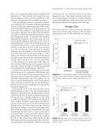
prerandomization air leak. The FocalSeal group had no
air leak detectable prior to chest closure in 92% of
patients compared with 29% in the control group
(p < .001). In the time from operation to hospital
discharge, 39% of the patients in the FocalSeal group had
no air leak versus 11% in the control group (p < .001)
(Figure 3-8). The mean time from skin closure to the last
detectable air leak was less in the FocalSeal group than in
controls (30.9 ± 4.8 h vs 52.3 ± 11.6 h, respectively;
p = .006). However, as in the previous studies, there was
not a statistical difference between the groups in time to
chest tube removal or length of hospital stay, although
the trend favored the FocalSeal group (Figure 3-9).
FocalSeal has also been used to seal air leaks that
develop during cardiac reoperations. Fifteen patients that
had air leaks recognized intraoperatively had the
pulmonary injuries treated with FocalSeal. All leaks were
controlled intraoperatively, and 73% of patients had air
leaks recognized postoperatively. Three of four patients
with a recurrent air leak had the air leak resolved in 3
days, but seal was never accomplished in one patient who
was immunosuppressed.
65
Interestingly, in all of the clinical trials, several
patients appeared to have no air leak intraoperatively as
assessed by submersion and controlled positive pressure
ventilation, and then developed air leaks postoperatively.
This may be due to improper application of the sealant
or ineffective adhesion of the sealant to the pulmonary
tissue. Another possibility is that negative intrathoracic
pressure from suction on chest tubes postoperatively
impeded closure of small air leaks. Some have therefore
advocated the avoidance of suction on chest tubes unless
there is both an air leak and a pneumothorax, with a goal
of removing the chest tubes as soon as drainage is
≤ 20 mL/h (John C. Wain, personal communication,
January 2003). The ability to seal most of the air leaks at
the time of chest closure along with avoidance of chest
tube suction may decrease postoperative chest tube dura-
tion and hospital stay, ultimately resulting in decreased
morbidity and cost. However, this has not been
supported by any of the studies currently in the litera-
ture. FocalSeal appears to be safe, but its efficacy depends
on the proper application, which can be tedious as well as
difficult, especially in poorly exposed areas of the lung.
Biologic Glue
The natural history of acute type A aortic dissection
carries an extremely grim prognosis without surgery,
with mortality rates of 38% in the first day and up to
Tissue Adhesives in Thoracic and Cardiovascular Surgery
/
55
FIGURE 3-8. Percentage of patients without air leaks intraoperatively
and from wound closure to hospital discharge for patients treated
with FocalSeal-L versus controls. Adapted from Wain JC et al.
62
p. 1626.
0
20
40
60
80
100
120
Patients without air leaks (%)
Control (
n
= 55)
FocalSeal (
n
= 177)
Intraoperative From Wound Closure
to Hospital Discharge
p
= < .001
p
= < .001
FIGURE 3-9. Mean time to last air leak in patients treated with
FocalSeal-L versus controls. NS = not significant. Adapted from Wain
JC et al.
62
p. 1627.
0
2
4
6
8
10
12
Mean Time (d)
Control (
n
= 55)
FocalSeal (
n
= 117)
T
T
p
= .006
p
= NS
p
= NS
T
T
T
T
From Skin
Closure to Last
Observed Air Leak
From Skin
Closure to Chest
Tube Removal
From Skin
Closure to
Hospital discharge
90% after 2 weeks from the onset of symptoms. The best
chance of survival in patients with this disease depends
on immediate diagnosis and emergent surgical interven-
tion, although reported mortality after surgery remains
10 to 20%. The dissection is occasionally limited to the
ascending aorta but often extends to the arch and
descending aorta. Proximal extension of the dissection
can involve the aortic valve or coronary arteries.
The friability of the remaining proximal and distal
aorta makes anastomosis extremely tenuous, and severe
bleeding or re-dissection can complicate the repair. Many
techniques to reinforce the aortic tissues have been advo-
cated, including the use of pledgeted sutures or sand-
wiching the separated layers of the aortic wall with
polytef strips prior to sewing on the graft. Several
authors have attributed improvements in outcomes in
their experiences to the use of biologic glues to adhere
the separated aortic wall layers, thus reinforcing the
tissues enough to hold sutures. The most frequently used
biologic glue for this indication has been GRF glue. In
1977, frustrated by the poor prognosis of treatment for
acute type A aortic dissection, Guilmet and colleagues
began using GRF glue clinically to seal the layers of the
aortic wall during the repair of acute type A aortic dissec-
tions.
66
Since then many surgeons have used GRF glue in
every case of acute type A aortic dissection. Although
randomized, controlled studies in this patient population
are impractical, surgeons advocating the use of GRF glue
report that significantly decreased bleeding and simplifi-
cation of the repair leads to decreased cardiopulmonary
bypass times and improved overall survival.
67
Some
continue to oversew and reinforce the native aorta with
polytef strips in addition to using the sealant, whereas
others have abandoned this technique and rely on the
GRF glue to reinforce the aorta for suturing to the graft.
Great care must be taken to avoid contamination of the
lumen with glue, especially near the coronary ostia
(Figure 3-10).
68
Reports of glue emboli are infrequent,
but these emboli can occur.
69
GRF glue is not approved by the FDA owing to
concerns about the toxicity of the formaldehyde compo-
nent.
70
Although GRF glue has been used extensively in
Europe and several studies have reported the benefits,
safety, and reliability of this sealant, recent reports of
reoperations owing to aortic medial necrosis of sites
previously repaired using this product are refocusing
attention on its potential toxicity. Bingley and colleagues
recently reported high rates of aortic regurgitation requir-
ing reoperation in patients who had the aortic root rein-
forced with GRF glue with resuspension of the aortic
valve.
71
Late aortic insufficiency occurred in 7 of 18
patients (39%), and 6 of these had re-dissection at the site
of the GRF glue reinforcement. Histologic findings were
consistent with tissue necrosis at the site of glue use
(Figure 3-11). Their conclusion was that this necrosis
could be attributed to either an improper glue application
56
/ Advanced Therapy in Thoracic Surgery
A
FIGURE 3-10. A–C, Suggested technique to reconstruct the aortic
root and proximal aortic arch using GRF glue. Gauze sponges are used
to prevent intraluminal glue. Adapted from and B and C reproduced
with permission from Laas J et al.
68
p. 227.
Other potential indications under investigation
include the use of tissue adhesives to avoid postoperative
adhesions, allow the local release of pharmacologic
agents, carry gene or protein therapeutic agents, or
enhance endothelialization of prosthetic or tissue-
engineered grafts. The clinical utility of tissue adhesives
has shown great promise over the past century. As the
technology and experience with tissue adhesives continue
to grow,we must expand our comprehension of the
proper use and limitations of these agents to take full
advantage of the clinical benefits they offer our patients.
References
1. Bergel S. Uber Wirkungen des Fibrins. Dtsch Med
Wo chenschr 1909;35:663–5.
2. Grey EG. Fibrin as a hemostatic in cerebral surgery. Surg
Gynecol Obstet 1915;21:452–4.
3. Harvey SC. The use of fibrin papers and forms in surgery.
Boston Med Surg J 1916;174:658–9.
4. Cronkite EP, Lozner EL, Deaver JM. Use of thrombin and
fibrinogen in skin grafting. JAMA 1944;124:976–8.
5. Blomback B, Blomback M. Purification of human and
bovine fibrinogen. Arkiv Kemi 1956;10:415–43.
6. Trott AT. Cyanoacrylate tissue adhesives: an advance in
wound care. JAMA 1997;277:1559–60.
7. Hino M, Ishiko O, Honda KI, et al. Transmission of symp-
tomatic parvovirus B19 infection by fibrin sealant used
during surgery. Br J Haematol 2000;108:194–5.
8. Reece TB, Maxey TS, Kron IL. A prospectus on tissue adhe-
sives. Am J Surg 2001;182:40–4.
9. Matras H, Dinges HP, Lassmann H, Mamoli B. Suture-free
interfascicular nerve transplantation in animal experi-
ments. Wien Med Wochenschr 1972;122:517–23.
10. Kuderna H, Matras H. Die klinische Anwendung der Klebung
von Nervenanastomosen bei der Rekonstruktion verletzter
peripherer Nerven. Wien Klin Wochenschr 1975;87:495–6.
11. Brands W, Beck M, Raute-Kreinsen U. Gewebeklebung der
rupturierten Milz mit hochkonzentriertem Human-
Fibrinogen. Z Kinderchir 1981;32:341.
12. Spangler HP, Holle J, Braun F, et al. Die Verklebung experi-
menteller Leberverletzungen mittels hochkonzentriertem
Fibrin. Acta Chir Austr 1975;7:89.
13. Spangler HP. Gewebeklebung und Iokale Blutstillung mit
Fibrinogen: Thrombin und Blutgerinnungsfaktor XIII.
Wien Klin Wochenschr 1976;88:1–18.
14. Akrami R, Kalmar P, Pokar H, Tilsner V. Abdichtung von
Kunststoffprothesen beim Ersatz der Aorta im thorakalen
Bereich. Thoraxchirurgie 1978;26:144–7.
15. Koveker G, de Vivie ER, Hellberg KD. Clinical experience
with fibrin glue in cardiac surgery. Thorac Cardiovasc Surg
1981;29:287–9.
16. Borst HB, Haverich A, Walterbusch G, Maatz W. Fibrin
adhesive: an important hemostatic adjunct in cardiovascu-
lar operations. J Thorac Cardiovasc Surg 1982;84:548–53.
17. Rousou J, Gonzalez-Lavin L, Cosgrove D, et al. Randomized
clinical trial of fibrin sealant in patients undergoing rester-
notomy or reoperation after cardiac operations: a multicen-
ter study. J Thorac Cardiovasc Surg 1989;97:194–203.
18. Kjaergard HK, Weis-Fogh US, Sorensen H, et al.
Autologous fibrin glue—preparation and clinical use in
thoracic surgery. Eur J Cardiothorac Surg 1992;6:52–4.
19. Mintz PD, Mayers L, Avery N, et al. Fibrin sealant: clinical
use and the development of the University of Virginia
Tissue Adhesive Center. Ann Clin Lab Sci 2001;31:108–18.
20. Spotnitz WD, Dalton MS, Baker JW, Nolan SP. Reduction of
perioperative hemorrhage by anterior mediastinal spray
application of fibrin glue during cardiac operations. Ann
Thorac Surg 1987;44:529–31.
21. Matthew TL, Spotnitz WD, Kron IL, et al. Four years’ expe-
rience with fibrin sealant in thoracic and cardiovascular
surgery. Ann Thorac Surg 1990;50:40–4.
22. Burgos R. Experience with fibrin sealant spray in cardiovas-
cular reoperations. Conference proceedings. Update and
future trends in fibrin sealing in surgical and nonsurgical
fields. Vienna: LBI Trauma; 1992. Abstract 193.
23. Huth C, Hoffmeister H-E. Use of fibrin glue
(Tissucol/Tisseel) to achieve hemostasis in patches and
suture lines in surgical repair of congenital heart defects. In:
Schlag G, Redl H, editors. Fibrin sealant in operative medi-
cine: thoracic surgery—cardiovascular surgery. Berlin:
Springer; 1986. p. 164–5.
24. Morikawa T. Tissue sealing. Am J Surg 2001;182:29–35S.
25. Berruyer M, Amiral J, Ffrench P, et al. Immunization by
bovine thrombin used with fibrin glue during cardiovascu-
lar operations: development of thrombin and factor V
inhibitors. J Thorac Cardiovasc Surg 1993;105:892–7.
26. Zumberg MS, Waples JM, Kao KJ, Lottenberg R.
Management of a patient with a mechanical aortic valve
and antibodies to both thrombin and factor V after repeat
exposure to fibrin sealant. Am J Hematol 2000;64:59–63.
27. Scheule M, Beierlein W, Wendel HP, et al. Aprotinin in
fibrin tissue adhesives induces specific antibody response
and increases antibody response of high-dose intravenous
application. J Thorac Cardiovas Surg 1999;118:348–53.
28. Fastenau DR, McIntyre JA. Immunochemical analysis of
polyspecific antibodies in patients exposed to bovine fibrin
sealant. Ann Thorac Surg 2000;69:1867–72.
29. Marek CA, Amiss LR, Morgan RF, et al. Acute thrombo-
genic effects of fibrin sealant on microvascular anastomoses
in a rat model. Ann Plast Surg 1998;41:415–9.
30. Turner AS, Parker D, Egbert B, et al. Evaluation of a novel
hemostatic device in an ovine parenchymal organ bleeding
model of normal and impaired hemostasis. J Biomed Mater
Res 2002;63:37–47.
58
/ Advanced Therapy in Thoracic Surgery
31. Prior JJ, Wallace DG, Harner A, Powers N. A sprayable
hemostat containing fibrillar collagen, bovine thrombin,
and autologous plasma. Ann Thorac Surg 1999;68:479–85.
32. The CoStasis Multi-center Collaborative Writing
Committee. A novel collagen-based composite offers effec-
tive hemostasis for multiple surgical indications: results of a
randomized controlled trial. Surgery 2001;129:445–50.
33. Sherman R, Chapman WC, Hannon G, Block JE. Control of
bone bleeding at the sternum and iliac crest donor sites
using a collagen-based composite combined with autolo-
gous plasma: results of a randomized controlled trial.
Orthopedics 2001;24:137–41.
34. Reuthebuch O, LaChat ML, Vogt P, et al. FloSeal: a new
hemostyptic agent in peripheral vascular surgery. Vasa
2000;29:204–6.
35. Oz MC, Cosgrove DM, Badduke BR, et al. Controlled clini-
cal trial of a novel hemostatic agent in cardiac surgery. Ann
Thorac Surg 2000;69:1376–82.
36. Wallace DG, Cruise GM, Rhee WM, et al. A tissue sealant
based on reactive multifunctional polyethylene glycol. J
Biomed Mater Res 2001;58:545–55.
37. Hill A, Estridge TD, Maroney M, et al. Treatment of suture
line bleeding with a novel synthetic surgical sealant in a
canine iliac PTFE graft model. J Biomed Mater Res
2001;58:308–12.
38. Glickman M, Cheissari A, Money S, et al. A polymeric
sealant inhibits anastomotic suture hole bleeding more
rapidly than Gelfoam/thrombin: results of a randomized
controlled trial. Arch Surg 2002;137:326–31.
39. Hewitt CW, Marra SW, Kann BR, et al. BioGlue surgical
adhesive for thoracic aortic repair during coagulopathy:
efficacy and histopathology. Ann Thorac Surg
2001;71:1609–12.
40. White JK, Titus JS, Tanabe H, et al. The use of a novel tissue
sealant as a hemostatic adjunct in cardiac surgery. Heart
Surg Forum 2000;3:56–61.
41. Kirsh MM, Rotman H, Behrendt DM, et al. Complications
of pulmonary resection. Ann Thorac Surg 1975;20:215–36.
42. Miller JI, Landreneau RJ, Wright CE, et al. A comparative
study of buttressed versus nonbuttressed staple line in
pulmonary resections. Ann Thorac Surg 2001;71:319–23.
43. Yano T, Yoloyama H, Fukuyama Y, et al. The current status
of post-operative complications and risk factors after a
pulmonary resection for primary lung cancer: a multivari-
ate analysis. Eur J Cardiothorac Surg 1997;11:445–9.
44. Lawrence GH, Ristroph R, Wood JA, Starr A. Methods for
avoiding a dire surgical complication: bronchopleural
fistula after pulmonary resection. Am J Surg
1982;144:136–40.
45. Weissberg D, Kaufman M. Suture closure versus stapling of
bronchial stump in 304 lung cancer operations. Scand J
Thorac Cardiovasc Surg 1992;26:125–7.
46. Ve nuta F, Rendina EA, De Giacomo T, et al Technique to
reduce air leaks after pulmonary lobectomy. Eur J
Cardiothorac Surg 1998;13:361–4.
47. El-Gamel A, Tsang GMK, Watson DCT. The threshold for
air leak: stapled versus sutured human bronchi, an experi-
mental study. Eur J Cardiothorac Surg 1999;15:7–10.
48. Cerfolio RJ, Bass C, Katholi CR. Prospective randomized
trial compares suction versus water seal for air leaks. Ann
Thorac Surg 2001;71:1613–7.
49. Rice TW, Kirby TJ. Prolonged air leak. Chest Surg Clin
North Am 1992;2:803–11.
50. Turk R, Weidringer JW, Hartel W, Blumel G. Closure of
lung leaks by fibrin gluing: experimental investigations and
clinical experience. Thorac Cardiovasc Surg 1983;31:185–6.
51. McCarthy PM, Trastek VF, Bell DG, et al. The effectiveness
of fibrin glue sealant for reducing experimental pulmonary
air leak. Ann Thorac Surg 1988;45:203–5.
52. Grunewald D. Intraoperative use of fibrin sealant in
pulmonary surgery: a prospective study on a series of 124
procedures. Ann Chir 1989;43:147–50.
53. Kjaergard H. Autologous fibrin glue: preparation and clini-
cal use in thoracic surgery. Eur J Cardiothorac Surg
1992;6:52–4.
54. Mouritzen C, Dromer M, Keinecke H-O. The effect of
fibrin gluing to seal bronchial and alveolar leakages after
pulmonary resections and decortications. Eur J
Cardiothorac Surg 1993;7:75–80.
55. Wong K, Goldstraw P. Effect of fibrin glue in the reduction
of postthoracotomy alveolar air leak. Ann Thorac Surg
1997;64:979–81.
56. Thistlethwaite PA, Luketich JD, Ferson PF, et al. Ablation of
persistent air leaks after thoracic procedures with fibrin
sealant. Ann Thorac Surg 1999;67:575–7.
57. Fleisher AG, Evans KG, Nelems B, Finley RJ. Effect of
routine fibrin glue use on the duration of air leaks after
lobectomy. Ann Thorac Surg 1990;49:133–4.
58. Izbicki JR, Kreusser T, Meier M, et al. Fibrin-glue-coated
collagen fleece in lung surgery: experimental comparison
with infrared coagulation and clinical experience. Thorac
Cardiovasc Surg 1994;42:306–9.
59. Wertzel H, Wagner B, Stricken L, et al. Experimental gluing
of lung parenchyma in rats. Thorac Cardiovasc Surg
1997;45:83–7.
60. Herget GW, Kassa M, Riede UN, et al. Experimental use of
an albumin-glutaraldehyde tissue adhesive for sealing
pulmonary parenchyma and bronchial anastomoses. Eur J
Cardiothorac Surg 2001;19:4–9.
61. Macchiarini P, Wain J, Almy S, Dartevelle P. Experimental
and clinical evaluation of a new synthetic, absorbable
sealant to reduce air leaks in thoracic operations. J Thorac
Cardiovasc Surg 1999;117:751–8.
Tissue Adhesives in Thoracic and Cardiovascular Surgery
/
59
60
/ Advanced Therapy in Thoracic Surgery
62. Wain JC, Kaiser LR, Johnstone DW, et al. Trial of a novel
synthetic sealant in preventing air leaks after lung resection.
Ann Thorac Surg 2001;71:1623–9.
63. Ranger WR, Halpin D, Sawhney AS, et al. Pneumostasis of
experimental air leaks with a new photopolymerized
synthetic tissue sealant. Am Surg 1997;63:788–95.
64. Porte HL, Jany T, Akkad R, et al. Randomized controlled
trial of a synthetic sealant for preventing alveolar air leaks
after lobectomy. Ann Thorac Surg 2001;71:1618–22.
65. Gillinov AM, Lytle BW. A novel synthetic sealant to treat air
leaks at cardiac reoperation. J Card Surg 2001;16:255–7.
66. Guilmet D, Bachet J, Goudot B, et al. Use of biological glue in
acute aortic dissection. Preliminary clinical results with a new
surgical technique. J Thorac Cardiovasc Surg 1979;77:516–21.
67. Bachet J, Goudot B, Dreyfus G, et al. The proper use of
glue: a 20-year experience with the GRF glue in acute aortic
dissection. J Card Surg 1997;12(2 Suppl):243–53.
68. Laas J, Jurmann MJ, Heinemann M, Borst HG. Advances in
aortic arch surgery. Ann Thorac Surg 1992;53:227–32.
69. Carrel T, Maurer M, Tkebuchava T, et al. Embolization of
biologic glue during repair of aortic dissection. Ann Thorac
Surg 1995;60:1118–20.
70. Fukunaga S, Karck M, Harringer W, et al. The use of
gelatin-resorcin-formalin glue in acute aortic dissection
type A. Eur J Cardiothoracic Surg 1999;15:564–70.
71. Bingley JA, Gardner MA, Stafford G, et al. Late complica-
tions of tissue glues in aortic surgery. Ann Thorac Surg
2000;69:1764–8.
72. Suehiro K, Hata T, Yoshitaka H, et al. Late aortic root redis-
section following surgical treatment for acute type A aortic
dissection using gelatin-resorcin-formalin glue. Jpn J
Thorac Cardiovasc Surg 2002;50:195–200.
73. Kirsch M, Ginat M, Lecerf L, et al. Aortic wall alterations
after use of gelatin-resorcinol-formalin glue. Ann Thorac
Surg 2002;73:642–4.
74. Katsumata T, Moorjani N, Vaccari G, Westaby S.
Mediastinal false aneurysm after thoracic aortic surgery.
Ann Thorac Surg 2000;70:547–52.
75. Raanani E, Latter DA, Errett LE, et al. Use of “BioGlue” in
aortic surgical repair. Ann Thorac Surg 2001;72:638–40.
76. Passage J, Jalali H, Tam RK, et al. BioGlue surgical adhe-
sive—an appraisal of its indications in cardiac surgery. Ann
Thorac Surg 2002;74:432–7.
77. Kucukaksu DS, Akgul A, Cagil K, Tasdemir O. Beneficial
effect of BioGlue surgical adhesive. Tex Heart Inst J
2000;27:307–8.
78. Kazui T, Washiyama N, Bashar AH, et al. Role of biologic
glue repair of proximal aortic dissection in the develop-
ment of early and midterm redissection of the aortic root.
Ann Thorac Surg 2001;72:509–14.
79. LeMaire SA, Schmittling ZC, Coselli JS, et al. BioGlue surgi-
cal adhesive impairs aortic growth and causes anastomotic
strictures. Ann Thorac Surg 2002;73:1500–5.
80. Erasmi AW, Sievers HH, Wolschlager C. Inflammatory
response after BioGlue application. Ann Thorac Surg
2002;73:1025–6.
81. Zimmermann T, Muhrer KH, Padberg W, Schwemmle K.
Closure of acute bronchial stump insufficiency with a
musculus latissimus dorsi flap. Thorac Cardiovasc Surg
1993;41:196–8.
82. Baumann WR, Ulmer JL, Ambrose PG, et al. Closure of a
bronchopleural fistula using decalcified human spongiosa
and a fibrin sealant. Ann Thorac Surg 1997;64:230–3.
83. Yasuda Y, Mori A, Kato H, et al. Intrathoracic fibrin glue for
postoperative pleuropulmonary fistula. Ann Thorac Surg
1991;51:242–4.
84. Musumeci F, Shukla V, Mignosa C, et al. Early repair of
postinfarction ventricular septal defect with gelatin-
resorcin-formol biological glue. Ann Thorac Surg
1996;62:486–8.
85. Robicsek F, Rielly JP, Marroum MC. The use of cyanoacry-
late adhesive (Krazy Glue) in cardiac surgery. J Card Surg
1994;9:353–6.
86. Lachapelle K, DeVarennes B, Ergina PL, Cecere R.
Sutureless patch technique for postinfarction left ventricu-
lar rupture. Ann Thorac Surg 2002;74:96–101.
87. Padro JM, Mesa JM, Silvestre J, et al. Subacute cardiac
rupture: repair with a sutureless technique. Ann Thorac
Surg 1993;55:20–3.
88. Fekete F, Gayet B, Panis Y. Apport de la colle de fibrine
dans le renforcement des anastomoses oesophagiennes.
Presse Med 1992;21:157–9.
89. Sierra DH. Fibrin sealant adhesive systems: a review of
their chemistry, material properties and clinical applica-
tions. J Biomater Appl 1993;7:309–51.
61
CHAPTER
4
MULTIMODALITY
MANAGEMENT OF
EARLY-STAGE
LUNG CANCER
KATHERINE M.W. P
ISTERS, MD
For patients with early-stage nonsmall cell lung cancer
(NSCLC), surgery remains the best treatment modality
for potential cure. Unfortunately, at the time of initial
presentation, the majority of patients with NSCLC have
disease that is not amenable to resection. For patients
who do undergo surgical resection with curative intent,
the 5-year survival rates are disappointing, ranging from
67% for T1N0 disease to 23% for patients with T1–3N2
disease extent.
1
The stage; tumor, node, and metastasis
(TNM) subsets; and 5-year survival rates for clinical and
pathologic staging are shown in Table 4-1. Efforts at
improving survival for patients with resectable NSCLC
have examined the use of combined modality therapy,
employing chemotherapy and/or radiation in the postop-
erative (adjuvant) or preoperative (neoadjuvant or
induction) settings.
Until recently, randomized trials of adjuvant therapy
have been disappointing, with the majority of trials
demonstrating no survival benefit. However, recent data
from randomized clinical trials has shown a survival
benefit and will be reviewed in this chapter. Chemo-
therapy administered prior to surgery or definitive irra-
diation has improved survival for patients with stage III
NSCLC.
2–5
The role of induction chemotherapy in
patients with early-stage (stage I and II) NSCLC is
currently under investigation.
Part I: Adjuvant Therapy
Radiation
Although postoperative radiation has been associated
with improved local control in patients with mediastinal
nodal involvement,
6
no trials have found an improve-
ment in overall survival. Many of the trials of postopera-
tive radiotherapy have not involved adequate numbers of
patients to detect small but clinically relevant survival
differences. A meta-analysis examining the effect of post-
operative radiotherapy was published in 1998.
7
This
analysis found a detrimental effect of postoperative
radiotherapy for patients with completely resected
NSCLC. A 21% relative increase in the risk of death asso-
ciated with radiotherapy (absolute reduction in survival
from 55 to 48% at 2 yr) was found. This effect was great-
est for patients with earlier-stage disease or minimal
nodal involvement. No patient subgroup defined by stage
or nodal status showed evidence of a clear benefit from
postoperative radiotherapy.
7
Although a decrease in local
recurrence was seen, the authors cautioned that this
effect was small and was outweighed by the adverse effect
of postoperative radiotherapy on survival. This meta-
analysis must be interpreted with caution as many of the
trials included used outdated radiotherapy techniques.
At present, the use of postoperative radiotherapy
should be restricted to those patients at highest risk for
TABLE 4-1. Survival Rates for Early-Stage NSCLC Based on
Clinical and Pathologic Staging
Stage TNM Classification 5-Year Survival (%)
Clinical Pathologic
IA T1N0M0 61 67
IB T2N0M0 38 57
IIA T1N1M0 34 55
IIB T2N1M0 24 39
T3N0M0 22 38
IIIA T3N1M0 9 25
T1–3N2M0 13 23
Adapted from Mountain CF.
1
NSCLC = nonsmall cell lung cancer; TNM = tumor, node, metastasis.
local recurrence (positive surgical margins, residual local
disease, or selected patients with multiple lymph node
involvement). Further research employing modern radio-
therapy techniques, such as conformal radiotherapy or
hyperfractionated radiotherapy, is warranted.
Chemotherapy
Initial trials exploring the use of postoperative chemother-
apy were conducted in the 1960s and 1970s.
8–12
No survival
benefit was found; however, these early trials were flawed
as factors such as histology, nodal involvement, perfor-
mance status, age, and intraoperative staging were not
considered in their design. Moreover, the chemotherapeu-
tic agents studied had minimal activity in NSCLC.
Platin-based Regimens
Many NSCLC adjuvant chemotherapy trials have exam-
ined the efficacy of cisplatin-based regimens. Only those
trials involving postoperative chemotherapy (and no
radiation) are reviewed in this chapter.
The Lung Cancer Study Group (LCSG) has conducted
two postoperative chemotherapy trials. The first trial,
LCSG trial 772, randomized patients with completely
resected stage II and III adenocarcinoma and large cell
cancer to receive postoperative CAP (cyclophosphamide,
doxorubicin [Adriamycin], cisplatin [Platinol]) chemo-
therapy or immunotherapy (intrapleural bacille
Calmette-Guérin and 18 mo of oral levamisole).
13
Disease-free survival was significantly prolonged in the
CAP arm (p = .018); however, the overall survival differ-
ence, although increased by 7 to 8 months (median), was
not statistically significant. The second LCSG trial
enrolled patients with completely resected T2N1 and
T2N0 NSCLC.
14
This trial randomized patients to four
courses of postoperative CAP chemotherapy or no post-
operative treatment. No difference in time to recurrence
or overall survival was found.
CAP chemotherapy was also evaluated in a trial
conducted in Finland.
15
This trial randomized 110
patients with completely resected T1–3N0 NSCLC to
postoperative CAP chemotherapy for six cycles or no
further therapy. In contrast to the LCSG studies, survival
at 10 years in the chemotherapy arm was significantly
better than for the control arm (61% vs 48%, p = .050).
Tw ice as many pneumonectomy patients were assigned
to the surgery-only arm, which may have influenced the
results of this study.
A trial conducted in Japan randomized patients with
completely resected stage III NSCLC to postoperative
vindesine and cisplatin chemotherapy versus control.
16
There was no difference in disease-free survival or overall
survival in this study.
Trials comparing postoperative chemotherapy with
surgery alone were reviewed as part of a meta-analysis
examining the role of chemotherapy in the treatment of
NSCLC.
17
The results showed considerable diversity and
evidence of a difference in direction of effect between the
predefined categories of chemotherapy (Table 4-2). The
results for long-term alkylating agents were consistent.
The hazard ratio estimates all favored surgery alone with
a combined hazard ratio of 1.15 (p = .005). This 15%
increase in the risk of death translated to an absolute
detriment of chemotherapy of 5% at 5 years. For regi-
mens containing cisplatin, the pattern of results was
consistent with most trials favoring chemotherapy. An
overall hazard ratio of 0.87 (p = .08), or a 13% reduction
in the risk of death was found. The absolute benefit from
cisplatin-based chemotherapy was 5% at 5 years. The
trials that were classified as using other regimens were
found to have an estimated hazard ratio of 0.89 in favor
of chemotherapy (p = .30), but there was insufficient
information to draw reliable conclusions.
Since the time of the meta-analysis, data from six addi-
tional studies has become available which has clarified the
role of postoperative adjuvant platin-based chemotherapy.
A small trial from Japan randomized 119 patients with
completely resected stage IIIA/N2 disease and randomized
patients to postoperative vindesine and cisplatin
chemotherapy for 3 cycles versus no further treatment. No
significant differences in overall survival were seen.
18
Investigators in Italy and the EORTC (European
Organisation for Research and Treatment of Cancer)
conducted a trial enrolling 1209 eligible patients with
completely resected stage I to III NSCLC and randomized
patients to postoperative mitomycin, vindesine and
cisplatin chemotherapy for 3 cycles versus no postopera-
tive chemotherapy.
19
Forty-three percent of patients
received postoperative radiotherapy. In the ALPI
(Adjuvant Lung Project Italy) trial, no significant differ-
ence in overall survival was seen with a hazard risk of
death of 0.96, 95% confidence intervals (CI) 0.81–1.13,
p = .589.
The IALT (International Adjuvant Lung Trial) results
were recently published and has been the largest trial of
postoperative adjuvant chemotherapy in NSCLC
conducted to date.
20
This trial randomized 1867 eligible
patients with completely resected stage I, II, and III
62
/ Advanced Therapy in Thoracic Surgery
TABLE 4-2 Meta-analysis: Postoperative Chemotherapy
Category Hazard Ratio (95% CI) p Value 5-Year Survival
(%)
Alkylating agents 1.15 (1.04–1.27) .005 Ϫ5
Other drugs 0.89 (0.72–1.11) .30 4
Cisplatin based 0.87 (0.74–1.02) .08 5
Adapted from Non-small Cell Lung Cancer Collaborative Group.
17
NSCLC to postoperative cisplatin-based chemotherapy
for 3 or 4 cycles versus no postoperative chemotherapy.
In addition to cisplatin, patients received either etopo-
side, vindesine, vinblastine or vinorelbine. In this trial,
27% of patients also received postoperative radiation.
This study found a 4% improvement in 5-year survival
favoring chemotherapy. This corresponded to a hazard
ratio of 0.86 (95% CI 0.76–0.98), with a statistically
significant p value of < .03.
The BLT (Big Lung Trial) was conducted in Great
Britain and randomized 381 patients with completely
resected stage I-III NSCLC to 3 cycles of postoperative
cisplatin-based chemotherapy.
21
In this study, 14% of
patients received postoperative radiation. No significant
differences in survival in the 1-year survival data
presented.
The NCIC (National Cancer Institute of Canada)
presented the results of their phase III randomized trial
of postoperative vinorelbine/cisplatin chemotherapy in
482 patients with completely resected stage IB and II
NSCLC at the annual meeting of the American Society of
Clinical Oncology (ASCO) 2004.
22
This trial found a 15%
improvement in the overall 5-year survival rate for those
patients randomized to received 4 cycles of postoperative
chemotherapy. The hazard rate was 0.70 (95% CI
0.52–0.92, p = .012).
Also presented at ASCO 2004 was the results of the
Cancer and Leukemia Group B (CALGB) randomized
study of postoperative paclitaxel and carboplatin for 4
cycles versus no further therapy in 344 patients with
completely resected stage IB (T2N0) NSCLC.
23
Like the
NCIC study, this trial found a marked benefit in favor of
postoperative chemotherapy with a 12% improvement in
survival at 4 years and a hazard rate of 0.62 (95% CI
0.41–0.95, p = .028).
These recent trial results have now changed the stan-
dard of care for patients with completely resected
NSCLC. Consistent reductions in the risk of death have
been observed in recent platin-based adjuvant
chemotherapy trials. Postoperative platin-based
chemotherapy should be recommended to completely
resected NSCLC patients with good performance status.
UFT Regimens
Studies examining the use of adjuvant oral fluorouracil
derivatives have been conducted in Japan. These trials
have examined the use of tegafur (FT) and UFT (Taiho
Pharmaceuticals, Japan) (a combination of tegafur and
uracil at a molar ratio of 1:4). The Chuba Oncology
group examined the effect of one cycle of postoperative
cisplatin and doxorubicin followed by oral UFT for 6
months on completely resected stage I to III NSCLC.
24
This trial did not stratify for known prognostic factors,
and there was an imbalance with respect to pathologic N
stage, with more advanced cases assigned to the
combined modality arm (p = .018). On an intention-to-
treat analysis, the overall 5-year survival rate was 62% for
surgery and chemotherapy versus 58% for surgery alone
(not significant). A reanalysis of the data incorporating
prognostic factors using the Cox proportional hazards
model was performed, and a significant difference in
overall and disease-free survival rates favoring the use of
adjuvant chemotherapy was found (p = .044 and p =
.036, respectively).
Wada and colleagues also evaluated the use of UFT in
the postoperative setting.
25
This trial enrolled 310
patients with completely resected NSCLC (stages I–III).
After surgery patients were randomly assigned to receive
one cycle of cisplatin and vindesine followed by oral UFT
for 1 year (CVUFT), 1 year of oral UFT, or no postopera-
tive therapy. The 5-year survival rates were 61% for
CVUFT, 64% for UFT, and 49% for the control group
(differences among the three groups: p = .053 by log-
rank test, and p = .044 by Wilcoxon rank sum test).
Adverse effects were generally mild.
The UFT administered in these studies was well toler-
ated and appeared to inhibit recurrence and prolong
survival when administered over 6 to 12 months follow-
ing surgery.
The single largest trial studying the effects of postop-
erative UFT therapy in resected NSCLC was conducted in
Japan.
26
This study randomized 999 patients with
completely resected stage I adenocarcinoma to either oral
UFT for 2 years or no postoperative treatment. There was
a survival benefit favoring the use of UFT, p = .04.
To xicity was minimal in this group of patients.
The results of a meta-analysis examining the effective-
ness of postoperative UFT were presented at ASCO
2004.
27
This meta-analysis included results from 2003
patients and was restricted to studies where patients
received postoperative UFT only. In this meta-analysis,
95% of patients had stage I disease, 84% had adenocarci-
noma, 45% were women, and the median age was 62
years. An overall benefit favoring the use of postoperative
UFT was seen with a hazard rate of 0.74 (95% CI
0.61–0.88, p = .001).
There is no confirmatory data concerning the use of
UFT in the postoperative NSCLC setting outside of
Japan. In addition, UFT is not available for use in the
United States.
Future trials of chemotherapy and surgery in
resectable NSCLC will likely focus on the incorporation
of targeted therapies and sequencing of modalities.
Multimodality Management of Early-Stage Lung Cancer
/
63
Part II: Preoperative Therapy
Chemotherapy
Numerous phase II trials of induction chemotherapy
followed by surgery for stage III NSCLC have been
conducted.
28–31
These trials will be discussed more exten-
sively in another chapter. In general, preoperative
cisplatin-based combination chemotherapy is feasible
and has higher response rates than have been previously
seen in the metastatic patient population. Treatment has
usually consisted of two to four induction chemotherapy
cycles. Some of the trials included attempted postopera-
tive chemotherapy and radiation. Major objective
response rates following chemotherapy have been as high
as 70 to 80%, with clinical complete responses occurring
in approximately 10% of patients. Complete resection
rates have ranged from 50 to 75%, and pathologic
complete responses (no viable tumor found in the resec-
tion specimen) have been found in approximately 5 to
15% of patients treated. Those patients who have been
found to have pathologic complete responses have been
noteworthy for significantly prolonged survival.
32
The
median survival rates in these trials were similar around
18 to 25 months, with 5-year survival rates in the range
of 25%. These figures compared favorably to historical
controls.
Two prospective, randomized trials comparing sur-
gery alone with induction chemotherapy and surgery
have been conducted in stage IIIA NSCLC.
2,3
One study
was conducted by Rosell and colleagues from the Uni-
versity of Barcelona and the other by Roth and colleagues
at M. D. Anderson Cancer Center. Both studies enrolled a
total of 60 patients (both trials were terminated early
after interim analyses indicated a significant survival
advantage in the chemotherapy arm). Cisplatin-based
chemotherapy was administered in both studies, and
both found a significant improvement in survival for
patients treated with induction chemotherapy.
Given the survival statistics following surgery alone
and the lack of evidence to support postoperative therapy
at that time, a multicenter phase II trial of induction
chemotherapy followed by surgery was undertaken (the
Bimodality Lung Oncology Team [BLOT] trial) in
patients with early stage NSCLC. Patients with clinical
stage IB (T2N0), II (T1–2N1, T3N0), and selected IIIA
(T3N1) disease received perioperative chemotherapy
consisting of paclitaxel (225 mg/m
2
,3 h infusion) and
carboplatin (area under the curve [AUC] = 6). The initial
cohort of 94 patients received two preoperative chemo-
therapy cycles and three cycles after surgery and has been
previously reported.
33
The BLOT trial had a second
cohort of 40 patients treated with three induction and
two postoperative chemotherapy cycles.
34
There were no
differences in age, gender, race, stage, or performance
status between the cohorts. The number of patients,
major radiographic response rate and 95% confidence
intervals to induction chemotherapy, operative mortality,
and 1- and 3-year survival rates are listed in Table 4–3.
This trial established the feasibility and safety of this
approach with encouraging survival rates compared with
historical controls.
1,33,34
Based on the phase II BLOT experience, a phase III
trial, (Southwest Oncology Group [SWOG] 9900), was
initiated to compare the bimodality approach (induction
paclitaxel and carboplatin plus surgery) to surgical resec-
tion alone in patients with early-stage NSCLC
(Figure 4–1). The primary objective of this trial was to
assess whether preoperative chemotherapy with pacli-
taxel and carboplatin for three cycles improved survival
compared with surgery alone in previously untreated
patients with clinical stage IB, II, and selected IIIA
NSCLC. Secondary objectives include a comparison of
time to progression, sites of relapse, operative mortality,
and toxicity between the two study arms. The response
rates and toxicities associated with the combination of
paclitaxel and carboplatin will also be evaluated. The
study planned to enroll 600 patients (300 in each arm) to
detect an improvement of 33% in median survival, or
increase in 5-year survival from 28 to 38%. Patients were
stratified by clinical stage IB/IIA versus IIB/IIIA and
randomized to induction chemotherapy followed by
surgery versus immediate surgery All patients entered
into the trial are to be followed for survival, recurrence,
and toxicity data. Accrual to this trial was suspended in
July 2004 after the results of the NCIC and CALGB adju-
vant trials were presented. Total accrual reached 354 out
of a planned 600 patients. No data regarding outcome is
available at this time.
Depierre and colleagues have reported the French
experience of a phase III randomized trial of induction
chemotherapy in early-stage NSCLC (stages IB, II, and
IIIA).
35
The aim of the study was to assess the impact of
induction chemotherapy prior to surgery on survival.
Three hundred fifty-five eligible patients were random-
64
/ Advanced Therapy in Thoracic Surgery
TABLE 4–3. Results of Phase II Bimodality Lung Oncology
Team Trial
Induction N Major Response Operative Survival
Chemotherapy (%) (95% CI) Mortality (%)
(%) 1 Year 3 Year
2 cycles 94 56 (46–67) 2 86 64
3 cycles 40 38 (23–54) 0 83 NA
Total 134 51 1 85 63
Adapted from Pisters K et al.
34
NA = not available.
5. Sause WT, Scott C, Taylor S, et al. Radiation Therapy
Oncology Group 88–08 and Eastern Cooperative Oncology
Group 4588: preliminary results of a phase III trial of
regionally advanced, unresectable non-small cell lung
cancer. J Natl Cancer Inst 1995;87:198–205.
6. The Lung Cancer Study Group. Effects of postoperative
mediastinal radiation on completely resected stage II and
stage III squamous cell carcinoma of the lung. N Engl J
Med 1986;315:1377–81.
7. PORT Meta-analysis Trialists Group. Postoperative radio-
therapy in non-small cell lung cancer: systematic review
and meta-analysis of individual patient data from nine
randomized controlled trials. Lancet 1998;352:257–63.
8. Slack N. Bronchogenic carcinoma: nitrogen mustard as a
surgical adjuvant and factor influencing survival. Cancer
1970;25:987–1002.
9. Higgins GA, Shields TW. Experience of the veterans admin-
istration surgical adjuvant group. In: Muggia FM,
Bozencweig M, editors. Lung cancer: progress in therapeutic
research. 11th ed. New York: Raven Press; 1979. p. 433–42.
10. Brunner KW, Marthaler T, Muller W. Effects of long-term
adjuvant chemotherapy with cyclophosphamide (NSC-
2627,2) for radically resected bronchogenic carcinoma.
Cancer Chemother Rep 1973;4:125–32.
11. Girling DJ, Stott H, Stephens RJ, et al. Fifteen-year follow-
up of all patients in a study of postoperative chemotherapy
for bronchial carcinoma. Br J Cancer 1985;52:867–73.
12. Shields TW, Higgins GA Jr, Humphrey EW, et al. Prolonged
intermittent adjuvant chemotherapy with CCNU and
hydroxyurea after resection of carcinoma of the lung.
Cancer 1982;50:1713–21.
13. Holmes EC, Gail M. Surgical adjuvant therapy for stage II
and stage III adenocarcinoma and large-cell undifferenti-
ated carcinoma. J Clin Oncol 1986;4:710–5.
14. Feld R, Rubinstein L, Thomas PA, and the Lung Cancer
Study Group. Adjuvant chemotherapy with cyclophos-
phamide, doxorubicin, and cisplatin in patients with
completely resected stage I NSCLC. J Natl Cancer Inst
1993;85:299–306.
15. Niiranen A, Niitamo-Korhonen S, Kouri M, et al. Adjuvant
chemotherapy after radical surgery for non-small cell lung
cancer: a randomized study. J Clin Oncol 1992;10:1927–32.
16. Ohta M, Tsuchiya R, Shimoyama M, et al. Adjuvant
chemotherapy for completely resected stage III non-small
cell lung cancer. J Thorac Cardiovasc Surg 1993;106:703–8.
17. Non-small Cell Lung Cancer Collaborative Group.
Chemotherapy in non-small cell lung cancer: a meta-analy-
sis using updated data on individual patients from 52
randomized clinical trials. BMJ 1995;311:899–909.
18. Ta da H, Tsuchiya R, Ichinose Y, et al. A randomized trial
comparing adjuvant chemotherapy versus surgery alone for
completely resected pN2 non-small cell lung cancer
(JCOG9304). Lung Cancer 2004; 43; 167–73.
19. Scagliotti SV, Fossati R, Torri V, et al. Randomized study of
adjuvant chemotherapy for completely resected stage I, II
or IIIA non-small cell lung cancer. J Natl Cancer Inst
2003;95:1453–61.
20. The International Adjuvant Lung Cancer Trial
Collaborative Group. Cisplatin-based adjuvant chemother-
apy in patients with completely resected non-small cell lung
cancer. New Engl J Med 2004;350:3351–60.
21. Waller D, Fairlamb DJ, Gower N, et al. The Big Lung Trial
(BLT): Determining the value of cispaltin-based
chemotherapy for all patinets with non-small cell lung
cancer. Preliminary results in the surgical setting [abstract
2543]. Proc Am Soc Clin Oncol 2003;22:632.
22. Winton TL, Livingston R, Johnson D, et al. A prospective
randomised trial of adjuvant vinorelbine and cisplatin in
completely resected stage IB and II non small cell lung
cancer Intergroup [abstract 7018] JBR.10. J Clin Oncol
2004;22(14 Suppl):621S.
23. Strauss, GM, Herndon J, Maddaus MA, et al. Randomized
clinical trial of adjuvant chemotherapy with paclitaxel and
carboplatin following resection in stage IB non-small cell
lung cancer (NSCLC): Report of Cancer and Leukemia
Group B (CALGB) Protocol 9633 [abstract 7019]. J Clin
Oncol 2004;22(14 Suppl):621S.
24. Imaizumi M and The Study Group of Adjuvant
Chemotherapy for Lung Cancer (Chuba, Japan). A
randomized trial of postoperative adjuvant chemotherapy
in non-small cell lung cancer (the second cooperative
study). Eur J Surg Oncol 1995;21:69–77.
25. Wada H, Hitomi S, Teramatsu T, et al. Adjuvant chemother-
apy after complete resection in non-small cell lung cancer. J
Clin Oncol 1996;14:1048–54.
26. Kato H, Ichinose Y, Ohta M, et al. A randomized trial of
adjuvant chemothearpy with uracil-tegafur for adenocarci-
noma of the lung. N Engl J Med 2004;350:1713–21.
27. Hamada C, Ohta M, Wada H et al. Survival benefit of oral
UFT of adjuvant chemtoherapy after comletely resected
non-small cell lung cancer [abstract 7002]. J Clin Oncol
2004;22(14 Suppl):617S.
28. Burkes RL, Ginsberg RJ, Shepherd FA, et al. Induction
chemotherapy with mitomycin, vindesine, and cisplatin for
stage III unresectable non-small cell lung cancer: results of
the Toronto phase II trial. J Clin Oncol 1992;10:580–6.
29. Darwish S, Minotti V, Crino L, et al. Neoadjuvant cisplatin
and etoposide for stage IIIA (clinical N2) non-small cell
lung cancer. Am J Clin Oncol 1994;17:64–7.
30. Martini N, Kris MG, Flehinger BJ, et al. Preoperative
chemotherapy for stage IIIA (N2) lung cancer: the
Memorial Sloan-Kettering experience with 136 patients.
Ann Thorac Surg 1993;55:1365–74.
31. Vokes EE, Bitran JD, Hoffman PC, et al. Neoadjuvant
vindesine, etoposide, and cisplatin for locally advanced
non-small cell lung cancer. Chest 1989;96:110–3.
66
/ Advanced Therapy in Thoracic Surgery
32. Pisters KMW, Kris MG, Gralla RJ, et al. Pathologic
complete response in advanced non-small cell lung cancer
following preoperative chemotherapy: implications for the
design of future non-small cell lung cancer combined
modality trials. J Clin Oncol 1993;11:1757–62.
33. Pisters KMW, Ginsberg RJ, Giroux DJ, et al. Induction
chemotherapy before surgery for early-stage lung cancer: a
novel approach. J Thorac Cardiovasc Surg 2000;119:429–39.
34. Pisters K, Ginsberg R, Giroux D, et al. Phase II Bimodality
Lung Oncology Team trial of induction paclitaxel/carbo-
platin in early stage non-small cell lung cancer: effect of
number of induction cycles, sites of relapse and survival
[abstract]. Proc Am Soc Clin Oncol 2001;20:323a.
35. Depierre A, Milleron B, Moro-Sibilot D, et al. Preoperative
chemotherapy followed by surgery compared with primary
surgery in resectable stage I (except T1N0), II, and IIIA
NSCLC. J Clin Oncol 2002;20:247–53.
36. Siegenthaler MP, Pisters KMW, Merriman KW, et al.
Preoperative chemotherapy for lung cancer does not
increase surgical morbidity. Ann Thorac Surg
2001;71:1105–12.
37. Martin J, Abolhoda A, Bains MS, et al. Long-term results of
combined modality therapy in resectable non-small cell lung
cancer [abstract]. Proc Am Soc Clin Oncol 2001;20:311a.
Multimodality Management of Early-Stage Lung Cancer
/
67
68
CHAPTER
5
ANATOMIC PULMONARY
RESECTIONS BY VIDEO
-
A
SSISTED THORACIC
SURGERY
ROBERT J. M
CKENNA
JR, MD
Video-assisted thoracic surgery (VATS) has developed to
the point at which standard thoracic procedures are
being performed on a regular basis with minimally inva-
sive surgery. Anatomic pulmonary resections by VATS
have been developed in the hope of reducing morbidity,
mortality, and hospital stay lengths, while allowing a
quicker return to regular activities for patients after
procedures that formerly required major incisions. There
is mounting evidence that VATS procedures do have
benefits over open procedures. This chapter describes the
techniques and results of VATS pulmonary resections.
Indications and Contraindications
Ta bles 5–1 and 5–2 show the indications and contraindi-
cations for a VATS lobectomy.
1
If a tumor is > 6 cm, then
it cannot be removed through the utility incision without
the ribs being spread. Any process that produces inflam-
mation or fibrosis, such as benign or malignant nodal
disease or preoperative chemotherapy or radiation, may
make an open procedure safer than a VATS approach. A
sleeve resection is challenging but can be performed with
VAT S .
2
General Approach for VATS Procedures
The general technique used is similar for all major VATS
pulmonary resections. Under one-lung general anesthe-
sia, the patient is placed in a full lateral, decubitus posi-
tion, as for a posterolateral thoracotomy. Good collapse
of the lung is imperative to give the surgeon adequate
exposure and enough room to operate in the closed
chest. A double-lumen tube usually provides better lung
collapse than does a bronchial blocker. The anesthesiolo-
gist stops ventilating the lung to be operated as soon as
the patient is positioned and the surgeon goes to scrub. If
the lung is not adequately collapsed when the surgeon
looks into the chest, then suction with a catheter or bron-
choscope in the main stem bronchus helps.
Incisions
The procedures are usually performed with either three
or four incisions. The surgeon stands on the anterior side
of the patient. The procedure starts with a 2 cm incision
in the midclavicular line in the sixth intercostal space.
Palpation through this incision confirms that there are
no significant adhesions in the lower part of the chest.
Either a finger or a ring forceps through this incision
pushes the diaphragm away from the chest wall to make
placement of the trocar safer by minimizing the chance
of injuring the liver on the right or the spleen on the left.
A trocar and thoracoscope are placed through the
eighth intercostal space to obtain the optimal panoramic
view of the thoracic cavity. This is in the midaxillary line
TABLE 5-1. Relative Contraindications for Video-Assisted
Thoracic Surgical Lobectomy
Nodal disease (benign or malignant)
Chest wall or mediastinal invasion (T3 or T4 stage)
Neoadjuvant chemotherapy
Neoadjuvant radiation therapy
Positive mediastinoscopy
TABLE 5-2. Indications for Video-Assisted Thoracic Surgical
Lobectomy
Clinical stage I lung cancer
Tumor size ≤ 6cm
Benign disease (giant bulla, bronchiectasis)
on the right side and slightly more posteriorly on the left
to avoid the pericardium and pericardial fat pad.
Preferred are the 5 mm thoracoscope because it causes
less trauma than the larger scopes, and the 30Њ lens
because it allows the surgeon to look around structures
better than a 0Њ lens.
The utility incision through which the surgeon
performs the operation is in the midaxillary line. It starts
at the anterior border of the latissimus dorsi muscle and
proceeds anteriorly for 4 to 6 cm. This location avoids
the long thoracic nerve that is located on the serratus
anterior muscle 1 cm posterior to the anterior border of
the latissimus. Precise placement of the location of this
incision is important for the ease of performing a VATS
resection. Through the midaxillary incision, a ring
forceps retracts the lung posteriorly so that the superior
pulmonary vein can be visualized. For an upper lobec-
tomy, the utility incision is placed directly up from the
vein. For a middle or lower lobectomy, the utility incision
is made one interspace lower. The ribs are not spread for
the procedure. A Weitlaner retractor holds the soft tissues
of the chest wall open to facilitate passage of instruments
into the chest, and so that suctioning in the chest does
not create negative pressure that causes the lung to
expand. A fourth incision is sometimes made in the
auscultatory triangle. This allows retraction of the lung
and provides a good angle for stapling some structures
(Table 5-3).
Localization of Lung Nodules by VATS
Through an understanding anatomy and computed
tomography (CT) scans, an experienced thoracic surgeon
should be able to find almost all lung nodules. The lung
is mobile and can be brought to a finger passed through
the utility incision. Occasionally, preoperative localiza-
tion of a lung nodule with a wire is helpful when a lung
mass is small (≤ 5 mm) or ≥ 2cm below the pleura.
3
Preoperatively, the radiologist places a hooked wire in the
nodule. Complications from this procedure are rare.
Wire localization has been performed more recently with
the increasing use of screening CT scans that find tiny
nodules that may be difficult to palpate.
General Technique for VATS Lobectomy
A lobectomy should follow the same procedures whether
it is performed with a thoracotomy or VATS, that is, an
anatomic resection with individual ligation of vessels and
the bronchus for the lobectomy and a lymph node
dissection or sampling.
1
Hilar Dissection
Vessels in the hilum are dissected sharply through the
utility incision with standard thoracotomy instruments
such as Metzenbaum scissors and DeBakey forceps.
Removal of hilar lymph nodes facilitates pathologic stag-
ing and enhances mobilization of vessels for transection
with a nonarticulating endoscopic stapler (EZ 35,
Ethicon, or Endo-GIA; US Surgical, Norwalk, CT).
Spreading a right-angled clamp widely behind the vessel
facilitates the passage of the stapler (Figure 5-1).
Alternatively, the surgeon can place a tie around a vessel.
A properly placed utility incision allows the surgeon to
tie extracorporeal knots and follow the tie with a finger
in the same fashion as for an open procedure.
Stapling Devices
The fissure, bronchus, and pulmonary vessels > 5 mm are
transected with an endoscopic stapler (Figures 5-2–5-4).
The vascular (20 mm) staples are used for the vessels, and
the green cartridge (48 mm) staples are used on the
Anatomic Pulmonary Resections by Video-Assisted Thoracic Surgery
/
69
TABLE 5-3. Incisions through Which the Stapler Is Passed*
Incision Tissue to Be Stapled
Utility Minor fissure
RUL bronchus
Inferior pulmonary vein
Midclavicular incision Major fissure
Minor fissure
Lower lobe artery
Inferior pulmonary vein
Lower lobe bronchus
Auscultatory triangle incision Superior pulmonary vein
Anterior trunk artery
RML artery
RML vein
LUL bronchus
LUL = left upper lobe; RML = right middle lobe; RUL = right upper lobe.
*To transect the various structures that need to be stapled for a VATS
lobtomy or pneumonectomy.
FIGURE 5-1. Right-angled clamp mobilizing the middle lobe vein. The
clamp is widely spread to allow easy passage of the stapler.
Anatomic Pulmonary Resections by Video-Assisted Thoracic Surgery
/
71
Simultaneous Stapling Lobectomy
VATS lobectomy without individual stapling of the
vessels and bronchus has been reported,
6
rather than
individual ligation, as described. Most surgeons believe
that a lobectomy should be performed with anatomic
dissection whether the procedure is performed as an
open or a VATS procedure.
Techniques for Specific Lobectomies
Right Upper Lobectomy
A right upper lobectomy begins with the dissection of
the superior pulmonary vein. Removal of hilar nodes
defines the middle and the upper lobe veins, which aids
definition of the anatomy for completion of the minor
fissure with the stapler. The completed minor fissure
creates a pathway for a vascular stapler from the auscul-
tatory triangle incision to the upper lobe vein. Removal
of lobar nodes along the artery provides exposure of the
arterial branches to the upper lobe. A vascular stapler
from the auscultatory triangle transects the anterior
trunk. Any additional, smaller arterial branches are tied
or clipped. A stapler from the midclavicular incision
further completes the minor fissure. This exposes the
posterior ascending artery. A lymph node between the
upper and intermediate lobe bronchi is removed. A
stapler from the midclavicular incision is placed on the
upper lobe bronchus. Finally, the remainder of the fissure
is completed with the stapler through the midclavicular
incision.
Middle Lobectomy
Middle lobectomy begins with hilar dissection to remove
hilar lymph nodes and mobilize the middle lobe vein.
The vein is small, so it can be tied, clipped, or stapled. A
stapler from the midclavicular incision then completes
the fissure between the middle and lower lobes. The
middle lobe is retracted superiorly. If there is a second
artery, this manuever exposes the artery so that it can be
tied. The bronchus is thus exposed for a stapler from the
auscultatory traingle. This exposes the middle lobe
artery, which can be tied or clipped. The final maneuver
is stapling the minor fissure through the utility thoraco-
tomy incision.
Lower Lobectomy with Complete Fissure
The approach for a lower lobectomy depends on the
completeness of the fissure. The operation is simpler
when the fissure is well developed. After opening the
pleura, the artery is mobilized in the fissure and tran-
sected with a stapler through the midclavicular incision.
Through the same incision, a stapler completes the major
fissure to the level of the transected artery. The surgeon
takes down the pulmonary ligament and harvests level 7
and 9 lymph nodes. Removal of the lymph node on the
superior edge of the inferior pulmonary vein and inci-
sion of the pleura on the anterior aspect of the inferior
pulmonary vein expose the vein for transection with a
vascular stapler. Lobar nodes are removed, the bronchus
is stapled, and the fissure is completed.
Lower Lobectomy with Incomplete Fissure
The operation for a lower lobectomy is different when
the fissure is poorly developed. First, the pulmonary liga-
ment is taken down and the inferior pulmonary vein is
mobilized and transected as noted above. The fissure is
completed between the middle and lower lobes. Superior
retraction of the lobe exposes the bronchus. Dissection
along the bronchus exposes the artery. Along the surface
of the artery, a plane is created for the placement of a
stapler to complete the fissure. Thus, the artery is
exposed and transected. The lobar nodes are removed;
the bronchus and the fissure are stapled.
Left Upper Lobectomy
The technique for a left upper lobectomy is similar to
that for a right upper lobectomy. The approach begins
anteriorly with a hilar dissection, stapling of the superior
pulmonary vein, and stapling of the anterior trunk of the
artery. A stapler through the midclavicular incision
completes the major fissure between the lingula and the
lower lobe to expose the lingular artery, which can be tied
from an anterior position or stapled from a posterior
position. The lobe is retracted superiorly to expose the
bronchus. The most dangerous part of a left upper lobec-
tomy is mobilization of the bronchus as a right-angled
clamp is passed between the bronchus and the artery.
After mobilization, the bronchus is stapled from a poste-
FIGURE 5-5. Level 10 nodes after the pleura has been incised along
the superior vena cava, azygos vein, and hilum.
72
/ Advanced Therapy in Thoracic Surgery
rior position. The remaining branches of the artery are
thus exposed. Dissection through the utility thoracotomy
incision mobilizes these arteries to be tied or clipped.
Finally, the fissure is closed with multiple firings of the
stapler through the midclavicular incision.
Left Lower Lobectomy
A left lower lobectomy is performed with the same tech-
nique as is used for a right lower lobectomy.
Pneumonectomy
A pneumonectomy on either side is simpler than a lobec-
tomy.The superior pulmonary vein is mobilized through
the utility incision and stapled through the midclavicular
or ascultatory incision. Lymph nodes are removed to
expose the artery. Concern about the endoscopic stapler
cutting without applying the staples on the vessel has led
surgeons to use either an endoscopic stapler with the
knife removed or a noncutting stapler. The inferior
pulmonary ligament is taken down so that the vein can
be exposed and stapled. Subcarinal nodes and peri-
cardium are separated from the main stem bronchus to
the level of the carina. Through the utility incision, a
30 mm TA stapler is then fired on the bronchus. If the
apex of the lung is passed first through the incision, an
entire lung can usually be removed through the same size
incision as is used to remove a lobe.
Results of VATS Lobectomy
Although there is no contemporary, randomized trial
comparing VATS and open lobectomies, there is mount-
ing evidence that a VATS approach offers the same opera-
tion with less morbidity and mortality. The literature
suggests that concerns regarding the safety of the proce-
dure appear to be unfounded. The acceptance of VATS
lobectomy has been slow because of a lack of knowledge
regarding the literature, the difficulty of performing VATS
resections, and not enough training for the procedure.
Results of VATS lobectomies and pneumonectomies
published in several larger, published series compare favor-
ably with those expected with thoracotomies
(Table 5-4).
5–12
Seven (0.7%) deaths in 1,232 patients were
caused by venous mesenteric infarct, myocardial infarc-
tion, respiratory failure, or unknown reasons. The inci-
dence of complications in these series varied from 10.0 to
21.9% for patients after VATS lobectomy. Complications
included the following: prolonged air leak (5–10%),
arrhythmias, pneumonia, respiratory failure, the need for a
transfusion (0–3%), and bronchial stump leak (0.36%).
There is no contemporary randomized trial to
compare VATS and open approaches for lobectomy.
Clinical trials groups have discussed conducting such a
trial, but investigators feel that it would not be feasable.
Comparisons of series suggest that the VATS approach
may have advantages. In the series shown in Table 5-4,
5–12
complication rates are lower for the VATS procedures
than in reported series for thoracotomy. One small,
randomized trial showed a significant benefit that
favored VATS.
13
Compared with patients who have
undergone a thoracotomy, patients who have undergone
VATS have better shoulder function,
14
a better 6-minute
walk, and less impairment of vital capacity.
15
A VATS
approach may be easier for older patients.
16
Conversion to Thoracotomy
Overall, conversion from VATS to a thoracotomy was
necessary in 119 of 1,232 operations (9.7%).
5–12
The inci-
dence for the individual series was 0 to 19.5%. In 70% of
the cases, the conversion to thoracotomy was prompted
for oncologic reasons, such as centrally located tumors
requiring vascular control, a sleeve resection, or unsus-
pected T3 tumors attached to the chest wall, diaphragm,
or superior vena cava. There were also nononcologic
reasons for conversion, such as abnormal, benign hilar
nodes and pleural symphysis.
Intraoperative Hemorrhage
A major concern for VATS procedures is that trying to
dissect a pulmonary vessel during a VATS procedure can
lead to bleeding that is difficult to control with limited
TABLE 5-4. VATS Lobectomies and Pneumonectomies
Study No. of Procedures Incidences of Cancer Incidences of Mortality (%) Length of Hospital Stay (d)
Lewis and Caccavale, 1998
6
200 171 0 3.07
Yim et al, 1998
7
214 168 1 (0.4) 6.8
Kaseda et al, 1998
5
145 103 1 (0.8) NA
Hermansson et al, 1998
8
30 15 0 4.4
Walker, 1998
9
150 123 3 (2) 7.2
Roviaro et al, 1998
10
169 142 1 (0.5) NA
Solaini et al, 2001
11
112 99 0 5.8
McKenna et al, 1998
12
212 212 1 (0.5) 4.6
Total 1,232 1,033 7 (0.7) 5.28
NA = not available; VATS = video-assisted thoracic surgery.
Anatomic Pulmonary Resections by Video-Assisted Thoracic Surgery
/
73
access. However, it appears that the risk is low when the
operation is performed by surgeons experienced in VATS.
At our institution, we keep a sponge stick available to
immediately apply pressure to control hemorrhage if
bleeding occurs. With the bleeding thus controlled, a
decision is made as to whether a thoracotomy is needed.
In these series, bleeding led to conversion to a thora-
cotomy in 10 cases (0.9%). No deaths resulted from the
bleeding episodes, and not all patients required transfu-
sion. A multi-institutional survey of 1,560 VATS lobec-
tomies reported by Mackinlay found that the only
intraoperative death was related to an intraoperative
myocardial infarction, not bleeding.
17
Postoperative Pain
Several studies now suggest that patients experience less
pain after a VATS lobectomy than after a lobectomy by
thoracotomy.
18–20
In patients who had a lobectomy done
by VATS (n = 83) or by thoracotomy (n = 110), the VATS
group averaged less morphine use than did the thoraco-
tomy group (57 vs 83 mg of morphine, p < .001).
18
In a
randomized, prospective trial of lobectomy in 67 patients
(47 by VATS and 23 by muscle-sparing thoracotomies),
Giudicelli and colleagues reported that postoperative
pain was significantly less (p < .02) after a VATS proce-
dure.
19
The incidence of post-thoracotomy pain
syndrome after VATS lobectomy (2.2%) is lower than
expected after thoracotomy.
1
A randomized trial showed
that patients experienced less pain and greater shoulder
strength in the first 6 months after VATS than after a
thoracotomy, but there was no difference after 1 year.
20
Tumor Seeding of the Incision
In these series, seeding of the VATS incisions has
occurred in 3 of 1,033 (0.3%) lobectomies performed for
cancer. The risk of tumor recurrence in a VATS incision
therefore appears to be low and can perhaps be even
lower with the use of proper bags to protect the incisions
during the removal of specimens.
21
Adequacy of Cancer Operation
Long-term disease-free survival is the ultimate measure
for the adequacy of any cancer operation. After VATS
lobectomy for cancer, 5-year survival has been reported
as 76 to 94%.
5–13
The cure rate for lung cancer does not
seem to be compromised when a complete cancer opera-
tion is performed by VATS. The immunologic impact of
a VATS lobectomy may be less than the immunologic
impact of an open procedure.
17
Robotics in Cardiothoracic Surgery
In cardiothoracic surgery, robotics have been used primar-
ily for cardiac procedures. The robot has been successfully
used for esophagectomy. A lobectomy is a complicated
procedure that involves firing the stapler multiple times, so
the addition of robotics technology would simply make
the current procedure more complicated.
Summary
In experienced hands, a VATS lobectomy appears to be a
safe procedure with low morbidity and mortality rates
that may be lower than those for a thoracotomy. A VATS
is a complete cancer operation that offers patients at least
the same survival as a lobectomy via a thoracotomy. The
procedure is not for all tumors or all thoracic surgeons.
References
1. McKenna RJ Jr. VATS lobectomy with mediastinal lymph
node sampling or dissection. Chest Surg Clin N Am
1995;4:223–32.
2. Santambrogio L, Cioffi U, De Simone M, et al. Video-
assisted sleeve lobectomy for mucoepidermoid carcinoma
of the left lower lobar bronchus: a case report [comment].
Chest 2002;121:635–6.
3. Mack MJ, Gordon MJ, Postma TW, et al. Techniques for
localization of pulmonary nodules for thoracoscopic resec-
tion. J Thorac Cardiovasc Surg 1993;106;550.
4. Nomori H, Ohtsuka T, Horio H, et al. Thoracoscopic lobec-
tomy for lung cancer with a largely fused fissure. Chest
2003;123:619–22.
5. Kaseda S, Aoki T, Hangai N. Video-assisted thoracic surgery
(VATS) lobectomy: the Japanese experience. Semin Thorac
Cardiovasc Surg 1998;10:300.
6. Lewis RJ, Caccavale RJ. Video-assisted thoracic surgical
non-rib spreading simultaneously stapled lobectomy
(VATS(n)SSL). Semin Thorac Cardiovasc Surg 1998;10:332.
7. Yim APC, Izzat MB, Lui HP, et al. Thoracoscopic major
lung resections: an Asian perspective. Semin Thorac
Cardiovasc Surg 1998;10:326.
8. Hermansson U, Konstantinov IE, Aren C. Video-assisted
thoracic surgery (VATS) lobectomy: the initial Swedish
experience. Semin Thorac Cardiovasc Surg 1998;10:285.
9. Walker WS. Video-assisted thoracic surgery (VATS) lobec-
tomy: the Edinburgh experience. Semin Thorac Cardiovasc
Surg 1998;10:291.
10. Roviaro G, Varoli F, Vergani C, Maciocco M. Video-assisted
thoracoscopic surgery (VATS) major pulmonary resections:
the Italian experience. Semin Thorac Cardiovasc Surg
1998;10:313.
11. Solaini L, Prusciano F, Bagioni P, et al. Video-assisted
thoracic surgery major pulmonary resections. Present expe-
rience. Eur J Cardiothoracic Surgery 2001;20:437–42.
12. McKenna RJ Jr, et al. VATS lobectomy: the Los Angeles
experience. Semin Thorac Cardiovasc Surg 1998;10:321.
74
/ Advanced Therapy in Thoracic Surgery
13. Hoksch B, Ablassmaier B, Walter M, Muller JM.
Complication rate after thoracoscopic and conventional
lobectomy. Zentralblatt fur Chirurgie 2003;128:106–10.
14. Li WW, Lee RL, Lee TW, et al. Impact of thoracic surgical
access on early shoulder function: video-assisted thoracic
surgery versus posterolateral thoracotomy. Eur J
Cardiothoracic Surgery 2003;23:390–6.
15. Nomori H, Ohtsuka T, Horio H, et al. Difference in the
impairment of vital capacity and 6-minute walking after a
lobectomy performed by thoracoscopic surgery, an anterior
limited thoracotomy, an anteroaxillary thoracotomy, and a
posterolateral thoracotomy. Surg Today 2003;33:7–12.
16. McKenna RJ Jr, Fischel RJ. VATS lobectomy and lymph
node dissection or sampling in eighty-year-old patients.
Chest 1994;106:1902.
17. Mackinlay TA. VATS lobectomy: an international survey.
Presented at the IVth International Symposium on
Thoracoscopy and Video-Assisted Thoracic Surgery; May
1997; Sao Paulo, Brazil.
18. Leaver HA, et al. Phagocyte activation after minimally inva-
sive and conventional pulmonary lobectomy. Eur J Clin
Invest 1996;26 Suppl 1:210.
19. Giudicelli R, Thomas P, Lonjon T, et al. Video-assisted
minithoracotomy versus muscle-sparing thoracotomy for
performing lobectomy. Ann Thorac Surg 1994;8:712.
20. Landreneau RJ, Mack MJ, Hazelrigg SR, et al. Prevalence of
chronic pain following pulmonary resection by thoraco-
tomy or video-assisted thoracic surgery. J Thorac
Cardiovasc Surg 1994;107:1079.
21. Downey RJ, McCormack P, LoCicero J III. Dissemination of
malignant tumors after video-assisted thoracic surgery: a
report of twenty-one cases. J Thorac Cardiovasc Surg
1996;111:954.
75
CHAPTER
6
RADIO FREQUENCY ABLATION
OF
THORACIC
MALIGNANCIES
SHARON THOMSEN
, MD
Heat treatment of human disease with cautery of open
wounds and ulcerating tumors has been reported and
observed among many societies since prehistoric times.
1,2
Currently, electrocautery, fulguration, and hot wire resec-
tion are popular methods used to treat superficial
tumors, small cancers, and scar tissue adhesions found
among various organ mucosal lumens and mesothelial
surfaces. The advent and frequent use of endoscopic and
laparoscopic techniques allow clinicians to explore the far
recesses of the body to attack superficial lesions with
minimally invasive procedures. In the past, thermal treat-
ments of deep-seated lesions in solid organs have
centered on total body or regional hyperthermia, in
which tissue temperatures are raised to 42ЊC for hours,
theoretically to kill heat-sensitive cancer cells while spar-
ing the more heat-tolerant normal cells.
3
Recent technologic advances in producing and
controlling heat generation by different energy sources
have sparked a new interest in using various delivery
methods to produce sufficient heat in deep-seated tissues
to thermally coagulate and lethally injure cells and
tissues, including cancers. This treatment approach is
called interstitial thermal therapy (ITT), and it uses
energy sources such as lasers, radio frequency and
microwave generators, and focused ultrasound transduc-
ers. Equally important to ITT are the exciting advances in
diagnostic imaging, including ultrasonography,
computed tomography (CT), magnetic resonance imag-
ing (MRI), and positron emission tomography (PET),
and the development of other feedback systems based on
tissue temperature and electric-conductivity changes.
These technologies allow real-time, high-resolution
monitoring of thermal lesion formation to control lesion
size and extent during treatment.
4–9
The general therapeutic concerns of ITT regardless of
the energy source include (1) distribution and extent of
lethal thermal injury in the cancer and the surrounding
tissues, (2) the delayed effects of the treatment on the
cancer and the patient, and (3) noninvasive methods of
determining therapeutic efficacy over time. The diagnos-
tic imaging technologies described above have been
found to be useful to monitor the post-treatment tissue
effects, responses, and lesion resolution and to detect
tumor eradication or recurrence over time.
The successes and limitations of ITT are related to
several engineering, physical, biologic, and medical
factors. The energies of nonionizing radiations, such as
light, microwave radiation, radio frequency radiation,
and focused ultrasound, are transformed into heat by the
interactions of the radiation or acoustic waves with the
tissues.
10–13
Therefore, the ultimate generation of heat
energy in the tissue depends on the following: (1) the
type of radiant or acoustic energy produced by the
source instrument, (2) the physical mechanisms that
transfer the energy from the source to the tissues, (3) the
delivered energy power or current, (4) the geometry of
the energy-delivery system, (5) the physical properties of
the tissues limiting the deposition of the energy, (6) the
mechanisms that transform the delivered energy to heat
energy within the tissues, (7) the physical and physiologic
properties of tissues that limit heat transfer within them,
(8) the tissue and organ anatomy, and (9) the acute,
intermediate, and delayed responses of the patient to
thermal injury.
1,14–17
Over the past 20 years, radio frequency interstitial
thermal therapies (RF-ITTs) for treatment of primary
and metastatic cancers in the liver or lung have been
tested and reported in preclinical phantom and animal
experiments, clinical case reports, and phase I clinical
trials.
4–6
The driving force for these new treatments has
been the need to find new, relatively less invasive meth-
ods by which either cure or palliation of local pulmonary
cancer growth could be accomplished in those patients
who cannot tolerate the rigors of surgery or systemic
chemotherapy. In these cases the therapeutic ideal is to
destroy the tumor and a small margin of surrounding
liver or lung tissue, while saving as much normal tissue
and function as possible, thus not compromising the
patient’s general health.
Monopolar RF-ITT of a lung lesion was first reported
in 1983.
18
A conducting wire, 120 cm long, was woven
into an unresectable 5 cm lung cancer, and radio
frequency power at 5 MHz was applied for 1 hour.
However, the tissue temperatures did not go above 42ЊC.
The nature and extent of tumor necrosis were not
reported at autopsy after the patient’s death of aspiration
pneumonia 2 months after placement of the wire.
However, it was noted that the surrounding noncancer-
ous lung tissue appeared normal.
Since that time, RF-ITT using needle monopolar elec-
trodes has been used with reasonable success in the treat-
ment of solid tumors in solid organs. The most
experience and success has been in the interstitial ther-
mal coagulation of primary and metastatic liver tumors
(over 3,000 cases).
19–21
It is with these tumors that the
quirks of RF-ITT instrumentation, probe design, energy
and heat distribution, and feedback mechanisms have
been tested, modified, and validated as demonstrated in
the excellent reviews of Goldberg and Dupuy.
4,5
The
results of the liver applications have led to the produc-
tion of reasonably reliable machines and delivery and
feedback systems that are now being applied to other
organs and tissues including the lung.
RF-ITT: Mechanisms of Tissue Heating
and Electrode Design
Radio frequency electromagnetic fields (usually ranging
from 500 kHz to 500 MHz) are delivered into tissues by
conductive electrodes (antennas) to form electric fields
in the tissues. In monopolar applications an applicator
electrode formed in some variation of needle arrays is
inserted into the target tissue. The electric current
disperses from the tips and sides of the exposed portions
of the applicator electrodes to flow through the body
tissues to the reference electrode, which is a relatively
large sheet of conductive material electrically coupled to
the skin surface (thighs or back) of the patient. The field
strength at any distance around a single-volume-point
electrode drops off proportional to 1/r
2
, and the power
delivered is proportional to 1/r
4
.Therefore, the applicator
electrodes are configured to different geometries that
govern the size and shape of the electric field and thus
the heated treatment volume (Figure 6-1).
7,11,22–24
Heat is generated by resistive dissipation (joule heat)
owing to movement of charged molecules such as ions
moving within the electric fields.
4,10,11,25
The heated treat-
ment volume is not determined only by the geometry of
the electrodes but also by the electric and thermal proper-
ties of the tissue that limit the distribution of these ener-
gies within the tissues.
7,8,18
It is to be remembered that the
tissue electric and thermal properties change as the heated
tissue desiccates (increasing impedance, decreasing elec-
tric conductivity, and decreasing heat transfer) during the
interstitial treatment.
11,15
Therefore, several modifications
of the electrodes have been implemented to better control
the creation of the electric field in the tissues, the thermal
lesion size, and the uniformity of the heating process.
4,26,27
The tissue effects of ITT including RF-ITT are due to
heat production sufficient to raise tissue temperatures to
a range of 60Њ to 90
Њ
C and maintain those temperatures
for some time interval that results in lethal thermal
damage to cells and tissues in the treatment volume.
Lethal thermal damage is the thermal denaturation of
proteins including enzymes, disruption of cellular
membranes and organelles, and loss of vital functions,
which lead to cell and tissue death.
1,28
Effective tissue heating in ITT usually takes several
seconds (for lasers) to several minutes (for radio frequency,
microwaves, and focused ultrasound). These time intervals
are characterized by the creation of heat gradients extend-
ing from the hot heat source volume to the cooler periph-
ery. The extent of the gradients depends on the power
delivered at the heat source, the thermal properties of the
native and heated tissues, and the blood flow in living
tissues leading to convective heat loss.
14
If the volume heat
76
/ Advanced Therapy in Thoracic Surgery
FIGURE 6-1. General configuration of radio frequency electrode
probes being used in radio frequency interstitial thermal therapy. The
applicator electrode probe diameters are usually about 17- or 18-
gauge trochars.
source is flat, parallel layers of damage zones develop along
the heat gradient that extends perpendicularly from the hot
surface to the cooler periphery of the underlying tissues. If
the volume heat source is a point, then the zones of thermal
damage form as concentric spherical bands extending from
the hot center along the heat gradients that radiate from the
hot center to form generally spherical, targetoid lesions.
Cylindrical volume heat sources behave as a series of point
sources along the long axis of the cylinders, thus producing
elliptical thermal lesions (Figure 6-2).
1,2,17,29–31
Distinct zones of pathologic thermal damage occur
along these thermal gradients. The thermal damage zones
and the mechanisms of their production have been
described pathologically and are useful to map and
measure thermal injury produced by various energy
sources and delivery instruments.
31
Some markers of
biologic thermal damage can be produced both in vitro
and in vivo, but others can only form in living tissues
with an intact blood flow and in animals and humans
that survive (Table 6-1).
In the past, the patient and cancer responses to ITT
have been hard to detect and measure clinically without
some kind of destructive diagnostic intervention such as
biopsy or excision.
30
Practically, the most important
histopathologic treatment marker to determine efficacy
at the time of treatment is the detection of the outer
boundary of the red thermal damage zone in the thermal
lesion. This boundary has been demonstrated to coincide
with the boundary of tissue necrosis, the “gold standard”
of tissue death, 2 to 4 days after heating. This coincidence
has been demonstrated in several different tissues in
numerous vertebrate species, including humans.
2,32
Other histopathologic methods of determining lethal
thermal damage have included using vital dyes to indicate
tissue viability and immunohistochemistry to detect
proteins associated with the cell cycle. But, these proce-
dures have their limitations, including the need to extract
tissue by biopsy or excision to map tissue death and some-
what cumbersome, multistep techniques.
33–35
Vital dyes that
depend on the oxidation-reduction reactions of certain
mitochondrial enzymes are useful to delineate relative
lethal thermal damage. However, besides requiring tissue,
the dyes have to be applied to fresh tissue immediately
after removal from the body. False-positive reactions can
occur because some moribund cells still contain active
mitochondrial membrane fragments at the time of stain-
ing but die later. False-negative reactions can result from
allowing the tissues to sit at room temperature for ≥ 1
hour prior to staining, the “magic time” during which the
mitochondrial enzymes remain active after severing of the
blood supply.
2,31,33,34,36–38
Immunohistochemistry is based on
the use of specific antibodies that bind with specific cellu-
lar or tissue antigens. Thermal coagulation of a protein
may not destroy its antibody-binding site; therefore, there
can be an immunologic localization of the antigen in dead
tissues. I have observed that immunologic localization of
cell cycle proteins in thermally coagulated tissues does not
imply viability.
Radio Frequency Ablation of Thoracic Malignancies
/
77
TABLE 6-1. Thermal Damage Zones
In Vitro/In Vivo In Vivo Only Survival Only
Ablation: removal of tissue solids Red thermal damage: thrombosis, Tissue lytic necrosis: outer boundary
hemostasis, hemorrhage, hyperhemia established at 2–3 d
Carbonization of tissue: carbon formation at Tissue lytic necrosis: enzymatic Wound healing: organization begins at 3–5 d, vascular
tissue/electrode interface degradation of tissues and fibrous granulation tissue formation begins at 4–6
d,
fibrous scar tissue formation begins at 5–7 d
Tissue water vaporization; steam vacuole formation,
tissue desiccation
Structural protein denaturation: cell shrinkage,
cell hyperchromasia, collagen hyalinization,
collagen birefringence loss
Vital enzyme protein denaturization: loss of
vital enzyme function
FIGURE 6-2. Heat gradient vectors and zones of heat damage from
different volume heat sources.
Pathologic Processes and Interpretation
of Diagnostic Images: Suggested
Correlations
Fortunately, recent advances in CT, MRI, and ultrasono-
graphic imaging are suggesting that almost real-time
imaging for monitoring thermal treatment is possi-
ble.
4,8,39–41
The addition of contrast agents that enhance
pathophysiologic events occurring as a result of the inter-
stitial heating may prove to be useful in the near future.
The few pathologic and image comparison studies done
in the past have been helpful, but more work has to be
done because image resolution has improved allowing
more accurate correlation to pathologic events. The
ultrasonographic, MR, and CT image changes reported
to be characteristic of the thermal lesion in ITT have
been related to the development of the peripheral red
damage zone in the treatment field. This damage zone is
formed by hemostasis, thrombosis, hyperhemia, edema,
and hemorrhage at the edge of the treatment field. The
outer damage zone image changes gradually diminish
over the following few days, probably due to the resolu-
tion of the hyperhemia and edema in these peripheral,
surviving tissues. In contrast, the images of the lethally
damaged cancer and adjacent tissues remain approxi-
mately the same size for a few weeks after treatment
because of the persistence of the peripheral damage
zones formed by hemostasis, thrombosis, and hemor-
rhage that surround the more central volumes of lytic
and thermal coagulation necrosis.
31
Ultimately, organiza-
tion and wound healing of the necrotic lesions are associ-
ated with gradual diminution and possible disappearance
of the abnormal image.Wound healing generally origi-
nates from the viable peripheral tissues with the infiltra-
tion of macrophages and inflammatory cells to clean up
the necrotic tissues (wound organization). Early lytic
necrosis in the peripheral lesion first occurs as the result
of the release of proteolytic lysosomal enzymes from the
heat-damaged cells. On the other hand, lytic necrosis of
the central thermal coagulum depends on the resump-
tion of blood flow into the coagulum and the delivery of
inflammatory cells with their proteolytic enzymes from
the outside because the thermally denatured, intrinsic
lytic enzymes of the cells of the coagulum no longer
function. Therefore, neovascularization (vascular granu-
lation tissue formation) also originating from the periph-
ery provides a constant supply of inflammatory cells to
the necrotic tissues over time. Because of these compli-
cated cellular logistics, total organization of the whole
coagulum can take several weeks to complete.
As neovascularization and wound organization progress
across the necrotic zones, they are soon followed by fibrous
granulation tissue formation replacing the necrotic tissue.
Last, fibrous scar tissue forms. The early stages of wound
healing are slow; thus, the image does not seem to change
in size until some of the bulk of the necrotic cancer and
tissue is removed during early organization and granula-
tion tissue formation. Later the fibrous scar tissue under-
goes remodeling and contraction with further shrinkage of
the lesion as long as there is no recurrence of the cancer.
32,42
Because of the persistent image density due to the
slow events of organization and healing of the wound,
the detection of recurrent cancer, especially cancer within
the treatment field, using currently available imaging
techniques is possible but fraught with problems.
Diagnostic CT and MRI with or without contrast can be
helpful.
4,5
Accurate interpretation is enhanced by the
availability of previous pre- and post-treatment images
for comparisons. If the outer boundary of the treatment
lesion shows increased irregularity and/or size, then
cancer recurrence should be suspected.
At this time, confirmation of recurrent cancer is best
accomplished by biopsy showing viable cancer cells or, in
those institutions with the facilities, PET scans demonstrat-
ing new cancer growth. However, biopsies, especially fine-
needle biopsies, are hampered by false-negative results
because of sampling error.
35
In addition, false-positive
histologic identification of “viable” cancer cells in the
central thermal coagulum is all too frequent. Thermal
coagulation “fixes” the cells by denaturation and deposition
of all proteins in situ. In addition, nuclear deoxyribonucleic
acid and nuclear and cytoplasmic ribonucleic acids are co-
deposited in situ with their associated proteins. These
substances still react with histologic dyes such as hema-
toxylin and eosin; thus, the cells of the central coagulum
appear to be normal or viable at the light microscopic level.
However, transmission electron microscopy shows coarse
aggregation of the denatured proteins and cytoplasmic
organelles, clumping of chromatin, and rupture of cellular
membranes, all providing unequivocal structural evidence
of dead cells (Figure 6-3). False-positive interpretations of
PET scans can result because of the increased metabolism
of the rapidly proliferating cells in the granulation tissue at
the periphery of the healing lesion.
5,27
RF-ITT of Malignant Lung Tumors
in Humans
RF-ITT of malignant lung tumors, that is, the thermal coag-
ulation of primary and metastatic lung cancers for cure or
palliation, is in its early stages. Nearly all the literature on the
treatment of human lung cancer consists of case reports or
small series (Table 6-2).
18,40,43–52
Nearly all cases involved
patients who had unresectable or multiple lung cancers or
who could not tolerate the rigors of surgical resection, full
field ionizing irradiation, or chemotherapy because of seri-
78
/ Advanced Therapy in Thoracic Surgery
peripheral tissue damage around the cancer, and (5) the
need to prevent distant thermal injuries (burns) associated
with the delivery trochars and reference electrodes.
7,8,11,27,40,53
Currently, three different delivery probes are being tested
in humans for applications in the lung. Their basic designs,
first applied and tested in liver lesions, include arrays of
multiple, parallel, internally cooled needle electrodes that
can deliver continuous or pulsed current (Radionics, Inc.,
Burlington, MA) and two instruments composed of radial
antenna arrays of thin metal tines deployed within the target
tissue (RITA Medical Systems, Inc., Mountain View, CA, and
Boston Scientific/ RadioTherapeutics, Inc., Mountain View,
CA). The deployed tines expand to assume either flared,
conical (RITA) or umbrella-shaped (RadioTherapeutics)
antennas.
4,5,21
The parallel needle electrodes in the Radionics instru-
ment are cooled to prevent desiccation, formation of water
vapor bubbles, and carbonization in tissues adjacent to the
needle. These pathophysiologic changes are responsible for
an early, sudden increase of tissue impedance that signals a
marked decrease in electric current conduction, thus
decreasing the electric field and treatment volumes. The
diameters of the treatment lesions have increased from
about 3 to 7 cm upon the use of the cooled parallel needle
electrodes.
26
More recently, the Radionics instrument has
been modified to deliver pulsed radio frequency as well as
continuous radio frequency currents.
27
All these modifica-
tions are used to cool the tissue to allow larger electric
fields to be created farther from the electrodes.
Attempts to obtain larger lesions have included infu-
sion of small amounts of saline solution around the appli-
cation electrode during RF-ITT.
53–55
The saline solution
maintains a “wet,” conductive environment that allows the
persistence and enlargement of the electric field around
the electrode. Larger thermal lesions have been produced
in livers and lungs of experimental animals receiving the
infusion when compared with lesions in animals with no
infusion. However, when tested in rabbit lungs, treat-
ment-related complications were more frequent in the
infused group (55.6%) than in the noninfused group
(20%). These observations suggest that saline infusion
during pulmonary RF-ITT may have deleterious effects
because the saline might leak into air spaces away from
the desired treatment volume and prevent careful control
of the thermal lesion size and extent.
The electric fields produced by the radial arrays of ray
electrodes (antennas) extend from the tine tips as well as
the exposed ray tines themselves to fill oval to umbrella-
shaped treatment volumes. Thermal lesion size is
controlled by using different-sized electrode arrays,
multiple placements and deployments of the electrodes
within and at the periphery of the cancers, and repeated
applications of radio frequency energy.
Automatic feedback systems are needed to control the
thermal lesion size and extent to vital structures and to
make sure the proper tissue temperatures for coagulation
are obtained. The feedback systems being used currently
involve a sudden increase in impedance (and decrease of
electric conductivity) owing to tissue desiccation in the
Radionics and RadioTherapeutics instruments, or tissue
temperature elevations to 90
Њ
C recorded by thermistors
embedded in the electrode tips of the RITA electrode.
Once the selected thresholds are reached, the power is
turned off automatically, although most machines can be
switched to a manual mode if needed. Currently most of
the above instruments produce acceptable therapeutic
thermal lesions over several minutes depending on the
physical properties of the tissues and the configurations
and placements of the electrodes.
Anatomic and Pathologic
Considerations of RF-ITT:
Observations from Preclinical and
Clinical Experiences
The more complete preclinical animal studies of RF-ITT
performed in surviving animals with experimental tumors
generally indicate good treatment responses with eradica-
tion or partial eradication of the cancers.
55,56
However,
survival follow-up including diagnostic imaging and
pathologic studies of RF-ITT lesions in lungs from normal
and tumor-bearing animals reveal that important factors
in the acute and survival treatment effects of RF-ITT are
(1) the functional anatomy of the lungs, (2) the location of
the thermal lesion, (3) the proximity of other tissues and
organs to the heating volume, (4) the electric properties of
the air-filled lung tissues surrounding the solid tumors,
and (5) modifications of radio frequency–delivery probes
and treatment environments.
40,54,55,57
Large blood vessels and bronchi enter into the hila of
the lungs and gradually decrease in size as they branch
into the lobes, segments, and peripheral pulmonary
parenchyma.
58,59
Pathologic and imaging studies in rabbits
treated with CT-guided percutaneous RF-ITT showed
that small and large pneumothoraxes were associated with
both central and peripheral thermal lesions.
52,55–57
The small
pneumothoraxes, which were not treated, produced no
symptoms, but the large ones were associated with
considerable morbidity in the animals. To date no massive
pneumothorax has been reported in human patients.
Thermal injury and persistent fibrous scarring of lung
parenchyma and pleura of adjacent lobes and thoracic
walls have been found in surviving animals and patients.
The injuries were detected clinically by patient complaints
of pleuritic pain, diagnostic images showing acute and
Radio Frequency Ablation of Thoracic Malignancies
/
81
chronic inflammatory changes in the pleura, and patho-
logic findings of pleuritis and pleural fibrosis at autopsy.
Gross and microscopic pathologic evaluations of RF-
ITT lesions in normal pigs demonstrate the role of
anatomy in the production of the complications of RF-
ITT in the lung.
42
Radial array electrodes were introduced
into the lungs through thoracotomy incisions without
continuous image guidance and followed for intervals up
to 28 days. Upon removal, the lungs were fixed by perfu-
sion though the bronchi with 10% buffered formalin.
Pulmonary hemorrhage and vascular thrombosis within
and around the thermal lesions, pulmonary infarction,
and chronic pulmonary atelectasis distal to the thermal
lesions were related to specific anatomic structures.
Pathologically, the acute pig thermal lesions were similar to
those seen in the rabbits by other investigators with a
central, light-colored, thermal coagulum and a peripheral
red rim of hemorrhage, thrombosis, hyperhemia, and
hemostasis (Figure 6-4).
54,55,57
At 3 days after treatment, the
red rim and central coagulum was prominent. In some
lesions the distal portion of the red rim was partially
obscured by segmental pulmonary hemorrhage distal to
thrombus-occluded pulmonary arteries and veins (Figures
6-5 and 6-6). By 7 days the boundary between the thermal
lesion and the surrounding normal tissues was formed of
inflammatory infiltrates, wound organization, and granu-
lation tissue formation, with these healing reactions origi-
nating from the viable tissues surrounding the lesions
(Figures 6-7 and 6-8). A wedge-shaped hemorrhagic
pulmonary infarction was associated with occlusive
thrombosis of a segmental artery and early fibrosis origi-
nating from the pleura (Figure 6-9). At 28 days portions of
unorganized thermal coagulum persisted in the center of
fibrous scar tissue that comprised the now-smaller thermal
lesion. Chronic postobstructive atelectasis distal to some
thermal lesions was associated with necrosis, fragmenta-
tion, and collapse of bronchial cartilage buried in fibrous
scar tissue within the thermal lesion (Figure 6-10).
Comparisons of the geometry and distributions of the
thermal lesions in the normal pig indicated that the radio
frequency thermal lesion boundaries can extend across
adjacent pleural surfaces and spaces to involve adjacent
organs and tissues (see Figure 6-7). The size and configu-
ration of the lesions suggest that both electric field
volumes and heat diffusion are factors in the formation
of these extended lesions. Similar thermal complications
have been reported in RF-ITT of the liver and adjacent
abdominal organs.
4,5
Vascular thrombosis was associated with transmural
thermal coagulation and necrosis of segmental blood vessel
walls as well as direct thermal coagulation of intraluminal
red blood cells in smaller vessels in the treatment lesions.
The segmental and subsegmental vascular thromboses in
the pig lesions were resolved by organization. The vascular
lumens were obstructed by scar tissue and/or contained
small tortuous vessels characteristic of recanalization.
I have observed that in some pigs, intraoperative fluo-
roscopy showed deflection of the electrode tines by the
walls of larger bronchi necessitating repeated placement of
the electrodes; this may account for the excessive tearing of
blood vessel walls, pleura, and parietal pleura with resultant
local hemorrhage and thrombosis. The pig lesions were not
made with continuous image guidance, which probably
increased the incidence of these complications in the
animals. These findings suggest that the placement of RF-
ITT electrodes could be significantly influenced or compro-
mised by the location of the target tumor adjacent to the
rigid walls of the larger hilar bronchi. The pathophysiologic
consequences of occlusive thrombosis and tearing of rela-
tively large segmental blood vessels such as pulmonary
infarction could prove to be a major limiting factor in the
RF-ITT treatment of more centrally placed lung cancers.
The segmental and subsegmental atelectasis extending
distal to the thermal lesion was present in some but not all
of the RF-ITT lesions in the pig. The collapsed bronchial
walls had been weakened by transmural necrosis of the
cartilage and soft tissues owing to direct thermal coagula-
tion within the thermal lesion. The bronchial lumens were
filled with cartilage fragments embedded in fibrous scar
tissue (see Figure 6-10). The resultant atelectasis seen at 28
days formed narrow, wedge-shaped, fibrotic lesions
containing residual collapsed alveoli and bronchioles in
the lung periphery. Again, the induction of bronchial wall
collapse and atelectasis could be limiting factors for RF-
ITT of lung tumors, especially for those patients with
severely compromised pulmonary function. Careful
consideration of the potential presence of a larger segmen-
tal bronchus within a tumor could be a major determinant
for radio frequency probe placement and treatment.
Clinical and experimental studies in RF-ITT reveal the
sparing of a thin layer of lung tissue around some large
arteries with flowing blood and around bronchi.
4,5,42,54
The
blood and air flow could be important sources of heat
dissipation by convection. These observations suggest
that cooling the air and increasing air flow during ITT
could be tested as a method to control thermal effect.
As stated before, electric impedance of tissues is an
important determinant in the formation of the electric
fields and extent of tissue coagulation in RF-ITT.
4,5,11,60,61
It
is especially relevant for pulmonary cancers and could
potentially be manipulated for designing specific treat-
ments in the lung. Lung parenchyma is filled with air and
has a greater electric impedance and lower electric
conductivity than the solid/necrotic tissues of most lung
cancers. The lung tissue surrounding the tumor can act
as an electric insulator and tends to confine a significant
82
/ Advanced Therapy in Thoracic Surgery
References
1. Pearce JA, Thomsen S. Rate process analysis of thermal
damage. In: Welch AJ, van Gemert MJC, editors. Optical
thermal response of laser-irradiated tissue. New York:
Plenum Press; 1995. p. 561–606.
2. Thomsen S. Qualitative and quantitative pathology of clini-
cally relevant thermal lesions. In: Ryan T, editor. Matching
the energy source to the clinical need. Vol CR-75.
Bellingham (WA): SPIE International Society of Optical
Engineering; 2000. p. 425–59.
3. Streffer C, van Beuningen D. The biolological basis of
hyperthermia and irradiation. Berlin: Springer; 1987.
4. Goldberg SN, Dupuy DE. Image-guided radiofrequency
tumor ablation: challenges and opportunities—part I. J
Vasc Interv Radiol 2001;12:1021–32.
5. Dupuy DE, Goldberg SN. Image-guided radiofrequency
tumor ablation: challenges and opportunities—part II. J
Vasc Interv Radiol 2001;12:1135–48.
6. Muller G, Roggan A. Laser-induced interstitial thermother-
apy. Bellingham (WA): SPIE International Society for
Optical Engineering; 1995.
7. Ryan T, editor. Matching the energy source to the clinical
need. Vol CR-75. Bellingham (WA): SPIE International
Society of Optical Engineers; 2000.
8. Ryan T, editor. Thermal treatment of tissue: energy delivery
and assessment. Vol 4247. Bellingham (WA): SPIE
International Society of Optical Engineers; 2001.
9. Skinner MG, Iizuka MN, Kolios M, Sherar MD. A theoreti-
cal comparison of energy sources—microwave, ultrasound
and laser for interestitial thermal therapy. Phys Med Biol
1998;43:3535–47.
10. Roussy G, Pearce JA. Foundations and industrial applica-
tions of microwaves and radio frequency waves: physical
and chemical processes. Chichester (UK): John Wiley and
Sons; 1995.
11. Pearce JA. Electrosurgery. London: Chapman and Hall Ltd;
1986.
12. Wells P. Physical principles of ultrasonic diagnosis. London:
Academic Press; 1976.
13. We lch AJ, van Gemert MJC. Optical-thermal response of
laser-irradiated tissue. New York: Plenum Press; 1995.
14. Orr C-S, Eberhart RC. Overview of heat transfer. In: Welch
AJ, van Gemert MJC , editors. Optical-thermal responses of
laser-irradiated tissue. New York: Plenum Press; 1995.
p. 367–84.
15. Va lvano JW. Tissue thermal properties and perfusion. In:
Welch AJ, van Gemert MJC , editors. Optical-thermal
response of laser-irradiated tissue. New York: Plenum Press;
1995. p. 445–88.
16. Valvano JW, Pearce J. Temperature measurements. In:
Welch AJ, van Gemert MJC , editors. Optical-thermal
response of laser-irradiated tissue. New York: Plenum Press;
1995. p. 489–534.
17. Thomsen S. Pathologic analysis of photothermal and
photomechnical effects of laser-tissue interactions.
Photochem Photobiol 1991;53:825–35.
18. Libby MB, Brezovich IA, Atkinson W, et al. Hyperthermia
with impanted electrodes: in vitro and in vivo correlations.
Int J Radiat Oncol Biol Phys 1983;9:373–82.
19. Curley S. Radiofrequency ablation of malignant liver
tumors. Oncologist 2001;6:14–23.
20. Parikh A, Curely SA, Fornage BD, et al. Radiofrequency abla-
tion of hepatic metastases. Semin Oncol 2002;29:168–82.
21. Neeman Z, Wood BJ. Radiofrequency ablation beyond the
liver. Tech Vasc Interv Radiol 2002;5:156–63.
22. Protsenko DE, Pearce JA. Thermoelectrical numerical
model of electrosurgical RF cutting. In: Ryan T, editor.
Thermal treatment of tissue: energy delivery and assess-
ment. Vol 4247. Bellingham (WA): SPIE International
Society of Optical Engineering; 2001. p. 203–9.
23. Pearce JA. Corneal reshaping by radio frequency current:
numerical model studies. In: Ryan T, editor. Thermal treat-
ment of tissue: energy delivery and assessment. Vol 4247.
Bellingham (WA): SPIE International Society of Optical
Engineering; 2001. p. 109–18.
24. Pearce JA, Thomsen S. Blood vessel architectural features
and their effect on thermal phenomena. In: Ryan T, editor.
Matching the energy source to the clinical need. Vol CR-75.
Bellingham (WA): SPIE International Society of Optical
Engineering; 2000. p. 231–77.
25. Licht S. Therapeutic heat and cold. Baltimore: Waverly
Press; 1972.
26. Goldberg SN, Sobiati L, Hahn PF, et al. Large-volume tissue
ablation with radio frequency by using clustered, internally
cooled electrode technique: laboratory and clinical experi-
ence in liver metastases. Radiology 1998;209:371–9.
27. Goldberg SN, Stein MC, Gazelle GS, et al. Percutaneous
radiofrequency tissue ablation: optimization of pulsed-
radiofrequency technique to increase coagulation necrosis.
J Vasc Interv Radiol 1999;10:907–16.
28. Kumar V, Cotran R, Robbins S. Robbins basic pathology.
Saunders: Philadelphia; 2003.
29. Mirza AN, Fornage BD, Sneige N, et al. Radiofrequency
ablation of solid tumors. Cancer J 2001;7:95–101.
30. Thomsen S. Identification of lethal thermal injury at the
time of phototherapy. In: Muller G, Roggan A, editors. Laser
induced interstitial thermal therapy. Bellingham (WA): SPIE
International Society of Optical Engineers; 1995. p. 459–67.
31. Thomsen S. Mapping of thermal injury in biologic tissues
using quantitative pathologic techniques. In: Ryan T, Wong
T, editors. Thermal treatment of tissue with image guid-
ance. Vol 3594. Bellingham (WA): SPIE International
Society of Optical Engineering; 1999. p. 82–95.
Radio Frequency Ablation of Thoracic Malignancies
/
89
32. Thomsen S, Ryan TP, Freeman LJ, DiFancesco MW.
Influence of different anatomies on healing of interstitial
thermal lesions in goat and pig breast. In: Ryan T, editor.
Thermal treatment of tissue: energy delivery and assess-
ment. Vol 4247. Bellingham (WA): SPIE International
Society of Optical Engineering; 2001. p. 69–75.
33. Bancroft JD, Cook HC. Manual of histological techniques
and their diagnostic application. New York: Churchill
Livingston; 1994.
34. Bancroft JD, Stevens A. Theory and practice of histological
techniques. New York: Churchill Livingstone; 1996.
35. Dail DH. Tissue sampling. In: Dail DH, Hammar SP,
editors. Pulmonary pathology. New York: Springer Verlag;
1994. p. 1–20.
36. Pearse AGE. Histochemistry. Vol 1. New York: Churchill
Livingston; 1991.
37. Pearse AGE. Histochemistry. Vol 2. New York: Churchill
Livingston; 1991.
38. Pearse AGE. Histochemistry. Vol 3. New York: Churchill
Livingston; 1995.
39. Morrison P, Joles F, Charous D, et al. MRI of laser-induced
interstitial thermal injury in an in vivo amimal liver model
with histologic correlation. J Magn Reson Imaging
1998;8:57–63.
40. Dupuy DE, Zagoria RJ, Akerle W, et al. Percutaneous abla-
tion of malignancies in the lung. AJR Am J Roentgenol
2000;174:57–9.
41. Chung AH. Thermal dosimetry of a focussed ultrasound
beam in vivo by magnetic resonance imaging. Med Phys
1999;26:2017–26.
42. Putnam JB, Thomsen S, Siegenthaler M. Therapeutic impli-
cations of heat-induced lung injury. In: Ryan T, editor.
Matching the energy source to the clinical need. Vol CR-75.
Bellingham (WA): SPIE International Society of Optical
Engineering; 2000. p. 139–160.
43. Chapman WC, Debelak JP, Pinson CW, et al. Hepatic
cryoablation but not radiofrequency ablation, results in
lung inflammation. Ann Surg 2000;231:752–61.
44. Herrera LJ, Fernando HC, Perry Y, et al. Radiofrequency
ablation of pulmonary malignant tumors in nonsurgical
candidates. J Thorac Cardiovasc Surg 2003;125:929–37.
45. Zagoria RJ, Chen M, Kavanagh PV, et al. Radiofrequency
ablation of lung metastases from renal cell carcinoma. J
Urol 2001;166:1827–8.
46. Highland AM, Mack P, Breen DJ. Radiofrequency thermal
ablation of a metastatic lung nodule. Eur Radiol
2002;12:166–70.
47. Kishi K, Nakamura H, Sudo A, et al. Tumor debulking by
radiofrequency associated with pulmonary carcinoma.
Lung Cancer 2002;38:317–20.
48. Marchand B, Perol M, Roche EDL, et al. Percutaneous
radiofrequency ablation of a lung metastasis: delayed cavi-
tation with no infection. J Comput Assist Tomogr
2002;26:1032–4.
49. Nishida T, Inoue K, Kawata Y, et al. Percutaneous radiofre-
quency ablation of lung neoplasms: a minimally invasive
strategy for inoperable patients. J Am Coll Surg
2002;195:426–7.
50. Rose SC, Fotoohi M, Levin DL, Harrell JH. Cerebral
microembolization during radiofrequency ablation of lung
malignancies. J Vasc Interv Radiol 2002;13:1051–4.
51. Schaefer O, Lohrmann C, Langer M. CT-guided radiofre-
quency ablation of a bronchogenic carcinoma. Br J Radiol
2003;76:268–70.
52. Steinke K, Habicht J, Thomsen S, et al. CT-guided radiofre-
quency ablation of a pulmonary metastasis followed by
surgical resection. Cardiovasc Interv Radiol
2002;25:543–46.
53. Vaughn C, Mychaskiw G II, Sewell P. Massive hemorrhage
during radiofrequency ablation of a pulmonary neoplasm.
Anesth Analg 2002;94:1149–51.
54. Goldberg SN, Ahmed M, Gazelle GS, et al. Radio-frequency
thermal ablation with NaCl solution injection: effect of
electrical conductivity on tissue heating and coagulation-
phantom and porcine liver study. Radiology
2001;219:157–65.
55. Lee JM, Kim SW, Li CA, et al. Saline-enhanced radiofre-
quency thermal ablation of the lung: a feasibility study in
rabbits. Korean J Radiol 2002;3:245–53.
56. Miao Y, Ni Y, Bosmans H, et al. Radiofrequency ablation for
eradication of pulmonary tumor in rabbits. J Surg Res
2001;99:265–71.
57. Miao Y, Ni Y, Bosmans H, et al. Radiofrequency ablation for
eradication of renal tumor in a rabbit model by using a
cooled-tip electrode technique. Ann Surg Oncol
2001;8:651–7.
58. Goldberg SN, Gazelle GS, Compton CC, McLoud TC.
Radiofrequency tissue ablation in the rabbit lung: efficacy
and complications. Acad Radiol 1995;2:776–84.
59. Wang N-S. Anatomy (respiratory organs). In: Dail DH,
Hammar SP, editors. Pulmonary pathology. 2nd ed. New
Yo rk:Springer-Verlag; 1994. p. 21–44.
60. Nagaishi C, Nagasawa N, Yamashita M, et al. Functional
anatomy and histology of the lung. In: Dial D, Hammar SP,
editors. Pulmonary pathology. Baltimore: University Park
Press; 1972. p. 1–295.
61. Ogan K, Jacomides L, Dolmatch B, et al. Percutaneous
radiofrequency ablation of renal tumors: technique, limita-
tions and morbidity. Urology 2002;60:954–8.
62. De W, Hulse JE, Walsh EP, Saul JP. Factors influencing
impedance during radiofrequency ablation in humans.
Chin Med J 1995;108:450–5.
90
/ Advanced Therapy in Thoracic Surgery