báo cáo khoa học: "PVP-coated silver nanoparticles block the transmission of cell-free and cell-associated HIV-1 in human cervical culture" ppsx
Bạn đang xem bản rút gọn của tài liệu. Xem và tải ngay bản đầy đủ của tài liệu tại đây (1.26 MB, 11 trang )
Lara et al. Journal of Nanobiotechnology 2010, 8:15
/>
Open Access
RESEARCH
PVP-coated silver nanoparticles block the
transmission of cell-free and cell-associated HIV-1
in human cervical culture
Research
Humberto H Lara*†, Liliana Ixtepan-Turrent†, Elsa N Garza-Treviño and Cristina Rodriguez-Padilla
Abstract
Background: Previous in vitro studies have demonstrated that polyvinylpyrrolidone coated silver nanoparticles (PVPcoated AgNPs) have antiviral activity against HIV-1 at non-cytotoxic concentrations. These particles also demonstrate
broad spectrum virucidal activity by preventing the interaction of HIV-1 gp120 and cellular CD4, thereby inhibiting
fusion or entry of the virus into the host cell. In this study, we evaluated the antiviral activity of PVP-coated AgNPs as a
potential topical vaginal microbicide to prevent transmission of HIV-1 infection using human cervical culture, an in vitro
model that simulates in vivo conditions.
Results: When formulated into a non-spermicidal gel (Replens) at a concentration of 0.15 mg/mL, PVP-coated AgNPs
prevented the transmission of cell-associated HIV-1 and cell-free HIV-1 isolates. Importantly, PVP-coated AgNPs were
not toxic to the explant, even when the cervical tissues were exposed continuously to 0.15 mg/mL of PVP-coated
AgNPs for 48 h. Only 1 min of PVP-coated AgNPs pretreatment to the explant was required to prevent transmission of
HIV-1. Pre-treatment of the cervical explant with 0.15 mg/mL PVP-coated AgNPs for 20 min followed by extensive
washing prevented the transmission of HIV-1 in this model for 48 h.
Conclusions: A formulation of PVP-coated AgNPs homogenized in Replens gel acts rapidly to inhibit HIV-1
transmission after 1 min and offers long-lasting protection of the cervical tissue from infection for 48 h, with no
evidence of cytotoxicity observed in the explants.
Based on this data, PVP-coated AgNPs are a promising microbicidal candidate for use in topical vaginal/cervical agents
to prevent HIV-1 transmission, and further research is warranted.
Background
Acquired immunodeficiency syndrome (AIDS), the disease caused by human immunodeficiency virus (HIV), is
responsible for over two million deaths per year. Highly
active anti-retroviral therapy (HAART), a treatment regimen that employs a cocktail of drugs to suppress HIV
infection, has significantly improved the quality of life
and life expectancy of millions of HIV-infected individuals. Numerous HIV-infected individuals are currently
treated with HAART, and these individuals harbor
chronic long-term infection; as a result, HIV eventually
* Correspondence:
1
Laboratorio de Inmunología y Virología, Departamento de Microbiología e
Inmunología, Facultad de Ciencias Biológicas, Universidad Autónoma de
Nuevo León, San Nicolas de los Garza, México
† Contributed equally
develops resistance to these drugs, resulting in a need to
change medication regimens and a subsequent increase
in the cost of treatment [1].
Worldwide, nearly half of all individuals living with HIV
are females who have acquired the virus through heterosexual exposure [2]. Although the use of prophylactic
agents during sexual intercourse can reduce the transmission of HIV-1, this option is not always feasible for many
women due to limited economic options and gender
inequality. Women cannot reliably negotiate the use of
condoms with their sexual partners [3-5], which leaves
them vulnerable to unwanted pregnancy and sexually
transmitted infections (STIs), including HIV [6,7].
Consequently, women urgently need infection prevention technology [8] that is within their personal control
[9,10]. As the clinical deployment of a safe and effective
Full list of author information is available at the end of the article
© 2010 Lara et al; licensee BioMed Central Ltd. This is an Open Access article distributed under the terms of the Creative Commons Attribution License ( which permits unrestricted use, distribution, and reproduction in any
medium, provided the original work is properly cited.
Lara et al. Journal of Nanobiotechnology 2010, 8:15
/>
HIV vaccine is likely to be years away, topical microbicide
formulations that are applied vaginally or rectally are
receiving increasing attention as an alternative strategy
for HIV prevention [11,12].
Infection prevention agents, such as vaginal microbicides, must be controlled by women [13] and provide a
defense against HIV infection. As such, a contraceptive
microbicide could help prevent unintended pregnancies
worldwide. To be a microbicide, these agents must be safe
and effective [14] following vaginal or rectal administration [15], should cause minimal or no genital symptoms
following long-term repeated usage [16], should act rapidly and should offer long-lasting protection from infection [17].
However, proper evaluation of the efficacy of such
agents in blocking HIV infection of female genital tissue
has been hampered by the lack of appropriate experimental models [18].
Previously demonstrated with a cervical tissue model,
the major target cells of infection reside below the genital
epithelium. As a result, HIV must cross this barrier to
establish infection. Immune activation due to inflammation secondary to venereal diseases enhances HIV infection of subepithelial cells, suggesting that genital
epithelial cells are not susceptible to HIV infection and
play no part in the transfer of infectious virus across the
epithelium. As a result, these cells may provide a barrier
to infection. They also demonstrated that virucidal agents
designed for topical vaginal use block HIV infection of
genital tissue. Such agents have major implications as
microbicides [18].However the application of microbicides directly to the cervical tissue can damage commensal vaginal flora and result in increased inflammation,[19]
leaving women susceptible to opportunistic infections
and HIV acquisition [20-22]. Therefore, it is necessary
that a microbicidal agent possess virucidal, bactericidal,
and anti-inflammatory activities. In addition, the treatment of sexually transmitted diseases may decrease the
infectivity of HIV-seropositive women by reducing their
exposure to HIV-1 in genital secretions [20].
Ideally, a retrovirucidal agent should fulfill several
requirements. First, it should act directly on the virus.
Dideoxynucleoside antivirals, such as AZT, require cellular metabolic activation and are, therefore, of little use in
this respect. Second, a retrovirucide should act at replication steps prior to the integration of proviral DNA into
the infected host cell's genome. Although protease inhibitors prevent maturation of newly synthesized viral particles, they are ineffective against pre-existing HIV
infection. Third, a retrovirucide should be able to be
absorbed by uninfected cells and provide protection from
infection by the residual active virus [23,24].
Silver ions have demonstrated activity against both bacteria and viruses. For example, AgNO3 has been widely
Page 2 of 11
used as a cauterizing agent for patients with aphthous
stomatitis [25,26], as a treatment of epistaxis in children
[27], and to stanch hemorrhages in cervices following
biopsies [28]. Additionally, AgNO3 has been used to prevent gonococcal ophthalmia neonatorum in newborns
for centuries [29]. Other agents derived from silver, such
as silver sulfadiazine (AgSD) cream, have been used by
physicians as topical treatments for burn wounds for the
past 60 years. During these treatments, erythema
decreases, whereas the expression of matrix metalloproteinases (MMPs) increases. This combination reduces
chronic inflammation without altering the patients' resistance to bacteria and, importantly, does so without
inducing scars.
Recent advances in nanotechnology have resulted in
the ability to produce pure silver as nanoparticles [30-35],
which are more efficient against HIV than silver ions
(AgSD and AgNO3) [36]. In addition, silver ions, silver
nanoparticles and silver nanocrystals are able to reduce
inflammation by altering the levels of cytokines involved
in the wound-healing process [37,38]. Decreased levels of
IL-10 and IL-6 may be important in preventing the formation of scars during wound repair [37]; as such, silver
nanoparticles may represent a possible microbicide alternative for the treatment of HIV-1 [39-42].
According to our previous in vitro results, polyvinylpyrrolidone coated silver nanoparticles (PVP-coated
AgNPs) inhibit HIV-1 infection (regardless of viral tropism or resistance profile) by binding to gp120 in a manner that prevents CD4-dependent virion binding, fusion,
and infection. As such, PVP-coated AgNPs block HIV-1
cell-free and cell-associated infection and act as a virucidal agent. As previously described, PVP-coated AgNPs
are an interesting virucidal candidate.
Therefore, we investigated the antiviral potency of
PVP-coated AgNPs in an in vitro human cervical tissuebased organ culture that simulates in vivo conditions [36].
We chose this model, as it included all of the natural
architecture found in vivo: stratified squamous epithelium, submucosa, and immune cells (Fig. 1) [23,24,43].
This model has been used to quantify inhibition of HIV
infection transmission throughout a cervical explant in a
non cytotoxic range of microbicide. In addition, this
model is useful for delimiting the time needed to observe
antiviral activity and for defining the duration of protection rendered against infection after application of the gel
on human tissue.
Results
Toxicity of PVP-coated AgNPs to the cervical tissue
To determine the toxic effect of PVP-coated AgNPs, we
analyzed the cervical stroma using hematoxylin and eosin
staining. First, we treated ecto-cervical tissues with 0.6,
0.3, 0.15, 0.1 and 0.05 mg/mL PVP-coated AgNPs for 48
Lara et al. Journal of Nanobiotechnology 2010, 8:15
/>
Page 3 of 11
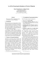
Figure 1 Human cervical culture model. a) To rule out possible leaks in the agarose seal, Dextran blue was added to the upper chamber on day 6
of culture, and its presence in the lower chamber was determined 20 h later to all Transwells used in the experiments and negative control well with
agarose only, b) other negative control with tissue alone without treatment and without challenge with virus and c) positive control well with tissue
alone infected with only with HIV-1 virus. d) Inhibition of HIV-1 transmission, Cervical tissue is treated with PVP-coated AgNPs at different concentrations in a Replens gel or RPMI + 10% FCS media, which was then infected with HIVIIIB. HIV transmission or inhibition of transmission across the mucosa
was determined in the lower chamber by formation of syncytia using indicator cells (MT-2).
h. The highest dose of PVP-coated AgNPs (0.6 mg/mL)
did not cause an acute inflammatory response in the cervical explant or induce cell death, as determined by the
histopathology with no signs of cell damage (no edema,
eosinophils or apoptosis). Compared to the negative control (no treatment), cervical tissue that had been incubated with 0.6, 0.3, 0.15, 0.1, and 0.05 mg/mL PVP-coated
AgNPs for 48 h showed mild lymphoid infiltration compared with the negative control (Fig 2a).
Next, we evaluated cell viability after 24 h of treatment
with various concentrations of PVP-coated AgNPs (0.6,
0.3, 0.15, 0.1 and 0.05 mg/mL) by comparing the percentage of viable cells in the cervical tissue without treatment
with PVP-coated AgNPs, relative to the positive control,
which was measured as the amount of ATP released from
viable cells using a luciferase-based assay. The values of
PVP-coated AgNPs chosen for toxicity studies exceeded
the amount necessary to inhibit transmission of HIV-1
infection in vitro trough the cervical explant [36]. Treatment with 0.3 mg/mL PVP-coated AgNPs was cytotoxic
in only 20% of the cells of the cervical explant, whereas a
dose of 0.6 mg/mL was cytotoxic to 23% of the cells. PVPcoated AgNPs formulated in Replens gel inhibited cell
viability by 5%, and Raft-media was cytotoxic to 18% of
cells (Fig. 2b). Raft-media has many antibiotics, which,
when combined, result in cytotoxicity.
Inhibition of cell-free and cell-associated HIV-1 viral
infection in the presence or absence of Replens gel
Results showed that two minutes of pre-treatment of the
cervical explant with 0.025 to 0.15 mg/mL of PVP-coated
AgNPs formulated in the Replens gel protected the cervical tissues from HIV-1IIIB infection; this inhibitory effect
was independent of the effect of the Replens gel alone. In
addition, PVP-coated AgNPs with or without the Replens
gel completely neutralized cell-free and cell-associated
HIV-1 transmission of infection through cervical tissues
at a dose of 0.15 mg/mL, although a similar result was
obtained at doses of 0.1 and 0.05 mg/mL Replens gel conferred protection in a dose-dependent manner, inhibiting
infection associated with the (H9+) cells more efficiently
than PVP-coated AgNPs in RPMI media containing 10%
FCS alone; the result was most significant at a dose of
0.025 mg/mL (Fig. 3).
Minimal time of exposure to PVP-coated AgNPs needed to
confer protection against the HIV-1 transmission of cellassociated infection in the cervical culture model
We evaluated the time required for 0.1 and 0.15 mg/mL
doses of PVP-coated AgNPs to block HIV-1IIIB infection
of cell-associated (H9 +) transmission through the cervical tissue. Complete protection occurred within one minute of pre-treatment with 0.15 mg/mL PVP-coated
AgNPs incubated for different times and after washing
Lara et al. Journal of Nanobiotechnology 2010, 8:15
/>
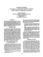
Figure 2 Toxicity of the PVP-coated AgNPs to the cervical tissue.
a) Normal squamous epithelium and the stroma of ecto-cervical tissues were exposed to Replens gel mixed with 0.15 mg/mL PVP-coated
AgNPs for 48 h. Ecto-cervical explants (5 mm) were exposed to either
Replens gel alone, which served as a control, or to PVP-coated AgNPs.
After 48 h of incubation in a 37°C humidified incubator, the tissues
were washed, embedded in paraffin, and stained with hematoxylin
and eosin. b) Cervical explants in the upper Transwell chambers were
exposed to Replens alone as a control, Raft-medium, or Replens gel
containing different concentrations of PVP-coated AgNPs (0.6, 0.3,
0.15, 0.1, 0.05 and 0 mg/mL). After 24 h, the medium containing PVPcoated AgNPs was removed and washed three times with culture media. Cell viability was measured by the CellTiter-Glo® assay. Graphs
show values of the means ± standard deviations from three separate
experiments. Graphs were created using the SigmaPlot 10.0 software.
away of the extracellular drug. Furthermore, PVP-coated
AgNPs completely blocked the T tropic wild type (HIV1IIIB) virus, the drug resistant viral isolate (AZT-RV), and
cell-associated HIV-1 (H9+ cells) transmission through
cervical tissue after one minute of pre-treatment (Fig. 4).
Duration of the protection time from HIV-1 infection
following 20 minutes pre-treatment of the cervical explants
with PVP-coated AgNPs
After 20 minutes of pre-treatment of the cervical explants
with 0.15 mg/mL PVP-coated AgNPs, the drug was
Page 4 of 11
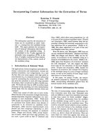
Figure 3 Inhibition of HIV-1 transmission with and without Replens gel using the cervical culture model. The upper chamber of
the Transwell with the cervical explant was exposed for 2 min to different concentrations of PVP-coated AgNPs (0.025, 0.05, 0.1 and 0.15 mg/
mL), either alone or mixed with the Replens gel. After thoroughly
washing extracellular PVP-coated AgNPs from the cervical explant,
cell-free (HIV-1IIIB) [(5 × 10⁵ TCID50)], or cell-associated virus (H9+) (5 ×
105 cells) were added. To evaluate inhibition of the HIV-1 infection, indicator cells (MT-2) in the lower chamber were cultured and formation
of syncytia was monitored for ten days. Graphs show values of the
means ± standard deviations from three separate experiments. Graphs
were created using the SigmaPlot 10.0 software.
removed and washed from the upper chamber, which
conferred almost total protection (90%) against HIV-1
transmission of infection for 48 h (Fig. 5), indicating a
long-lasting protective effect by the PVP-coated AgNPs
in the cervical explant.
Discussion
The development of non-toxic microbicides effective
against the transmission of cell-free and cell-associated
virus, which have long-lasting efficacy on the treated tissue [17,44] and are rapidly acting [45], is a highly desirable approach to the prevention of HIV-1 transmission
during sexual intercourse. Inhibiting the transmission of
Lara et al. Journal of Nanobiotechnology 2010, 8:15
/>
Figure 4 Time needed for PVP-coated AgNPs to confer protection
from the transmission of HIV-1 through the cervical. Cervical explants were pretreated for 1, 15 and 30 minutes with 0.1 or 0.15 mg/mL
PVP-coated AgNPs. After thoroughly washing extracellular PVP-coated
AgNPs from the cervical tissue, cell-associated virus (H9+) (5 × 10⁵ cells)
was added to the upper chamber of the Transwell. Indicator cells (MT2) were cultured in the lower chamber to evaluate the inhibition of HIVIIIB infection. Graphs show values of the means ± standard deviations
from three separate experiments. Graphs were created using the SigmaPlot 10.0 software.
HIV in vivo will likely require a combination of microbicidal products that provide broad anti-viral effects and
prevent the development of HIV strains resistant to the
microbicides [46]. It is clear that the development of a
topical vaginal microbicide is technically, ethically, and
culturally complicated. However, the number of lives
saved with such an agent may exceed the risks involved
[47].
In fact, microbicides could have the potential to eliminate drug-resistant bacteria, in addition to sexually transmitted diseases that cause inflammation. Previous studies
have reported that silver ions and silver nanoparticles
exert anti-inflammatory effects, induce lymphoproliferation, and inhibit bacterial and HIV-1 infection. These
characteristics make silver nanoparticles of interest in
microbicide research [48,49].
The mechanism of the antiviral action of PVP-coated
AgNPs as an HIV-1 virucidal agent has been previously
established by Lara HH et al. First, studies have revealed
that PVP-coated AgNPs inactivate HIV-1 and block viral
entry through gp120-CD4 interaction. Second, PVPcoated AgNPs (1.0-2.5 mg/mL) efficiently block the
fusion of HL2/3 and HeLa CD4 cells in a dose-dependent
manner. Third, PVP-coated AgNPs act as an effective
broad-spectrum microbicide against cell-free virus (i.e.,
laboratory strains, clinical isolates, T- and M-tropic
strains, and resistant strains), as well as the cell-associ-
Page 5 of 11
ated virus. Fourth, PVP-coated AgNPs are effective virucides, as they inactivate HIV particles in a short period of
time, exerting their activity at an early stage of viral replication (i.e., entry or fusion) and at post-entry stages [36].
Recent studies have shown that silver nanoparticles are
capable of being internalized into cells and can penetrate
through skin cells (HEKs) [50]. Other authors have
described the localization of PVP-coated AgNPs only in
the superficial layers of the stratum corneum, a result
similar to that found in a static cell diffusion study [51].
Other nanoparticles have not been shown to penetrate
into the deeper epidermis [52,53].
Finally, previous studies report that silver nanoparticles
and silver nanocrystals suppress the expression of TNF-α,
which is a cytokine that plays a pivotal role in HIV-1
pathogenesis by up-regulating the transcription of HIV-1
[48,49]. It also prevents inflammation and, as such, may
enhance wound healing in vivo [54]. Moreover, inflammation produces immune activation, enhancing HIV-1
infection of subepithelial cells of the human cervical tissue. Consequently, an agent that prevents inflammation
should inhibit the transmission of HIV infection by
impeding the enhancement of HIV infection [18].
Based on the previous studies mentioned above, the
purpose of this study was to demonstrate the ability of
PVP-coated AgNPs to inhibit HIV-1 transmission of
infection in a rapid manner with long-lasting effects and
efficiently throughout the human cervical explant in an in
vitro model that simulates in vivo conditions.
For these studies, we used a model of a human cervical
explant, which contained the natural in vivo tissue architecture of stratified squamous epithelium, submucosa,
and immune cells (Fig. 1). In this model, the infectious
virus is transmitted across the mucosal barrier by both
cell-free and cell-associated HIV-1 [7]. Although some
researchers have questioned whether HIV transmission
in this model is a result of leakage around the polarized
cervical tissues [55], these concerns were rebutted [56]
based on additional data not shown in the original
description of the model. This model has also been used
by Greenhead and colleagues to study the effects of various microbicides [57].
PVP-coated AgNPs that were formulated using Replens
gel were more effective as a virucide compared to the
PVP-coated AgNPs dissolved in RPMI+10% FCS media.
This increased activity is due to the ability of this gel to
diffuse the PVP-coated AgNPs more homogenously into
the cervical tissue as compared to the medium [6] (Fig. 3),
even though RPMI with FCS prevents agglomeration of
PVP-coated AgNPs [58]
We demonstrated that pre-treatment of cervical tissues
with PVP-coated AgNPs neutralized the transmission of
HIV-1 using human cervical explants. Specifically, we
found that 0.15 mg/mL PVP-coated AgNPs inhibited
Lara et al. Journal of Nanobiotechnology 2010, 8:15
/>
Page 6 of 11
Figure 5 Protection from HIV-1 infection following pre-treatment of the cervical explant with PVP-coated AgNPs. a) Cervical explants were
exposed to 0.1 or 0.15 mg/mL PVP-coated AgNPs in RPMI + 10% FCS media for 20 minutes. After thoroughly washing extracellular PVP-coated AgNPs
from the cervical explant and after 1 minute, 24 h, 48 h and 72 h, cell-free virus (HIV-1IIIB) [(5 × 10⁵ TCID50)] was added to the upper chamber. To verify
the neutralization of HIV-1 transmission, we cultured the indicator cells (MT-2) in the lower chamber and evaluated inhibition of the HIV-1 infection.
b) Cervical explants were exposed to HIV-1 in the absence of PVP-coated AgNPs as a control and to 0.1 or 0.15 mg/mL of PVP-coated AgNPs as pretreatment. Graphs show values of the means ± standard deviations from three separate experiments. Graphs were created using the SigmaPlot 10.0
software.
infection by HIV-IIIB and HIV-AZT-RV cell-free viruses as
well as cell-associated infection at doses that were not
toxic to the human cervical tissue. In addition, treatment
of the cervical tissue with 0.15 mg/mL PVP-coated
AgNPs augmented the number of lymphocytes relative to
the control (Fig 2a). The increased proliferation of lymphocytes was presumably due to activation of the
immune system, which was induced by the continuous
expression of death factors (mutations of Fas-L or CD95).
This resulted in activation of lymphocytes, including
CD4 T cells, CD8 CTL, or APC and turned them into
effectors of apoptosis, leading to the destruction of
healthy, non-infected cells [59-61]. Silver ions and silver
nanoparticles improve wound healing by reducing
inflammation, inducing the proliferation of lymphocytes
[37] and inhibiting bacterial and HIV-1 infection; thus,
PVP-coated AgNPs are a potential therapeutic agent
against the dissemination of drug-resistant bacteria,
thereby providing protection from sexually transmitted
diseases. Importantly, we demonstrated that high concentrations of PVP-coated AgNPs (0.3 and 0.6 mg/mL)
were only cytotoxic to a small population of cells, affecting the viability of 20-23% of the cells in the cervical
explant, which correlates with low off-target cytotoxic
effects (Fig. 2).
An ideal microbicide should act rapidly [45]; in accordance with this, we observed that one minute of exposure
to PVP-coated AgNPs (0.15 mg/mL) was the minimal
time necessary to achieve protection of the cervical tissue
against the transmission of infection by cell-free and cellassociated viruses (Fig. 4). Previously, in vitro studies
demonstrated that when PVP-coated AgNPs (2.5-5 mg/
mL) were dissolved in RPMI+10% FCS media, they conferred partial protection (50%) from HIV-1 cell-associated infection in a dose-dependent manner [36]. In
further support of PVP-coated AgNPs as microbicides,
PVP-coated AgNPs were also effective in the presence of
Lara et al. Journal of Nanobiotechnology 2010, 8:15
/>
cell-associated infection, even under 'non-optimal' conditions.
First-generation microbicides are only effective for a
few hours and, therefore, require administration shortly
before coitus [62]. Previously, we reported that treatment
with PVP-coated AgNPs in concentrations ranging from
0.031 mg/mL to 5.0 mg/mL for one minute reduced the
transmission of HIV-1 infection in PBMC and H9+ cells
by 20%-30% and that this protection lasted for several
hours [36].
Using the cervical explant model, we evaluated the
long-term effectiveness of PVP-coated AgNPs, which is
an important pharmacodynamic parameter investigated
during the development of a topical microbicide agent. It
contributes to the choice of antiviral dosing regimens and
is defined as the length of time that infection is suppressed following brief exposure to the antimicrobial
agent. Ideally, a microbicide should remain effective for
several hours after topical application [63]. In this study,
the transmission of HIV-1 infectivity through the cervical
explant was inhibited in almost all cases when PVPcoated AgNPs were formulated in the Replens gel. Additionally, PVP-coated AgNPs (0.15 mg/mL) that were
applied to the cervical tissue for 20 min in a gel formulation were able to abolish HIV-1 transmission for a period
of 48 h after the gel was removed and washed thoroughly;
after this period of time, the HIV-1 was added to the cervical explant at different times until 72 hours to evaluate
the duration of protection to the tissue (Fig. 5). These
results are in accordance with our previous findings that
showed pre-treatment of uninfected cells with PVPcoated AgNPs conferred protection from acquiring HIV1 in vitro, even in the absence of extracellular drug [36]. A
dose of 0.15 mg/mL PVP-coated AgNPs represents a
threshold level necessary for inhibition of transmission,
even after 48 h (Fig 5). PVP-coated AgNPs were able to
confer protection for similar lengths of time compared to
other microbicides, including UC781 [64,65,23,24,66].
In comparison to various viral entry inhibitors, PVPcoated AgNPs offer many advantages. For example,
although dextrin sulfate reduced the ability of virus (HIV1HSBc2) to infect cells in vitro by 77%, it did not protect
cells against the R5 virus (HIV-1 JRCSF) [67]. Further,
although nonoxynol-9 is a microbicide that is active
against a wide range of pathogens, it is potentially cytotoxic to host cells. In contrast to these compounds, PVPcoated AgNPs have low cytotoxicity, protect cervical tissue against HIV infection in a manner independent of coreceptors [36] and could possibly reduce inflammation.
As such, PVP-coated AgNPs are an ideal microbicide to
study [68].
Our hypotheses concerning the inactivation of HIV-1
transmission throughout the cervical explant model
using the PVP-coated AgNPs is that the drug acts as a
potent virucidal agent that attaches at the viral mem-
Page 7 of 11
brane, [36,69] and may protect the natural barrier of the
genital epithelium, therefore inactivating the ability of the
HIV virus to reach the target cells that reside below.
Thus, when the HIV virus crosses the genital epithelium,
it is already inactivated and unable to transfer infection to
the target cells that reside in the subepithelium [36], as
evidenced by an absence of infection (absence of syncytia
on indicator cells) on the lower chamber of the cervical
model after treatment with PVP-coated AgNPs.
In addition to their virucidal activity, PVP-coated
AgNPs also impair the ability of the HIV-1 virus to
develop resistance [36]. Importantly, these nanoparticles
have potent activity against most strains of HIV and provide broad protection against other STIs. These compounds are stable at room temperature, accessible in
terms of cost, and have demonstrated in vitro safety
[36,70,71].
Conclusions
Previous in vitro studies evaluating PVP-coated AgNPs as
potential virucidal agents have revealed that these compounds, in addition to providing broad-spectrum bactericidal and HIV-1 virucidal activity, also blocked the
infection of cell-free and cell-associated HIV-1 [72]. The
gel formulation of PVP-coated AgNPs is probably the
first microbicide with broad spectrum virucidal, bactericidal, and anti-inflammatory properties in cervical tissue
[36,54,73].
Our results show that PVP-coated AgNPs function as
potential microbicides with virucidal properties that are
capable of preventing the transmission of HIV-1 in a
human cervical tissue explant model when used at a nontoxic dosage range. PVP-coated AgNPs protect against
infection transmission of cell-free and cell-associated
HIV-1, acting within one minute after the treatment of
the cervical tissue. Importantly, after 20 minutes of pretreatment with PVP-coated AgNPs and subsequent
washing, the cervical culture remained protected against
infection with HIV-1 for as long as 48 h, demonstrating
long-lasting protection. This feature is necessary for a
topical vaginal microbicide to ensure protection many
hours after gel application and even more so after the gel
is washed away [17].
However, further studies are necessary to evaluate the
potential toxicities (i.e., genetic, reproductive, and carcinogenic toxicities) and long-term side effects associated
with the use of PVP-coated AgNPs as an inhibitor of
HIV-1 infection. Studies evaluating hypersensitivity/photosensitivity and condom integrity are also necessary [7].
Methods
Silver Compounds
Commercially manufactured 30-50 nm spherical silver
nanoparticles surface-coated with 0.2 wt% PVP (PVPcoated AgNPs) were used for these studies (NanoAmor,
Lara et al. Journal of Nanobiotechnology 2010, 8:15
/>
Houston, TX). Stock solutions of PVP-coated AgNPs
were prepared in RPMI 1640 cell culture media with 10%
FCS.
Serial dilutions of the stock solution were made using
RPMI + 10% FCS media.
HIV-1 isolates and cell culture
The following reagents were obtained from the AIDS
Research and Reference Reagent Program, AIDS Division, the National Institute of Allergies and Infectious
Diseases and the National Institute of Health and Collaborators: MT-2 (from Dr. Douglas Richman), H9+ cells
(from Dr. Robert Gallo), HeLa-CD4-LTR-β-gal cells and
the viral strains HIV-1IIIB and HIV-1AZT-RV. HIV-1IIIB was
propagated by sub-culturing in the MT-2 and H9+ cells,
according to the DAIDS Virology Manual for HIV Laboratories. Aliquots of the cell-free supernatants from virulent cultures were used for viral inoculation. MT-2 and
H9+ cells were cultured in RPMI 1640 (Sigma-Aldrich),
supplemented with 10% fetal calf serum (FCS) and antibiotics. Commercially manufactured 30-50 nm PVP-coated
AgNPs were used in these studies (NanoAmor, Houston,
TX). A stock solution was prepared in RPMI culture
media enriched with 10% fetal calf serum (FCS), which
prevents agglomeration. Serial dilutions of the stock solution yielded four different solutions with concentrations
ranging from 0.025 to 0.6 mg/mL All work related to
HIV-1 and cell culture manipulation was done in a biosafety level 3 (BSL-3) laboratory at the Laboratorio de
Inmunología y Virología, Universidad Autonoma de
Nuevo Leon, Mexico.
Formulation of Replens gel/PVP-coated AgNPs
PVP-coated AgNPs were formulated in a non-spermicidal Replens gel (3% glycerin, 0.08% sorbic acid, 1% carbopol 940, 4% liquid paraffin and 16% 1 N NaOH) [24].
Gels containing PVP-coated AgNPs at serial concentrations from 0.025- 0.6 mg/mL were added to the upper
chambers of the cervical culture model.
Page 8 of 11
reagent) and positive control (explant without treatment).
A Veritas microplate luminometer from Turner Biosystems (Model 9100-002) was used in these experiments.
Cytotoxicity was evaluated in a dose-dependent manner
and was based on the percentage of viable cells relative to
the positive control.
Cervical explant model
Ecto-cervical tissue was collected from HIV-1-negative,
pre-menopausal women undergoing planned therapeutic
hysterectomies after their informed consent was
obtained. All tissues were processed for organ culture
within 5 hours of the completion of surgery. Tissue samples were soaked in a concentrated antibiotic wash solution (20,000 U/mL penicillin and streptomycin, 250 μg/
mL fungizone, and 120 U/mL nystatin) for 10 min. The
tissues were then washed three times in Raft-media 21
(Dulbecco's modified Eagle medium supplemented with
25% Ham's F12 medium, 0.1 nM cholera toxin, 5 μg/mL
apo-transferrin, 4 mg/m/L hydrocortisone, 0.5 ng/mL
EGF, 10% FBS and 10,000 U/mL penicillin, and streptomycin) and cut into 0.4 × 0.5 cm pieces. A piece of tissue
with the epithelial layer oriented on top was placed in the
top chamber of a 12-well Transwell plate, a permeable tissue culture support that uses microporous membranes.
These permeable wells permit cells to uptake and secrete
molecules on both their basal and apical surfaces and,
thereby, carry out metabolic activities in a more physiological fashion. A 3% solution of agarose in Hank's
medium was added to the area surrounding the tissue in
the top well, which upon solidification created a tight seal
around the tissue. Cervical and vaginal explants, comprising epithelial and stromal tissues, were kept at 37°C in
a humidified atmosphere containing 5% CO2 [23,74].
To rule out possible leaks in the agarose seal, Dextran
blue was added to the upper chamber on day 6 of culture,
and its presence in the lower chamber was determined 20
h later[13].
Toxicity of PVP-coated AgNPs to the cervical tissue
Inhibition of cell-free and cell-associated HIV-1 viral
infection in the presence or absence of Replens gel
The effect of various concentrations of PVP-coated
AgNPs (0.6, 0.3, 0.15, 0.1 and 0.05 mg/mL) on cervical
explant tissues for 48 h was examined by histochemistry,
as previously described [23,24]. The viability of cervical
biopsies was quantified after 24 h of exposure to Replens
gel mixed with 0.6, 0.3, 0.15, 0.1 and 0.05 mg/mL PVPcoated AgNPs, using the CellTiter-Glo® luminescent cell
viability assay (Promega Cat. G7572). Microtiter plates
were incubated at 37°C in a 5% CO2 humidified atmosphere for 24 h and were used to determine the number
of viable cells in a culture by quantification of ATP. All
assays were run in parallel according to the producer's
protocol and included both a negative (measure of only
In these experiments we used an in vitro cervical tissuebased organ culture model that was developed to study
the heterosexual transmission of HIV-1 infection simulating in vivo conditions.
The upper chamber of the Transwell with cervical
explant was pre-treated for 2 min at different concentrations of PVP-coated AgNPs (0.025, 0.05, 0.1 and 0.15 mg/
mL), either alone or in formulation with the Replens gel,
followed by thorough washing of the extracellular PVPcoated AgNPs from the cervical explant. Cell-free (HIV1IIIB) [(5 × 10⁵ TCID50)] or cell-associated virus (H9+) (5 ×
105 cells) was added to the cervical explant in the upper
chamber. To evaluate the inhibition of HIV-1 transmis-
Lara et al. Journal of Nanobiotechnology 2010, 8:15
/>
sion through the cervical explant, indicator cells (MT-2)
were cultured in the lower chamber and were monitored
for the formation of syncytia for ten days, as previously
described [13,75,76]. A positive virus control (cervical
explant infected with HIV-1 without treatment) must
produce observable syncytia within seven days of incubation, which reflects the presence of infection. The first
reading of the plate must be made by day three. Negative
control wells (cervical explants not infected with HIV-1)
must not develop syncytia, which reflect an absence of
infection. If either control does not react as expected, the
assay is suspect and should be repeated.
In the case of the MT-2 cells, half of the media were
changed for new RPMI+10% FCS media with MT-2 uninfected cells every three days, and the formation of syncytia was monitored for ten days. The cytopathic effects of
the viral infection of MT2 cells were analyzed by microscopic assessment of syncytia formation. These latter
data were obtained by analysis of duplicate samples by
two independent observers [13,75,76].
Minimal time of exposure to PVP-coated AgNPs needed to
confer protection from HIV-1 transmission of cellassociated infection in the cervical culture model
To define the minimal time of exposure needed to confer
protection to the cervical explant from transmission of
infection, RPMI+10% FCS media containing 0, 0.1 and
0.15 mg/mL PVP-coated AgNPs was added to the upper
chambers, and 1, 15, or 30 minutes later, the medium was
removed. The upper chambers were then washed three
times with the culture media. H9+ (5 × 10⁵ cells) were
then added to the upper chamber of the Transwell to
evaluate inhibition of HIV-1 cell-associated transmission.
Target cells (MT- 2) were added to the lower chambers.
In the case of the MT-2 cells, half of the media were
replenished with the new media and added to the MT-2
uninfected cells every three days. The formation of syncytia was monitored for ten days.
Duration of time of protection from HIV-1 infection
following 20 minutes of pre-treatment of the cervical
explants with PVP-coated AgNPs
Five millimeter circular pieces of ectocervical tissues
were placed in the top chambers of a 12-well Transwell
sealed with 3% agarose with the epithelial layer oriented
on top. Media containing PVP-coated AgNPs was added
to the upper chambers, and after 20 min of treatment, the
RPMI+10% FCS media containing PVP-coated AgNPs in
the upper chambers was removed from ectocervical tissues on the upper chambers and was then washed thoroughly three times with media. After washing of the top
chambers, HIV-1IIIB [(5 × 10⁵ TCID50)] was added after 1
min, 24 h, 48 h and 72 h. Indicator cells (MT- 2) were
Page 9 of 11
added to the lower chambers to measure the percentage
of inhibition of infection transmission through the cervical explant to the lower chamber where the indicator cells
are cultured. With respect to the culture of MT-2 cells,
half of the media were exchanged for new media and
added to the MT-2 uninfected cells every three days. The
formation of syncytia was monitored for ten days after
infection.
MT-2 infectivity assay
MT-2 cells were added as indicator cells to monitor the
transmission of HIV-1 infectivity to the lower chambers
as soon as the HIV-1 was added to infect the cervical tissue of the upper chamber with or without PVP-coated
AgNPs formulated into gel or RPMI+10% FCS media. In
the case of MT-2 cells, half of the media was replenished
with the new media and added to the MT-2 uninfected
cells every three days. The formation of syncytia was
monitored every day for ten days. For a positive control
on cervical tissue, only HIVIIIB was added without treatment with PVP-coated AgNPs, syncytia were counted for
all cells in the tissue. For the negative control, only cervical tissue without HIVIIIB and PVP-coated AgNPs were
used. Syncytia were counted in the lower chamber; for
the negative control, all cells were expected to be without
syncytia.
The percentages of cells with tissue showing signs of
inhibition of HIV-1 infection transmission were evaluated with respect to the positive control. The cytopathic
effects of the viral infection of MT2 cells were analyzed
by microscopic assessment of syncytial formation. These
latter data were obtained by analysis of duplicate samples
by two independent observers [75-77].
Statistical analysis
Graphs show values of the means ± standard deviations
from three separate experiments. Graphs were created
using the SigmaPlot 10.0 software.
Competing interests
The authors declare that they have no competing interests.
Authors' contributions
All authors read and approved the final manuscript. H.H.L. participated in the
conception and experimental design of the in vitro HIV-1 manipulation and
infection assays, in the analysis and interpretation of the data, and in the writing and revision of this report. L.I-T. participated in designing the in vivo cervical
tissue model and helped analyze and interpret the results. H.H.L. and L.I-T.
made equal contributions to this study working in the cervical model, working
designing, and authoring E.N.G-T. participated in the analysis and interpretation of the data and in writing and revising this report. C.R-P. participated in the
experimental design of this study.
Acknowledgements
The following funding sources supported our experiments: the Programa de
Apoyo a la Investigacion en Ciencia y Tecnologia (PAICyT) of the Universidad
Autonoma de Nuevo Leon, Mexico, and the Consejo Nacional de Ciencia y Tecnologia (CONACyT) of Mexico.
Lara et al. Journal of Nanobiotechnology 2010, 8:15
/>
Author Details
Laboratorio de Inmunología y Virología, Departamento de Microbiología e
Inmunología, Facultad de Ciencias Biológicas, Universidad Autónoma de
Nuevo León, San Nicolas de los Garza, México
Received: 12 April 2010 Accepted: 13 July 2010
Published: 13 July 2010
© 2010 Lara et Access from: />This is an Openal; licensee BioMed Central Ltd. the terms of the Creative Commons Attribution License ( which permits unrestricted use, distribution, and reproduction in any medium, provided the original work is properly cited.
Journal of Nanobiotechnology distributed under
article is available article 2010, 8:15
References
1. Ray N: Maraviroc in the treatment of HIV infection. Drug Des Devel Ther
2009, 2:151-161.
2. Walker PR, Worobey M, Rambaut A, Holmes EC, Pybus OG: Epidemiology:
Sexual transmission of HIV in Africa. Nature 2003, 422:679.
3. Elias C, Coggins C: Acceptability research on female-controlled barrier
methods to prevent heterosexual transmission of HIV: Where have we
been? Where are we going? J Womens Health Gend Based Med 2001,
10:163-173.
4. Kalichman SC, Williams EA, Cherry C, Belcher L, Nachimson D: Sexual
coercion, domestic violence, and negotiating condom use among lowincome African American women. J Womens Health 1998, 7:371-378.
5. van der Straten A, King R, Grinstead O, Serufilira A, Allen S: Couple
communication, sexual coercion and HIV risk reduction in Kigali,
Rwanda. AIDS 1995, 9:935-944.
6. Balzarini J, Naesens L, Verbeken E, Laga M, Van DL, Parniak M, Van ML,
Anne J, De CE: Preclinical studies on thiocarboxanilide UC-781 as a
virucidal agent. AIDS 1998, 12:1129-1138.
7. McGowan I: Microbicides: a new frontier in HIV prevention. Biologicals
2006, 34:241-255.
8. Fowler MG, Melnick SL, Mathieson BJ: Women and HIV. Epidemiology
and global overview. Obstet Gynecol Clin North Am 1997, 24:705-729.
9. Elias CJ, Coggins C: Female-controlled methods to prevent sexual
transmission of HIV. AIDS 1996, 10(Suppl 3):S43-S51.
10. Brown H: Marvellous microbicides. Intravaginal gels could save millions
of lives, but first someone has to prove that they work. Lancet 2004,
363:1042-1043.
11. Cutler B, Justman J: Vaginal microbicides and the prevention of HIV
transmission. Lancet Infect Dis 2008, 8:685-697.
12. Pauwels R, De CE: Development of vaginal microbicides for the
prevention of heterosexual transmission of HIV. J Acquir Immune Defic
Syndr Hum Retrovirol 1996, 11:211-221.
13. Zussman A, Lara L, Lara HH, Bentwich Z, Borkow G: Blocking of cell-free
and cell-associated HIV-1 transmission through human cervix organ
culture with UC781. AIDS 2003, 17:653-661.
14. Moscicki AB: Vaginal microbicides: where are we and where are we
going? J Infect Chemother 2008, 14:337-341.
15. Severy LJ, Tolley E, Woodsong C, Guest G: A framework for examining the
sustained acceptability of microbicides. AIDS Behav 2005, 9:121-131.
16. D'Cruz OJ, Uckun FM: Clinical development of microbicides for the
prevention of HIV infection. Curr Pharm Des 2004, 10:315-336.
17. Weeks MR, Mosack KE, Abbott M, Sylla LN, Valdes B, Prince M: Microbicide
acceptability among high-risk urban U.S. women: experiences and
perceptions of sexually transmitted HIV prevention. Sex Transm Dis
2004, 31:682-690.
18. Greenhead P, Hayes P, Watts PS, Laing KG, Griffin GE, Shattock RJ:
Parameters of human immunodeficiency virus infection of human
cervical tissue and inhibition by vaginal virucides. J Virol 2000,
74:5577-5586.
19. Sadeghi-Nejad H, Wasserman M, Weidner W, Richardson D, Goldmeier D:
Sexually transmitted diseases and sexual function. J Sex Med 2010,
7:389-413.
20. Mcclelland RS, Wang CC, Mandaliya K, Overbaugh J, Reiner MT, Panteleeff
DD, Lavreys L, Ndinya-Achola J, Bwayo JJ, Kreiss JK: Treatment of cervicitis
is associated with decreased cervical shedding of HIV-1. AIDS 2001,
15:105-110.
21. Coleman JS, Hitti J, Bukusi EA, Mwachari C, Muliro A, Nguti R, Gausman R,
Jensen S, Patton D, Lockhart D, Coombs R, Cohen CR: Infectious
correlates of HIV-1 shedding in the female upper and lower genital
tracts. AIDS 2007, 21:755-759.
22. Weiler AM, Li Q, Duan L, Kaizu M, Weisgrau KL, Friedrich TC, Reynolds MR,
Haase AT, Rakasz EG: Genital ulcers facilitate rapid viral entry and
dissemination following intravaginal inoculation with cell-associated
simian immunodeficiency virus SIVmac239. J Virol 2008, 82:4154-4158.
Page 10 of 11
23. Collins KB, Patterson BK, Naus GJ, Landers DV, Gupta P: Development of
an in vitro organ culture model to study transmission of HIV-1 in the
female genital tract. Nat Med 2000, 6:475-479.
24. Zussman A, Lara L, Lara HH, Bentwich Z, Borkow G: Blocking of cell-free
and cell-associated HIV-1 transmission through human cervix organ
culture with UC781. AIDS 2003, 17:653-661.
25. Alidaee MR, Taheri A, Mansoori P, Ghodsi SZ: Silver nitrate cautery in
aphthous stomatitis: a randomized controlled trial. Br J Dermatol 2005,
153:521-525.
26. Tanweer F, Hanif J: Re: Silver nitrate cauterisation, does concentration
matter? Clin Otolaryngol 2008, 33:503-504.
27. Link TR, Conley SF, Flanary V, Kerschner JE: Bilateral epistaxis in children:
efficacy of bilateral septal cauterization with silver nitrate. Int J Pediatr
Otorhinolaryngol 2006, 70:1439-1442.
28. Lowe DG, Levison DA, Crocker PR, Shepherd JH: Silver deposition in the
cervix after application of silver nitrate as a cauterising agent. J Clin
Pathol 1988, 41:871-874.
29. Hoyme UB: Clinical Significance of Crede's Prophylaxis in Germany at
Present. Infect Dis Obstet Gynecol 1993, 1:32-36.
30. Lok CN, Ho CM, Chen R, He QY, Yu WY, Sun H, Tam PK, Chiu JF: Che CM:
Silver nanoparticles: partial oxidation and antibacterial activities. J Biol
Inorg Chem 2007, 12:527-534.
31. Kim JS, Kuk E, Yu KN, Kim JH, Park SJ, Lee HJ, Kim SH, Park YK, Park YH,
Hwang CY, Kim YK, Lee YS, Jeong DH, Cho MH: Antimicrobial effects of
silver nanoparticles. Nanomedicine 2007, 3:95-101.
32. Morones JR, Elechiguerra JL, Camacho A, Holt K, Kouri JB, Tapia J, Yacaman
MJ: The bactericidal effect of silver nanoparticles. Nanotechnology
2005, 16:2346-2353.
33. Shahverdi AR, Fakhimi A, Shahverdi HR, Minaian S: Synthesis and effect of
silver nanoparticles on the antibacterial activity of different antibiotics
against Staphylococcus aureus and Escherichia coli. Nanomedicine
2007, 3:168-171.
34. Sondi I, Salopek-Sondi B: Silver nanoparticles as antimicrobial agent: a
case study on E. coli as a model for Gram-negative bacteria. J Colloid
Interface Sci 2004, 275:177-182.
35. Lara HH, Ayala-Nuñez NV, Ixtepan-Turrent L, Rodriguez-Padilla C:
Bactericidal effect of silver nanoparticles against multidrug-resistant
bacteria. World Journal of Microbiology and Biotechnology 2009.
36. Lara HH, Ayala-Nuñez NV, Ixtepan-Turrent L, Rodriguez-Padilla C: Mode of
antiviral action of silver nanoparticles against HIV-1. J
Nanobiotechnology 2010.
37. Tian J, Wong KK, Ho CM, Lok CN, Yu WY, Che CM, Chiu JF, Tam PK: Topical
delivery of silver nanoparticles promotes wound healing.
ChemMedChem 2007, 2:129-136.
38. Dunn K, Edwards-Jones V: The role of Acticoat with nanocrystalline
silver in the management of burns. Burns 2004, 30(Suppl 1):S1-S9.
39. Neurath AR, Strick N, Li YY, Debnath AK: Cellulose acetate phthalate, a
common pharmaceutical excipient, inactivates HIV-1 and blocks the
coreceptor binding site on the virus envelope glycoprotein gp120.
BMC Infect Dis 2001, 1:17.
40. Elechiguerra JL, Burt JL, Morones JR, Camacho-Bragado A, Gao X, Lara HH,
Yacaman MJ: Interaction of silver nanoparticles with HIV-1. J
Nanobiotechnology 2005, 3:6.
41. Ayala-Nuñez NV, Lara HH, Ixtepan-Turrent L, Rodriguez-Padilla C: Silver
Nanoparticles Toxicity and Bactericidal Effect Against MethicillinResistant Staphylococcus aureus: Nanoscale Does Matter. J
Nanobiotechnology 2009.
42. Borkow G, Lara HH, Covington CY, Nyamathi A, Gabbay J: Deactivation of
human immunodeficiency virus type 1 in medium by copper oxidecontaining filters. Antimicrob Agents Chemother 2008, 52:518-525.
43. Cummins JE Jr, Guarner J, Flowers L, Guenthner PC, Bartlett J, Morken T,
Grohskopf LA, Paxton L, Dezzutti CS: Preclinical testing of candidate
topical microbicides for anti-human immunodeficiency virus type 1
activity and tissue toxicity in a human cervical explant culture.
Antimicrob Agents Chemother 2007, 51:1770-1779.
44. Terrazas-Aranda K, Van HY, Hazuda D, Lewi P, Costi R, Di SR, Cara A,
Vanham G: Human immunodeficiency virus type 1 (HIV-1) integration:
a potential target for microbicides to prevent cell-free or cellassociated HIV-1 infection. Antimicrob Agents Chemother 2008,
52:2544-2554.
45. Klasse PJ, Shattock R, Moore JP: Antiretroviral drug-based microbicides
to prevent HIV-1 sexual transmission. Annu Rev Med 2008, 59:455-471.
Lara et al. Journal of Nanobiotechnology 2010, 8:15
/>
46. Sexton A, Harman S, Shattock RJ, Ma JK: Design, expression, and
characterization of a multivalent, combination HIV microbicide. FASEB
J 2009, 23:3590-3600.
47. Cutler B, Justman J: Vaginal microbicides and the prevention of HIV
transmission. Lancet Infect Dis 2008, 8:685-697.
48. Bhol KC, Schechter PJ: Topical nanocrystalline silver cream suppresses
inflammatory cytokines and induces apoptosis of inflammatory cells in
a murine model of allergic contact dermatitis. Br J Dermatol 2005,
152:1235-1242.
49. Sibbald RG, Contreras-Ruiz J, Coutts P, Fierheller M, Rothman A, Woo K:
Bacteriology, inflammation, and healing a study of nanocrystalline
silver dressings in chronic venous leg ulcers. Adv Skin Wound Care 2007,
20:549-558.
50. Arora S, Jain J, Rajwade JM, Paknikar KM: Cellular responses induced by
silver nanoparticles: In vitro studies. Toxicol Lett 2008, 179:93-100.
51. Larese FF, D'Agostin F, Crosera M, Adami G, Renzi N, Bovenzi M, Maina G:
Human skin penetration of silver nanoparticles through intact and
damaged skin. Toxicology 2009, 255:33-37.
52. Larese FF, D'Agostin F, Crosera M, Adami G, Renzi N, Bovenzi M, Maina G:
Human skin penetration of silver nanoparticles through intact and
damaged skin. Toxicology 2009, 255:33-37.
53. Zhang LW, Monteiro-Riviere NA: Assessment of quantum dot
penetration into intact, tape-stripped, abraded and flexed rat skin.
Skin Pharmacol Physiol 2008, 21:166-180.
54. Tian J, Wong KK, Ho CM, Lok CN, Yu WY, Che CM, Chiu JF, Tam PK: Topical
delivery of silver nanoparticles promotes wound healing.
ChemMedChem 2007, 2:129-136.
55. Shattock RJ, Griffin GE, Gorodeski GI: In vitro models of mucosal HIV
transmission. Nat Med 2000, 6:607-608.
56. Gupta P, Collins K, Patterson B, Naus G, Landers D: Reply to 'In vitro
models of mucosal HIV transmission'. Nat Med 2000, 6:607-608.
57. Greenhead P, Hayes P, Watts PS, Laing KG, Griffin GE, Shattock RJ:
Parameters of human immunodeficiency virus infection of human
cervical tissue and inhibition by vaginal virucides. J Virol 2000,
74:5577-5586.
58. Greulich C, Kittler S, Epple M, Muhr G, Koller M: Studies on the
biocompatibility and the interaction of silver nanoparticles with
human mesenchymal stem cells (hMSCs). Langenbecks Arch Surg 2009,
394:495-502.
59. Gougeon ML, Lecoeur H, Dulioust A, Enouf MG, Crouvoiser M, Goujard C,
Debord T, Montagnier L: Programmed cell death in peripheral
lymphocytes from HIV-infected persons: increased susceptibility to
apoptosis of CD4 and CD8 T cells correlates with lymphocyte
activation and with disease progression. J Immunol 1996,
156:3509-3520.
60. Poon VK, Burd A: In vitro cytotoxity of silver: implication for clinical
wound care. Burns 2004, 30:140-147.
61. Wright JB, Lam K, Buret AG, Olson ME, Burrell RE: Early healing events in a
porcine model of contaminated wounds: effects of nanocrystalline
silver on matrix metalloproteinases, cell apoptosis, and healing.
Wound Repair Regen 2002, 10:141-151.
62. Benkop-Schnurch A, Hornof M: Intravaginal drug delivery
systems:Design, Challenges and Solutions. Adv Drug Deliv Rev 2003,
4:241-254.
63. Mauck C, Rosenberg Z, Van DL: Recommendations for the clinical
development of topical microbicides: an update. AIDS 2001,
15:857-868.
64. Lynch G, Low L, Li S, Sloane A, Adams S, Parish C, Kemp B, Cunningham AL:
Sulfated polyanions prevent HIV infection of lymphocytes by
disruption of the CD4-gp120 interaction, but do not inhibit monocyte
infection. J Leukoc Biol 1994, 56:266-272.
65. Harrison PF, Rosenberg Z, Bowcut J: Topical microbicides for disease
prevention: status and challenges. Clin Infect Dis 2003, 36:1290-1294.
66. Dezzutti CS, James VN, Ramos A, Sullivan ST, Siddig A, Bush TJ, Grohskopf
LA, Paxton L, Subbarao S, Hart CE: In vitro comparison of topical
microbicides for prevention of human immunodeficiency virus type 1
transmission. Antimicrob Agents Chemother 2004, 48:3834-3844.
67. Moulard M, Lortat-Jacob H, Mondor I, Roca G, Wyatt R, Sodroski J, Zhao L,
Olson W, Kwong PD, Sattentau QJ: Selective interactions of polyanions
with basic surfaces on human immunodeficiency virus type 1 gp120. J
Virol 2000, 74:1948-1960.
Page 11 of 11
68. Moscicki AB: Vaginal microbicides: where are we and where are we
going? J Infect Chemother 2008, 14:337-341.
69. Elechiguerra JL, Burt JL, Morones JR, Camacho-Bragado A, Gao X, Lara HH,
Yacaman MJ: Interaction of silver nanoparticles with HIV-1. J
Nanobiotechnology 2005, 3:6.
70. Rohan LC, Sassi AB: Vaginal drug delivery systems for HIV prevention.
AAPS J 2009, 11:78-87.
71. Elechiguerra JL, Burt JL, Morones JR, Camacho-Bragado A, Gao X, Lara HH,
Yacaman MJ: Interaction of silver nanoparticles with HIV-1. J
Nanobiotechnology 2005, 3:6.
72. Ndesendo VM, Pillay V, Choonara YE, Buchmann E, Bayever DN, Meyer LC:
A review of current intravaginal drug delivery approaches employed
for the prophylaxis of HIV/AIDS and prevention of sexually transmitted
infections. AAPS PharmSciTech 2008, 9:505-520.
73. Bhol KC, Schechter PJ: Topical nanocrystalline silver cream suppresses
inflammatory cytokines and induces apoptosis of inflammatory cells in
a murine model of allergic contact dermatitis. Br J Dermatol 2005,
152:1235-1242.
74. Balzarini J, Brouwer WG, Dao DC, Osika EM, De CE: Identification of novel
thiocarboxanilide derivatives that suppress a variety of drug-resistant
mutant human immunodeficiency virus type 1 strains at a potency
similar to that for wild-type virus. Antimicrob Agents Chemother 1996,
40:1454-1466.
75. Borkow G, Vijayabaskar V, Lara HH, Kalinkovich A, Lapidot A: Structureactivity relationship of neomycin, paromomycin, and neaminearginine conjugates, targeting HIV-1 gp120-CXCR4 binding step.
Antiviral Res 2003, 60:181-192.
76. Borkow G, Lara HH, Lapidot A: Mutations in gp41 and gp120 of HIV-1
isolates resistant to hexa-arginine neomycin B conjugate. Biochem
Biophys Res Commun 2003, 312:1047-1052.
77. Montefiori DC, Robinson WE Jr, Schuffman SS, Mitchell WM: Evaluation of
antiviral drugs and neutralizing antibodies to human
immunodeficiency virus by a rapid and sensitive microtiter infection
assay. J Clin Microbiol 1988, 26:231-235.
doi: 10.1186/1477-3155-8-15
Cite this article as: Lara et al., PVP-coated silver nanoparticles block the
transmission of cell-free and cell-associated HIV-1 in human cervical culture
Journal of Nanobiotechnology 2010, 8:15