Báo cáo khoa học: "Adhesions due to peritoneal carcinomatosis caused by a renal carcinoma leading to mechanical gastric outlet obstruction: a case report" pps
Bạn đang xem bản rút gọn của tài liệu. Xem và tải ngay bản đầy đủ của tài liệu tại đây (1.16 MB, 4 trang )
CAS E REP O R T Open Access
Adhesions due to peritoneal carcinomatosis
caused by a renal carcinoma leading to
mechanical gastric outlet obstruction: a case
report
Filippo Mocciaro
1*
, Gabriele Curcio
1
, Ilaria Tarantino
1
, Luca Barresi
1
, Gaetano Burgio
2
, Salvatore Gruttadauria
3
,
Settimo Caruso
4
and Mario Traina
1
Abstract
Introduction: Gastric outlet obstruction is a clinical syndrome caused by a variety of mechanical obstructions.
Peptic ulcer disease used to be responsible for most gastric outlet obstruction, but in the last 40 years the
prevalence of malignant tumors has risen significantly. Adhesive disease is an infrequent and insidious cause of
mechanical gastric outlet obstruction.
Case presentation: We report the case of a 78-year -old Caucasian man who had a clinical history of a right
nephrectomy for malignancy three years earlier and who was admitted for a severe gastric outlet obstruction
(score of 1) confirmed both by an upper endoscopy and by a fluoroscopic view after contrast injection. A
computed tomography scan and a laparotomy, with omental biopsies, showed a peritoneal carcinomatosis with
the devel opment of abdominal adhesions that prompted an abnormal gastric rotation around the perpendicular
axis of his antrum with a dislocation in the empty space of his right kidney. Sympto ms disappeared after surgical
bypass through a gastrojejunostomy.
Conclusions: Our patient experienced a very rare complication characterized by the development of adhesions
due to peritoneal carcinomatosis caused by a renal carcinoma treated with nephrectomy. These adhesions
prompted an abnormal dislocation of his antrum, as an internal hernia, in the empty space of his right kidney.
Introduction
Gastric outlet obstruction (GOO) is a clinical syndrome
caused by a variety of mechanical obstructions (for
example, malignancy, peptic ulcer disease, Crohn dis-
ease, and chronic pancreatitis). GOO is typically charac-
terized by epigastric abdominal pain, early post-prandial
vomiting with or without nausea, and weight loss.
Before 1970, peptic ulcer disease was responsible for
most GOO, but since the introduction of proton pump
inhibitors in clinical practice 40 years ago, the preva-
lence of malignant tumors as the cause of GOO has
risen to between 50% and 80% of all cases [1]. Adhesive
disease from p revious surgery is an infreque nt cause of
GOO but is a common cause of small bowel obstruc-
tions [2].
Case presentation
A 78-year-old Caucasian man, referred to our institute
by another hospital, was examined in our out-patient
clinic for frequent episodes of post-prandial vomiting in
theprevious30days.Thehospitalreferredhimwitha
clinical and endoscopi cal suspicion of gastric lymphoma
(severe stricture of his gastric antrum), although the
results of his biops y analysis were negative. A computed
tomography scan confirmed the findings seen on upper
endoscopy but offered no clear explanation of its nature.
His clinical history included a right nephrectomy for
malignancy three years earlier, although he underwent
no chemotherapy. At examination, he appeared thin and
malnourished and had a Gastric Outlet Obstruction
* Correspondence:
1
Gastroenterology Unit, IsMeTT, UPMC, Via Tricomi 1, Palermo 90100, Italy
Full list of author information is available at the end of the article
Mocciaro et al. Journal of Medical Case Reports 2011, 5:306
/>JOURNAL OF MEDICAL
CASE REPORTS
© 2011 Mocciaro et al; licensee BioMed Central Lt d. This is an Open Access article distributed unde r the terms of the Creative
Commons Attribution License ( which permi ts unrestricted use, distribution, and
reproduction in any medium, provided the original work is properly cited.
Scoring System (GOOSS) score of 1 (0 = no oral i ntake,
1 = liquids only, 2 = soft f oods, and 3 = solid food/full
diet) [ 3]. His blood pressure, heart rate, and blood cell
count were normal. His serum c reatinine was high,
although his electrolytes were within the normal range.
No other significantly abnormal serum values were
observed. We decided, on the basis of this evidence, to
repeat the upper endoscopy in order to evaluate the
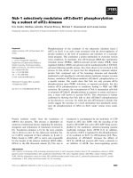
stricture. His stomach appeared normal except in the
corpus-antrum region, where his mucosa seemed con-
gested in a significant narrowing of his lumen (Figure
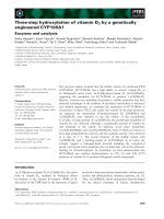
1). The duodenum cannulation was difficult because of
severe angulations of his antrum, which were confirmed
by fluoroscopic view after contrast injection through the
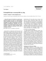
scope (Figure 2). At e ndoscopic ultrasound, performed
with a 20 MHz UM-3R radial scanning ultrasonic
miniprobe (Olympus Corporation, Tokyo, Japan)
inserted in a therapeutic gastroscope (GIF-1TQ160;
Olympus America Inc., Melville, NY, USA), the nar-
rowed area appeared with mild thickening of his mucosa
but with normal stratification of his gast ric wall (Figure
3). All of his biopsy results were negative on pathologi-
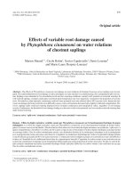
cal a nalysis. On a planned computed tomography scan,
the bulb and the second portion of his duodenum
appeared raised and inclined back toward his residual
right kidney area (Figure 4). Widespread involvement of
his peritoneum with irregular and nodular thickening
was also observed. To resolve the GOO and obtain large
omental biopsies, it was decided, in agreement with the
surgeon, that our patient undergo a laparotomy with
surgical bypass through a gastrojejunostomy. On biopsy,
the final diagnosis of the pathologist was poorly differ-
entiated omental carcinomatosis, probably related to the
previous right renal carcinoma. Seven days after the
operation, our p atient’ s status was good, with regular
transit through the gastrojejunostomy at fluoroscopy.
He restarted oral feeding (GOOSS score = 3) without
vomiting or other symptoms and, according to the
oncologist, started chemotherapy for carcinomatosis.
Discussion
Symptomatic adhe sions after surgery are frequent (25%
of readmissions in the first post-operative year) [2], and
the ris ks increase considerably in the presence of perito-
neal carcinomatosis [4]. However, adhesive disease can
serve a s an axis for gastric rotation around the long or
the perpendicular axis of the stomach.
To the best of our knowledge, no data on the develop-
ment of post-nephrectomy adhesions in patients
Figure 1 Narrowing of lumen at upper endoscopy.
Figure 2 Fluoroscopic view shows angulations of the antrum before and after contrast injection through a scope.
Mocciaro et al. Journal of Medical Case Reports 2011, 5:306
/>Page 2 of 4
operated on for renal malignancy have been published.
In a 10-year study of 871 living-donor nephrectomies,
less than 1% of patients experienced major complica-
tions and a mere 8% developed minor complications.
There were no reports of adhesive disease [5]. A recent
meta-analysis on laparoscopic versus open live-donor
nephrectomy showed that laparoscopy is safer and
found no development of adhesive disease after either
type of surgery [6]. There is an interesting case report
on an internal hernia in the retroperitoneum at the site
of a previous nephrectomy in a 25-year-old living donor
who developed signs and symptoms of part ial small
bowel obstruction [7].
In the long-term post-nep hrectomy fo llow-up of
patients with renal malignancy, the major concern is
metastatic disease. The greatest risk of recurrence fol-
lowing resection for renal cell carci noma is within t hree
to five years after the operation, with predominant lung,
bone, liver, brain, and local-regional involvement [8].
However, recurrence can occur anywhere, including the
peritoneum, e ven if it i s largely reported to be a conse-
quence of ovarian, colonic, or hepatic malignancie s. It is
Figure 3 Endoscopic ultrasound shows mild thickening of the mucosa with normal stratification of the gastric wall.
Figure 4 Multi-detector computed tomography (MDCT) multi-planar reconstruction shows herniation of the duodenum into the renal
space (white arrows).
Mocciaro et al. Journal of Medical Case Reports 2011, 5:306
/>Page 3 of 4
associated with a poor prognosis , limited treat ment [9],
and the develo pment of adhesions with obst ructive
symptoms [4].
Our patient experienced a very rare complication
characterized by the development of adhesion s due to
peritoneal carcinomatosis caused by a previous renal
carcinoma treated wit h nephrectomy but not che-
motherapy. These adhesions prompted an abnormal gas-
tric rotation around the perpendicular axis of his
antrum, with a dislocation, as an internal hernia, in the
empty space of his right kidney. This case is interesting
for two reasons: (a) GOO can occur as a late adhesive
complication after abdominal surgery o r peritoneal car-
cinomatosis or both, and (b) despite the low frequency
of incidence, a late metastasis from renal carcinoma can
involve the peritoneum witho ut ascites but with severe
obstructive symptoms.
Conclusions
This report highlights the importance of regular out-
patient visits in patients with a history of neoplasms,
even if they have undergone surgery and e specially if
they have not been treated with chemotherapy. Parti cu-
lar attention should be paid to new obstructive symp-
toms as possible consequences of late post-surgical or
unusual peritoneal metastatic complications.
Consent
Written informed consent was obtained from the patient
for publicatio n of this case report and any accompany-
ing images. A copy of the written consent is available
for review by the Editor-in-Chief of this journal.
Abbreviations
GOO: gastric outlet obstruction; GOOSS: Gastric Outlet Obstruction Scoring
System.
Acknowledgements
We thank Warren Blumberg for editorial assistance.
Author details
1
Gastroenterology Unit, IsMeTT, UPMC, Via Tricomi 1, Palermo 90100, Italy.
2
Intensive Care Unit, IsMeTT, UPMC, Via Tricomi 1, Palermo 90100, Italy.
3
Surgery Unit, IsMeTT, UPMC, Via Tricomi 1, Palermo 90100, Italy.
4
Radiology
Unit, IsMeTT, UPMC, Via Tricomi 1, Palermo 90100, Italy.
Authors’ contributions
FM collected the data and wrote the article. GC, IT, and LB were involved in
drafting the manuscript and revising it critically for important intellectual
content. GB, SG, and SC were involved in revising the manuscript critically
for important intellectual content. MT was involved in revising the
manuscript critically for important intellectual content and gave final
approval of the version to be published. All authors read and approved the
final manuscript.
Competing interests
The authors declare that they have no competing interests.
Received: 3 November 2010 Accepted: 13 July 2011
Published: 13 July 2011
References
1. Chowdhury A, Dhali GK, Banerjee PK: Etiology of gastric outlet
obstruction. Am J Gastroenterol 1996, 91:1679.
2. Parker MC, Ellis H, Moran BJ, Thompson JN, Wilson MS, Menzies D,
McGuire A, Lower AM, Hawthorn RJ, O’Briena F, Buchan S, Crowe AM:
Postoperative adhesions: ten-year follow-up of 12,584 patients
undergoing lower abdominal surgery. Dis Colon Rectum 2001, 44:822.
3. Adler DG, Baron TH: Endoscopic palliation of malignant gastric outlet
obstruction using self-expanding metal stents: experience in 36 patients.
Am J Gastroenterol 2002, 97:72-78.
4. Idelevich E, Kashtan H, Mavor E, Brenner B: Small bowel obstruction
caused by secondary tumors. Surg Oncol 2006, 15:29-32.
5. Johnson EM, Remucal MJ, Gillingham KJ, Dahms RA, Najarian JS, Matas AJ:
Complications and risks of living donor nephrectomy. Transplantation
1997, 64:1124.
6. Nanidis TG, Antcliffe D, Kokkinos C, Borysiewicz CA, Darzi AW, Tekkis PP,
Papalois VE: Laparoscopic versus open live donor nephrectomy in renal
transplantation: a meta-analysis. Ann Surg 2008, 247:58.
7. Knoepp L, Smith M, Huey J, Mancino A, Barber H: Complication after
laparoscopic donor nephrectomy: a case report and review.
Transplantation 1999, 68:449.
8. Ljungberg B, Alamdari FI, Rasmuson T, Roos G: Follow-up guidelines for
nonmetastatic renal cell carcinoma based on the occurrence of
metastases after radical nephrectomy. BJU Int 1999, 84:405-411.
9. Davies JM, O’Neil B: Peritoneal carcinomatosis of gastrointestinal origin:
natural history and treatment options. Expert Opin Investig Drugs 2009,
18:913-919.
doi:10.1186/1752-1947-5-306
Cite this article as: Mocciaro et al.: Adhesions due to peritoneal
carcinomatosis caused by a renal carcinoma leading to mechanical
gastric outlet obstruction: a case report. Journal of Medical Case Reports
2011 5:306.
Submit your next manuscript to BioMed Central
and take full advantage of:
• Convenient online submission
• Thorough peer review
• No space constraints or color figure charges
• Immediate publication on acceptance
• Inclusion in PubMed, CAS, Scopus and Google Scholar
• Research which is freely available for redistribution
Submit your manuscript at
www.biomedcentral.com/submit
Mocciaro et al. Journal of Medical Case Reports 2011, 5:306
/>Page 4 of 4