Báo cáo y học: "Mast cell activation disease: a concise practical guide for diagnostic workup and therapeutic options" ppt
Bạn đang xem bản rút gọn của tài liệu. Xem và tải ngay bản đầy đủ của tài liệu tại đây (360.44 KB, 8 trang )
REVIEW Open Access
Mast cell activation disease: a concise practical
guide for diagnostic workup and therapeutic
options
Gerhard J Molderings
1*
, Stefan Brettner
2
, Jürgen Homann
3
, Lawrence B Afrin
4
Abstract
Mast cell activation disease comprises disorders characterized by accumulation of genetically altered mast cells
and/or abnormal release of these cells’ mediators, affecting functions in potentially every organ system, often
without causing abnormalities in routine laboratory or radiologic testing. In most cases of mast cell activation
disease, diagnosis is possible by relatively non-invasive investigation. Effective therapy often consists simply of
antihistamines and mast cell membrane-stabilising compounds supplemented with medications targeted at
specific symptoms and complications. Mast cell activation disease is now appreciated to likely be considerably
prevalent and thus should be considered routinely in the differential diagnosis of patients with chronic multisystem
polymorbidity or patients in whom a definitively diagnosed major illness does not well account for the entirety of
the patient’s presentation.
Introduction
The term mast cell activation disease (MCAD) den ote s
a collection of disorders characterized by (1) accumula-
tion of pathological mast cells in potentially any or all
organs and tissues and/or (2) aberrant release of variable
subsets of mast cell mediators. A classification has been
proposed which differentiates several types and sub-
classes of MCAD (Table 1). The traditionally recognized
subclass termed systemic mastocytosis (SM) includes dis-
orders characterized by certain pathological immunohis-
tochemical and mutational findings (the WHO criteria;
Table 2; [1,2]) which are divided into several subtypes
(Table 1). On the other hand, mast cell activation syn-
drome (MCAS) presents a complex clinical picture of
multiple mast cell mediator-induced symptoms, failure
to meet the WHO criteria for diagnosis of SM, and
exclusion of relevant differential diagnoses [1,3-5].
Symptoms observed in patients with MCAS are little, if
any, different from those seen in patients with SM [6-8].
Patients present var iable and often fluctuating patterns
of symptoms (Table 3; [9-15]) which depend on the
tissue responses to mast cell mediators released both
spontaneously and in response to trigger stimuli.
A rare variant of MCAD is mast cell leukemia (MCL;
Table 1). This aggressive mast cell neoplasm is defined
by increased numbers of mast cells in bone marrow
smears (≥20%) and by circulating mast cells (reviewed in
[2]). Patients typically suffer from rapidly progressive
organopathy involving the liver, bone marrow and other
organs. The bone marrow typically shows a diffuse,
dense infiltration with mast cells. In typical MCL, mast
cell s account for more than 10% of blood leukocytes. In
a smaller group of patients, pancytopenia occurs and
mast cells account for less than 10% (aleukemic variant
of MCL). The prognosis in MCL is poor. Most patients
survive less than 1 year and respond poorly to cytore-
ductive drugs or chemotherapy.
Mast cell activation disease in general has long been
thought to be rare. However, although SM and MCL as
def ined by the WHO criteria are truly rare, recent find-
ings suggest MCAS is a fairly common disorder. Evi-
dence has been presented for a causal involvement of
pathologically active mast cells not only in the patho-
genesis of SM and MCAS but also in the etiology of
idiopathic anaphylaxis [16-18], interstitial cystitis [19],
some subsets of fibromyalgia [20,21] and some subsets
of irritable bowel syndrome [22-24].
* Correspondence:
1
Institute of Human Genetics, University Hospital of Bonn, Sigmund-Freud-
Str. 25, D-53127 Bonn, Germany
Full list of author information is available at the end of the article
Molderings et al. Journal of Hematology & Oncology 2011, 4:10
/>JOURNAL OF HEMATOLOGY
& ONCOLOGY
© 2011 Molderings et al; licensee BioMed Central Ltd. This is an Open Access article dis tributed under the terms of the Creative
Commons Attribution License ( , which permits unrestricted use, distri bution, and
reproduction in any medium, provided the original work is properly cited.
Pathogenesis
Mutations in kinas es (particularly in the tyrosine kinase
Kit) and in enzymes and receptors (JAK2, PDGFRa,
RASGRP4, Src-kinases, c-Cbl-e ncoded E3 ligase, hista-
mine H4 receptor) which are crucially involved in the
regulation of mast cell activity have been identified as
necessary to establish a clonal mast cell popu lati on, but
other abnormalities yet to be determined must be added
for the development of a clinically symptomatic disease
([7,8,25,26]; further references therein). The observations
that the same KIT mutation (e.g. D816V) can be asso-
ciated with both good prognosisaswellasprogression
to advanced disease [27] and that the D816V mutation
has also been detected in healthy subjects [28] highli ght
the potential role of ot her factors in determining the
progression/outcome of the disease. Recent findings sug-
gest that the immunohistochemical and morphological
alterations which constitute the WHO criteria for SM
(formation of mast cell clusters; spindle-shaped mor-
phology of mast cells; expression of CD25 on mast cells;
Table 2) are causally related to and specific for the
occurrence of a mutation in codon 816 of tyrosine
kinase Kit in the affected mast cells [6,29-31]. Another
aspect that lim its the diagnostic value of this mutation
is that during progression of SM the Kit mutant D816V
may disappear ([32]; own unpublished observation).
Taken together, the recent genetic findings suggest that
theclinicallydifferentsubtypes of MCAD (encompass-
ing SM, MCL, and MCAS) should be more accurately
regarded as varying presentations of a common generic
root process of mast cell dysfunction than as distinct
diseases [4,7,8,11].
Clinical diagnostics
MCAD is first suspected on clinical grounds, based on
recognition of compatible mast cell mediator-related
symptoms and, in some, identification of typical skin
lesions. The clinical presentation of MCAD is very
diverse, since due to both the widespread distribution of
mast cells and the great heterogeneity of aberrant med-
iator expression patterns, symptoms can occur in vir-
tually all organs and tissues (Table 3). Moreover,
symptoms often occur in a temporally staggered fashion,
waxing and waning over years to decades. Symptom s
often initially manifest during adolescence or even child-
hood or infancy but are recognized only in retrospect as
MCAD-related. Clinical features and courses vary
greatlyandrangefromveryindolentwithnormallife
expectancy to highly aggressive with reduced survival
times. Physical examination should include inspection
for a large assortment of types of skin lesions, testing
Table 1 Classification of mast cell activation disease
(modified from [2-4])
Mast cell activation disease
(MCAD)
Mast cell activation syndrome
(MCAS)
Systemic mastocytosis (SM)
defined by the WHO criteria • Indolent systemic mastocytosis
• Isolated bone marrow mastocytosis
• Smoldering systemic mastocytosis
• Systemic mastocytosis with an
associated clonal hematologic non-
mast cell lineage disease
• Aggressive systemic mastocytosis
Mast cell leukemia (MCL)
Table 2 Criteria proposed to define mast cell activation
disease (for references, see text)
Criteria to define mast cell
activation syndrome
WHO criteria to define systemic
mastocytosis
Major criteria Major criterion
1. Multifocal or disseminated
dense infiltrates of mast cells in
bone marrow biopsies and/or in
sections of other extracutaneous
organ(s) (e.g., gastrointestinal tract
biopsies; CD117-, tryptase- and
CD25-stained)
Multifocal dense infiltrates of mast
cells (>15 mast cells in aggregates)
in bone marrow biopsies and/or in
sections of other extracutaneous
organ(s) (CD117-, tryptase- and
CD25-stained)
2. Unique constellation of clinical
complaints as a result of a
pathologically increased mast cell
activity (mast cell mediator release
syndrome)
Minor criteria Minor criteria
1. Mast cells in bone marrow or
other extracutaneous organ(s)
show an abnormal morphology
(>25%) in bone marrow smears or
in histologies
1. Mast cells in bone marrow or
other extracutaneous organ(s)
show an abnormal morphology
(>25%) in bone marrow smears or
in histologies
2. Mast cells in bone marrow
express CD2 and/or CD25
2. Mast cells in bone marrow
express CD2 and/or CD25
3. Detection of genetic changes in
mast cells from blood, bone
marrow or extracutaneous organs
for which an impact on the state
of activity of affected mast cells in
terms of an increased activity has
been proved.
3. c-kit mutation in tyrosine kinase
at codon 816 in mast cells in
extracutaneous organ(s)
4. Evidence of a pathologically
increased release of mast cell
mediators by determination of the
content of
4. Serum total tryptase >20 ng/ml
(does not apply in patients who
have associated hematologic non-
mast-cell lineage disease)
• tryptase in blood
• N-methylhistamine in urine
• heparin in blood
• chromogranin A in blood
• other mast cell-specific
mediators (e.g., leukotrienes,
prostaglandin D
2
)
The diagnosis mast cell activation syndrome is made if both major criteria or
the second criterion and at least one minor criterion are fulfilled. According to
the WHO criteria [1], the diagnosis systemic mastocytosis is established if the
major criterion and at least one minor criterion or at least three minor criteria
are fulfilled.
Molderings et al. Journal of Hematology & Oncology 2011, 4:10
/>Page 2 of 8
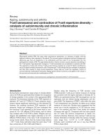
for dermatographism (Darier’s sign), and palpating for
hepatosplenomegaly and lympha denopathy. A diagnostic
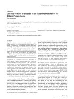
algorithm is shown in Figure 1. Recognition of a mast
cell mediator rele ase syndrome, i.e. a pattern of symp-
toms caused by the unregulated increased release of
mediators from mast cells, can be aided by use of a vali-
dated checklist [2,11,12,33] which lists the complaint
complexes to be considered. In addition to the detecti on
of the characteristic clinical constellation of findings, it
must be investigated whether levels of the mast cell-spe-
cific mediators tryptase, histamine, and heparin are ele-
vated in the blood, whether the excretion of the
histamine metabolite methylhistamine into the urine is
incre ased, and whether mast cell activi ty-related eosino-
philia, basophilia or monocytosis in the blood can be
observed. Other useful markers fairly specific to mast
cells include s erum chromogranin A (in the absence of
cardiac and renal failure, neuroendocrine cancer, and
proton pump inhibitor use) and serum and urinary leu-
kotriene and prostaglandin isoforms (e.g., leukotriene E
4
,
prostaglandin D
2
,andprostaglandin9a ,11bPGF
2
).
Together with a characteristic clinical presentation,
abnormal markers can be of diagnostic, therapeutic and
prognostic relevance. However, it remains unsettled
whether demonst ration of an elevation of mast cell
activity markers is absolutely necessary for diagnosis of
MCAD because (1) many conditions (e.g., degrading
enzymes, complexing molecules, tissue pH) may attenu-
ate or impede spill-over of exocytosed mediators from
tissues into the blood, (2) only a handful of the more
than 60 releasable mast cell mediators can be detected
by routine commercial techniques, and (3) mediator
release syndrome may be due to an amplification cas-
cade of basophil, eosinophil, and general leukocyte acti-
vation induced by liberation of only a few mast cell
mediators [34] which, again, may not be detectable by
present techniques.
When relevant differential diagnoses of a mast cell
activation disease (Table 4) which may present mast cell
mediator-induced symptoms by activation of normal
mast cells (e.g., allergy) or as result of non-mast-cell-
specific expression of mediators (e.g., neuroendocrine
cancer) are excluded, the cause of the mast cell media-
tor release syndrome must lie in the uncontrolled
increase in activ ity of pathologically altered mast cells.
Patients with most types of MCAD often initially enjoy
symptom-free intervals interspersed amongst sympto-
matic periods. Over time, symptom-free intervals
shorten, and finally symptoms become chronic with
intensity which fluctuates but with an overall trend
toward steadily increasi ng intensity. Following the pro-
posed revised diagnostic criteria (Table 2; [3-5,9,35]),
MCAD is diagnosed if either both major criteria or one
major criterion and at least one minor criterion are met.
After clinical diagnosis, a bone marrow biopsy is usually
recommended because based on current information it
cannot be predicted whether the genetic alterations
inducing pathological mast cell activity in affected mast
cell s have not also induced disturbances in hematopoie-
tic non-mast cell lineages. SM due to codon 816 muta-
tions has been shown to be associated with myeloid
neoplasms (and, less frequently, with B-cell neoplasms)
frequently enough to warrant routine marrow biopsy
when SM is suspected (e.g., serum tryptase elevation per
the WHO criteria, frequent unprovoked anaphylactoid
events). The frequency of discov ery of associated hema-
tologic neoplasms on marrow biopsy at the time of diag-
nosis of MCAS remains unclear but in our experience
appears very low. However, a byproduct of marrow
biopsy is that immunohistochemical analysis of the spe-
cimen may permit the classification of the mast cell acti-
vation disease as SM defined by the WHO criteria or as
MCAS (Table 2). In this context, it has to be considered
that due to the typically patch y distribution of mast cell
Table 3 Frequent signs and clinical symptoms ascribed to
episodic unregulated release of mast cell mediators
(modified from [12]; further references therein; an
exhaustive survey is given in [50])
Signs and
Symptoms
Abdominal abdominal pain, intestinal cramping and bloating,
diarrhea and/or obstipation, nausea, non-cardiac
chest pain, Helicobacter pylori-negative gastritis,
malabsorption
Oropharyngeal burning pain, aphthae
Respiratory cough, asthma-like symptoms, dyspnea, rhinitis,
sinusitis
Ophthalmologic conjunctivitis, difficulty in focusing
Hepatic splenomegaly, hyperbilirubinemia, elevation of liver
transaminases, hypercholesterolemia
Splenomegaly
Lymphadenopathy
Cardiovascular tachycardia, blood pressure irregularity
(hypotension and/or hypertension), syncope, hot
flush
Neuropsychiatric headache, neuropathic pain, polyneuropathy,
decreased attention span, difficulty in
concentration, forgetfulness, anxiety, sleeplessness,
organic brain syndrome, vertigo, lightheadedness,
tinnitus
Cutaneous urticaria pigmentosa, hives, efflorescences with/
without pruritus, telangiectasia, flushing,
angioedema
Abnormal
bleeding
Musculoskeletal muscle pain, osteoporosis/osteopenia, bone pain,
migratory arthritis
Interstitial cystitis
Constitutional fatigue, asthenia, fever, environmental sensitivities
Molderings et al. Journal of Hematology & Oncology 2011, 4:10
/>Page 3 of 8
Table 4 Diseases which should be considered as differential diagnoses of mast cell activation disease, since they may
mimick or may be associated with mast cell activation (diagnostic procedure of choice in parentheses)
Endocrinologic disorders Diabetes mellitus (laboratory determination)
Pancreatic endocrine tumours (gastrinoma, insulinoma, glucagonoma, somatostatinoma, VIPoma; laboratory
determination, medical history)
Porphyria (laboratory determination)
Disorders of the thyroid gland (laboratory determination)
Morbus Fabry (clinical picture, molecular genetic investigation)
Gastrointestinal disorders Helicobacter-positive gastritis (gastroscopy, biopsy)
Infectious enteritis (stool examination)
Eosinophilic gastroenteritis (endoscopy, biopsy)
Parasitic infections (stool examination)
Inflammatory bowel disease (endoscopy, biopsy)
Celiac disease (endoscopy, biopsy, laboratory determination)
Primary lactose intolerance (molecular genetic investigation)
Microscopic colitis (endoscopy, biopsy)
Amyloidosis (endoscopy, biopsy)
Intestinal obstructions by adhesions, volvulus and other reasons (medical history, imaging methods, laparoscopy)
Hepatitis (laboratory determination)
Cholelithiasis (imaging methods)
Hereditary hyperbilirubinemia (laboratory determination)
Immunological/neoplastic
diseases
Carcinoid tumour (medical history, laboratory determination)
Pheochromocytoma (medical history, laboratory determination)
Primary gastrointestinal allergy (medical history)
Hypereosinophilic syndrome (laboratory determination)
Hereditary angioedema (medical history, laboratory determination)
Vasculitis (medical history, laboratory determination)
Intestinal lymphoma (imaging methods)
Figure 1 Diagnostic algorithm.
Molderings et al. Journal of Hematology & Oncology 2011, 4:10
/>Page 4 of 8
infiltration in the bones a single marrow biopsy fails to
find systemic mastocytosis in the marrow approximately
one-sixth of the time [36].
An aggressive course of MCAD is characterized and
defined by organopathy caused by pathologic infiltration
of various organs by neoplastic mast cells inducing an
impairment of organ function. Organopathy due to mast
cell infiltration is indicated by findings termed C-find-
ings: (1) significant cytopenia(s); (2) hepatomegaly with
impairment of liver function due to mast cell infiltra-
tion, often with ascites; (3) splenomegaly with hypers-
plenism; (4) malabsorption with hypoalbuminemia and
weight loss; (5) life-threatening impairment of organ
function in other organ systems; (6) osteolyses and/or
severe osteoporosis with pathologic fractures. Urticaria
pigmentosa-like skin lesions are usually absent. In con-
trast to MCL, the bone marrow smear shows fewer than
20% mast cells (reviewed in [2]). Mast cell infiltration
with organomegaly but without end organ dysfunction
(hepatomegaly, splenomegaly, lymphadenopathy, bone
marrow alterations) is a B-finding and may occur in a
subvariant of SM (smoldering SM) with high mast cell
burden.
Treatment of mast cell activation diseases
The cornerstone of therapy is avoidance of identifiable
triggers for mast cell degranulation such as animal
venoms, extremes of temperature, mechanical irritation,
alcohol, or medications (e.g., aspirin, radiocontrast
agents, certain anesthetic agents). Individual patients
may have variable tolerance patterns and avoidance lists,
butitalsoisnotuncommontohavenoidentifiable,
reliable triggers.
Drug treatment of MCAD patients is highly individua-
lized. Curative therapies are not avail-able, and each
MCAD patient should b e treated in accordance with his
symptoms and complications. Irrespective of the specific
clinical presentation of MCAD, evidence-based therapy
consists of trigger avoidance, antihistamines, and mast
cell membrane-stabilising compounds (basic therapy,
Table 5) supplemented as needed by medicatio ns target-
ing individual mast cell mediator-induced symptoms or
complications (symptomatic therapy, Table 5). First hints
of success with any given therapy are usually seen within
4 weeks once sui table do sing has been achieved Multiple
simultaneous changes in the medication regimen are dis-
couraged since such can confound identification of the
Table 5 Treatment options for mast cell activation disease
Basic therapy (continuous oral combination
therapy to reduce mast cell activity)
• H
1
-histamine receptor antagonist (to block activating H
1
-histamine receptors on mast cells; to
antagonize H
1
-histamine receptor-mediated symptoms)
• H
2
- histamine receptor antagonist (to block activating H
2
-histamine receptors on mast cells;
to antagonize H
2
-histamine receptor-mediated symptoms)
• Cromolyn sodium (stabilising mast cells)
• Slow-release Vitamin C (increased degradation of histamine; inhibition of mast cell
degranulation; not more than 750 mg/day)
• If necessary, ketotifen to stabilise mast cells and to block activating H
1
-histamine receptors on
mast cells
Symptomatic treatment options (orally as
needed)
• Headache⇒ paracetamol; metamizole; flupirtine
• Diarrhea⇒ colestyramine; nystatin; montelukast; 5-HT
3
receptor inhibitors (eg. ondansetron);
incremental doses (50-350 mg/day; extreme caution because of the possibility to induce mast
cell degranulation) of acetylsalicylic acid; (in steps test each drug for 5 days until improvement
of diarrhea)
• Colicky abdominal paindue to distinct meteorism ⇒ metamizole; butylscopolamine
• Nausea⇒ metoclopramide; dimenhydrinate; 5-HT
3
receptor inhibitors; icatibant
• Respiratory symptoms(mainly increased production of viscous mucus and obstruction with
compulsive throat clearing) ⇒ montelukast; urgent: short-acting ß-sympathomimetic
• Gastric complaints⇒ proton pump inhibitors (de-escalating dose finding)
• Osteoporosis, osteolysis, bone pain⇒ biphosphonates ([51]; vitamin D plus calcium
application is second-line treatment in MCAD patients because of limited reported success and
an increased risk for developing kidney and ureter stones; [52])
• Non-cardiac chest pain⇒ when needed, additional dose of a H
2
-histamine receptor
antagonist; also, proton pump inhibitors for proven gastroesophageal reflux
• Tachycardia⇒ verapamil; AT1-receptor antagonists; ivabradin
• Neuropathic pain and paresthesia⇒ a-lipoic acid
• Interstitial cystitis⇒ pentosan, amphetamines
• Sleep-onset insomnia/sleep-maintenance insomnia⇒ triazolam/oxazepam
• Conjunctivitis⇒ exclusion of a secondary disease; otherwise preservative-free eye drops with
glucocorticoids for brief courses
• Hypercholesterolemia⇒ (does not depend on the composition of the diet) therapeutic trial
with HMG-CoA reductase inhibitors (frequently ineffective)
• Elevated prostaglandin levels, persistant flushing⇒ incremental doses of acetylsalicylic acid
(50-350 mg/day; extreme caution because of the possibility to induce mast cell degranulation)
All drugs should be tested for tolerance in a low single dose before therapeutic use, if their tolerance in the patient is not known from an earlier application.
Molderings et al. Journal of Hematology & Oncology 2011, 4:10
/>Page 5 of 8
specific therapy res ponsible for a given improvement (or
deterioration). Ineffective or harmful agents should be
stopped promptly. If symptoms are resistant to therapy,
as a next therapeutic step toward reducing mast cell
activity and thereby decreasing mediator release, treat-
ment with prednisone, ciclosporine (cyclosporine A), low
dose methotrexate or azathi oprine can be c onsidered.
Recently, anti-IgE treatment with the humanized murine
monoclonal antibody omalizumab has alleviated high
intensity symptoms of MCAD [37]. Since treatment with
omalizumab has an acceptable risk- benefit profile, it
should be considered in cases of MCAD resistant to evi-
dence-based therapy. Recently, molecularly targeted ther-
apybytyrosinekinaseinhibitorssuchasimatinib
mesylate, dasatinib and midostaurin has been investi-
gated. As with all drugs used in therapy of MCAD, their
therapeutic success seems to be strongly dependent on
the individual patient. In formal stu dies in SM patients,
although the kinase inhibitors reduced mast cell burden
as reflected by histological normalization in bone marrow
and improved laborat ory surrogate markers, at best only
partial improvement of mediator-related symptoms was
achieved [38-41]. However, in some case reports, imati-
nib and dasatinib have been significantly effective at
relieving symptoms. In spite of potential significant
adverse effects of these drugs, a therapeutic trial may be
justified in individual cases at an early stage. Given that
PI3K/AKT/mTOR is one of the downstream signalling
pathways upregulated by activated Kit, in theory mTOR
inhibitors (e.g., sirolimus, temsirolimus, everolimus) may
have utility in MCAD, but to date the one trial of this
notion (everoli mus in SM) showed no signific ant clinical
activity [42].
A difficult situation is the occurrence of life-threaten-
ing anaphylaxis in patients with MCAD. If anaphylaxis
is provoked by a known allergen, especially hymenoptera
venom, immunotherapy should be considered with
recognition of potential risks [43-45]. In case of repeated
life-threatening anaphylactoid episodes, the self-adminis-
tration of epinephrine on demand has been recom-
mended as an appropriate approach.
In patients with high-grade variants of MCAD (pre-
sence of C-findings) and a progressive clini-cal course,
cytoreductive drugs are recommended and are pre-
scribed together with anti-mediator-type drugs [46,47].
Potential therapeutic options are interferon-a and 2-
chlorodeoxyadenosine (2-CdA, cladribine). Interferon-a
is frequently combined with prednisone and is com-
monly used as first-line cytoreductive therapy for
aggressive SM. It ameliorates SM-related organopathy in
a proportion of cases but is associated with conside rable
adverse effects (e.g., flu-like symptoms, myelosuppres-
sion, depression, hypothyroidism), which may limit its
use in MCAD [48,49]. PEGylated interferon-a has been
shown to be as efficacious as, and less toxic than the
non-PEGylated form in some chronic myeloproliferative
diseases, but it has not been specifically studied in
MCAD. 2-Chlorodeoxyadenosine (2-CdA) is generally
reserved for last choice treatment of patients with
aggressive SM who are either refractory or intolerant to
interferon-a. Potential toxicities of 2-CdA include signif-
icant and potentially prolonged myelosuppression and
lymphopenia with increased risk of opportunistic infec-
tions. Patients who fail interferon-a and 2-Cd A therapy
are candidates for experimental drugs. However, such
therapeutic maneuvers and their potential beneficial
effects have to be balanced against the long-term risk
and serious side effects of these therapies (often immu-
nosuppressive or/and mutagenic). Polychemotherapy
including intensive induction regimens of the kind used
in treating acute myeloid leukemia, as well as high-dose
therapy with stem cell rescue, represent investigational
approaches restricted to rare, selected patients. A variety
of other agents have been reported to have in-vitro
activity against at least some MCAD-associated muta-
tions [3] and may have a future role in the treatment of
this disease.
No tools yet exist to predict which specific therapeutic
regimen will be optimal for the individual MCAD
patient. However, especially in non-aggressive disease
(comprising the great majority of patients), at least par-
tial improvement is usually attainable with one regimen
or another, and thus the practitioner is obligated to per-
sist with therapeutic trials until no options remain.
Finally, although clinical trials in MCAD are rare, enrol-
ment in such must be a priority.
Conclusions
MCAD comprises disorders affecting functions in poten-
tially every organ system by abnor mal release of media-
tors from and/or accumulation of genetically altered
mast cells. There is evidence that MCAD is a disorder
with considerable prevalence and thus should be consid-
ered routinely in the differential diagnosis of patients
with chronic multisystem polymorbidity of unknown
cause. In most cases of MCAD, diagnosis is possible b y
relatively non-invasive investigation. Effective therapy
often consists simply of antihistamines and mast cell
membrane-stabilising compounds supplemented with
medications targeted at specific symptoms and
complications.
Acknowledgements
Publication of this article was supported by the B.Braun-Stiftung (Germany)
and the Förderclub Mastzellforschung e.V. (Germany).
Author details
1
Institute of Human Genetics, University Hospital of Bonn, Sigmund-Freud-
Str. 25, D-53127 Bonn, Germany.
2
Department of Oncology, Hematology and
Molderings et al. Journal of Hematology & Oncology 2011, 4:10
/>Page 6 of 8
Palliative Care, Kreiskrankenhaus Waldbröl, Dr Goldenburgen-Str. 10, D-51545
Waldbröl, Germany.
3
Department of Internal Medicine, Evangelische Kliniken
Bonn, Waldkrankenhaus, Waldstrasse 73, D-53177 Bonn, Germany.
4
Division
of Hematology/Oncology, Medical University of South Carolina, Charleston,
South Carolina, USA.
Authors’ contributions
All authors have equally contributed to draft the manuscript. All authors
read and approved the final manuscript.
Competing interests
The authors declare that they have no competing interests.
Received: 20 January 2011 Accepted: 22 March 2011
Published: 22 March 2011
References
1. Valent P, Horny HP, Escribano L, Longley BJ, Li CY, Schwartz LB, Marone G,
Nuñez R, Akin C, Sotlar K, Sperr WR, Wolff K, Brunning RD, Parwaresch RM,
Austen KF, Lennert K, Metcalfe DD, Vardiman JW, Bennett JM: Diagnostic
criteria and classification of mastocytosis: a consensus proposal. Leuk Res
2001, 25:603-625.
2. Valent P, Akin C, Escribano L, Födinger M, Hartmann K, Brockow K,
Castells M, Sperr WR, Kluin-Nelemans HC, Hamdy NA, Lortholary O, Robyn J,
van Doormaal J, Sotlar K, Hauswirth AW, Arock M, Hermine O, Hellmann A,
Triggiani M, Niedoszytko M, Schwartz LB, Orfao A, Horny HP, Metcalfe DD:
Standards and standardization in mastocytosis: consensus statements
on diagnostics, treatment recommendations and response criteria. Eur J
Clin Invest 2007, 37:435-453.
3. Homann J, Kolck UW, Ehnes A, Frieling T, Raithel M, Molderings GJ:
Systemic mastocytosis - definition of an internal disease. Med Klin
(Munich) 2010, 105:544-553.
4. Akin C, Valent P, Metcalfe DD: Mast cell activation syndrome: Proposed
diagnostic criteria. J Allergy Clin Immunol 2010, 126:1099-1104.
5. Hamilton MJ, Castells M, Greenberger NJ: Mast cell activation syndrome -
a newly recognized cause of recurrent acute abdominal pain.
Gastroenterology 2010, 138(Suppl 1):S89.
6. Alvarez-Twose I, González de Olano D, Sánchez-Muñoz L, Matito A, Esteban-
López MI, Vega A, Mateo MB, Alonso Díaz de Durana MD, de la Hoz B, Del
Pozo Gil MD, Caballero T, Rosado A, Sánchez Matas I, Teodósio C, Jara-
Acevedo M, Mollejo M, García-Montero A, Orfao A, Escribano L: Clinical,
biological, and molecular characteristics of clonal mast cell disorders
presenting with systemic mast cell activation symptoms. J Allergy Clin
Immunol 2010, 125:1269-1278.
7. Molderings GJ, Kolck UW, Scheurlen C, Brüss M, Homann J, von Kügelgen I:
Multiple novel alterations in Kit tyrosine kinase in patients with
gastrointestinally pronounced systemic mast cell activation disorder.
Scand J Gastroenterol 2007, 42:1045-1053.
8. Molderings GJ, Meis K, Kolck UW, Homann J, Frieling T: Comparative
analysis of mutation of tyrosine kinase kit in mast cells from patients
with systemic mast cell activation syndrome and healthy subjects.
Immunogenetics 2010, 62:721-727.
9. Horan RF, Austen KF: Systemic mastocytosis: retrospective review of a
decade’s clinical experience at the brigham and the women’s hospital.
Invest Dermatol 1991, 96:5S-14S.
10. Castells M: Mast cell mediators in allergic inflammation and
mastocytosis. Immunol Allergy Clin North Am 2006, 26:465-485.
11. Hermine O, Lortholary O, Leventhal PS, Catteau A, Soppelsa F, Baude C,
Cohen-Akenine A, Palmérini F, Hanssens K, Yang Y, Sobol H, Fraytag S,
Ghez D, Suarez F, Barete S, Casassus P, Sans B, Arock M, Kinet JP,
Dubreuil P, Moussy A: Case-control cohort study of patients’ perceptions
of disability in mastocytosis. PLoS ONE 2008, 3:e2266.
12. Alfter K, von Kügelgen I, Haenisch B, Frieling T, Hülsdonk A, Haars U,
Rolfs A, Noe G, Kolck UW, Homann J, Molderings GJ: New aspects of liver
abnormalities as part of the systemic mast cell activation syndrome.
Liver Int 2009, 29:181-186.
13. Escribano L, Alvarez-Twose I, Sánchez-Muñoz L, Garcia-Montero A, Núñez R,
Almeida J, Jara-Acevedo M, Teodósio C, García-Cosío M, Bellas C, Orfao A:
Prognosis in adult indolent systemic mastocytosis: A long-term study of
the Spanish Network on Mastocytosis in a series of 145 patients.
J Allergy Clin Immunol 2009, 124:514-521.
14. Lim KH, Pardanani A, Butterfield JH, Li CY, Tefferi A: Cytoreductive therapy
in 108 adults with systemic mastocytosis: Outcome analysis and
response prediction during treatment with interferon-alpha,
hydroxyurea,
imatinib mesylate or 2-chlorodeoxyadenosine. Am J
Hematol 2009, 84:790-794.
15. Lundequist A, Pejler G: Biological implications of preformed mast cell
mediators. Cell Mol Life Sci 2011, 68:965-675.
16. Akin C, Scott LM, Kocabas CN, Kushnir-Sukhov N, Brittain E, Noel P,
Metcalfe DD: Demonstration of an aberrant mast cell population with
clonal markers in a subset of patients with “idiopathic” anaphylaxis.
Blood 2007, 110:2331-2333.
17. Gonzalez de Olano D, de la Hoz Caballer B, Nunez Lopez R, Sanchez
Munoz L, Cuevas Agustin M, Dieguez MC, Alvarez Twose I, Castells MC,
Escribano Mora L: Prevalence of allergy and anaphylactic symptoms in
210 adult and pediatric patients with mastocytosis in Spain: a study of
the Spanish network on mastocytosis (REMA). Clin Exp Allergy 2007,
37:1547-1555.
18. Brockow K, Jofer C, Behrendt H, Ring J: Anaphylaxis in patients with
mastocytosis: a study on history, clinical features and risk factors in 120
patients. Allergy 2008, 63:226-232.
19. Sant GR, Kempuraj D, Marchand JE, Theoharides TC: The mast cell in
interstitial cystitis: role in pathophysiology and pathogenesis. Urology
2007, 69(4 Suppl):34-40.
20. Enestrom S, Bengtsson A, Frodin T: Dermal IgG deposits and increase of
mast cells in patients with fibromyalgia - relevant findings or
epiphenomena? Scand J Rheumatol 1997, 26:308-313.
21. Lucas HJ, Brauch CM, Settas L, Theoharides TC: Fibromyalgia - new
concepts of pathogenesis and treatment. Int J Immunopathol Pharmacol
2006, 19:5-9.
22. Kolck UW: Investigations on the pathogenesis of the systemic mast cell
activation syndrome and its impact on heart function. Bonn, University,
medical thesis 2009 [ URN:
nbn:de:hbz:5N-19064.
23. Nickel JC, Tripp DA, Pontari MA, Moldwin RM, Mayer R, Carr LK,
Doggweiler R, Yang CC, Whitcomb D, Mishra N, Nordling J: Phenotypic
associations between interstitial cystitis/painfil bladder syndrome and
irritable bowel syndrtome, fibromyalgia, chronis fatigue syndrome: a
case control study. J Urology 2009, 181(Suppl):19.
24. Frieling T, Meis K, Kolck UW, Homann J, Hülsdonk A, Haars U, Hertfelder H-J,
Oldenburg J, Seidel H, Molderings GJ: Evidence for mast cell activation in
patients with therapy-resistant irritable bowel syndrome. Z Gastroenterol
2011, 49:191-194.
25. Orfao A, Garcia-Montero AC, Sanchez L, Escribano L: REMA: Recent
advances in the understanding of mastocytosis: the role of KIT
mutations. Br J Haematol 2007, 138:12-30.
26. Bodemer C, Hermine O, Palmérini F, Yang Y, Grandpeix-Guyodo C,
Leventhal PS, Hadj-Rabia S, Nasca L, Georgin-Lavialle S, Cohen-Akenine A,
Launay JM, Barete S, Feger F, Arock M, Catteau B, Sans B, Stalder JF,
Skowron F, Thomas L, Lorette G, Plantin P, Bordigoni P, Lortholary O, de
Prost Y, Moussy A, Sobol H, Dubreuil P: Pediatric mastocytosis is a clonal
disease associated with D(816)V and other activating c-KIT mutations.
J Invest Dermatol 2010, 130
:804-815.
27.
Garcia-Montero AC, Jara-Acevedo M, Teodosio C, Sanchez ML, Nunez R,
Prados A, Aldanondo I, Sanchez L, Dominguez M, Botana LM, Sanchez-
Jimenez F, Sotlar K, Almeida J, Escribano L, Orfao A: KIT mutation in mast
cells and other bone marrow hematopoietic cell lineages in systemic
mast cell disorders: a prospective study of the Spanish Network on
Mastocytosis (REMA) in a series of 113 patients. Blood 2006,
108:2366-2372.
28. Lawley W, Hird H, Mallinder P, McKenna S, Hargadon B, Murray A,
Bradding P: Detection of an activating c-kit mutation by real-time PCR in
patients with anaphylaxis. Mutation Res 2005, 572:1-13.
29. Mayerhofer M, Gleixner KV, Hoelbl A, Florian S, Hoermann G, Aichberger KJ,
Bilban M, Esterbauer H, Krauth MT, Sperr WR, Longley JB, Kralovics R,
Moriggl R, Zappulla J, Liblau RS, Schwarzinger I, Sexl V, Sillaber C, Valent P:
Unique effects of KIT D816V in BaF3 cells: induction of cluster formation,
histamine synthesis, and early mast cell differentiation antigens. J
Immunol 2008, 180:5466-5476.
30. Teodosio C, García-Montero AC, Jara-Acevedo M, Sánchez-Muñoz L, Alvarez-
Twose I, Núñez R, Schwartz LB, Walls AF, Escribano L, Orfao A: Mast cells
from different molecular and prognostic subtypes of systemic
Molderings et al. Journal of Hematology & Oncology 2011, 4:10
/>Page 7 of 8
mastocytosis display distinct immunopheno-types. J Allergy Clin Immunol
2010, 125:719-726.
31. Yang Y, Létard S, Borge L: Pediatric mastocytosis-associated KIT
extracellular domain mutations exhibit different functional and signaling
properties compared with KIT-phosphotransferase domain mutations.
Blood 2010, 116:1114-1123.
32. Gotlib J, Berubé C, Growney JD, Chen CC, George TI, Williams C, Kajiguchi T,
Ruan J, Lilleberg SL, Durocher JA, Lichy JH, Wang Y, Cohen PS, Arber DA,
Heinrich MC, Neckers L, Galli SJ, Gilliland DG, Coutré SE: Activity of the
tyrosine kinase inhibitor PKC412 in a patient with mast cell leukemia
with the D816V KIT mutation. Blood 2005, 106:2865-2870.
33. Molderings GJ, Kolck U, Scheurlen C, Brüss M, Frieling T, Raithel M,
Homann J: Systemic mast cell disease with gastrointestinal symptoms - a
diagnostic questionnaire. Dtsch Med Wochenschr 2006, 131:2095-2100.
34. Theoharides TC, Kempuraj D, Tagen M, Conti P, Kalogeromitros D:
Differential release of mast cell mediators and the pathogenesis of
inflammation. Immunol Rev 2007, 217:65-78.
35. Roberts LJ, Oates JA: Biochemical diagnosis of systemic mast cell
disorders. J Invest Dermatol 1991, 96(3 Suppl):19S-24S.
36. Butterfield JH, Li C-Y: Bone marrow biopsies for the diagnosis of systemic
mastocytosis: is one biopsy sufficient? Am J Clin Path 2004, 121:264-267.
37. Molderings GJ, Raithel M, Kratz F, Azemar M, Haenisch B, Harzer S,
Homann J: Omalizumab treatment of systemic mast cell activation
disease: experiences from four cases. Intern Med 2011, 50:1-5.
38. Droogendijk HJ, Kluin-Nelemans HJC, van Doormaal JJ, Oranje AR, van de
Loosdrecht AA, van Daele PLA: Imatinib mesylate in the treatment of
systemic mastocytosis - A phase II trial. Cancer 2006, 107:345-351.
39. Gotlib J, George TI, Corless C, Linder A, Ruddell A, Akin C, DeAngelo DJ,
Kepten I, Lanza C, Heinemann H, Yin O, Gallagher N, Graubert T: The Kit
tyrosine kinase inhibitor midostaurine (PKC412) exhibits a high response
rate in aggressive systemic mastocytosis (ASM): interim results of a
phase II trial. Blood 2008, 110:1039A.
40. Verstovsek S, Tefferi A, Cortes J, O’Brien S, Garcia-Manero G, Pardanani A,
Akin C, Faderl S, Manshouri T, Thomas D, Kantarjian H: Phase II study of
dasatinib in Philadelphia chromosome-negative acute and chronic
myeloid diseases, including systemic mastocytosis. Clin Cancer Res 2008,
14:3906-3915.
41. Vega-Ruiz A, Cortes JE, Sever M, Manshouri T, Quintás-Cardama A, Luthra R,
Kantarjian HM, Verstovsek S: Phase II study of imatinib mesylate as
therapy for patients with systemic mastocytosis. Leuk Res 2009,
33:1481-1484.
42. Parikh SA, Kantarjian HM, Richie MA, Cortes JE, Verstovsek S: Experience
with everolimus (RAD001), an oral mammalian target of rapamycin
inhibitor, in patients with systemic mastocytosis. Leuk Lymph 2010,
51:269-274.
43. de Olano DG, Twose IA, Esteban López MI, Muñoz LS, Alonso Díaz de
Durana MD, Castro AV, Montero AG, González-Mancebo E, González TB,
Herrero Gil MD, Fernández-Rivas M, Orfao A, de la Hoz Caballer B,
Castells MC, Mora LE: Safety and effectiveness of immunotherapy in
patients with indolent systemic mastocytosis presenting with
Hymenoptera venom anaphylaxis. J Allergy Clin Immunol 2008,
121:519-526.
44. Ruëff F, Przybilla B, Biló MB, Müller U, Scheipl F, Aberer W, Birnbaum J,
Bodzenta-Lukaszyk A, Bonifazi F, Bucher C, Campi P, Darsow U, Egger C,
Haeberli G, Hawranek T, Körner M, Kucharewicz I, Küchenhoff H, Lang R,
Quercia O, Reider N, Severino M, Sticherling M, Sturm GJ, Wüthrich B:
Predictors of severe systemic anaphylactic reactions in patients with
Hymenoptera venom allergy: importance of baseline serum tryptase-a
study of the European Academy of Allergology and Clinical Immunology
Interest Group on Insect Venom Hypersensitivity. J Allergy Clin Immunol
2009, 124:1047-1054.
45. Bonadonna P, Zanotti R, Müller U: Mastocytosis and insect venom allergy.
Curr Opin Allergy Clin Immunol 2010, 10:347-353.
46. Valent P, Sperr WR, Akin C: How I treat patients with advanced systemic
mastocytosis. Blood 2010, 116:5812-5817.
47. Lim KH, Tefferi A, Lasho TL, Finke C, Patnaik M, Butterfield JH, McClure RF,
Li CY, Pardanani A: Systemic mastocytosis in 342 consecutive adults:
survival studies and prognostic factors. Blood 2009, 113:5727-5736.
48. Simon J, Lortholary O, Caillat-Vigneron N, Raphael M, Martin A, Briere J,
Barete S, Hermine O, Casussus P: Interest of interferon alpha in systemic
mastocytosis. The french experience and review of the literature. Pathol
Biol 2004, 52:294-299.
49. Butterfield JH: Interferon treatment for hypereosinophilic syndromes ans
systemic mastocytosis. Acta haematol 2005, 114:26-40.
50. Afrin LB: Polycythemia from mast cell activation disorder: lessons from
incongruence of symptoms vs. test results. Am J Med Sci 2011.
51. Barete S, Assous N, de Gennes C, Grandpeix C, Feger F, Palmerini F,
Dubreuil P, Arock M, Roux C, Launay JM, Fraitag S, Canioni D, Billemont B,
Suarez F, Lanternier F, Lortholary O, Hermine O, Francès C: Systemic
mastocytosis and bone involvement in a cohort of 75 patients. Ann
Rheum Dis 2010, 69:1838-1841.
52. Molderings GJ, Solleder G, Kolck UW, Homann J, Schröder D, von
Kügelgen I, Vorreuther R: Ureteral stones due to systemic mastocytosis:
diagnostic and therapeutic characteristics. Urol Res 2009, 37:227-229.
doi:10.1186/1756-8722-4-10
Cite this article as: Molderings et al.: Mast cell activation disease: a
concise practical guide for diagnostic workup and therapeutic options.
Journal of Hematology & Oncology 2011 4:10.
Submit your next manuscript to BioMed Central
and take full advantage of:
• Convenient online submission
• Thorough peer review
• No space constraints or color figure charges
• Immediate publication on acceptance
• Inclusion in PubMed, CAS, Scopus and Google Scholar
• Research which is freely available for redistribution
Submit your manuscript at
www.biomedcentral.com/submit
Molderings et al. Journal of Hematology & Oncology 2011, 4:10
/>Page 8 of 8