Introduction to ENVIRONMENTAL TOXICOLOGY Impacts of Chemicals Upon Ecological Systems - CHAPTER 8 pdf
Bạn đang xem bản rút gọn của tài liệu. Xem và tải ngay bản đầy đủ của tài liệu tại đây (84.56 KB, 14 trang )
CHAPTER
8
Heavy Metals
Pollution caused by heavy metals is now a worldwide phenomenon. Among the
many heavy metals, lead (Pb), mercury (Hg), cadmium (Cd), arsenic (As), chromium
(Cr), zinc (Zn), and copper (Cu) are of most concern, although the last three metals
are essential nutrients in animal and human nutrition. These metals are widely used
in industry, particularly in metal-working or metal-plating, and in such products as
batteries and electronics. They also are used in the production of jewelry, paint
pigments, pottery glazes, inks, dyes, rubber, plastics, pesticides, and even in medi-
cines. These metals enter the environment wherever they are produced, used, and
ultimately discarded.
Heavy metals are very toxic because, as ions or in compound forms, they are soluble
in water and may be readily absorbed into living organisms. After absorption, these
metals can bind to vital cellular components such as structural proteins, enzymes, and
nucleic acids, and interfere with their functioning. In humans, some of these metals,
even in small amounts, can cause severe physiological and health effects.
In this chapter, we will consider Pb, Cd, and Hg, the three heavy metals widely
recognized as the most toxic in our environment.
LEAD
Lead (Pb) is one of the ancient metals and has been used by humans for several
thousands of years. Pb plays an important role in the economy of all industrialized
countries in the world. In the U.S., the industrial consumption of Pb is estimated to
be about 1.3 million tons per year, with a concomitant annual emission of about
600,000 tons of Pb into the environment (NAS 1980). Additional amounts are added
through mining, smelting, manufacturing, disposal, and recycling processes. Fur-
thermore, until recently huge amounts of Pb and its compounds had been emitted
into the atmosphere as a result of leaded gasoline combustion. Consequently, Pb is
ubiquitous in our environment.
Because Pb is toxic to humans at high doses, levels of exposure encountered by
some members of the population constitute a serious public health problem (NAS).
© 1999 by CRC Press LLC
The importance of Pb as an environmental pollutant is apparent since the Environ-
mental Protection Agency (EPA) has designated Pb as one of the six criteria air
pollutants.
Properties and Uses
Lead has a low melting point (326°C). It is a soft, malleable metal and it can
be easily formed into a variety of shapes. It also can form alloys with many other
metals. Other important industrial products containing Pb include pipes, paints,
solders, glass, pottery glazes, rubber, plastics, and insecticides.
Exposure
Atmospheric Lead
Sources of atmospheric Pb include lead smelters, burning of coal and materials
containing Pb, refining of scrap, wind blown from soils, and lead alkyls from
gasoline. Effluents from smokestacks and other gaseous emissions from smelters
and refining processes can distribute significant quantities of Pb into the air, soils,
and vegetation growing nearby. However, the most common source of Pb contam-
ination in ambient air until recently was the exhaust from automobiles. Tetraethyl
lead was introduced as an antiknock agent in gasoline in the 1920s and since then
has played an increasingly important role as an atmospheric pollutant. Following
the mandatory use of unleaded gasoline and improved industrial emission control,
atmospheric Pb emission has decreased dramatically. According to an EPA report,
Pb emission from major emission sources in the U.S. decreased from 56,000 to 7100
metric tons per year between 1981 and 1990 (EPA 1991). While the atmospheric
Pb pollution problem in other developed countries likewise has been significantly
reduced, a similar trend has not occurred in many third-world countries.
Waterborne Lead
Surface waters may contain significant amounts of Pb when subjected to some
special contamination. About 14% of representative drinking water supplies (i.e.,
piped drinking water) were found to contain more than 10 mg/l in a 1963–1965
survey. Less than 1% was found to be in excess of 30 mg/l. On the other hand,
rainwater collected near a busy highway may contain as much as 50 mg/l.
Another serious problem related to waterborne Pb is lead shot left in the North
America’s lakes and ponds. A large number of waterfowl in the U.S. are poisoned
or killed following ingestion of the shot.
Lead in Food
Food has long been a major source of Pb intake for animals and humans. Animals
may ingest Pb-contaminated vegetation and become intoxicated. In humans, Pb may
© 1999 by CRC Press LLC
be ingested through Pb-contaminated containers or Pb pottery glazes. Researchers
suggest that some Roman emperors might have become ill and even died from Pb
poisoning by drinking wines contaminated with high levels of Pb.
Vegetation growing near highways has been shown to accumulate high amounts
of Pb deposited from automobile exhaust (Lagerwerff et al. 1973; Khalid et al. 1996).
Pica, children’s craving for unnatural foods, is thought responsible for the chronic
Pb poisoning among many poor urban children, as they eat flaking paint from the
walls of old houses. About 27 million housing units were built before 1940 when
Pb was in common use (Lin-Fu 1982). Lead paint poses a major threat for children
and is one of the major public health problems that many communities face.
Lead in Soils
Lead and other metals can impact soils and biota by deposition from polluted
air. Stack emission from smelters (Little and Martin 1972) and emission from
automobile exhaust systems along highways are examples. Pb contamination due to
mine wastes also is an important problem in areas surrounding metal mines. Earlier
reports indicate that about 50% of the Pb liberated from motor vehicles in the U.S.
was deposited within 30 m of the roadways (Ryan 1976) and the remainder was
scattered over large areas. Lead accumulation in soils near roads varies with traffic
volume and decreases rapidly with distance from the road. For example, Pb con-
centrations of 128 to 700 ppm are found in soil adjacent to 12 highways in the
Minneapolis-St. Paul area (Ryan 1976). These levels were much greater than the
reported value of 10 to 15 ppm in unpolluted rural soils. Grass collected near an
intersection of two heavily traveled highways near Denver, CO contained as much
as 3000 ppm Pb, while vegetable samples from gardens less than 50 ft from roads
in Canandaigua, NY averaged 115 ppm Pb (range: < 10 ppm to 700 ppm).
In an attempt to assess the effect of the mandatory use of unleaded gasoline in
new automobiles on Pb concentrations in highway soils, Byrd et al. (1983) studied
Pb concentrations in soils along U.S. Interstate 20 in northeast Louisiana and
observed that the concentrations increased from 1973 to 1974 but decreased from
1975 to 1979. They concluded that the mandatory use of unleaded gasoline had
significantly reduced the Pb concentrations in soils near highways.
Lead Toxicity
Effect on Plants
Plants exposed to high levels of Pb from ambient air and soils can accumulate
the metal and manifest toxicity. The toxicity and presence of other trace metals vary
greatly among plant species. Based on
in vitro
studies, toxicity sequences have been
determined for several species. Barley plants were shown to be more sensitive to
Pb than to Cr, Cd, Ni, or Zn (Oberlander and Roth 1978), and exposure to relatively
high levels of Pb was shown to inhibit seed germination (Koeppe 1977; Yu 1991).
The effect of Pb on germination, however, was found to be less severe compared to
© 1999 by CRC Press LLC
several other metals such as Cd, As, and Hg (Koeppe; Fargasova 1994). It is important
to note that, following plant uptake, Pb moves into the food chain and thus can affect
animals and humans.
Effect on Animals
The effect of Pb on freshwater fish varies depending on the species of fish.
Goldfish, for example, are relatively resistant to Pb, presumably due to their abundant
gill secretion. As mentioned above, following the ingestion of expended lead shot
in lakes or in the field, more than one million birds are estimated killed each year
in the U.S. Lead absorbed by the bird paralyzes the gizzard leading to starvation,
and death usually follows within several weeks after the exposure.
Effect on Humans
Daily intake of Pb in humans is estimated to range from 20 mg to 400 mg per
person. The FAO/WHO Expert Committee established a Provisional Tolerable
Weekly Intake (PTWI) of 3000 mg, approximately 500 mg/day. Only one-half of
this amount appears to be safe for children. About 5 to 15% of ingested Pb is
absorbed. This amounts to 15 to 25 mg/day and represents two-thirds of the total
absorbed Pb. By contrast, about 20 to 40% of the inhaled Pb is absorbed, amounting
to about 8 mg/day, or one-third of the total absorbed Pb.
The considerably higher blood Pb levels in industrial populations reflect wide-
spread environmental Pb pollution. However, data obtained from the Second
National Health and Nutrition Examination Survey (NHANES II) indicate that there
has been a reduction in the overall mean blood-lead level of the U.S. population
during the period 1976 through 1980, from 15.8 mg/dl to 10.0 mg/dl (Lin-Fu 1982).
It is suggested that an increased use of unleaded gasoline by the U.S. population
may be responsible for the observed decrease.
Lead is one of the systemic poisons in that once absorbed into the circulatory
system, it is distributed throughout the body where it causes serious health effects.
Manifested effects of Pb poisoning include nausea, anorexia, severe abdominal
cramps, weight loss, anemia, renal tubular dysfunction, muscle aches, and joint
pains. Lead can pass the placental barrier and may reach the fetus, resulting in
miscarriages, abortions, and stillbirths.
Through interaction with cellular components of brain cells, Pb also adversely
affects the central nervous system (CNS). Clinical symptoms such as encephalopa-
thy, convulsions, and delirium may occur. In severe cases coma and death may
follow. These injuries are often reflected by behavioral disturbances observed in Pb-
poisoned victims.
It is estimated that approximately 90% of Pb absorbed by humans is deposited
in the bone (Aufderheide and Wittmers 1992). Bone, however, is no longer consid-
ered a sink for Pb in the body. Rather, it is recognized as a two-way process of
active influx and efflux of Pb to and from the bone and blood stream (Silbergeld
et al. 1993). As a result, bone acts like a reservoir for Pb, thus influencing the
exposure of the metal in the body.
© 1999 by CRC Press LLC
Although there is evidence that both inorganic and organic lead compounds are
carcinogenic in experimental animals (Cherlewski 1979; Blake and Mann 1983), no
conclusive evidence has been reported in humans.
Biochemical Effect
Lead is taken up and transported in plants (Cannon and Bowles 1962) and can
decrease cell division at very low concentrations. Lead inhibits the electron transport
in corn mitochondria, especially when phosphate is present (Koeppe and Miller
1970).
Lead, as mentioned above, is a systemic poison and can induce deleterious effects
in living organisms. The biochemical effect of Pb is complex and, in certain areas,
its mode of action remains unclear. Several well-established biochemical effects are
discussed here. First, as an electropositive metal, Pb has a high affinity for the
sulfhydryl (SH) group. Enzymes that depend on the SH group as the active site are,
therefore, inhibited by Pb. In this case, Pb reacts with the SH group on the enzyme
molecule to form mercaptide, leading to inactivation of the enzyme. The following
reaction depicts such a relationship:
2RSH + Pb
2+
→→
→→
R–S–Pb–S–R + 2H
+
Examples of the sulfhydryl-dependent enzymes include adenyl cyclase and
aminotransferases. Adenyl cyclase catalyzes the conversion of ATP to cyclic AMP
needed in brain neurotransmission. Aminotransferases are involved in transamination
and thus important in amino acid metabolism.
Second, divalent Pb is similar in many aspects to Ca and may exert a competitive
action on body processes such as mitochondrial respiration and neurological func-
tions. Lead can compete with Ca for entry at the presynaptic receptor. Since Ca
evokes the release of acetylcholine across the synapse, this inhibition manifests itself
in the form of decreased endplate potential. The miniature endplate potential release
of subthreshold levels of acetylcholine has been shown to be increased (Barton et al.
1978). The close chemical similarity between Pb and Ca may partially account for
the fact that they seem interchangeable in biological systems and that 90% or more
of the total body burden of Pb is found in the skeleton.
Third, Pb can interact with nucleic acids, leading to either decreased or increased
protein synthesis. Lead has been shown to reduce the ability of t-RNA to bind
ribosomes. The effect of Pb on nucleic acids, therefore, has important biological
implications (Barton et al. 1978).
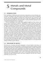
Finally, it is widely known that Pb impairs the formation of red blood cells. The
mechanism involved in the impairment is that Pb inhibits both
δ
-aminolevulinic acid
dehydratase (ALA-D)(Hernberg et al. 1970) and ferrochelatase (Tephly et al. 1978).
These are two key enzymes involved in heme biosynthesis. ALA-D catalyzes the
conversion of
δ
-aminolevulinic acid into porphobilinogen (PBG), whereas ferroche-
latase is responsible for catalyzing the incorporation of Fe
2+
into protoporphyrin IX
to form heme (Figure. 8.1). Lead inhibition of the two enzymes appears to be due
to its interaction with Zn and Fe required in the process.
© 1999 by CRC Press LLC
CADMIUM
Cadmium (Cd) is a transition metal in Group IIb along with Zn and Hg. It is
frequently associated with Zn. The U.S. is the world’s largest producer of cadmium,
with an annual output of about 5000 short tons. Mexico is an important producer
of Cd-bearing dusts and fumes, but most of these are smelted in the U.S.
Properties and Uses
Cadmium is a silver-white metal with an atomic weight of 112.4 and a low
melting point of 321°C. It is malleable and can be rolled out into sheets. The metal
unites with the majority of the heavy metals to form alloys. It is readily oxidized to
the +2 oxidation state, producing the colorless Cd
2+
ion. Cadmium persists in the
environment with a half-life of 10 to 25 years.
About two-thirds of all Cd produced is used in the plating of steel, Fe, Cu, brass,
and other alloys to protect them from corrosion. Other uses include solders and
electrical parts, pigments, plastics, rubber, pesticides, galvanized iron, etc. Special
uses of Cd include aircraft manufacturing and semi-conductors. Because Cd strongly
absorbs neutrons, it is also used in the control rods in nuclear reactors.
Exposure
General sources of exposure to Cd include air, water, and food. Atmospheric
emission of Cd may arise from such activities as mining and metallurgical process-
ing, combustion of fossil fuel, textile printing, application of fertilizers and fungi-
cides, recycling of ferrous scraps and motor oils, disposal and incineration of Cd-
containing products (e.g., plastics), and tobacco smoke.
The major nonoccupational routes of human Cd exposure are through ingestion
and inhalation. Ambient air is not a significant source of Cd exposure for the majority
of the U.S. population. Nearly all airborne Cd is due to human activities and thus
the highest concentrations are found in industrialized cities and in the vicinity of
smelting operations (Fleischer 1974). While aerial deposition is an important route
of mobility for Cd, airborne routes of exposure are not as important as soil and water
routes.
Tobacco in all of its forms contains appreciable amounts of Cd, and tobacco
smoke is one of the largest single sources of Cd exposure to humans. Since the
absorption of Cd in the lungs is much greater than that from the gastrointestinal
tract, smoking contributes significantly to the total body burden. Each cigarette on
Figure
8.1
Steps in heme synthesis inhibited by lead.
© 1999 by CRC Press LLC
the average contains approximately 1.5 to 2.0 mg of Cd, of which 70% passes into
the smoke.
Waterborne Cd is probably the largest problem because it is common in the
aquatic environment. Many Cd-containing wastes end up in lakes and marine water.
Wastes from Pb mines, various chemical industries, motor oils, and rubber tires are
some examples.
Cadmium pollution of soils can occur from several sources; a major one being the
deposition of municipal sewage sludge onto agricultural soils. Other sources of Cd
pollution are through rainfall and dry precipitation, as well as phosphate fertilizers.
Food consumption accounts for the largest sources of exposure to Cd by animals
and humans, primarily because of the ability of plants to bioaccumulate Cd at high
rates. In addition, aquatic organisms can potentially accumulate large amounts of Cd.
Cadmium Toxicity
Effect on Plants
Cadmium is accumulated by all plants. The extent of Cd accumulation, however,
varies markedly with species and variety. Soil pH is the most important factor
controlling Cd uptake by plants, with lower pH favoring its uptake. Tobacco plants
have been shown to absorb high levels of Cd from the soil (Bache 1985). Phytotox-
icity of Cd is manifested by stunting, chlorosis, reduction in photosynthesis, wilting,
and necrosis. Like Pb, Cd inhibits seed germination under laboratory conditions
(Koeppe 1977; Yu 1991; Fargasova 1994). Seedlings exposed to solutions of Cd
salts exhibit decreased root elongation and development.
Effects on Animals and Humans
Cadmium is toxic in small amounts and there is no evidence that Cd has any
useful biological function. Among the sources of exposure to Cd mentioned above,
exposure through airborne Cd is minimal to the general population, with the excep-
tion of tobacco smokers. Cadmium in drinking water, although a major source, rarely
becomes a serious problem. On the average, potable waters contain about 10 ppb
Cd. This amounts to an uptake of about 20 to 30 µg/day, based on daily water
consumption of 2 to 3 liters (Friberg 1974).
Daily intake of Cd from food is estimated at 35 to 90 µg. When dietary exposure
reaches critical concentrations, estimated to be about 250 to 300 µg/day, toxicity
symptoms are manifested. Cadmium intakes of Japanese farmers suffering from the
widely known “itai-itai” disease were reported to be from 600 to 1000 µg/day. The
disease was caused by ingestion of rice highly contaminated with Cd. The rice
paddies received water discharged from upstream Zn mines. Many of the victims
died as a result of the disease.
Once absorbed, Cd readily shows up in the blood plasma, bound in albumin
(Nordberg 1985). The bound Cd is shortly taken up by tissues, preferentially by the
liver. The Cd in the liver apparently cycles, bound with metallothionein (MT),
through the blood, kidney, and, to a small amount, bone and muscle tissue.
© 1999 by CRC Press LLC
The excretion of Cd appears minimal under normal exposure. Loss in the urine
accounts for the major route of Cd excretion, whereas only minute amounts are
excreted in the feces. As mentioned above, absorbed Cd persists in body tissues.
The long-term excretion rate of Cd is only 0.005% per day beginning after about
50 years of age (Friberg 1974).
Although dietary intake is the means by which humans are most highly exposed
to Cd, inhalation of Cd is more dangerous than ingestion. This is because, through
inhalation, the body’s organ is directly and intimately exposed to the metal. Further-
more, 25 to 40% of inhaled Cd from the air is retained, while only 5 to 10% of
ingested Cd is absorbed. Inhaled Cd may cause emphysema and pneumonitis, while
ingested Cd may result in disturbances in the gastrointestinal tract, vomiting, pro-
teinuria, osteomalacia, liver dysfunction, kidney damage manifested by anemia, and
hypertension. Cadmium is also known to be embryotoxic.
Biochemical Effect
Cadmium has been shown to impair many plant cellular functions, such as
photophosphorylation, succinate oxidation, ATP synthesis, mitochondrial NADH
oxidation, and electron transport (Nriagu 1980). Cadmium is a potent enzyme
inhibitor, affecting a variety of plant enzymes such as PEP carboxylase, lipase,
invertase (Yu 1998), and others. Extensive reports are available concerning Cd-
dependent inhibition of enzymes from animals and humans. Alkaline phosphatase
and ATPases of myosin and pulmonary alveolar macrophage cells are examples.
Two mechanisms appear to be involved in enzyme inhibition. One is through binding
to SH groups on the enzyme molecule; another is through competing with Zn and
displacing it from metalloenzymes. Naturally, Cd also can bind with SH-containing
ligands in the membrane and other cell constituents, causing structural and functional
disruptions. For instance, by inducing damage to mitochondria, Cd can uncouple
oxidative phosphorylation and impair energy metabolism of the cell. At moderate
levels, Cd toxicity is related to its antimetabolite activities toward essential metals
such as Zn, Cu, Se, and Fe. In mammals, the impact caused by Cd is thus influenced
by the relative intakes of these and other metals and vice versa (Hamilton and Valberg
1974). In addition, dietary protein has been shown to be related to the toxicity of
ingested Cd. A low protein diet results in an increased absorption of Cd and thus
increased toxicity.
MERCURY
Mercury (Hg) is the only common metal that is liquid at room temperature. It
is rare in the Earth’s crust (0.1 to 1 ppm). Although several forms occur, the principal
ore is cinnabar, HgS. Elemental Hg yields as cinnabar is “roasted” and the resulting
Hg vapor condensed. Some inorganic and organic Hg compounds are extremely
toxic. A number of episodes leading to many fatalities occurred in different countries
in recent years as a result of exposure to the metal or its compounds.
© 1999 by CRC Press LLC
Properties and Uses
Mercury (atomic number 80, atomic weight 200.59) has a high specific gravity,
13.6 times that of water. Its boiling point is 357°C, which is relatively low, and this
property leads to easy separation from its ores and amalgams. Its freezing point is
–39°C, the lowest for any metal. Mercury has a long liquid range of 396°C and it
expands uniformly over this range. This linear expansion, together with the fact that
Hg does not wet glass, makes the metal useful in thermometers. Mercury has the
highest volatility of any metal. Its good electrical conductivity makes it exceptionally
useful in electrical sealed switches and relays. Many metals dissolve in mercury to
form amalgams (alloys).
In the U.S. the largest user of Hg is the chlor-alkali industry in which chlorine
and caustic soda are produced by the electrolysis of a salt (NaCl) solution. Mercury
is widely used in barometers, Hg batteries, and other electrical apparatus. Many of
its compounds are used as catalysts in industrial chemistry, and Hg vapor is utilized
in UV spectrophotometers. High-pressure mercury-vapor lamps are now widely
installed for street and highway lighting, and Hg compounds are added to paints as
preservatives. Formerly, certain Hg compounds were widely used as pesticides in
agriculture. Mercury has no known biological role and, as mentioned above, the
metal and its compounds are toxic to all living organisms.
Sources of Mercury Pollution
Mercury contamination of the environment is caused by both natural and
man-made sources. Natural sources include volcanic action and erosion of mer-
cury-containing sediments. Some of the ways humans contaminate the environment
with Hg is through mining, transporting and processing mercury ores; dumping
industrial wastes into rivers and lakes; combustion of fossil fuels (e.g., Hg content
of coal is about 1 ppm), pulp, and paper; use of mercury compounds as seed dressings
in agriculture; and exhaust from metal smelters.
Toxicity
Effect on Plants
All plants appear to concentrate traces of Hg. The concentration of Hg in plants
depends on deposits in the soil, plant species, and locality. Like Pb and Cd discussed
previously, Hg can have a deleterious effect on different species of plants. It is
particularly toxic to barley plants, more so than Pb, Cr, Cd, Ni, and Zn (Oberlander
and Roth 1978). Mercury, similar to Pb and Cd, impairs germination, as manifested
by depressed root elongation and shoot growth (Yu 1998).
Effect on Animals
Freshwater and marine organisms and their predators normally contain more Hg
than terrestrial animals. Levels in top predatory fish are higher. Fish may accumulate
© 1999 by CRC Press LLC
Hg in excess of the 0.5 mg/g FDA guideline depending on various factors. This
accumulation is part of a dynamic process in which an organism strives to maintain
equilibrium between intake and elimination. Numerous analyses have demonstrated
that a majority of the tissue Hg in most fish is in the form of methylmercury (Westoo
1973). The Hg accumulated in fish comes primarily through absorption from the
water across the gill or through the food chain, although some higher species may
convert inorganic Hg into methylmercury. Some Hg also is taken up through the
mucous layer and/or skin.
The metabolic rate of the fish and the mercury concentration in the aquatic
ecosystem appear to be more important factors in bioaccumulation than age or
exposure rate. Since increased temperature enhances the metabolic rate, more Hg is
concentrated in the summer than in the winter. The toxicity of Hg and other heavy
metals to fish is increased with increase in temperature. The 96-h LC
50
of Hg for
freshwater crayfish (
Procambarus clarkii, Girard
) was found to be 0.79 mg/l at
20°C, 0.35 mg/l at 24°C, and 0.14 mg/l at 28°C (Del Ramo et al. 1987).
Wild birds concentrate the highest levels of Hg in the kidney and liver with less
in the muscle tissues. Swedish ornithologists observed the first Hg-related ecological
problems during 1950s. Many species of birds declined both in numbers and breeding
success, while Hg levels increased in the feathers of several species of seed-eating
birds. In the US. and Canada, elevated levels of Hg also were found in seed-eating
birds and their predators, presumably through eating Hg-treated seed dressings. In
1970 both countries banned alkylmercurial seed dressings, and the levels decreased
in game birds that do not feed on aquatic organisms. However, where phenylmercuric
seed dressings continue to be applied in the U.S., pheasants and other wild birds
can still accumulate relatively high levels of Hg.
Effect on Human Health
There is no indication that mercury compounds in the concentrations and forms
found in either the atmosphere or drinking water supplies contribute significantly to the
methylmercury burden in the human body. The available data shows that almost all the
methylmercury in the human diet comes from fish, other seafood, and possibly red meat.
The two major Japanese outbreaks of methylmercury poisoning in Minamata
Bay and in Niigata were caused by industrial discharge of methylmercury and other
mercury compounds into Minamata Bay and into the Agano River, resulting in
accumulation of methylmercury in fish and shellfish. The median total Hg level in
fish caught in Minamata Bay at the time of the epidemic was estimated as 11 mg/g
fresh weight. More than 700 cases of methylmercury poisoning were identified in
Minamata and more than 500 in Niigata (WHO 1975).
The critical organ concentration may differ for different stages of the human life
cycle. The developing fetal (and newborn) brain may be the most sensitive organ
(i.e., critical organ) in terms of human methylmercury toxicity. During the Japanese
Minamata outbreak, 23 infants with severe psychomotor signs of brain damage were
shown. They were born to mothers who had consumed fish taken from waters known
to be heavily contaminated with effluent containing methylmercury.
© 1999 by CRC Press LLC
Perhaps the greatest source of danger in industrial and research laboratories lies
in the inhalation of Hg vapor. Mercury vapor can diffuse through alveolar membrane
and reach the brain whereby the vapor may interfere with coordination. The relative
toxicity of various compounds toward tissue depends on their relative ease of for-
mation of the Hg
2+
ion.
The biological half-life of Hg is estimated to be 70 d. A critical daily intake was
estimated to be 300 mg Hg as methylmercury for an average 70-kg man. Chronic
Hg poisoning may result from exposure to small amounts of Hg over extended
periods of time, such as may occur in industries which use Hg or its salts. The
symptoms include salivation, loss of appetite, anemia, gingivitis, excessive irritation
of tissues, nutritional disturbances, and renal damage. Acute Hg poisoning results
from ingestion of soluble Hg salts. Mercuric chloride precipitates all proteins with
which it comes in contact. Vomiting usually occurs a few minutes after ingestion.
The victim experiences extreme salivation and thirst, nausea, severe gastrointestinal
irritation, and abdominal pain. Loss of fluids and electrolytes occurs.
Biochemical Effect
Similar to those of Pb and Cd, the ultimate effects of Hg in the body are inhibition
of enzyme activity and cell damage. Inhibition of a large variety of enzyme systems
by Hg has been reported (Boyer et al. 1959). The particular reactivity of Hg with
thiol ligands has further confirmed the selective affinity of this metal to react with
the SH group, as shown with methylmercury in the following:
RSH + CH
3
Hg
+
→→
→→
R–S–Hg–CH
3
+ H
+
Mercury is known to affect the metabolism of mineral elements such as Na and
K by increasing the latters’ permeability. Mercury also inhibits active transport
mechanism through dissipation of normal cation gradient; destroys mitochondrial
apparatus; causes swelling of cells leading to lysis; decreases
α
- and
γ
-globulins
while increasing
β
-globulin, suggesting liver dysfunction; decreases DNA content
in cells; and adversely affects chromosomes and mitosis, leading to mutagenesis.
Metallothionein, a protein receptor present in kidney tissue, tends to bind actively
with Hg. Thus, it is suggested that metallothionein exercises a protective effect
(Clarkson 1972). When the metallothionein receptors are saturated with Hg, mor-
phologic damage becomes manifested. Furthermore, metallothionein content in the
kidneys increases with repeated Hg exposure, suggesting an adaptive mechanism.
It is widely recognized that dietary selenium (Se) exhibits a protective effect against
Hg toxicity (Sumino et al. 1977). Reduction of the lethal and neurotoxic effects of
methylmercury compounds has been noted. The reason for the protective action of Se
is not very clear. The interaction of methylmercury with SH groups is considered the
natural biological sink for the Hg compound. Approximately 95% of the methylmercury
bound to fish protein has been shown to be part of the methylmercury-cysteinyl coor-
dination complex. The selenohydryl group has been shown to bind methylmercury 100
times more tightly than the SH group (Sugiura et al. 1976).
© 1999 by CRC Press LLC
In addition to Se, vitamin E is also known to protect against the toxic effect of
methylmercury. However, a much higher concentration of this vitamin is required
to provide the same level of protection as with Se.
REFERENCES AND SUGGESTED READINGS
Anon. 1978. An Assessment of Mercury in the Environment. National Research Council-
National Academy of Science, Washington, D.C.
Anon. 1980. Lead in the Human Environment. National Research Council-National Academy
of Science, Washington, D.C.
Anon. 1991. National Air Quality and Emission Trends Report, U.S. Environmental Protection
Agency, Washington, D.C.
Aufderheide, A.C. and L.E. Wittmers. 1992. Selected aspects of the spatial distribution of
lead in bone.
NeuroToxicol
. 13: 809-820.
Bache, C.A. 1985. Cadmium and nickel in mainstream particulates of cigarettes containing
tobacco grown on a low-cadmium soil-sludge mixture.
J. Toxicol. Environ. Health
. 16:
315-319.
Barton, J., M. Conrad, L. Harrison, and S. Nuby. 1978. Effects of calcium on the absorption
and retention of lead.
J. Lab. Clin. Med
. 91: 366-376.
Blake, K.C.H. and M. Mann. 1983. Effect of calcium and phosphorus on the gastrointestinal
absorption of lead in man.
J. Lab. Clin. Med
. 91: 366-376.
Boyer, P.D., H. Lardy, and K. Myrback. 1959.
The Enzymes
, 2nd ed., Vol. 1, Academic Press,
New York.
Byrd, D.S., J.T. Gilmore, and R.H. Lea. 1983. Effect of decreased use of lead in gasoline on
the soil of a highway.
Environ. Sci. Technol
. 17: 121-123.
Cannon, H. and J. Bowles. 1962. Contamination by tetraethyl lead.
Science
137: 765-766.
Cherlewski, F. 1979. Influence of dietary zinc on lead toxicity during gestation and lactation
in the female rat.
J. Nutr
. 19: 1703-1709.
Clarkson, T.W. 1972. The pharmacology of mercury compounds.
Ann. Rev
.
Pharmacol
. 12:
375.
Cooper, G.P., J.W. Suszkie, and R.S. Manalis. 1984. Heavy metals: effects on synaptic
transmission.
NeuroToxicol
. 5: 247-266.
Del Ramo, J., J. Diaz-Mayans, A. Torreblanca, and A. Nunez. 1987. Effects of temperature
on the acute toxicity of heavy metals (Cr, Cd, and Hg) to the freshwater crayfish,
Procambarus clarkii (Girard). Bull. Env. Contam. Toxicol
. 38: 736-741.
Fargasova, A. 1994. Effect of Pb, Cd, Hg, As, and Cr on germination and root growth of
Sinapis alba
seeds.
Bull. Environ. Contam. Toxicol
. 52: 452-456.
Fleischer, J. 1974. Environmental impact of cadmium: a review by the panel of hazardous
trace substances.
Environ. Health Perspect.
7: 253.
Friberg, L. 1974.
Cadmium in the Environment.
CRC Press, Cleveland, Ohio, p. 248.
Hamilton, D.L. and L.S. Valberg. 1974. Relationship between cadmium and iron absorption.
Am. J. Physiol
. 227: 1033-1037.
Hernberg, S., J. Nikkanen, G. Mellin, and H. Lilius. 1970.
δ
-aminolevulinic acid dehydrase
as a measure of lead exposure.
Arch. Environ. Health
21: 140-145.
Khalid, F., M.Z. Iqbal, and M.S. Qureshi. 1996. Concentration of heavy metals determined
in leaves and soil from various areas of Karachi City.
Environ. Sci
. 4: 213-219.
Koeppe, D.E. 1977. The uptake, distribution, and effect of cadmium and lead in plants.
Sci.
Total Environ
. 7: 197-206.
© 1999 by CRC Press LLC
Koeppe, D.E. and R.J. Miller. 1970. Lead effects on corn mitochondrial respiration.
Science
167: 1376-1378.
Lagerwerff, J.V., W.H. Armiger, and A.W. Specht. 1973. Uptake of lead by alfalfa and corn
from soil and air.
Soil Sci
. 115: 455-460.
Lin-Fu, J.S. 1982. Children and lead: new findings and concerns.
N.E. J. Med
. 307: 615-616.
Little, P. and M.H. Martin. 1972. A survey of zinc, lead, and cadmium in soil and natural
vegetation around a smelting complex.
Environ. Pollut
. 3: 241-243.
Nordberg, G.F., T. Kjellstrom, and M. Nordberg, Eds. 1985.
Cadmium and Health: A Toxi-
cological and Epidemiological Appraisal,
Vol. I, Exposure Dose and Metabolism, CRC
Press, Boca Raton, FL.
Nriagu, J.O. 1980.
Cadmium in the Environment,
Part 1
,
Environmental cycling, Wiley-
Interscience, New York.
Oberlander, H.E. and K. Roth. 1978. The effect of heavy metal chromium, nickel, copper,
zinc, cadmium, mercury, and lead on the intake and deposition of calcium and phosphate
in young Barley plants.
J. Plant Nutr. Manure, Soil Sci.
141: 107-116.
Ryan, J. 1976. Proceedings of National Conference on Disposal of Residuals on Land, St.
Louis, MO.
Silbergeld, E.K., J. Sauk, M. Somerman, A. Todd, F. MeNeil, B. Fowler, A. Fontaine, and J.
van Buren. 1993. Lead in bone: storage site, exposure source, and target organ.
Neuro-
Toxicol
. 14: 225-236.
Sugiura, Y., Y. Hojo, Y. Tamai, and H. Tanaka. 1976. Selenium protection against mercury
toxicity. Binding of methylmercury by the selenohydryl-containing ligand.
J. Am. Chem
.
98: 2339-2341.
Sumino, K., R. Yamamoto, and S. Kitamura. 1977. A role of selenium against methylmercury
toxicity.
Nature
268: 73-74.
Tephly, T.R., G. Wagner, R. Sedman, and W. Piper. 1978. Effects of metals on heme biosyn-
thesis and metabolism.
Fed. Proc.
37: 35-39.
Westoo, G. 1973. Methylmercury as a percentage of total mercury in flesh and viscera of
salmon and sea trout of various ages.
Science
181: 567-568.
World Health Organization Report. 1976.
Environmental Health Criteria 1, Mercury
. Feb.
1975 WHO meeting, Geneva.
Yu, M H. 1991. Effects of lead, copper, zinc, and cadmium on growth and soluble sugars in
germinating mung bean seeds.
Abstr. 12th Ann. Meet. Soc. Environ. Toxicol. Chem
. p.169.
Yu, M H. 1998. Personal communication.
STUDY QUESTIONS
1. Why are heavy metals toxic to organisms?
2. List four sources of lead exposure. Explain a source of Pb for each of the four
major exposure pathways.
3. Characterize the mandatory use of unleaded gasoline on the extent of Pb contam-
ination.
4. How does Pb affect plants? Nonhuman animals?
5. Which human systems are affected by Pb poisoning? Why would human bone be
a tissue of interest in Pb toxicity?
6. Describe four biochemical effects of Pb.
7. Cadmium exposure to animals and plants is largest from what source? What other
sources exist for Cd exposure?
8. List several effects of Cd on plants.
© 1999 by CRC Press LLC
9. Why is inhaled Cd more dangerous than ingested Cd?
10. List the biochemical effects of Cd.
11. What are the biological roles of Hg?
12. What are the toxic effects of Hg on plants? On nonhuman animals?
13. What are the effects of temperature on Hg bioaccumulation in animals? Why?
14. What is the major source of methylmercury in the human diet?
15. What are the biochemical effects of Hg in animals?
16. Discuss several biochemical protective mechanisms against Hg toxicity.
© 1999 by CRC Press LLC