Endocrinology Basic and Clinical Principles - part 4 pptx
Bạn đang xem bản rút gọn của tài liệu. Xem và tải ngay bản đầy đủ của tài liệu tại đây (955.61 KB, 45 trang )
Chapter 8 / Neuroendocrine–Immune Interface 123
initiating factors and their regulation will provide tar-
gets for novel therapies.
SELECTED READINGS
Buckingham JC, Cowell A-M, Gillies G, Herbison AE, Steel JH.
The neuroendocrine system: anatomy, physiology and responses
to stress. In: Buckingham JC, Cowell A-M, Gillies G, eds. Stress,
Stress Hormones and the Immune System. Chichester, UK: John
Wiley & Sons, 1997:9–47.
Chikanza IC. Perturbations of arginine vasopressin secretion during
inflammatory stress. Pathophysiologic implications. Ann NY
Acad Sci 2000;917:825–834.
Elenkov IJ. Systemic stress-induced Th2 shift and its clinical impli-
cations. Int Rev Neurobiol 2002;52:163–186.
Harbuz M. Neuroendocrinology of autoimmunity. Int Rev Neurobiol
2002;52:133–161.
Harbuz MS, Jessop DS. Is there a defect in cortisol production in
rheumatoid arthritis? Rheumatology 1999;38:298–302.
Harbuz MS, Jessop DS. Stress and inflammatory disease: widening
roles for serotonin and substance P. Stress 2001;4:57–70.
Li XF, Mitchell JC, Wood S, Coen CW, Lightman SL, O’Byrne KT.
The effect of oestradiol and progesterone on hypoglycaemic
stress-induced suppression of pulsatile luteinising hormone
release and on corticotropin releasing hormone mRNA expres-
sion in the rat. J Neuroendocrinol 2003;15:468–476.
Lightman SL, Windle RJ, Ma X-M, Harbuz MS, Shanks N, Julian
MD, Wood SA, Kershaw YM, Ingram CD. Dynamic control of
HPA function and its contribution to adaptive plasticity of the
stress response. In: Yamashita Y, et al., eds. Control Mecha-
nisms of Stress and Emotion: Neuroendocrine-Based Studies.
Amsterdam, The Netherlands: Elsevier, 1999:111–125.
Munck A, Guyre PM, Holbrook NJ. Physiological functions of
glucocorticoids in stress and their relation to pharmacological
actions. Endocr Rev 1984;5:25–44.
Tilders FJ, Schmidt ED, Hoogendijk WJ, Swaab DF. Delayed ef-
fects of stress and immune activation. Baillieres Best Pract Res
Clin Endocrinol Metab 1999;13:523–540.
Chapter 9 / Insect Hormones 125
INSECTS/PLANTS/COMPARATIVE
PART
III
126 Part III / Insects / Plants / Comparative
Chapter 9 / Insect Hormones 127
127
From: Endocrinology: Basic and Clinical Principles, Second Edition
(S. Melmed and P. M. Conn, eds.) © Humana Press Inc., Totowa, NJ
9
Insect Hormones
Lawrence I. Gilbert, PhD
CONTENTS
INTRODUCTION
PTTH AND PROTHORACIC GLAND ACTIVATION
ECDYSTEROIDS
JUVENILE HORMONES
CONCLUSION
erpillars) of the gypsy moth, demonstrated that the
insect brain released a substance (hormone) that con-
trols insect molting, i.e., the secretion of a new and larger
cuticle, to allow growth, and the digestion and shedding
of the old cuticle (ecdysis). When the brain was extir-
pated 10 d or more after the final larval–larval molt,
pupation ensued, and brainless but otherwise normal
moths emerged. If brain extirpation occurred <10 d after
the last larval molt, the larvae failed to metamorphose to
the pupal stage, although they survived for weeks. These
and other studies led Kopec´ to conclude that the brain
liberated some substance into the hemolymph (blood)
that is essential for the larval-pupal molt and that it is
released about 10 d after the last larval molt. This was
the cornerstone of the field of neuroendocrinology.
In the 1930s and 1940s, the giants of the field
extended research on this brain factor, and the source of
the factor was shown to be specific protocerebral neuro-
secretory cells. We now know that the brain factor acts
on glands in the prothorax of the insect to elicit synthesis
and secretion of a steroidal prohormone, an ecdysteroid,
that is ultimately responsible for eliciting the molting
process. The current name for this neurohormone is
prothoracicotropic hormone (PTTH) (Fig. 1).
On the basis of subsequent microsurgical studies, it
was shown that glands attached to the brain, the corpora
allata, were the source of a hormone (juvenile hormone
[JH]) that controls the quality of the molt, i.e., whether
1. INTRODUCTION
Recent estimates place the number of insect species
at 2–20 million, more by far than the total of all other
animals and plants on Earth. Although insects affect the
human condition in a variety of ways, primarily as pol-
linators, competitors for agricultural products, and vec-
tors of disease, their sheer diversity and numbers make
this class of arthropods worthy of study. Indeed, insects
have become the model of choice for a variety of
research endeavors in genetics, biochemistry, develop-
mental biology, endocrinology, and so forth. Because
they are encased in a semirigid exoskeleton (cuticle),
insects and other arthropods must shed this cuticle
periodically (molt) in order to grow and undergo meta-
morphosis. Although insect molting and metamorpho-
sis have been scrutinized since the time of Aristotle, the
exact control mechanisms have remained elusive. How-
ever, research on insect hormones has contributed sig-
nificantly to the general field of endocrinology.
The now accepted dogma that the nervous system not
only controls target organs via action potentials and
neurotransmitters, but is also, in a sense, an endocrine
system (hence, the term neuroendocrinology) was first
conceptualized on the basis of data derived from studies
on insect development. It was more than eight decades
ago that Stefen Kopec´ (1922), working on larvae (cat-
128 Part III / Insects / Plants / Comparative
it be larval–larval, larval–pupal, or pupal–adult. Its role
is to favor the synthesis of larval (juvenile) structures
and inhibit differentiation (metamorphosis) to the pupal
and/or adult stages. Although the action of JH is con-
nected to that of the molting hormone and it therefore
does not, in a sense, act as an independent agent in con-
trolling growth processes, it does act alone in many adult
insects as a gonadotropic hormone. Thus, the three major
glands controlling insect growth and development are
the brain, prothoracic glands, and corpora allata, their
respective secretions being a neuropeptide, a steroid,
and sesquiterpenoid compounds (Fig. 2).
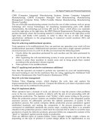
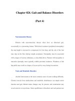
Fig. 1. Endocrine control of metamorphosis. Most of the data contributing to this scheme were derived from studies on silkworms
and the tobacco hornworm, Manduca sexta, although the scheme applies to all insects in a general sense. Note that in the case of
Manduca, JH acid rather than JH is released from the corpus allatum toward the end of the last larval stage.
Chapter 9 / Insect Hormones 129
Figure 1 is a generalized scheme for the Lepidop-
tera (moths and butterflies) and the details may not per-
tain to all insects. Specific neurosecretory cells (the
prothoracicotropes) synthesize PTTH as a prohormone
that is cleaved to the true PTTH as it is transported along
the axons to the corpora allata, where it is stored in axon
endings and ultimately released into the hemolymph.
Once released, PTTH acts on the prothoracic glands to
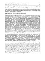
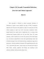
Fig. 2. Hormones and related molecules that play critical roles in control of molting and metamorphosis. (A) The structure of Bombyx
PTTH. The upper diagram indicates the predicted organization of the initial translation product. The lower diagram shows the location
of inter- and intracellular disulfide bonds. (B) Structure of cholesterol and some major ecdysteroids. (C) Structure of various JHs and
methyl farnesoate. JH I and JH II are almost entirely restricted to the Lepidoptera, JHB
3
to the cyclorraphan Diptera, whereas JH III
is ubiquitous in insects.
130 Part III / Insects / Plants / Comparative
stimulate ecdysteroid synthesis. In the Lepidoptera, this
stimulation results in the enhanced biosynthesis of
3-dehydroecdysone (3dE), which is converted into
ecdysone (E) by a hemolymph ketoreductase and from
that into 20-hydroxyecdysone (20E) in target cells,
20E being the principal molting hormone of insects.
Additionally, as Fig. 1 notes, the corpora allata synthe-
size and secrete JH, which is bound to a hemolymph-
binding protein (JHBP), transported to target tissues,
and acts in concert with 20E to determine the quality of
the molt. Although this process typifies the endocrine
control of molting in most insects, the exact molecular
mechanisms are conjectural, although great strides have
been made in recent years and are the subject of the
remainder of this chapter.
2. PTTH AND PROTHORACIC
GLAND ACTIVATION
2.1. Chemistry and Role
Almost all studies on PTTH action have been per-
formed on larvae and pupae of the tobacco hornworm,
Manduca sexta. This PTTH structure, as well as that of
four other lepidopteran PTTHs, has been elucidated by
direct sequencing or by deducing the structure after
having cloned the gene. The first of these was the PTTH
of the commercial silkworm, Bombyx mori. After more
than 30 yr of study using several million Bombyx brains,
Ishizaki and Suzuki (1992) purified and characterized
the Bombyx PTTH (Fig. 2) and showed that it is synthe-
sized as a prohormone of 224 amino acids and then
cleaved to form the mature neurohormone, a homodimer
(approx 26 kDa) containing inter- and intramonomer
disulfide binds, the latter requisite for hormone activity.
The Bombyx PTTH antibody reacts with putative
prothoracicotropes in a variety of insects, including
Manduca and Drosophila, as judged by immunocy-
tochemical and immunogold analyses, but it is physi-
ologically inactive in these species. Thus, there is likely
high specificity in the epitopes of the PTTH neuropep-
tide that are required for interaction with a putative cell
membrane receptor in the target glands (i.e., the protho-
racic glands).
Correlations have been reported between PTTH lev-
els in the hemolymph and the molting hormone titer for
both Manduca and Bombyx and, in both cases, reflect
subsequent increases in the ecdysteroid titer. In
Manduca, there are two PTTH peaks during the fifth
(final) larval stage as well as two ecdysteroid surges.
The first is responsible for a small increase in
ecdysteroid titer at about d 3.5 of the 9-d fifth instar
(stage) when the JH titer is at its nadir and also for a
change in commitment (reprogramming), so that when
challenged by a larger ecdysteroid surge 4 d later, tar-
get cells respond by synthesizing pupal rather than
larval structures. Thus, these two ecdysteriod (and
PTTH) peaks are primarily responsible for metamor-
phosis, and they must be elicited in a very precise
manner in the absence of JH. Indeed, the precision of
the molting process has contributed significantly to the
success enjoyed by insects on this planet during the
past half billion years.
The prothoracicotropes apparently receive, directly
or indirectly, information from the insect’s external
(photoperiod, temperature) and internal environment
(state of nutrition), and when the appropriate conditions
are met, they release PTTH from their termini in the
corpus allatum. How and where these influences are
sensed and then “transmitted” to the neurons that syn-
thesize PTTH is not known.
2.2. Action via Second-Messenger Systems
The only confirmed targets of PTTH are the paired
prothoracic glands, which have been well studied in
Manduca, each gland composed of about 220 mono-
typic cells surrounded by a basal lamina. Although no
candidate PTTH receptor(s) has yet been reported in the
prothoracic glands of any insect, the PTTH-prothoracic
gland axis has many similarities to vertebrate steroid
hormone–producing pathways, such as the adrenocorti-
cotropic hormone (ACTH)-adrenal gland system. By
analogy, it is probable that PTTH binds to a receptor that
spans the plasma membrane multiple times, contains an
extracellular ligand-binding domain, and has an intrac-
ellular domain that binds G protein heterotrimers.
PTTH stimulates increased ecdysteroid production
in the prothoracic glands via a cascade of events that
has yet to be elucidated completely (Fig. 3). Studies in
the 1960s revealed a correlation between circulating
ecdysteroid titers and adenylate cyclase activity in the
prothoracic gland, suggesting a role for cyclic adenos-
ine monophosphate (cAMP), and also that at some
developmental periods a cAMP-independent pathway
might be involved. In the Manduca prothoracic gland,
calcium is clearly pivotal in the response to PTTH.
Glands incubated in Ca
2+
-free medium with a calcium
chelator or a calcium channel blocker exhibit a greatly
attenuated production of cAMP and ecdysteroids in
response to PTTH. More recent studies have impli-
cated the mobilization of internal as well as external
Ca
2+
stores in the PTTH response and have demon-
strated a striking rise in the Ca
2+
levels of prothoracic
gland cells within a few seconds of PTTH administra-
tion in vitro.
Composite observations suggest that PTTH-depen-
dent cAMP production by prothoracic glands is gener-
ated by a Ca
2+
-calmodulin-sensitive adenylate cyclase.
Chapter 9 / Insect Hormones 131
The interaction between calmodulin and G protein (pre-
sumably G
sα
) is complicated and varies during the final
instar. In the first half of this period, calmodulin acti-
vates prothoracic gland adenylate cyclase and facilitates
G protein activation of adenylate cyclase. Subsequently,
prothoracic gland G protein activation of adenylate
cyclase is refractory to the presence of calmodulin in
such assays. Calcium still apparently plays a role in the
PTTH transductory cascade after the first half of the
fifth instar, since incubation of pupal glands in Ca
2+
-
free medium inhibits PTTH-stimulated ecdysteroido-
genesis, and higher levels of Ca
2+
-calmodulin can
still activate adenylate cyclase in prothoracic gland
membrane preparations. Regardless of the complicated,
developmentally dynamic relationships among calcium,
calmodulin, G proteins, and adenylate cyclase, it is clear
that PTTH elicits increased cAMP formation in protho-
racic glands leading to activation of a cAMP-dependent
protein kinase (protein kinase A [PKA]) and subsequent
protein phosphorylation.
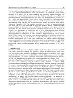
Fig. 3. A signal transductory cascade in the prothoracic glands of M. sexta is elicited by PTTH and results in enhanced synthesis and
secretion of ecdysteroid, namely, 3-dehydroecdysone. ER = Endoplasmic reticulum; IP
3
= inositol triphosphate; PLCβ = phospho-
lipase Cβ; PIP2 = phosphatidylinositol-4,5-bisphosphate; DAG = diacylglycerol; PKC = protein kinase C; ATP = adenosine triph-
osphate. (Graphics by R. Rybczynski reproduced with permission.)
132 Part III / Insects / Plants / Comparative
PTTH-stimulated PKA activity appears to be neces-
sary for PTTH-stimulated ecdysteroidogenesis, because
such ecdysteroid synthesis by prothoracic glands chal-
lenged with a PKA-inhibiting cAMP analog is sub-
stantially inhibited. Several PTTH-dependent protein
phosphorylations have been described for Manduca
prothoracic glands including a mitogen-activated pro-
tein kinase (MAPK), such as extracellular-regulated
kinase (ERK), as well as S6 kinase, the most striking
and consistent of these phosphoproteins being the
ribosomal protein S6, the phosphorylation of which
has been correlated with increased translation of spe-
cific mRNAs in several mammalian cell types. In
Manduca, rapamycin inhibits both PTTH-stimulated
S6 phosphorylation and ecdysteroidogenesis, suggest-
ing that S6 is an integral player in the PTTH
transductory cascade. Consistent with this view are the
observations that PTTH-stimulated S6 phosphoryla-
tion can be readily detected before the PTTH-stimu-
lated increase in ecdysteroid synthesis occurs and that
S6 is phosphorylated multiple times in a dose- and
time-dependent manner.
Over the last several years, a number of studies have
revealed that PTTH preparations or cAMP analogs
stimulate general protein synthesis in the Manduca pro-
thoracic gland via a branch of the transductory cascade
that is distinct from that leading to the activation of
ecdysteroidogenesis. PTTH may, therefore, modulate
or control the growth status of the prothoracic gland,
perhaps independently of its ability to elicit ecdyste-
roidogenesis, and could play a role in regulating the
levels of ecdysteroidogenic enzymes, analogous to pep-
tide regulation of enzymes responsible for vertebrate
steroid hormone synthesis. Additional factors, such as
JH, could determine whether PTTH stimulates or inhib-
its gland growth, ecdysteroid synthesis, or both.
Protein synthesis is required for ACTH stimulation
of steroidogenesis in the adrenal cortex as well as for
the Manduca prothoracic gland response to PTTH. It is
therefore likely that in both the adrenal cortex and
prothoracic glands, the phosphorylation state of ribo-
somal S6 is critical to the relationship between protein
synthesis and steroidogenesis. Presumably, the PKA-
promoted multiple phosphorylation of ribosomal S6
imparts information to the translational machinery to
synthesize specific proteins, which, in turn, regulate
some rate-limiting step in ecdysteroid biosynthesis.
An interesting outcome of this work is the close anal-
ogy observed between control of the insect and mamma-
lian steroidogenic systems. It is obviously a “successful”
system in an evolutionary sense, since insects appeared
on Earth several hundred million years before mam-
mals, and the ancestors of both groups diverged at least
100 million yr before that. Although it is interesting that
such divergent groups of animals use the same types of
molecules as hormones (peptides, steroids), it is extraor-
dinary that they regulate the synthesis of their steroid
hormones in an almost identical manner.
3. ECDYSTEROIDS
3.1. Structure-Activity Relationships
That ecdysteroids, particularly 20E, elicit the molt is
no longer in question and has been established as a cen-
tral dogma of the field. What may not be so obvious is
that in contrast to vertebrate systems, almost the entire
insect is the target of ecdysteroids, e.g., regulation of the
growth of motor neurons, control of choriogenesis,
stimulation of the growth and development of imaginal
disks, initiation of the breakdown of larval structures
during metamorphosis, and induction of the deposition
of cuticle by the epidermis.
Just recently microarray and computational analy-
ses demonstrated that the 20E regulatory network
reaches far beyond the molting process in Drosophila
melanogaster. The data are based on mutations of the
20E (EcR) receptor and indicate that in the metamor-
phosis of the midgut, genes that encode a variety of
factors are activated by this network and that genes
involved in cell cycling are also dependent on 20E for
their activation.
It is fitting that recent breakthroughs on the mecha-
nism of action of ecdysteroids (see Section 3.3.) were
accomplished using Drosophila, because it was a bio-
assay developed with another fly that was so well uti-
lized for the initial crystallization of E and then 20E
four decades ago. Since that time, a host of ecdysteroids
(Fig. 2B), their precursors, and their metabolites have
been identified. We know that the cis-A-B ring junction
is essential for molting hormone activity regardless of
whether a hydrogen atom or a hydroxyl group is the 5β
substituent, as is the 6-oxo-7-ene system in the B ring.
The 3β- and 14α-hydroxyl groups are required for high
activity in vivo, whereas the presence or absence of
hydroxyls at C-2, C-5, or C-11 does not appear to affect
biologic activity. The only essential feature of the side
chair appears to be the 22β
F
-hydroxyl.
Although E was the first of the ecdysteroids to be
crystallized and characterized and thought to be the
insect molting hormone 40 yr ago, it is actually con-
verted into the principal molting hormone, 20E, by
tissues peripheral to the prothoracic glands (Fig. 1), a
reaction mediated by an E 20-monooxygenase. In some
insects, particularly the Lepidoptera, as exemplified
by Manduca, the major if not sole ecdysteroid synthe-
sized and secreted by the prothoracic glands is 3dE
(Fig. 2B), which is converted into E by a ketoreductase
Chapter 9 / Insect Hormones 133
in the hemolymph, with the resulting E then hydroxy-
lated to 20E in target tissues.
3.2. Biosynthesis
In most organisms, every carbon atom in cholesterol
(Fig. 2B) is derived from either the methyl-or carboxylol-
carbon of acetate, but insects (and other arthropods) are
incapable of this synthesis owing to one or more meta-
bolic blocks between acetate and cholesterol. Thus, ste-
rols are required in the diet.
The first step in the conversion of cholesterol into E
via 3dE is the stereospecific removal of the 7β-hydro-
gen to form 7-dehydrocholesterol (7dC), a sterol rel-
egated to the prothoracic glands of Manduca and other
Lepidoptera. This cholesterol 7,8-desaturating activity
in the prothoracic glands of Manduca is cytochrome P-
450 dependent, perhaps via 7β-hydrocholesterol. When
[
3
H]7dC is incubated with prothoracic glands in vitro,
there is excellent conversion into both 3dE and E, with
the kinetics of conversion highly dependent on develop-
mental stage and experimental paradigm. The
desaturation to 7dC is probably not PTTH dependent,
but the neuropeptide (via S6) may initiate the modula-
tion of enzyme activity responsible for the transforma-
tion of 7dC to the next, yet unidentified sterol in the E
biosynthetic pathway.
There are a number of postulated intermediates
between 7dC and 3dE, such as 5α-sterol intermediates,
3-oxo-∆
4
intermediates, and ∆
7
-5α-6α-epoxide inter-
mediates, but their intermediacy remains conjectural.
By contrast, more is known about the terminal hydroxy-
lations necessary for the synthesis of the polyhydro-
xylated ecdysteroids. The enzymes responsible for
mediating the hydroxylations at C-2, C-22, and C-25
appear to be classic cytochrome P-450 enzymes, the
former two being mitochondrial and the latter micro-
somal. The sequence of hydroxylation is C-25, C-22,
and C-2.
Very recently, studies on a series of Drosophila
embryonic lethal mutations have allowed the cloning
and characterization of those genes encoding the P-450
enzymes responsible for the terminal hydroxylations
leading to the production of E and the monooxygenase
that mediates the conversion of E into 20E (Gilbert,
2004). In those studies, advantage was taken of the
availability of the fly database (Drosophila genome
project), and the fact that these so-called Halloween
genes (disembodied, shade, shadow, phantom) were
mapped in the 1980s to specific chromosome loci, and
had been shown to regulate embryonic processes that
may be attributed to low titers of molting hormone. By
identifying these genes in the fly database, sequencing
them, transfecting coding regions into a cell line, and
using these cell lines for more classic biochemical
analysis, all four genes that encode P-450 enzymes that
mediate the last four hydroxylations in 20E biosynthe-
sis have been identified and characterized (see the struc-
ture of cholesterol and 20E in Fig. 2B; hydroxylations
at C-2, C-20, C-22, and C-25).
Once formed, 3dE is converted into E through the
mediation of a hemolymph ketoreductase in the Lepi-
doptera, and the E is then transformed into 20E at
peripheral (target) tissues. In the case of flies such as
Drosophila, the prothoracic gland cells produce E rather
than 3dE, and the intermediary step mediated by the
ketoreductase is not needed in these insects. The evolu-
tionary significance of this difference in the product of
the prothoracic gland cells is not known. The complete
biosynthetic scheme has not been elucidated owing to
the difficulty of identifying the extremely short-lived
intermediates from minute quantities of tissues and the
less than handful of laboratories actively engaged in
such investigations; however, perhaps with an exten-
sion of the paradigms utilized for the a forementioned
Halloween genes, the details of the complete E biosyn-
thetic pathway may be known in the near future. With-
out the entire sequence of reactions in hand, it is not yet
possible to identify those rate-limiting reactions that
may be controlled by hormones (PTTH), neuromodu-
lators, or the nervous system.
3.3. Ecdysteroid Receptors
Several cell types in the higher flies and other insects
contain polytene (giant) chromosomes, whose struc-
ture and ease of examination led to the field of Droso-
phila cytogenetics. At specific developmental stages,
discrete regions of these chromosomes undergo puff-
ing, a phenomenon now known to be the morphologic
manifestation of gene activity, i.e., mRNA synthesis.
Forty-four years ago Clever and Karlson (1960)
showed that 20E could elicit a stage-specific puffing
pattern in the salivary gland chromosomes of the midge
Chironomus tentans, the first unequivocal demonstra-
tion that steroid hormones act at the level of the gene.
This discovery was followed by an exhaustive analysis
of salivary gland polytene chromosome puffing during
the development of Drosophila by Ashburner and col-
leagues, which involved the testing of E and 20E on the
puffing pattern. This led to the “Ashburner Model” of
ecdysteroid hormone action. In this model, an intrac-
ellular receptor-20E complex elicits elevated tran-
scription of “early puff” genes and, at the same time,
represses the transcription of the “late puff” genes.
Subsequently, the gene products of the “early puff”
genes act on the “late puff” genes to stimulate tran-
scriptional activity while feeding back on the “early
puff” genes, resulting in puff regression. This model
134 Part III / Insects / Plants / Comparative
has withstood the test of time, and several of the “early
puff” gene products have been shown to be transcrip-
tion factors and members of the steroid/thyroid hor-
mone receptor superfamily (nuclear receptor super-
family; see Chapters 2 and 4). Indeed, one gene product,
E75, was the probe utilized that led to the isolation of
the Drosophila ecdysone receptor gene (EcR) by the
Hogness Laboratory a few years ago.
The gene product of EcR binds to the proper response
elements and to radiolabeled ecdysteroid but requires a
heterodimeric partner to fulfill its function (Fig. 4). This
critical element is also a member of the nuclear receptor
superfamily, ultraspiracle (Usp), which is the Droso-
phila homolog of retinoid × receptor (RXR) which forms
heterodimers with a variety of mammalian hormones.
The Drosophila heterodimer is stabilized by endo-
genous 20E, and there are indications that the applica-
tion of exogenous hormone will increase the amount or
affinity of EcR in target cells, although it is not known
if this effect is at the level of transcription or translation.
It is of interest that EcR exists in at least three isoforms
that differ from one another in the transactivation
domain, and there is some tissue and developmental
specificity, although the exact reason for the existence
of isoforms remains conjectural. Their presence cer-
tainly suggests that there are as yet unidentified trans-
acting factors with roles in ecdysteroid action. Indirect
evidence also suggests that EcR is not monogamous
(i.e., can form heterodimeric relationships with gene
products other than Usp). Finally, there is a plethora of
data indicating that 20E is not the only ecdysteroid with
molting hormone activity, and that certain prohormones
and “metabolites” of 20E may be hormones in their own
right and perhaps interact with specific isoforms of EcR
in the EcR-USP complex. As in the field of steroid hor-
mone receptors in general, little is known about the
“docking” of the ecdysteroid-receptor complex with the
hormone response element and enhanced gene activity
in the form of specific mRNA synthesis (puffing).
4. JUVENILE HORMONES
4.1. Chemistry
The development of structures that distinguish adult
forms from larval forms is regulated by a complex inter-
action between JH and the ecdysteroids. The JHs are a
unique group of sesquiterpenoid compounds that have
Fig. 4. Activation of ecdysone receptor (EcR), mostly factual but some theoretical (e.g., hsp 90). (Graphics by R. Rybczynski
reproduced with permission.)
Chapter 9 / Insect Hormones 135
been identified definitively only in insects (Fig. 2C) and
one plant species, although their structural proximity to
retinoids is obvious. At least six JHs have been identi-
fied from various insect orders (Fig. 2C). JH III appears
to occur in all orders and is the principal product of the
corpus allatum in most, with the notable exceptions of
the Lepidoptera, in which JH I and JH II may have sig-
nificant roles, and the Diptera. In Drosophila, the
bisepoxide of JH III, JHB
3
, is predominant and the sole
JH in some species of flies.
The absolute configurations of the epoxide group of
only some of the JHs have been resolved (Fig. 2). There
are chiral centers at the 10 position of JH III and at the
10 and 11 positions of the other JHs. In addition, JHB
3
from Diptera possesses two chiral centers, at positions
6, 7, and 10. At present, the absolute configurations are
known only for JH I, 4-Me-JH I, JHB
3
, and JH III. This
is important because the unnatural enantiomers appear
to be less biologically active or are degraded at different
rates by esterolytic enzymes than are the natural enan-
tiomers.
JH acids are also produced by the corpora allata of
Manduca larvae. The glands lose their ability to methy-
late JH I and II acid during the final larval stage as a
result of the disappearance or inactivation of the methyl
transferase enzyme, and thus produce large quantities of
these JH acids, which are released into the hemolymph
(see Section 4.3., discussion of methoprene acid).
4.2. Biosynthesis and Degradation
The JHs are synthesized in the corpora allata from
acetate (JH III) and/or propionate (higher JH homologs).
The biosynthetic pathway for JH III is identical to that
for vertebrate sterol biosynthesis until the production of
farnesyl pyrophosphate. As noted previously, insects do
not produce cholesterol and related steroids de novo;
rather, JH is the product of this pathway. It is notewor-
thy that there is significant sequence similarity between
the HMGCoA reductase, the enzyme responsible for the
conversion of HMGCoA into mevalonate, of the insect
corpus allatum and that of vertebrate liver, a principal
site of de novo sterol biosynthesis, suggesting that this
pathway to farnesyl pyrophosphate is of ancient origin.
The formation of the side chains in the “modified”
homologs involves differential utilization of substrates,
including propionate and acetate, to give rise to both C-
5 and C-6 pyrophosphate intermediates. Condensation
of two C-6 units plus one C-5 unit results in the forma-
tion of JH I, whereas that of one C-6 unit plus two C-5
units produces JH II.
The hemolymph JH titer must reflect both the rate of
production and the rate of degradation. This estimate is
clouded by the presence of JH-specific-binding proteins
in the hemolymph, whose function has been hypo-
thesized to be the protection of JH from degradation by
both general and specific hemolymph esterases. JH-
specific epoxide hydrolases, capable of hydrating the
epoxide function to the diol, also play a role in the cata-
bolism of JH.
4.3. Postulated Action
The JH titer is believed to be the primary endocrine
factor influencing the “quality” of developmental events
during metamorphosis (e.g., in Lepidoptera, the nature
of the molt-larval-larval, larval-pupal, or pupal-adult)
(Fig. 1). It is generally assumed that the absence (or near
absence) of JH is required for metamorphosis in holom-
etabolous insects (Fig. 1). Therefore, JH defines the
outcome of molts, both metamorphic and nonmeta-
morphic, and can therefore be regarded as the metamor-
phic hormone of insects.
Although there are still no unequivocal data showing
the existence of a JH receptor, there is a multitude of
observations that JH can modulate larval and pupal gene
activity elicited by the molting hormone (i.e., does not
act in the absence of ecdysteroids). There is increasing
evidence that JH also acts at the level of the cell mem-
brane via a classic second-messenger system (see Chap-
ter 3), as it modulates the uptake of vitellogenin from
the hemolymph into the developing oocyte. Therefore,
JH may have multivalent roles and modes of action, as
does, e.g., progesterone. In preadult stages, JH has an
obvious role in preventing precocious development and
eliciting larval or pupal syntheses. The prevailing opin-
ion is that JH acts as a “competency determinant;” that
is, it affects the target cell’s competence to respond to
20E. The mechanism by which JH accomplishes this
task is unknown, but it is surely one of the most intri-
guing problems in endocrinology and developmental
biology. Further, very recent work has established that
a well-known JH analog, methoprene, as well as its acid
metabolite, can activate RXR in vertebrate cells, but
that only the metabolite can bind RXR, indicating that
methoprene must be metabolized before it is active in
this system. This suggests that perhaps in the case of JH,
it is a metabolite (JH acid?) that binds to the receptor,
whereas past failures in the search for a receptor utilized
the native JH.
5. CONCLUSION
In this abbreviated review, only the essence of the
field could be discussed, and there was no opportunity
to detail the >50 peptide hormones or hormone-like
peptides that have been described in recent years, sev-
eral of which appear to be identical to vertebrate hor-
mones (e.g., insulin) and others that deal with a variety
136 Part III / Insects / Plants / Comparative
of homeostatic mechanisms (e.g., hypo- and hypergly-
cemic, hypolipemic, adipokinetic). For the most part,
every vertebrate peptide hormone has an immunocyto-
chemically similar (or identical) counterpart in insects,
as have estrogens, progesterone, and so on. Therefore,
these hormones that play such strategic roles in the life
of higher organisms were “discovered” by insects or
ancestors of the insects. Thus, the hormones, second-
messenger systems, receptor mechanisms, neuroendo-
crine axis, biosynthetic mechanisms, and so forth are all
of very ancient lineage, and their basic essence has been
well preserved. With the current use of a genetic organ-
ism (Drosophila) to study endocrine paradigms, we
can look forward to future findings that should allow
insights into the myriad of endocrine mechanisms that
have survived severe evolutionary pressures.
ACKNOWLEDGMENTS
I thank Megan Edwards for clerical work, Susan
Whitfield for reproducing the figures, and Dr. Robert
Rybczynski for the graphics for Figs. 3 and 4.Research
from the Gilbert Laboratory was supported by grants
from the National Science Foundation and the National
Institutes of Health. The Halloween gene work is being
supported by NSF grant IBN 0130825.
REFERENCES
Clever U, Karlson P. Induktion von Puff Veränderungen in der
Speicheldrüsenchromosomen von Chironomus tentans durch
ecdson. Exp Cell Res 1960;20:623–626.
Gilbert LI, Iatrou K, Gill S, eds. Comprehensive Molecular Insect
Science, vol. 3. Amsterdam, The Netherlands: Elsevier, 2004.
Gilbert LI, Rybczynski R, Tobe S. Endocrine cascade in insect
metamorphosis. In: Gilbert LI, Tata JR, Atkinson BG, eds. Meta-
morphosis: Post-Embryonic Reprogramming of Gene Expres-
sion in Amphibian and Insect Cells. San Diego, CA: Academic,
1996:59–107.
Ishizaki H, Suzuki A. Brain secretory peptides of the silkmoth
Bombyx mori: prothoracicotropic hormone and bombyxin.
In: Joose J, Buijs RM, Tilders FJH, eds. Progress in Brain
Research, vol. 92, Amsterdam, The Netherlands: Elsevier,
1992:1–14.
Kopec´ S. Studies on the necessity of the brain for the inception of
insect metamorphosis. Biol Bull 1922;42:323–342.
Li T-R, White KP. Tissue-specific gene expression and ecdysone-
regulated genomic networks in Drosophila. Dev Cell 2003; 5:59–
71.
SELECTED READINGS
Gilbert LI. Halloween genes encode P450 enzymes that mediate
steroid hormone biosynthesis in Drosophila melanogaster. Mol
Cell Endocrinol. 2004;215:1–10.
Gilbert LI, Combest WL, Smith WA, Meller VH, Rountree DB.
Neuropeptides, second messengers and insect molting.
BioEssays 1988;8:153–157.
Gilbert LI, Rybczynski R, Warren JT. Control and biochemical
nature of the ecdysteroidogenic pathway. Ann.Rev. Entomology
2002;47,883–916.
Harmon MA, Boehm MF, Heyman RA, Mangelsdorf DJ. Activation
of mammalian retinoid x receptors by the insect growth regulator
methoprene. Proc Natl Acad Sci USA 1995;92:615–619.
Henrich V, Rybczynski R, Gilbert LI. Peptide hormones, steroid
hormones and puffs: Mechanisms and models in insect develop-
ment. In: Litwack G., ed. Vitamins and Hormones, vol. 55. San
Diego, CA: Academic, 1999:73–125.
Koelle MR, Talbot WS, Segraves WA, Bender MT, Cherbas P,
Hogness DS. The Drosophila EcR gene encodes an ecdysone
receptor, a new member of the steroid receptor superfamily. Cell
1991;67:59–77.
Riddiford LM. Cellular and molecular actions of juvenile hormone.
I. General considerations and prematamorphic actions. Adv In-
sect Physiol 1994; 24:213–274.
Song Q, Gilbert LI. Multiple phosphorylation of ribosomal protein
S6 and specific protein synthesis are required for prothor-
acicotropic hormone-stimulated ecdysteroid biosynthesis in the
prothoracic glands of Manduca sexta. Insect Biochem Mol Biol
1995;25:591–602
Chapter 10 / Signal Transduction Pathways in Plants 137
137
From: Endocrinology: Basic and Clinical Principles, Second Edition
(S. Melmed and P. M. Conn, eds.) © Humana Press Inc., Totowa, NJ
10
Phytohormones and Signal
Transduction Pathways in Plants
William Teale, PhD, Ivan Paponov, PhD,
Olaf Tietz, PhD, and Klaus Palme, PhD
CONTENTS
INTRODUCTION
SIGNAL TRANSDUCTION PATHWAYS OF PLANTS
ROLE OF PHYTOHORMONES
AUXINS
AUXIN PERCEPTION
EFFECT OF AUXIN SIGNALING ON GENE EXPRESSION
GIBBERELLINS
CYTOKININS
BRASSINOSTEROIDS
ABSCISIC ACID
ETHYLENE
PERSPECTIVE
plants to increase their reproductive potential and are
consequences of the idiosyncrasies of a plant’s cellular
signaling mechanisms.
2. SIGNAL TRANSDUCTION
PATHWAYS OF PLANTS
The emergence of complete genome sequences from
strategic eukaryotic models has allowed the compara-
tive analysis of plant and animal signal transduction
pathways. In both cases, this analysis has offered insight
into the features of specific signaling pathways that were
not achievable at the time the previous edition of this
book was published. It is now hoped that by looking
closely at the emerging differences between analogous
signaling pathways in plants and animals, it will be
possible to identify their relationship to the divergence
of the two lineages. Excellent recent reviews on this
INTRODUCTION
Since the divergence of plants and animals about
1.5 billion yr ago, the signal transduction pathways in
both kingdoms have been subjected to very different
selection pressures. These fundamental differences
have influenced the evolution of both the signaling
molecules themselves and the mechanisms by which
signals are relayed. Among these differences, a plant’s
ability to continuously form new organs during its
postembryonic development, the increased frequency
of high degrees of both ploidy and gene duplication in
many higher plants, and the multicellular haploid
gametophytes of more primitive plants could be par-
ticularly significant. Particular developmental pro-
cesses, such as totipotency (the ability of a plant to
regenerate itself from vegetative tissue), have enabled
138 Part III / Insects / Plants / Comparative
topic have been published over the past 5 yr (Cock et al.,
2002; Wendehenne et al., 2001). Here we give a brief
overview of selected examples in order to illustrate some
interesting features of plant signaling pathways and then
discuss these pathways in the context of novel develop-
ments in plant evolution.
As a result of photoautotrophism, the evolution of
plants has been constrained by the absence of mobility
and the presence of relatively rigid cell walls. The cap-
ture and integration of chloroplasts from bacterial
progenitors profoundly influenced the signaling mecha-
nisms of modern plants. Not surprisingly, for sessile
photosynthetic organisms able to sense carefully their
fluctuating environment, the developmental pathways
of plants are irrevocably and necessarily linked to the
perception of external cues. Temperature, light, touch,
water, and gravity can all activate endogenous develop-
mental programs. Of these, light has an especially
important role, not only as the energy source for photo-
synthesis, but also as a stimulus for many developmen-
tal processes throughout the life cycle of plants, from
seed germination to flowering. Consequently, plants
have the richest array of light-sensing mechanisms of
any group of organisms. These photoreceptors are able
to measure not only the intensity but also the quality of
light available to the plant. Phytochromes, e.g., are the
photoreceptors for red and far-red light responses (Nagy
and Schäfer, 2002). They are red-light-activated serine/
threonine kinases that exist in two photointerconvertible
forms. On stimulation with red light, they move from
the cytosol to the nucleus, where they interact with pro-
teins such as the helix-loop-helix transcription factor
phytochrome-interacting factor (PIF3; Martinez-Garcia
et al., 2000). These proteins then bind to light-respon-
sive promoter elements leading to transcription, thereby
achieving light-regulated gene activation (Tyagi and
Gaur, 2003). Thus, phytochrome signaling involves
both nuclear and cytosolic interactions.
Comparative genomic analysis of plant genomes
from species such as Arabidopsis thaliana (thale cress)
and Oryza sativa (rice) has revealed many signaling
compounds that are highly conserved between animals
and plants. The reiteration of core signaling mecha-
nisms in plants and animals suggests that overall differ-
ences between the two kingdoms evolved via the
modification of basic ancestral pathways. However,
this basic similarity is found in combination with many
novel elements or motifs. Overall organizational prin-
ciples are shared among plants and animals, indicating
that a core of conserved signaling genes and pathways
is used repeatedly in many different developmental
contexts. RAS genes are a good example to illustrate
this argument. RAS genes belong to the small guanosine
5´-triphosphatase protein family. They are master regu-
lators of numerous cellular processes including signal-
ing, cargo transport, and nuclear transport. They are
regarded as molecular switches that alternate between
an active and an inactive state, thereby ensuring the
flow of information at the expense of guanosine 5´-
triphosphate. This molecular switch appears to have
been developed early, and throughout evolution, it has
been adapted to a variety of tasks. Small G proteins are
classified in five families: the RAS (according to the
oncogene Ras from
rat sarcoma virus), the RAB (Ras
of brain), the ARF (ADP ribosylation factor), the
RAN (
Ras-related nuclear protein), and the RHO (Ras
homologous) family. They interact with partner pro-
teins (effectors) to form dynamic complexes regulating
a plethora of crucial cellular processes. In plants, how-
ever, no RAS genes, but only members of the RAB, ARF,
and the RAN families have been found. An additional
plant-specific family of small G proteins is named ROP,
for
RHO of plants (Vernoud et al., 2003). Apparently,
only members of those families that play intricate roles
in metabolite transport and cell polarity control have
been conserved in plants. It is conceivable that the
sessile nature of plants demands tight control over
secretory pathways to enable and precisely adjust the
cell elongation processes. In this case, homeostatic
control of cellular membrane compartments, transport
of macromolecules between intracellular compart-
ments and the extracellular space, and nuclear transport
would have added importance for the evolutionary suc-
cess of plants.
Despite conservation of the basic secretory machin-
ery between plants and other eukaryotes, several recent
findings suggest distinct structural and functional dif-
ferences in plants. It is therefore expected that the sys-
tematic functional analysis of key players of plant
secretion will uncover novel insights into the processes
by which the formation of transport vesicles and intra-
cellular trafficking by internal and external cues are
controlled, and by which vesicles are delivered to target
membranes.
Ultimately, from analysis of these processes, research-
ers will learn important lessons on how plant cells
control apical and basal cell polarity. Moreover, such
approaches will not only uncover important aspects of
the organizational blueprint of the plant secretory path-
way, but also reveal fundamental functional differences
between plants and other eukaryotes and indicate how
these differences relate specifically to the relationship
between form and function in plants. Analysis of the
plant cargo delivery system provides privileged views
not just into unique aspects of secretion control, but also
into many other plant-specific processes, such as hor-
Chapter 10 / Signal Transduction Pathways in Plants 139
monal control of growth, gravitropic and phototropic
responses, establishment and maintenance of cell polar-
ity, cell differentiation, mediation of disease resistance,
and fruit ripening. In the long term, insight into these
fundamental processes will be important for many bio-
technological applications.
3. ROLE OF PHYTOHORMONES
Auxin, cytokinin, abscisic acid (ABA), gibberellin
(GA), and ethylene are the five classic hormone path-
ways that appear very early in plant evolution and have
been adapted to functional uses in many contexts of
plant development (Fig. 1). Brassinosteroids are a rela-
tively recent addition to this list, but must also be con-
sidered as potent plant growth regulators. These
phytohormones are secondary metabolites that play
physiological roles at specific stages of a plant’s life-
cycle. They are typically considered in terms of three
sequential events: their biosynthesis, their perception,
and the signals that are subsequently initiated as a con-
sequence. The effects of a phytohormone are commonly
demonstrated either by their exogenous application to
a growing plant, or by the inhibition or exaggeration of
their influence in mutant plants. Such plants may be
affected in the rate of biosynthesis of a particular hor-
mone, in the sensing of a hormone’s presence, or in the
subsequent transduction of a downstream signaling cas-
cade. Phytohormones represent integral components of
the mechanisms by which a plant regulates both its own
development and its response to the wide variety of
stimuli it receives from its environment. Since Charles
and Francis Darwin first attributed the bending of a
grass coleoptile toward light to the action of a growth
mediator, research into the biosynthesis and mode of
action of phytohormones has developed into one of the
most widely studied aspects of plant biology (Davies,
1995).
We now give an overview of the current understand-
ing of both how higher (seed-bearing) plants perceive
phytohormones and how this perception is translated
into a physiological response. Plants, owing to their
sessile nature, cannot move autonomously in response
to environmental stimuli in the same way as many ani-
mals can. As already inferred, this restraint has been
overcome, at least in part, by the extension of the role
of hormones from that of regulator (either metabolic or
developmental) into the means by which a response to
environmental stimuli are elicited. For example,
Darwin’s first experiments on coleoptile bending rep-
resent the attempt of a young grass shoot to increase its
photosynthetic capacity. It was subsequently demon-
strated that the response is mediated by production of
indole-acetic acid (IAA) (a member of the auxin class
of phytohormones) in the shoot tip, followed by asym-
metrical redistribution throughout the growing plant.
Cells respond to the concentration of IAA by elongat-
ing in a dose-responsive manner, producing a physi-
ologic response.
Fig. 1. Phytohormones: chemical structure and properties.
140 Part III / Insects / Plants / Comparative
4. AUXINS
Auxins are vital mediators of developmental and
physiological responses in plants and a paradigm for plant
growth regulators. They regulate apical-basal polarity in
embryonic development; apical dominance in shoots;
induction of lateral and adventitious roots; vascular tissue
differentiation; and cell growth in both stems and coleop-
tiles, including the asymmetric growth associated with
phototropic and gravitropic responses (Davies, 1995).
Concentration, perception, and the effect that signal-
ing has on gene expression are central issues when con-
sidering the phytohormone signaling pathways that
affect growth and regulation. In relation to auxin, it has
been suggested that efflux-mediated gradients are the
underlying driving force for the formation of all plant
organs, regardless of their developmental origin and
fate. An attractive theory is therefore that the relative
concentration of auxin is particularly important in plant
development. Both the concentration of auxin in any
one cell and the steepness of the auxin concentration
gradient over a group of cells are determined by the rate
of auxin synthesis in source cells, the rate of its transport
through a tissue, and the overall rate of its degradation
or conjugation (the majority of auxin present in any one
cell exists as biologically inactive conjugate).
Auxin is transported from the shoot downward. How-
ever, the prevailing model of the initiation of auxin gra-
dients in the apical meristem has been questioned by the
demonstration that all parts of young plants can synthe-
size IAA, thus potentially diminishing the importance
of polar auxin transport (Ljung et al., 2001).
In the 1920s, Cholodny and Went independently sug-
gested the chemiosmotic hypothesis of auxin transport,
which was later refined by Rubery and Sheldrake (1974)
and Raven (1975). The theory predicts the existence of
an auxin efflux carrier that actively and asymmetrically
redistributes auxin in root and stem tissue on gravitropic
or phototropic stimulation.
Auxin movement both into and out of cells requires
specialized carriers (Friml and Palme, 2002). Several
Arabidopsis genes encoding putative auxin carriers
have been identified during the past decade. The amino
acid permease-like gene AUX1 and the family of bacte-
rial transporter-like PIN genes encode putative auxin
influx (Bennett et al., 1996) and efflux carriers, respec-
tively. Characterization of the first putative auxin efflux
carrier PIN1 (Gälweiler et al., 1998) gave context to
auxin’s asymmetric localization. PIN1 encodes a 622-
amino-acid protein with 12 predicted transmembrane-
spanning segments (Fig. 2). It shares similarity with a
group of transporters from bacteria of the major facili-
tator class, evidence supporting a transport function
(Gälweiler et al., 1998; Pao et al., 1998). A search of the
Arabidopsis genome for genes with homology to PIN1
revealed another seven genes belonging to the same
family. Similar sequences have been found in all other
plants now sequenced, but not in animals, indicating
that PIN proteins have evolved exclusively in plants.
Based on genetic evidence, PIN proteins are strong
candidates for either the auxin efflux carrier itself or an
important regulatory component of the efflux machin-
ery (Palme and Gälweiler, 1999). More important, the
distributions of PIN1 and other PIN proteins in the
plasma membrane of auxin-transporting cells of stems
and roots were found to be dynamic and asymmetric
according to the direction of auxin flux (Friml et al.,
2002b; Gälweiler et al., 1998; Geldner et al., 2001;
Müller et al., 1998; Steinmann et al., 1999) (Fig. 3).
Auxin gradients in plant tissue appear to be sink driven;
gradient formation seems to be regulated by auxin trans-
port (rather than degradation) machinery. For example,
the formation of a maximum auxin concentration at the
Arabidopsis root apex depends on the activity of PIN4
(Friml et al., 2002a).
It is likely that the activity of the efflux complex is
regulated by phosphorylation (Delbarre et al., 1998).
Auxin efflux was found to be more sensitive to the spe-
cific transport inhibitor N-1-naphthylphthalamic acid
(NPA) in seedlings of an Arabidopsis mutant named
rcn1 (“root curl in NPA”) than in the wild type. The
RCN1 gene encodes a subunit of protein phosphatase
2A (Garbers et al., 1996). Furthermore, the mutant can
be phenocopied with a phosphatase inhibitor (Deruere
et al., 1999). The protein kinase PINOID enhances polar
auxin transport (Benjamins et al., 2001) and is another
potential component of the hypothetical auxin-efflux
complex.
5. AUXIN PERCEPTION
According to the widely accepted theory, phytohor-
mone signaling begins with the perception of free hor-
mone by a specific receptor. In the case of auxin, there
is evidence for multiple sites of auxin perception. It
therefore appears that, at least initially, the auxin signal
can transduce through more than one signaling path-
way.
To date, the best-characterized auxin-binding pro-
tein is ABP1 (Napier et al., 2002), which was origi-
nally identified, purified, and cloned from maize
(Hesse et al., 1989; Löbler and Klämbt, 1985). The
high binding constant of auxin and ABP1 has inspired
much research, however, the protein has no homology
to any other known receptor family, and it is ubiqui-
tous in vascular plants, including the pteridophytes and
bryophytes (Napier et al., 2002). A KDEL retention
motif at the C-terminus of ABP1 ensures an ER loca-
Chapter 10 / Signal Transduction Pathways in Plants 141
tion (Henderson et al., 1997; Tian et al., 1995); how-
ever, some ABP1 does pass along the constitutive
secretion pathway to the plasma membrane and cell
surface (Diekmann et al., 1995; Henderson et al.,
1997). The ER location makes the characterization of
ABP1 more complex because most of the physiologi-
cal data demonstrate activity of ABP1 on the plasma
membrane. Here, auxin is able to control several cel-
lular responses, including tobacco mesophyll proto-
plast hyperpolarization (Leblanc et al., 1999a, 1999b),
tobacco mesophyll protoplast division (Fellner et al.,
1996), expansion of tobacco and maize cells in culture
(Jones et al., 1998), and maize protoplast swelling
(Steffens et al., 2001). These effects can be inhibited
by the application of anti-ABP1 antibodies, which are
unable to enter the cell. It has therefore been concluded
that ABP1 is able to elicit a physiological response in
the presence of auxin at the surface of the plasma mem-
brane. A functional role of ABP1 inside the ER has not
been shown; these data may be reconsidered, however,
because auxin efflux carriers are now known to cycle
continuously in membrane vesicles between the
plasma membrane and the endosome (Geldner et al.,
2001). There is considerable speculation about the pos-
sible role of auxin transporters in auxin signaling. It is
possible that measurement of the flux of auxin through
either influx or efflux carriers (or both) monitors auxin
level in the cell. It is also possible that specific trans-
porter family members no longer act as transporters
but have evolved a receptor function (Friml and Palme,
2002). Sugar sensing is important for plants and yeast
to report the carbohydrate status within cells and out-
side of cells. It has been demonstrated that some pro-
teins that show transporter topology do not transport
sugars but sense sugar outside of cells and control tran-
scription of sugar transporters that control the sugar
homeostasis in cells (Lalonde et al., 1999). A similar
mechanism for auxin perception is conceivable.
Fig. 2. Predicted AtPIN1 protein structure.
Fig. 3. AtPIN1 (inner arrows) and AtPIN2 (outer arrows) in
Arabidopsis root tip. Arrows indicate the direction of Auxin
fluxes in marked cell files.
6. EFFECT OF AUXIN
SIGNALING ON GENE EXPRESSION
Auxin-dependent transcriptional activation can occur
within minutes of a signal being perceived (Abel and
Theologis, 1996). In the nucleus, the regulation of gene
expression by auxin can be mediated by the action of two
families of auxin-induced proteins: the Aux/IAA pro-
teins and the auxin response factors (ARFs) (Hagen and
Guilfoyle, 2002). ARFs bind to auxin response promoter
elements upstream of genes and activate or repress their
transcription. Aux/IAA proteins can dimerize with ARF
proteins, thus inhibiting their activity (Tiwari et al.,
142 Part III / Insects / Plants / Comparative
2003). However, they have very short half-lives, ranging
from a few minutes to a few hours (Abel et al., 1994;
Gray et al., 2001). A normal auxin response is dependent
on this rapid turnover of Aux/IAA proteins, as it lowers
the concentration of the inhibitory ARF-Aux/IAA dimer
(Ulmasov et al., 1997; Worley et al., 2000).
Aux/IAA proteins are found in all higher plants and
are characterized by four highly conserved domains
(Abel and Theologis, 1996; Guilfoyle et al., 1998). In
yeast two-hybrid assays, their dimerization with ARF
proteins has been shown to involve two of these domains
(which are similar to those of the ARF proteins)
(Ulmasov et al., 1997). Another domain, domain II, is
crucial for Aux/IAA function, with many lines of evi-
dence demonstrating that it is the target for Aux/IAA
protein destabilization, ensuring the rapid turnover
required for a normal auxin response. The fusion of Aux/
IAA proteins to reporter proteins, such as luciferase or
β-glucuronase (GUS), results in the destabilization of
the reporter protein (Gray et al. 2001;Worley et al., 2000).
This indicates that Aux/IAA proteins contain a transfer-
able destabilization sequence. A nonfunctional domain
II, as found in the auxin-resistant mutants axr3-1, axr2-
1, and shy2, dramatically increases the protein’s half-life
and prevents the ARF proteins from functioning (Gray
et al., 2001; Ouellet et al., 2001; Worley et al., 2000). The
stabilization of an Aux/IAA-reporter fusion by inhibi-
tors of the 26S proteasome indicates that auxin signaling
requires SCF
TIR1
-mediated turnover of Aux/IAA pro-
teins.
7. GIBBERELLINS
GAs are a large group of diterpenes comprising well
over a hundred members. However, only a handful can
elicit a physiological response, the others being repre-
sentative of a large and complicated web of biosynthetic
pathways from ent-kaurene, the product of the first dedi-
cated step of GA biosynthesis. These biosynthetic path-
ways are now well understood. GAs regulate a wide
range of physiological processes, including cell divi-
sion and cell elongation, and are crucial to the control of
processes as diverse as germination, stem elongation,
flowering, fruit ripening, and senescence.
The last 5 yr have seen a dramatic increase in our
understanding of the processes involved in GA signal
transduction (Gomi and Matsuoka, 2003), however,
the exact mechanisms by which a plant’s response to
GA is brought about remain unclear. A class of tran-
scription factors, the DELLA proteins, has emerged as
a central mediator of many GA responses, although
they probably do not bind DNA directly (Dill et al.,
2001). It was decided that these proteins (named after
a five-amino-acid N-terminal motif) are important
components of the GA signal transduction pathway
after the analysis of a number of dwarfed mutants from
a range of species (Sun, 2000). An important example
(and the mutant from which the first member of the
family was cloned) is the gai1-1 mutant of Arabidopsis.
Plants with a dominant mutation at the GAI allele dis-
play a semidwarf phenotype, insensitive to the exogen-
ous application of GA (Peng and Harberd, 1993). All
results indicate that the DELLA proteins are negative
regulators of GA signaling.
Altogether there are five DELLA proteins in
Arabidopsis (GAI, RGA, RGL1, RGL2, and RGL3),
but only one in rice (SLR1) and barley (SLN1). They
share a high degree of sequence homology and belong
to a wider group of plant transcription factors called the
GRAS family. All of these proteins share the same basic
structure, with an N-terminal GA-signal-perception
domain, a serine/threonine-rich domain, a leucine zip-
per, and a C-terminal regulatory domain conserved
among GRAS proteins. This structure has been used to
suggest a model for the mode of action of SLR1 where
the protein exists as a dimer, the subunits linked by
the leucine zipper (Itoh et al., 2002). The active form
(receiving no GA signal) represses the GA response via
the C-terminus, which is deactivated when a GA signal
is bound by the DELLA domain.
As is the case for auxin, the GA receptor is still
unknown. However, there is evidence that the trans-
duction of the GA signal from the plasma membrane
to the nucleus involves G proteins (Ueguchi-Tanaka
et al., 2000). A range of secondary messengers have
also been shown to be involved in this process, but the
exact role of many remains unclear (Sun, 2000). As in
animals, it has recently emerged that the post-transla-
tional modification of proteins by the addition of O-
linked N-acetylglucosamine could be involved in
signaling processes (Thornton et al., 1999). Analysis
of two mutants of Arabidopsis, partially rescued by the
application of exogenous GA, has revealed two puta-
tive O-GlcNAc transferases (by homology with known
enzymatic sequences) thought to be involved in the
GA signaling pathway (Hartweck et al., 2002). In ani-
mals, the transferase ability has been shown to com-
pete with phosphorylation of serine and threonine
residues, suggesting a possible mechanism for their
mode of action in plants, and a link to DELLA protein
function.
8. CYTOKININS
Cytokinins play a major role in many different devel-
opmental and physiological processes in plants, such
as cell division, regulation of root and shoot growth
and branching, chloroplast development, leaf senes-
Chapter 10 / Signal Transduction Pathways in Plants 143
cence, nutrient mobilization, biomass distribution,
stress response, and pathogen resistance. In contrast to
our understanding of auxin and GA signal transduc-
tion, proteins have been identified that function as
cytokinin receptors. Activation tagging experiments in
Arabidopsis identified CKI1, a gene encoding a recep-
tor histidine kinase, whose overexpression was seen to
induce typical cytokinin responses (Kakimoto, 1996).
Although CKI1 is able to activate the cytokinin signal-
ing pathway, it does not bind cytokinins directly
(Hwang and Sheen, 2001). Nevertheless, the discov-
ery of CKI1 suggested that the cytokinin transduction
pathway in higher plants could be similar to the pro-
karyotic two-component system. This hypothesis was
proved by the identification of CRE1/AHK4, another
histidine kinase, as the first cytokinin receptor (Inoue
et al., 2001; Suzuki et al., 2001; Ueguchi et al., 2001).
The availability of the Arabidopsis genomic sequence
led to the identification of two further cytokinin recep-
tors (AHK2 and AHK3).
After cytokinin perception, the resulting signal is
transmitted by a multistep phosphorelay system through
a complex form of the two-component signaling path-
ways that has been well characterized in prokaryotes
and lower eukaryotes. Functional evidence for cytoki-
nin sensing by the receptor CRE1/AHK4 was obtained
in elegant complementation experiments in yeast and
Escherichia coli, which rendered these heterologous
hosts cytokinin sensitive. Two other histidine kinases,
AHK2 and AHK3, have been shown to be active in the
same complementation test system and to give proto-
plasts cytokinin sensitivity (Hwang and Sheen, 2001;
Yamada et al., 2001), indicating that these two proteins
are also cytokinin receptors. Each receptor comprises an
N-terminal extracellular domain, a membrane anchor,
and a C-terminal transmitter domain, capable of auto-
phosphorylation. The three cytokinin receptor genes
differ in their expression pattern. CRE1/AHK4 is mainly
expressed in the roots, whereas AHK2 and AHK3 are
present in all major organs (Inoue et al., 2001; Ueguchi
et al., 2001). This tissue-specific expression of cytoki-
nin receptors could be an additional layer of control to
the perception of cytokinin.
Considering the large number of response-regulator
genes associated with the two-component signaling
sytem (22 in Arabidopsis), it has been suggested that
they could both have different functions, using differ-
ent targets in addition to participating in cross talk with
other hormones. There are accumulating data demon-
strating that in order to understand the growth responses
to cytokinins, it is important to understand such cross
talk between cytokinins and nutrients as well as cyto-
kinins and other phytohormones. Moore et al. (2003)
showed that application of cytokinins as well as the use
of transgenic Arabidopsis lines with constitutive
cytokinin signaling could overcome the glucose repres-
sion response. The insensitivity of Arabidopsis glu-
cose insensitive2 (gin2) to auxin and hypersensitivity
to cytokinin could be the clue to understanding the
antagonistic interaction between cytokinins and auxin
and its dependency on the glucose status of tissues.
9. BRASSINOSTEROIDS
Steroids are important signaling molecules in plants
as well as in animals. Since the discovery of brassi-
nolide in 1979, brassinosteroids (BRs) have been shown
to be important at a number of stages of a plant’s devel-
opment, including stem elongation, germination and
senescence. BRs retain the basic four-ring structure of
many steroid hormones; like animal steroid hormones,
they are synthesized from cholesterol. Two mutants
deficient in the biosynthesis of BRs, DET2 and CPD,
develop as if grown under light when grown in the dark
(Chory et al., 1991; Li and Chory, 1997). This demon-
strates that, in addition to these other crucial processes,
BRs play an important role in photomorphogenesis.
The identification of a BR receptor has been a recent
significant advance in phytohormone biology; this
section focuses on the mechanism by which BRs are
initially sensed by the cell, before highlighting some
interesting similarities between the perception and
mode of action of BRs and the regulation of develop-
ment in animals. In animals, steroids receptors are
nuclear located (Marcinkowska and Wiedlocha, 2002);
in plants, no nuclear steroid receptors have been found,
indicating that plant cells have evolved a different
method to receive the BR signal.
The bri mutant of Arabidopsis shows a dwarfed phe-
notype similar to that of mutants deficient in the bio-
synthesis of BRs. BRI1 was thought to be involved in
signaling owing to the mutant plants’ unresponsive-
ness to applied brassinolide. Cloning of the bri1 gene
revealed
a leucine-rich repeat (LRR) receptor-like
kinase, an
immediate candidate for the BR receptor (Li
and Chory,
1997). BRI1 was subsequently shown to be
located at the plasma membrane (Friedrichsen et al.,
2000), and to have a relatively high affinity for bio-
active BR (Wang et al., 2001). LRR receptor kinases
contain three domains: an extracellular domain com-
prising several leucine-rich repeating sections (in the
case of BRI1, 25), a transmembrane section and a
cytoplasmic kinase domain. BRI1 also contains a 70-
amino-acid island in the LRR domain, necessary for
the protein’s function (Li and Chory, 1997). The use of
chimeric proteins has demonstrated that the BRI1
extracellular domain was both necessary and sufficient
144 Part III / Insects / Plants / Comparative
for the translation of a specific set of genes. When
fused to the intracellular kinase domain of a similar
receptor-like kinase, a protein involved triggering a
plant’s defensive response to pathogens, activation of
the extracellular BRI1 domain and, hence, defense-
related gene expression could be induced by BR (He et
al., 2000). The LRR-receptor kinases are members of
a very large class of proteins in Arabidopsis compris-
ing 174 members with diverse function. Only a small
number have been ascribed a function, including
CLAVATA (involved in meristem development) and
ERECTA (involved in organogenesis) (Dievart and
Clark, 2003). The functions of LRR-receptor kinases
are diverse. In Arabidopsis, three BRI-like proteins
share high homology and are similar to protein se-
quences found in monocotyledonous species. Of these,
BRL1 and BRL3 are able to rescue the bri1 mutation,
suggesting a closely related function.
The cloning of a homolog of BRI1 in tomato revealed
an intriguing overlap of function of the tBRI1 (BRI of
tomato) protein. Systemin is a peptide signal impor-
tant in pathogen-defense responses in plants, acting by
amplifying the induction of the jasmonate signaling
pathway. It was discovered that the same receptor
(called SR160 in the context of systemin) was respon-
sible for both BR and systemin signaling (Montoya
et al., 2002; Szekeres, 2003). This dual receptor func-
tion has also been observed in animals, with the hor-
mone progesterone able to inhibit specifically the
peptide oxytocin from binding to its receptor, a uterine
G protein–coupled receptor (Grazzini et al., 1998). It
has been suggested that BR could bind to its receptor
while simultaneously bound to a specific protein,
owing to sequence homology to animal steroid-bind-
ing proteins being found in the Arabidopsis genome
(Li and Chory, 1997), and the fact that, in general,
LRRs mediate protein-protein interactions rather than
smaller ligand binding.
10. ABSCISIC ACID
Plants control water balance with a range of strate-
gies. For most land plants, the anatomy of leaves is
centered on a balance between minimizing water loss
and maximizing both exposure to the sun and the rate
of diffusion of molecular oxygen away from and car-
bon dioxide into photosynthetic cells. This balance is
essential for maintaining the flux of electrons that pass
through the light-dependent reactions of photosynthe-
sis. The leaf is an organ that is necessarily exposed to
relatively high levels of sunlight and, therefore, of
water loss through evaporation from pores (stomata).
ABA is the phytohormone which regulates the open-
ing and closing of stomata. It does this by controlling
the turgor pressure inside the two surrounding banana-
shaped guard cells. Much of the work on ABA signal-
ing has been focused on guard cells and mutants
affecting their function. However, ABA influences
both physiological (gene expression in response to salt
stress and drought) and developmental (e.g., germina-
tion and seedling development) processes. ABA seems
to affect many different signaling pathways, some-
times with a high degree of redundancy; the extent to
which it mediates cross talk between environmental
and developmental stimuli is currently the subject of
concentrated research.
The amount of free ABA able to elicit a response is
thought to be dependent on many factors. These include
movement of ABA through the plant, the relative rates
of ABA synthesis and catabolism, and the concentration
of ABA in the leaf symplast.
It has become clear that the ABA signal is transduced
through a number of secondary messengers, among
them lipid-derived signals, H
2
O
2
, G proteins, and nitric
oxide (Himmelbach et al., 2003). The varying cellular
concentrations of these compounds unite to influence
indirectly the cytosolic concentration of Ca
2+
, the cen-
tral factor in many ABA signals (McAinsh et al., 1997).
It is thought that ABA has two modes of action: the first
“nongenomic” effect is able to change the turgor pres-
sure in guard cells by altering the plasma membrane’s
permeability to ions, and the second acts via changing
the transcription levels of ABA-responsive genes. It
is thought that both processes are reliant on alterations
in the intracellular concentration of Ca
2+
; however, it is
not yet fully understood to what extent the pathways are
separated.
Despite a long history of research, the nature of the
initial ABA receptor remains elusive. However, it is
widely believed that the initial event in the signaling
cascade is the binding of ABA to either a membrane-
bound or a cytosolic receptor. In many cases, this bind-
ing results in the activation of Ca
2+
-influx channels
resulting in the ABA-mediated increase in intracellu-
lar Ca
2+
concentration (Murata et al., 2001). Another
important factor in this process is the altering perme-
ability of the tonoplast to Ca
2+
; this is influenced by
intracellular lipid-derived signals and cyclic adeno-
sine 5´-diphosphate–ribose concentration, the latter
dependent on the Ca
2+
concentration itself (Wu et al.,
2003). The overall increase in intracellular Ca
2+
results
first in the inhibition of K
+
-influx channels and, sec-
ond, in the activation of K
+
-efflux channels and the
inhibition of H
+
-adenosine triphosphatase. Therefore
it can be seen that ABA initiates a complicated
mesh of interconnecting signals, resulting in a physi-
ologic response (Finkelstein et al., 2002).
Chapter 10 / Signal Transduction Pathways in Plants 145
11. ETHYLENE
It has long been known that exposure to ethylene
elicits a well-characterized response in seedlings. The
so-called triple response, a signature of ethylene signal-
ing, comprises an increase in the girth of hypocotyl and
root as well as the formation of an apical hook. This
well-defined phenotype has been the basis of ethylene
research, which is used to identify mutants in the ethyl-
ene signaling pathway. Ethylene has been shown to be
important in a wide range of processes including fruit
ripening, senescence, and defense response. The ethyl-
ene signaling pathway is relatively well characterized,
and it has also been shown that ethylene signaling is
intrinsically linked to many other phytohormonal sig-
naling pathways.
There are five ethylene receptors in Arabidopsis:
ETR1, ETR2, ERS1, ERS2, and EIN4 (Stepanova and
Ecker, 2000). They are all histidine kinases, the same
class of two-component regulatory system as is found in
the cytokinin signaling pathway. The ethylene recep-
tors can be most easily classified in two ways. In the
first, they are grouped into those with (ETR1, ETR2,
and EIN4) and without (ERS1 and ERS2) a receiver
domain, the domain that receives the phosphotransfer
from the histidine kinase. It is thought that a signal could
be transduced via a dimer of receptors (Hall et al., 2000);
it has been suggested that the proteins without a receiver
domain operate in a receptor complex. The second and
more common classification groups the ethylene recep-
tors according to their structure. Subfamily I (ETR1 and
ERS1) has three membrane-spanning regions, whereas
subfamily II (ETR2, ERS2, and EIN4) has four mem-
brane-spanning regions and lacks conserved residues in
the histidine kinase domain. Interestingly, the pheno-
type of receptor-deficient mutants can be rescued with
an ETR1 protein with an inactivated histidine kinase
domain. This work suggests that the ethylene receptor
complex can transduce a signal by a mechanism other
than histidine kinase–dependent phosphotransfer
(Wang et al., 2003). Although it can also homodimerize
(Schaller and Bleecker, 1995), ETR1 has been shown to
interact directly with CTR, a protein similar to the Raf
family of mitogen-activated protein kinase (MAPK)
kinases . The receptor complex has been shown to be
located at the endoplasmic reticular membrane (Gao
et al., 2003), and negative regulation by a MAPK signal-
ing cascade has been demonstrated in Arabidopsis and
Medicago (Ouaked et al., 2003). This provide another
hint as to the complex relationship between what have
been traditionally regarded as discrete phytohormone
signaling pathways. Understanding the significance of
such integration will be a major challenge in the coming
years.
12. PERSPECTIVE
Over the last decade, tremendous progress has been
made using genetic analysis of the model plant
Arabidopsis. This has allowed researchers to dissect
developmental programs as well as hormonal and envi-
ronmental responses, including light regulation and
plant-pathogen interactions. This postgenomic era, in
which numerous other plant genomes will be fully
sequenced (e.g., rice, Medicago, poplar), will bring both
comparative genomic analysis and biosystems-oriented
approaches likely to uncover the regulatory pathways
underlying the amazing biosynthetic capacity of plants.
This will enable not only basic research but also plant
biotechnology to increase the range of plant products
available to researchers, providing the potential to cre-
ate a safer environment.
REFERENCES
Abel S, Oeller PW, Theologis A. Early auxin-induced genes encode
short-lived nuclear proteins. Proc Natl Acad Sci USA 1994;91:
326–330.
Abel S, Theologis A. Early genes and auxin action. Plant Physiol
1996;111:9–17.
Benjamins R, Quint A, Weijers D, Hooykaas P, Offringa R. The
PINOID protein kinase regulates organ development in
Arabidopsis by enhancing polar auxin transport. Development
2001;128:4057–4067.
Bennett MJ, Marchant A, Green HG, May ST, Ward SP, Millner PA,
Walker AR, Schulz B, Feldmann KA. Arabidopsis AUX1 gene:
a permease-like regulator of root gravitropism. Science 1996;
273:948–950.
Chory J, Nagpal P, Peto CA. Phenotypic and genetic analysis of
DET2 a new mutant that affects light-regulated seedling devel-
opment in Arabidopsis. Plant Cell 1991;3:445–460.
Cock JM, Vanoosthuyse V, Gaude T. Receptor kinase signalling in
plants and animals: distinct molecular systems with mechanistic
similarities. Curr Opin Cell Biol. 2002;14:230–236.
Davies PJ. The plant hormones: their nature, occurrence, and func-
tions. In: Davies PJ., ed. Plant Hormones: Physiology, Biochem-
istry and Molecular Biology, 2nd Ed. Dordrecht, Netherlands:
Kluwer Academic Publishers, 1995:1–12.
Delbarre A, Muller P, Guern J. Short-lived and phosporylated pro-
teins contribute to carrier-mediated efflux, but not to influx, of
Auxin in suspension-cultured Tobacco cells. Plant Physiol
1998; 116:833–844.
Deruere J, Jackson K, Garbers C, Soll D, DeLong A. The RCN1-
encoded A subunit of protein phosphatase 2A increases phos-
phatase activity in vivo. Plant J 1999;20:389–399.
Dievart A., Clark SE. Using mutant alleles to determine the structure
and function of leucine-rich repeat receptor-like kinases. Curr
Opin Plant Biol. 2003;6:507–516.
Dill A, Jung HS, Sun TP. The DELLA motif is essential for gibber-
ellin-induced degradation of RGA. Proc Natl Acad Sci USA 2001;
98:14,162–14,167.
Fellner M, Ephritikhine G, Barbierbrygoo H, Guern J. An antobody
raised to a maize auxin-binding protein has inhibitory effects on
cell division of tobacco mesophyl protoplasts. Plant Physiol Bio-
chem 1996;34:133–138.
Finkelstein RR, Gampala SSL, Rock CD. Abscisic acid signaling in
seeds and seedlings. Plant Cell 2002;14:15–45.
Friml J, Benkova E, Ikram, Blilou I,Wisniewska J, Hamann T, Ljung
K, Wood S, Sandberg G, Scheres B, Palme K. AtPIN4 mediates
146 Part III / Insects / Plants / Comparative
sink-driven gradients and root patterning in Arabidopsis. Cell
2002b;108:661–673.
Friml J, Palme K. Polar auxin transport—old questions and new
concepts? Plant Mol Biol 2002;49:273–284.
Friml J, Wisniewska J, Benkova E, Mendgen K, Palme K. Lateral
relocation of auxin efflux regulator PIN3 mediates tropism in
Arabidopsis. Nature 2002a;415:806–809.
Gälweiler L, Guan C, Müller A, Wisman E, Mendgen K, Yephremov
A, Palme K. Regulation of polar auxin transport by AtPIN1 in
Arabidopsis vascular tissue. Science 1998;282:2226–2230.
Gao ZY, Chen YF, Randlett MD, Zhao XC, Findell JL, Kieber JJ,
Schaller GE. Localization of the Raf-like kinase CTR1 to the
endoplasmic reticulum of Arabidopsis through participation in
ethylene receptor signaling complexes. J Biol Chem 2003;
278:34,725–34,732.
Garbers C, Delong A, Deruere J, Bernasconi P, Soll D. A mutation
in Arabidopsis 2A phosphatase regulatory subunit affects auxin
transport in Arabidopsis. EMBO J 1996;15:2115–2124.
Geldner N, Friml J, Stierhof YD, Jürgens G, Palme K. Auxin trans-
port inhibitors block PIN1 cycling and and vesicle trafficking.
Nature 2001;413:425–428.
Gomi K, Matsuoka M. Gibberellin signalling pathway. Curr Opin
Plant Biol 2003;6:489–493.
Gomi K, Matsuoka M. Gibberellin signalling pathway. Curr Opin
Plant Biol 2003;6:489–493.
Gray WM, Kepinski S, Rouse D, Leyser O, Estelle M. Auxin regu-
lates SCFTIR1-dependent degradation of AUX/IAA proteins.
Nature 2001;414:271–276.
Grazzini E, Guillon G, Mouillac B, Zingg HH. Inhibition of oxyto-
cin receptor function by direct binding of progesterone. Nature
1998;392:509–512.
Guilfoyle T, Hagen G, Ulmasov T, Murfett J. How does auxin turn
on genes? Plant Physiol. 1998;118:341–347.
Hagen G, Guilfoyle T. Auxin-responsive gene expression: genes,
promoters and regulatory factors. Plant Mol Biol 2002;49:
373–385.
Hall AE, Findell JL, Schaller GE, Sisler EC, Bleecker AB. Ethylene
perception by the ERS1 protein in Arabidopsis. Plant Physiol
2000;123:1449–1457.
Hartweck LM, Scott CL, Olszewski NE. Two O-linked N-
acetylglucosamine transferase genes of Arabidopsis thaliana L.
Heynh. Have overlapping functions necessary for gamete and
seed development. Genetics 2002;161:1279–1291.
He Z, Wang ZY, Li J, Zhu Q, Lamb C, Ronald P, Chory J. Perception
of brassinosteroids by the extracellular domain of the receptor
kinase BRI1. Science 2000;288:2360–2363.
Henderson J, Bauly J M, Ashford DA, Oliver SC, Hawes CR, Lazarus
CM, Venis MA, Napier RM. Retention of maize auxin-binding
protein in the endoplasmic reticulum: Quantifying escape and
the role of auxin. Planta 1997;202:313–323.
Hesse T, Feldwisch J, Balshuesemann D, Bauw G, Puype M,
Vandekerckhove J, Löbler M, Klämbt D, Schell J, Palme K.
Molecular cloning and structural analysis of a gene from Zea
mays L. coding for a putative receptor for the plant hormone
auxin. EMBO J 1989;8:2453–2462.
Himmelbach A, Yang Y, Grill E. Relay and control of abscisic acid
signaling. Curr Opin Plant Biol 2003;6:470–479.
Hwang I, Sheen J. Two-component circuitry in Arabidopsis cytoki-
nin signal transduction. Nature 2001;413:383–389.
Inoue T, Higuchi M, Hashimoto Y, Seki M, Kobayashi M, Kato T,
Tabata S, Shinozaki K, Kakimoto T. Identification of CRE1 as a
cytokinin receptor from Arabidopsis. Nature 2001;409:1060–
1063.
Itoh H, Ueguchi-Tanaka M., Sato Y, Ashikari M, Matsuoka M. The
gibberellin signaling pathway is regulated by the appearance and
disappearance of SLENDER RICE1 in nuclei. Plant Cell 2002;
14:57–70.
Jones AM, Im KH, Savka MA, Wu MJ, DeWitt NG, Shillito R, Binns
AN. Auxin-dependent cell expansion mediated by overexpressed
auxin-binding protein 1. Science 1998;282:1114–1117.
Kakimoto T. CKI1, a cystidine kinase homolog implicated in cyto-
kinin signal transduction. Science 1996;274:982–985.
Lalonde S, Boles E, Hellmann H, Barker L, Patrick JW, Frommer
WB, Ward J. The dual function of sugar carriers: transport and
sugar sensing. Plant Cell 1999;11:707–726.
Leblanc N, David K, Grosclaude J, Pradier JM, Barbier-Brygoo H,
Labiau S, Perrot-Rechenmann C. A novel immunological approach
establishes that the auxin-binding protein, Nt-abp1, is an element
involved in auxin signaling at the plasma membrane. J Biol Chem
1999b;274:28,314–28,320.
Leblanc N, Perrot-Rechenmann C, Barbier-Brygoo H. The auxin-
binding protein Nt-ERabp1 alone activates an auxin-like trans-
duction pathway. FEBS Lett 1999a;449:57–60.
Li JM. Brassinosteroids signal through two receptor-like kinases.
Curr Opin Plant Biol 2003;6:494–499.
Li JM, Chory J. A putative leucine-rich repeat receptor kinase
involved in brassinosteroid signal transduction. Cell 1997;90:
929–938.
Li JM, Nam KH. Regulation of brassinosteroid signaling by a GSK3/
SHAGGY-like kinase. Science 2002;295:1299–1301
Li J, Wen JQ, Lease KA, Doke JT, Tax FE, Walker JC. BAK1, an
Arabidopsis LRR receptor-like protein kinase, interacts with BRI1
and modulates brassinosteroid signaling. Cell 2002;110:213–222.
Ljung K, Bhalerao RP, Sandberg G. Sites and homeostatic control
of auxin biosynthesis in Arabidopsis during vegetative growth.
Plant J. 2001;28:465–474.
Löbler M., Klämbt D. Auxin-binding protein from coleoptile mem-
branes of corn Zea mays cultivar Mutin 1. Purification by immu-
nological methods and characterization. J Biol Chem 1985;260:
9848–9853.
Marcinkowska E, Wiedlocha A. Steroid signal transduction acti-
vated at the cell membrane: from plants to animals. Acta Biochem
Pol 2002;49:735–745.
Martinez-Garcia JF, Huq E, Quail PH. Direct targeting of light sig-
nals to a promoter element-bound transcription factor. Science
2000;288 (5467):859–863.
McAinsh MR, Brownlee C, Hetherington AM. Calcium ions as sec-
ond messengers in guard cell signal transduction. Physiol Plant
1997;100:16–29.
Montoya T, Nomura T, Farrar K, Kaneta T, Yokota T, Bishop GJ.
Cloning the tomato curl3 gene highlights the putative dual role
of the leucine-rich repeat receptor kinase tBRI1/SR160 in plant
steroid hormone and peptide hormone signaling. Plant Cell 2002;
14:3163–3176.
Moore B, Zhou L, Rolland F, Hall Q, Cheng WH, Liu YX, Hwang
I, Jones T, Sheen J. Role of the Arabidopsis glucose sensor HXK1
in nutrient, light, and hormonal signaling. Science 2003;300:
332–336.
Müller A, Guan C, Gälweiler L, Tänzler P, Huijser P, Marchant A,
Parry G, Bennett M, Wisman E, Palme K. AtPIN2 defines a locus
of Arabidopsis for root gravitropism control. EMBO J 1998;17:
6903–6911.
Murata Y, Pei ZM, Mori IC, Schroeder J. Abscisic acid activation of
plasma membrane Ca2+ channels in guard cells requires cytoso-
lic NAD (P)H and is differentially disrupted upstream and down-
stream of reactive oxygen species production in abi1-1 and abi2-1
protein phosphatase 2C mutants. Plant Cell 2001;13:2513–2523.
Nagy F, Schäfer E. Phytochromes control photomorphogenesis by
differentially regulated, interacting signaling pathways in higher
plants. Ann Rev Plant Biol 2002;53:329–355.
Napier RM, David KM, Perrot-Rechenmann C. A short history of
auxin-binding proteins. Plant Mol Biol 2002;49:339–348.
Ouaked F, Rozhon W, Lecourieux D, Hirt H. A MAPK pathway me-
diates ethylene signaling in plants. EMBO J 2003;22:1282–1288.
Chapter 10 / Signal Transduction Pathways in Plants 147
Ouellet F, Overvoorde PJ, Theologis A. IAA17/AXR3: biochemi-
cal insight into an auxin mutant phenotype. Plant Cell 2001;13:
829–842.
Pao S, Paulsen I, Saier MH Jr. Major Facilitator Supperfamily.
Microbiol Mol Biol Rev 1998;62:1–34.
Peng JR, Harberd NP. Derivative alleles of the Arabidopsis Gibber-
ellin-insensitive (Gai) mutation confer a wild-type phenotype.
Plant Cell 1993;5:351–360.
Schaller GE, Bleecker AB. Ethylene-binding sites generated in
yeast expressing the Arabidopsis ETR1 gene. Science 1995;
270:1809–1811.
Sheen J. Mutational analysis of protein phosphatase 2C involved
in abscisic acid signal transduction in higher plants. Proc Natl
Acad Sci USA 1998;95:975–980.
Steinmann T, Geldner N, Grebe M, Mangold S, Jackson CL, Paris
S, Gälweiler L, Palme K, Jürgens G. Coordinated polar localisa-
tion of auxin efflux carrier PIN1 by GNOM ARF GEF. Science
1999;286:316–318.
Steffens B, Feckler C, Palme K, Christian M, Böttger M, Lüthen H.
The auxin signal for protoplast swelling is perceived by extracel-
lular ABP1. Plant J. 2001;27:591–599.
Stepanova AN, Ecker JR. Ethylene signaling: from mutants to mol-
ecules. Curr Opin Plant Biol 2000;3:353–360.
Sun TP. Gibberellin signal transduction. Curr Opin Plant Biol 2000;
3:374–380.
Suzuki T, Miwa K, Ishikawa K, Yamada H, Aiba H, Mizuno T. The
Arabidopsis sensor His-kinase, AHK4, can respond to cytoki-
nins. Plant Cell Physiol 2001;42 :107–113.
Szekeres M. Brassinosteroid and systemin: two hormones perceived
by the same receptor. Trends Plant Sci 2003;8:102–104.
Tian HC, Klambt D, Jones AM. Auxin-binding protein 1 does not
bind auxin within the ER despite this being the predominant
subcellular location for this hormone receptor. J Biol Chem 1995;
270:26,962–26,969.
Tiwari SB, Hagen G, Guilfoyle T. The roles of auxin response factor
domains in auxin-responsive transcription. Plant Cell 2003;15:
533–543.
Thornton TM, Swain SM, Olszewski NE. Gibberellin signal trans-
duction presents the SPY who O- GlcNAc’d me. Trends Plant
Sci 1996;4:424–428.
Tyagi AK, Gaur T. Light regulation of nuclear photosynthetic genes
in higher plants. Crit Rev Plant Sci 2003;22:417–452.
Ueguchi C, Sato S, Kato T, Tabata S. The AHK4 gene involved in
the cytokinin-signaling pathway as a direct receptor molecule
in Arabidopsis thaliana. Plant Cell Physiol 2001;42:751–
755.
Ueguchi-Tanaka M, Fujisawa Y, Kobayashi M, Ashikari M, Iwasaki
Y, Kitano H, Matsuoka M. Rice dwarf mutant d1, which is defec-
tive in the alpha subunit of the heterotrimeric G protein, affects
gibberellin signal transduction. Proc Natl Acad Sci USA 2000;
97:11,638–11,643.
Ulmasov T, Murfett J, Hagen G, Guilfoyle TJ. AUX/IAA proteins
repress expression of reporter genes containing natural and
highly active synthetic auxun response elements. Plant Cell
1997;9:1963–1971.
Vernoud V, Horton AC, Yang ZB, Nielsen E. Analysis of the small
GTPase gene superfamily of Arabidopsis. Plant Physiol 2003;
131:1191–1208.
Wang ZY, Seto H, Fujioka S, Yoshida S, Chory J. BRI1 is a critical
component of a plasma-membrane receptor for plant steroids.
Nature 2001;410:380–383.
Wang WY, Hall AE, O’Malley R, Bleecker AB. Canonical histidine
kinase activity of the transmitter domain of the ETR1 ethylene
receptor from Arabidopsis is not required for signal transmis-
sion. Proc Natl Acad Sci USA 2003;100:352–357.
Wendehenne D, Pugin A, Klessig DF, Durner J. Nitric oxide: com-
parative synthesis and signaling in animal and plant cells. Trends
Plant Sci 2001;6:177–183.
Worley CK, Zenser N, Ramos J, Rouse D, Leyser O, Theologis A,
Callis J. Degradation of Aux/IAA proteins is essential for normal
auxin signalling. Plant J 2000;21:553–562.
Wu Y, Sanchez JP, Lopez-Molina L, Himmelbach A, Grill E, Chua
NH. The abi1-1 mutation blocks ABA signaling downstream of
cADPR action. Plant J 2003;34:307–315.