Emergencies and Complications in Gastroenterology - part 6 potx
Bạn đang xem bản rút gọn của tài liệu. Xem và tải ngay bản đầy đủ của tài liệu tại đây (150.39 KB, 10 trang )
50
Dig Dis 2003;21:46–53
Farthing
Fig. 1.
Intestinal fluid balance.
Acute Intestinal Failure
Chronic intestinal failure may occur in a variety of set-
tings including severe motility disorders (systemic sclero-
sis, intestinal pseudo-obstruction), radiation injury, and
occasionally malignancy, but the most common cause of
admission to an intestinal failure unit is small bowel
Crohn’s disease. These conditions may require long-term
intravenous nutrition (IVN), but this can usually be
planned and thus cannot be considered a medical emer-
gency [28, 29]. However, intestinal failure may develop
acutely presenting initially with major problems in fluid
and electrolyte balance with substantial losses of other
cations. This situation most commonly arises following
massive intestinal resection for mesenteric infarction, vol-
vulus, Crohn’s disease or desmoid tumours [30].
Two major groups of patients with intestinal failure
have emerged; those with a high jejunostomy in which the
colon, ileum and part of the jejunum have been resected
and patients with a jejuno-colic anastomosis in which all
or a substantial part of the colon remains in situ [28, 29,
31]. The risk of developing intestinal failure or the short
bowel syndrome is determined not by length of bowel that
is removed but by how much remains. The length of nor-
mal small intestine varies widely between individuals
when measured at laparotomy, ranging from 320 to
846 cm, with a mean of about 500 cm. The length of the
remaining intestine is a good predictor of future needs
with respect to fluid, electrolyte and nutritional support
[32].
Pathophysiology of Intestinal Failure
Although the intestinal loss in intestinal failure can be
extremely high, sometimes in excess of 5 litres/24 h and
resembling a secretory diarrhoea, the fundamental prob-
lem is failure of absorption. Failure to absorb fluid and
electrolytes, particularly sodium and magnesium, results
in the most clinically important deficits during the initial
phase of the illness. There are however qualitative and
quantitative differences between the anatomical variants
of intestinal failure in respect of the fluid, electrolyte and
nutritional losses that occur.
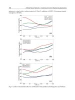
The greatest fluid losses generally occur in jejunostomy
patients because of failure to re-absorb secretions from
the proximal gut. Nine litres of fluid enter the jejunum
every 24 h (fig. 1) but the jejunum has only a limited
capacity for retrieval. There is a moderately good correla-
tion between the length of remaining small intestine and
the ability to obtain a net positive balance of fluid and
electrolytes. It has been possible to classify patients with a
high jejunostomy into those that are net absorbers, that is
jejunal efflux is always less than oral intake, and net secre-
tors, in which jejunal efflux always exceeds oral intake.
Net absorbers generally have a residual length of
1 100 cm, whereas net secretors generally have ! 100 cm
(fig. 2) [32]. These observations have important sequelae
when planning fluid and electrolyte supplements. Net
secretors virtually always require intravenous fluid and
electrolyte support, whereas net absorbers can usually
manage on oral supplements with some surviving solely
on a normal diet. Carbohydrate absorption is also closely
related to the length of residual jejunum [33]. An addi-
tional factor that probably contributes to fluid losses in
patients with a jejunostomy is the rapid gastric emptying
of liquids.
In patients with an intact colon, fluid and electrolyte
balance is easier to maintain and it has been estimated
that the colon is equivalent to 50 cm of small intestine
with respect to sodium and water absorption [34]. The
presence of the colon can make the difference between a
life-long dependency on IVN and the ability to survive on
a normal diet or possibly a normal diet supplemented
with oral supplements. Magnesium deficiency is also less
common in patients with a colon [29]. The colon is also
important for energy retrieval of malabsorbed carbohy-
drate amounting to up to 500 kcal/24 h [35]. Patients with
a colon are however more likely to develop oxalate renal
Severe IBD: Medical Management
Dig Dis 2003;21:46–53
51
Fig. 2.
Daily oral intake and intestinal ef-
fluent. IVN = Intravenous nutrition; IVF =
intravenous fluid; OS = oral supplements
[adapted from 30].
stones due to enhanced oxalate absorption from the colon
[34].
Initial Management of Acute Intestinal Failure
The rational management of intestinal failure depends
on an assessment of fluid, electrolyte and nutrient losses.
The aims of this assessment are two-fold, namely to rapid-
ly correct any major deficiencies that have occurred dur-
ing the early phase of the condition and secondly to plan
the long-term management, particularly to predict wheth-
er or not there will be a need for IVN [28, 29].
Although intestinal losses in excess of 2 litres/24 h are
often indicative that some form of intravenous support
will be required, it is essential that the initial assessment
be carried out when the patient is fluid and electrolyte
replete. Patients may have been drinking vast quantities
of low sodium liquids in an attempt to deal with thirst
promoted by dehydration and hyponatraemia. This will
exacerbate sodium and magnesium deficiency and in-
crease intestinal effluent.
It is advisable therefore to stabilise the situation by giv-
ing appropriate volumes of intravenous saline to rehy-
drate until body weight is stable and confirm that there is
adequate sodium in the urine (1 20 mmol/l). When rehy-
dration and sodium repletion is achieved, the patient can
then be progressively transferred to a normal diet and
intestinal effluent volume (or weight) assessed. If intesti-
nal losses continue to exceed 2 litres/24 h then it is highly
likely that intravenous replacement of saline will be
required, and as losses approach 3–4 litres/day then this
will be essential. If losses are less than 2 litres/24 h, it is
likely that fluid and electrolyte homeostasis can be main-
tained orally, but such patients may require supplementa-
tion with 1–2 litres of a high sodium (1 90 mmol/l) glu-
cose-electrolyte solution [36, 37]. Many of the commer-
cially available oral rehydration solutions have inade-
quate sodium concentrations for patients with a high out-
put jejunostomy. It may be necessary to make up an
appropriate solution in the home or hospital pharmacy.
In patients with intestinal effluents exceeding 2 litres/
24 h there is always the risk of magnesium deficiency [28].
The risk is substantially reduced when the colon is
retained. Deficiency should be screened for during the ini-
tial assessment by measuring plasma magnesium concen-
tration although deficiency may be apparent clinically
with symptoms in the peripheral and central nervous sys-
tem including paraesthesiae, tetany, lassitude, depression
and occasionally convulsions. There may also be muscle
weakness. In symptomatic cases of magnesium deficien-
cy, potassium and calcium concentrations are also re-
52
Dig Dis 2003;21:46–53
Farthing
duced. In severe acute deficiency, magnesium sulphate
should be given intravenously with careful monitoring of
plasma magnesium concentration. Many patients with a
chronically high intestinal effluent will require replace-
ment on a regular basis, magnesium oxide (12–24 mmol/
24 h) being the preferred preparation.
Potassium deficiency is uncommon in intestinal fail-
ure and is usually only seen when there is ! 50 cm of resid-
ual small intestine. Hypokalaemia in jejunostomy pa-
tients may be indicative of sodium depletion as a result of
either secondary hyperaldosteronism or a magnesium def-
icit.
Drug Therapy to Reduce Intestinal Effluent
Pharmacological approaches to reducing intestinal ef-
fluent are only modestly effective and in general are
unable to change a patient’s status from being dependent
on IVN or IV fluids to an individual who can survive on
oral intake alone. However, a reduction in effluent can be
achieved by either improving intestinal absorption or by
inhibiting intestinal secretion. Synthetic opioid drugs
such as loperamide or the opiate, codeine phosphate, are
the first-line medications to be evaluated. Although it has
been difficult to unequivocally demonstrate efficacy be-
cause of the relatively small numbers of patients that are
available for inclusion in clinical trials, detailed balance
studies in an individual patient clearly show beneficial
effects with respect to reducing sodium and fluid loss [38].
An alternative approach is to use the somatostatin ana-
logue, octreotide, that slows intestinal transit and reduces
gastric, pancreatic and biliary secretion. A variety of small
studies have shown that octreotide reduces intestinal out-
put and some have also shown a reduction in sodium and
potassium loss [39]. These effects have been sustained
long term and no major adverse effects have been re-
ported. Unfortunately these effects are insufficient to con-
vert a patient from being a net secretor to a net absorber
or render a patient no longer dependent on IVN. How-
ever, reducing intravenous fluid requirements for a pa-
tient will decrease the time that the individual needs to be
connected to the infusion system.
An alternative approach to reducing secretion into the
gut is to use an H
2
-receptor antagonist or a proton pump
inhibitor [40]. The efficacy of these drugs is probably
within the same range as octreotide although responses in
individual patients may be idiosyncratic and it therefore
worthwhile beginning in a hierarchical way with the acid
inhibitors and then moving on to octreotide to determine
whether additional benefits can be achieved.
References
1 Farthing MJG, McLean AM: Pancolitis and
toxic megacolon; in Gassull MA, Obrador A,
Chantar C (eds): Management of Inflammatory
Bowel Disease. Prous Science Publishers, 1994,
pp 195–209.
2 Farthing MJG: Infectious diarrhea; in Irvine
EJ, Hunt RH (eds): Evidence-Based Gastroen-
terology. Hamilton, BC Decker, 2001, pp 323–
341.
3 Prantera C, Lorenzetti T, Cerro P, et al: The
plain abdominal film accurately estimates ex-
tent of active ulcerative colitis. J Clin Gas-
troenterol 1991;13:231–234.
4 Bartram CI: Radiology in the current assess-
ment of ulcerative colitis. Gastrointest Radiol
1977;1:383–392.
5 Vernia P, Colaneri O, Tomei E, Caprilla R:
Intestinal gas in ulcerative colitis. Dis Colon
Rectum 1979;22:346–349.
6 Bartram CI, Preston DM, Lennard-Jones JE:
The ‘air enema’ in acute colitis. Gastrointest
Radiol 1983;8:61–65.
7 Halpert RD: Toxic dilation of the colon. Radiol
Clin North Am 1987;25:147–155.
8 Hata J, Haruma K, Suenga K, et al: Ultrasonic
assessment of inflammatory bowel disease. Am
J Gastroentrol 1992;87:443–447.
9 Gore RM: CT of inflammatory bowel disease.
Radiol Clin North Am 1989;27:717–730.
10 Meyers S, Janowitz HD: Systemic corticoste-
roid therapy of ulcerative colitis. Gastroenter-
ology 1985;89:1189–1191.
11 Truelove SC, Jewell DP: Intensive intravenous
regimen for severe attacks of ulcerative colitis.
Lancet 1974;i:1067–1070.
12 Jarnerot G, Rolny P, Sandberg-Gertzen H: In-
tensive intravenous treatment of ulcerative co-
litis. Gastroenterology 1985;89:1005–1013.
13 Lennard-Jones JE, Ritchie JK, Hilder W, Spic-
er CC: Assessment of severity in colitis: A pre-
liminary study. Gut 1975;16:579–584
14 Travis SPL, Farrant JM, Nolan DJ, Mortensen
NM, Kettlewell MGW, Jewell DP: Predicting
outcome in severe ulcerative colitis. Gut 1996;
38:905–910.
15 Lichtiger S, Present DH: Preliminary report:
Cyclosporin in treatment of severe active ulcer-
ative colitis. Lancet 1990;336:16–19.
16 Loftus CG, Loftus EV, Sandborn WJ: Cyclo-
sporin for refractory ulcerative colitis. Gut
2002;52:172–173.
17 Binder SC, et al: Toxic megacolon in ulcerative
colitis. Gastroenterology 1974;66:909–915.
18 Odyniec NA, Judd ES, Sauer WG: Toxic mega-
colon in ulcerative colitis. Gastroenterology
1974;66:909.
19 Grant CS, Dozois RR: Toxic megacolon: Ulti-
mate fate of patients after successful medical
management. Am J Surg 1984;147:106–110.
20 DeDombal FT, Watts JMK, Watkinson G,
Goligher JC: Intraperitoneal perforation of the
colon in ulcerative colitis. Proc R Soc Med
1965;58:713–715.
21 Odyniec ND, Dudd ES, Sauer WG: Toxic
megacolon: Significant improvement in surgi-
cal management. Arch Surg 1967;94:638–643.
22 Goligher JC, et al: Early surgery in the manage-
ment of severe ulcerative colitis. Br Med J
1967;iii:193.
23 Sirinek KR, et al: Total proctocolectomy and
ileostomy: Procedure of choice for acute toxic
megacolon. Arch Surg 1985;112:518.
24 Podolsky DK: Inflammatory bowel disease. N
Engl J Med 2002;347:417–429.
25 Heuschkel RB, Menache CC, Megerian JT,
Baird AE: Enteral nutrition and corticosteroids
in the treatment of acute Crohn’s disease in
children. J Pediatr Gastroenterol Nutr 2000;
31:8–15.
Severe IBD: Medical Management
Dig Dis 2003;21:46–53
53
26 Ballinger AB, Azooz O, El-Haj T, Poole S,
Farthing MJG: Growth failure occurs through
a decrease in insulin-like growth factor-1 which
is independent of undernutrition in a rat model
of colitis. Gut 2000;46:694–700.
27 Griffiths AM, Ohlsson A, Sherman PM, Suth-
erland LR: Meta-analysis of enteral nutrition as
a primary treatment of active Crohn’s disease.
Gastroenterology 1995,32:1056–1067.
28 Lennard-Jones JE: Practical management of
the short bowel. Aliment Pharmacol Ther
1994;8:563–577.
29 Farthing MJG: Intestinal failure; in Farthing
MJG (ed): Clinical Challenges in Gastroenter-
ology. London, Dunitz, 1996, pp 86–103.
30 Nightingale JMD, Walker ER, Burnham WR,
et al: Short bowel syndrome. Digestion 1990;
45(suppl 1):77–83.
31 Nightingale JMD: The short bowel. Eur J Gas-
trohepatol 1995;7:514–520.
32 Nightingale JMD, Lennard-Jones JE, Walker
ER, Farthing MJG: Jejunal efflux in short-bow-
el syndrome. Lancet 1990;336:765–768.
33 Rodrigues CA, Lennard-Jones JE, Thompson
DG, Farthing MJG: Energy absorption as a
measure of intestinal failure in the short bowel
syndrome. Gut 1989;30:176–183.
34 Nightingale JMD, Lennard-Jones JE, Gertner
DJ, Wood SR, Bartram CI: Colonic preserva-
tion reduces the need for parenteral therapy,
increases the incidence of renal stones but does
not change the high prevalence of gallstones in
patients with a short bowel. Gut 1992;33:
1493–1497.
35 Nordgaard I, Hansen BS, Mortensen PB: Colon
as a digestive jejunostomy output in patients
with severe short-bowel syndrome. Lancet
1994;343:373–376.
36 Lennard-Jones JE: Oral rehydration solutions
in short bowel syndrome. Clin Ther 1990;
12(suppl A):129–137.
37 Nightingale JMD, Lennard-Jones JE, Walker
ER, Farthing MJG: Oral salt supplements to
compensate for jejunstomy losses: Comparison
of sodium chloride capsules, glucose electrolyte
solution and glucose polymer electrolyte solu-
tion (Maxijul). Gut 1992;33:759–761.
38 Rodrigues CA, Lennard-Jones JE, Walker ER,
Thompson DG, Farthing MJG: The effects of
octreotide, soy polysaccharide, codeine and lo-
peramide on nutrient, fluid and electrolyte ab-
sorption in the short bowel syndrome. Aliment
Pharmacol Ther 1989;3:159–169.
39 Nightingale JMD, Walker ER, Burnham WR,
Farthing MJG, Lennard-Jones JE: Octreotide
(a somatostatin analogue) improves the quality
of life in some patients with a short intestine.
Aliment Pharmacol Ther 1989;3:367–373.
40 Nightingale JMD, Walker ER, Farthing MJG,
Lennard-Jones JE: Effect of omeprazole on in-
testinal output in the short-bowel syndrome.
Aliment Pharmacol Ther 1991;5:405–412.
Review Article
Dig Dis 2003;21:54–62
DOI: 10.1159/000071340
Surgical Treatment of Severe
Inflammatory Bowel Diseases
Christine Leowardi Gundi Heuschen Peter Kienle Udo Heuschen
Jan Schmidt
Department of Surgery, University of Heidelberg, Germany
Jan Schmidt, MD
Department of Surgery, University of Heidelberg
Kirschnerstrase 1, DE–69120 Heidelberg (Germany)
Tel. +49 6221 566204, Fax +49 6221 565781
ABC
Fax + 41 61 306 12 34
www.karger.com
© 2003 S. Karger AG, Basel
0257–2753/03/0211–0054$19.50/0
Accessible online at:
www.karger.com/ddi
Key Words
Crohn’s disease
W Ulcerative colitis W Surgical treatment
Abstract
Surgical treatment of severe inflammatory bowel dis-
eases is required in failed medical treatment, in emer-
gencies and for complications. Indications for surgery
and operative techniques have changed significantly
over the last few years. There is a clear tendency towards
earlier and less invasive surgical interventions per-
formed in specialized and experienced centers. Im-
proved quality of life of patients with Crohn’s disease or
ulcerative colitis after surgical therapy supports an ear-
lier consideration of the surgical treatment option. A
close cooperation with the involved gastroenterologist is
mandatory in this context.
Copyright © 2003 S. Karger AG, Basel
Introduction
Ulcerative colitis (UC) and Crohn’s disease (CD) are
inflammatory disorders of the gastrointestinal tract of
unknown etiology. Both diseases are primarily a domain
of conservative medicine. However, about one third of
patients with CD or UC do not respond to conventional
medical treatment. In this subgroup of patients with
severe inflammatory bowel diseases, surgery can lead to a
significant relief of symptoms and in UC patients even
cure the disease.
Crohn’s Disease
CD is an idiopathic, chronic inflammatory disease of
the gastrointestinal tract that primarily affects the small
intestine and colon, which may be caused by environmen-
tal and genetic factors.
The incidence rate varies between different geographi-
cal regions, with an average of 3–6 cases/100,000/year [1].
There is a typical ‘bimodal’ age distribution at diagnosis
with a first peak between the age of 15 and 30 and a sec-
ond peak later in life in the sixth or seventh decade.
Regarding the gender distribution, several studies de-
scribed a slight female predominance, with an increased
risk for women of about 20–30%. CD appears to be asso-
ciated with a significant genetic predisposition with an
increased relative risk for first-degree relatives of affected
patients between the age of 18 and 36. Proven risk factors
are smoking [2], oral contraception [3] and a high socio-
economic status.
Surgical Treatment of Inflammatory Bowel
Diseases
Dig Dis 2003;21:54–62
55
The etiology of CD is still unknown, but three funda-
mental theories are presently being discussed [4–6]: (1) an
impaired intestinal epithelial barrier function with a loss
of tolerance towards intraluminal antigens; (2) a dis-
turbed immunological response in the intestinal wall
towards ubiquitous luminal antigens, and (3) a specific
infection.
CD is a transmural, predominantly submucosal in-
flammatory disease that most commonly affects the distal
ileum and colon but may occur in any part of the gastroin-
testinal tract. Macroscopically, segments of affected bow-
el are characteristically sharply demarcated from adjacent
normal bowel (‘skip lesions’). Transmural inflammation
leads to bowel wall thickening and lymph edema and can
result in extensive fibrosis with strictures. Patchy, muco-
sal longitudinal and transverse ulcers with intervening
mucosal edema can develop which then appear as the typ-
ical cobblestone relief. Often the attached mesentery is
markedly thickened and lymph edematous with adher-
ence of the inflamed segment to neighboring organs,
forming conglomerates with sometimes interenteric or
blind fistulas and abscesses. Mesenteric fat typically ex-
tends on over the serosal surface of the bowel. Microscopi-
cally, there are submucosal edemas, lymphoid aggrega-
tions, lymphoplasmacellular infiltrates, ulcers and fibro-
sis with influx and proliferation of macrophages. Nonca-
seating granulomas with multinucleated giant cells are
detectable in up to 60% of patients.
Clinical Symptoms and Complications
Clinical symptoms vary with the location of the in-
flamed region. Chronic diarrhea with abdominal pain,
fever, anorexia, weight loss, and a right lower quadrant
mass or fullness are the most common presenting fea-
tures. Many patients are first seen with an acute abdomen
due to intestinal obstruction, sometimes simulating acute
appendicitis. In the selected surgical setting, there is an
increased percentage of patients with perianal fistulas.
Extraintestinal manifestations include joints (arthritis),
skin (pyoderma gangrenosum), kidneys and the urinary
tract (stones, fistulas), gallbladder and bile ducts (stones,
sclerosing cholangitis).
Due to the varying locations of the disease, the devel-
opment of complications has a wide spectrum. Intestinal
bleeding, perforation, obstructions, development of ente-
roenteric, enterovesical, retroperitoneal, or enterocuta-
neous fistulas, and abscess formations are common com-
plications in CD, often requiring surgical intervention.
The risk of developing a CD-associated carcinoma is
increased about 5- to 6-fold [7].
Surgical Therapy
The mainstay of CD treatment remains medical thera-
py, which is beyond the scope of this review. Interested
readers are referred to the literature [8] or the Cochrane
Library (www.update-software.com).
Patients suffering from severe CD require surgery ei-
ther to manage complications or in case of failure of medi-
cal treatment. 2,070 cases with CD were treated at the
Surgical Department of the University of Heidelberg
between 1982 and January 2003.
Surgery, as well as conservative medical treatment,
cannot cure the disease. However, more than 90% of all
patients treated surgically in our institution declared that
they experienced a complete remission of symptoms
(68%), or a significant relief of complaints. Nevertheless,
the recurrence rate in the following 10–15 years in these
patients was still high (50%) [9]. Most of these recurrences
can be effectively treated with a further operation. The
former widespread fear of a ‘short-bowel syndrome’ is
now unfounded. The modern principles of Crohn’s sur-
gery restrict resection to inflamed sections only without
so-called ‘security margins’ as practised in cancer surgery
[10]. Short fibrotic strictures can be treated with stricturo-
plasty, also known as ‘conservative surgery’. Minimally
invasive techniques can now be used in a large number of
cases. Therefore, surgical therapy should be considered
early in the treatment of symptomatic stenoses, fistulas,
septic complications and situations refractory to conser-
vative treatment. Furthermore, complications of long-
term therapy with glucocorticoids or immunosuppres-
sants, as well as malignant transformation may be
avoided by surgical treatment.
Specific Indications for Surgery
Controversy still remains regarding the right time for
surgery. A major reason for early surgical intervention is
the high rate of symptomatic relief after surgery. Further-
more, the resected bowel parts are mostly without func-
tion. Opponents of this concept state that delayed surgery
is associated with fewer resections and therefore a lower
risk of short-bowel syndrome. We believe that time of sur-
gery should be based on the clinical symptoms. It is
important to consider the preoperative medication with
its side effects and the potential increase of perioperative
complications due to the medications. The application of
these principles should lead to a reasonable decision
regarding the time of surgery with a maximum relief of
complaints and a minimum incidence of surgery-related
disadvantages. These principles, however, are not yet ade-
quately considered. Scott and Hughes [11] found that
56
Dig Dis 2003;21:54–62
Leowardi/Heuschen/Kienle/Heuschen/
Schmidt
Fig. 1.
Stricturoplasty: After opening the bowel on the anti-mesenter-
ic aspect of the loop, proximal and distal to the stricture, sutures are
placed in such a way as to change the longitudinal incision into a
transverse one.
74% of all operated patients would have preferred an ear-
lier operation if they had known the postoperative result
beforehand. After having taken the decision for an opera-
tion, a ‘Crohn staging’ should be performed to evaluate
affected areas and to determine an individual surgical
concept.
Preoperative Investigations
A detailed patient’s history and clinical examination,
including rectal examination, are mandatory. The whole
gastrointestinal tract should be examined thoroughly to
evaluate all sites of possible Crohn manifestations preop-
eratively. Sonography can show thickening of the bowel
wall, fistulas or abscesses. Gastroduodenoscopy and co-
lonoscopy are standard preoperative investigations. Dis-
tal small bowel affection may often be identified by colon-
oscopy if intubation of the terminal ileum is possible.
Proximal small bowel involvement can be evaluated by
barium meal or hydro-MRI. In Heidelberg, hydro-MRI
with filling of small bowel and colon with water is done to
evaluate the extent of the disease. This investigation can
at the same time assess direct affection of the colon and
small bowel, as well as extraluminal findings, such as fis-
tulas and abscesses in one step without radiation exposure
[12]. For verification of fistulas or abscesses, proctoscopy
or rectoscopy complemented by endosonography are es-
sential to assess rectal mucosa and fistula morphology.
Sometimes fistulography or barium enema are useful.
Stenosis and Obstruction
Patients with acute symptoms of bowel obstruction
should be nil per os and should be nourished and rehy-
drated parenterally. Inflammatory stenoses are primarily
treated conservatively with glucocorticoids. Surgical ther-
apy of stenoses, strictures or other obstructions depends
on the localization of the affected areas. The most fre-
quently performed operation for CD is the resection of the
ileocecal region or isolated small bowel resection. In short
strictures, not exceeding 8–10 cm stricturoplasty (Hei-
neke-Mikulicz) can be performed (fig. 1).
This indication is well suited for a minimally inva-
sive procedure, alternatively median laparotomy is per-
formed. Stenoses of the colon can sometimes be problem-
atic, because the recurrence rate is higher in Crohn’s coli-
tis than in small bowel affections. However, the basic
principle remains the same: ‘resect as much as necessary,
but as little as possible’. Bypass operations of Crohn’s
associated conglomerate tumors have been abandoned
due to blind-loop problems, neoplastic transformation
and septic complications.
Abscesses
In the majority of the cases, abscesses in CD are the
result of sealed perforations of the bowel. The most fre-
quent location of these abscesses is the lower right abdo-
men and the perianal region. Most of the abscesses can be
treated by interventional drainage. After achieving con-
trol of the septic situation, patients can then undergo elec-
tive surgery with resection of the affected segment later.
Sometimes, especially when multiple interenteric or mul-
tilocular abscesses are present, surgical drainage is neces-
sary. Perianal fistulas and abscesses distal to the sphincter
can be incised and drained perineally. Perirectal abscesses
proximal to the sphincter and levator muscle should be
drained through the abdomen due to the risk of persisting
translevatoric or transsphincteric fistulas. In the presence
of a visible fistula proximal to the sphincter and simulta-
neous severe inflammation of the rectum, a protective
ileostomy should be considered.
Fistulas
Fistulas mostly originate from primarily CD affected
segments of the gastrointestinal tract. There is often a ste-
nosis distal to the inflamed segment increasing the intra-
luminal pressure in the transmurally inflamed bowel wall,
predisposing to fistula formation. These fistulas can pene-
trate all neighboring structures and organs. In the worst
case a complex system of communicating fistulas and
abscesses with consecutive secondary affection of other
Surgical Treatment of Inflammatory Bowel
Diseases
Dig Dis 2003;21:54–62
57
organs develops. To outline the distribution of different
fistulas, see table 1.
Internal Fistulas
About one third of all CD patients develop an internal
fistula as described above [13]. Interenteric such as ileo-
sigmoidal fistulas are the most common ones. This situa-
tion is not necessarily an indication for surgery. The ter-
minal ileum is often the primarily affected organ, the sig-
moid or other diseased bowel is only involved secondari-
ly. If the stenosis of the terminal ileum is symptomatic,
the therapy of choice is the resection of the terminal ileum
with excision of fistula opening in the sigmoid or other
affected bowel segments. An absolute indication for sur-
gery is a blind-ending retroperitoneal fistula. This is often
the origin of a psoas abscess and various other secondary
affections of different organs with further complications.
Enterovesical fistulas are also an absolute indication for
operative treatment. These fistulas can lead to life-threat-
ening recurrent ascending urinary tract infections.
Several other types of internal fistulas can occur, but
they are less frequent.
Enterocutaneous Fistulas
Enterocutaneous fistulas generally originate from the
terminal ileum or from an anastomosis from previous
operations. Colocutaneous fistulas are more difficult to
treat. An uncomplicated enterocutaneous fistula itself is
not necessarily an indication for surgery. However, it is
associated with an increased risk for additional fistulas
and abscesses and is an indicator for active, often stenos-
ing, CD in the organ of origin. This usually results in the
affected organ having to be resected and the fistula tract
excised. Anastomotic recurrence of CD is treated by
resection of the frequently stenotic anastomosis.
Perianal Fistulas
Five to 10% of all CD patients and 40–60% of surgical-
ly treated patients show perianal fistulas. An aggressive
operative therapy should only be performed if the patient
has significant complaints, because perianal fistulas tend
to recur. If surgical therapy is undertaken, the anal sphinc-
ter should be treated with utmost care. In this context it
sometimes can be necessary to construct a temporary pro-
tective stoma. Incision and drainage of abscesses and the
placement of a Seton, however, is often sufficient to stabi-
lize the local situation and prevent recurrent abscesses.
For infrasphincteric or submucous fistulas, an open-
lay technique together with adequate medical treatment
should be used. Inter- or transsphincteric fistulas originat-
Table 1.
Surgical interventions in patients
with Crohn’s disease in the Surgical Depart-
ment of the University of Heidelberg, 1982–
2000
Resections
Small bowel 224
Ileocecal region 254
Anastomoses 207
Colon 53
Hemicolectomy 95
Subtotal colectomy 99
Proctocolectomy/proctectomy 64
Fistulas
Interenteric 216
Enterocutaneous 84
Enterogenital 67
Enterovesical 35
Retroperitoneal 35
Anal 260
Others
Abscess 156
Ureterolysis 20
Explorative lap. 22
Lavage 29
Endosc. intervention 36
Others 178
Reconstruction
Stricturoplasty 175
Mucosa flap 159
Omentoplasty 83
Reconstruction of continuity 14
Ileostomy closure 111
Colostomy closure 6
Pouch formation 4
Deviation
Ileostomy 251
Colostomy 39
Hartmann operation 19
Intestinal bypass 3
Gastroenterostomy 6
ing in the anal canal are more difficult to treat. A careful
excision of the fistula in an open-lay technique, the suture
of the sphincter and a mucosa flap covering the internal
fistula opening is the treatment of choice. Suprasphincter-
ic or translevatoric fistulas often do not heal without tem-
porary stool deviation. Associated abscesses should be
incised and drained, followed by the construction of a
protective loop ileostomy. After reduction of inflamma-
tion by local and systemic anti-inflammatory therapy,
excision and mucosa flap or even rectal resection should
58
Dig Dis 2003;21:54–62
Leowardi/Heuschen/Kienle/Heuschen/
Schmidt
Table 2.
Morbidity of 1,941 operations be-
tween 1981 and 9/2002 in patients with CD
No complications 87%
Mortality 0.5%
Relaparotomy 4.7%
Anastomotic leaks 1.5%
Abscess 1.5%
Ileus 0.7%
Others 1.0%
Other septic complications 3.9%
Others 2.8%
follow. Recto-vaginal fistulas should be treated by elective
excision, mucosa flap and reconstructive levatorplasty, in
most cases under temporary stoma protection [14].
Emergency Indications for Surgery in CD
Fulminant or Toxic Colitis. Similar to UC, Crohn’s
colitis can also take a fulminant course. Surgical therapy
should be urgently undertaken if the patient’s condition
fails to improve under intensive care medicine. After
72 h, mortality increases significantly [15]. Partial colec-
tomy with a terminal ileostomy, followed by secondary
reconstruction of continuity, is the therapy of choice in
most cases.
Perforation. 1–3% of all surgically treated CD patients
suffer free perforations of the small or large bowel [16].
They usually present with an acute abdomen and free air
in the abdomen on plain X-ray. An immediate operation
with resection of the perforated bowel and, if present,
with the associated stenotic bowel segment is obligatory.
Preferably discontinuity resections should be performed,
especially in severe peritonitis where the mortality rate
after primary anastomoses is significantly increased [17].
Hemorrhage. A massive life-threatening hemorrhage is
the reason for 1–13% of all surgical emergencies in CD
patients. It occurs more often in young men and often
originates in the terminal ileum. An immediate mesenter-
icography can usually localize the source of the bleeding
and warrants a precise resection [18]. In such a situation
we leave the angiocatheter in place and inject isosulphan
blue in the operating room to specifically identify the
bleeding bowel segment that needs to be resected.
Operative Technique
The basic principle is the minimal possible resection to
achieve a defined goal. A resection with unaffected mar-
gins has not been shown to have a beneficial effect [10, 19,
20]. Resective surgery for CD can now also be performed
using a laparoscopic approach. The potential advantages
associated with laparoscopic intestinal surgery include
less postoperative pain, and wound infections, quicker
resumption of oral feeding, a reduced hospital stay and
earlier return to work. Other advantages such as less post-
operative intra-abdominal adhesions and improved cos-
metic results may be particularly attractive in patients
who are likely to undergo multiple operations during their
lifetime [21]. No differences in recurrence rate or in dis-
ease-free interval were noted between groups of patients
operated on with an open technique or laparoscopically
[22]. If the surgeon has enough experience in minimal
invasive surgery, primary surgery should be performed
with a laparoscopically assisted technique. Suitable opera-
tions are ileocecal, small bowel and colon resections, stric-
turoplasty and stoma construction.
There is no agreement in the literature as to which type
of anastomosis is preferable. In our institution, we used to
perform one-layered end-to-end anastomosis with inter-
rupted sutures. We have now changed to a two-layered
running suture technique (either end-to-end or end-to-
side with 5/0 PDS suture) because we feel that this is safer
with a lower leak rate.
Postoperative Morbidity and Mortality
Between 1981 and September 2002, 1,941 operations
were performed on patients with CD at the Surgical
Department of Heidelberg. Overall morbidity was 12.5%,
including all major complications requiring a surgical
reintervention; mortality was 0.5% (table 2).
Ulcerative Colitis
UC is a chronic, idiopathic inflammatory and ulcer-
ative disease of the rectal and colonic mucosa of unknown
etiology. UC usually extends from the distal rectum to the
more proximal segments of the colon and most common-
ly affects only the mucosa, rarely deeper layers of the
bowel.
The incidence in North and Central Europe, as well as
in North America, is 2–8 cases/100,000/year. Age at diag-
nosis has two peaks with a first peak between the age of 20
and 30 years and a second one at the age of 60. Women
seem to be affected slightly more often and the incidence
in Jewish people is higher than in non-Jewish [23].
Although the etiology of UC remains unknown, several
possible factors are presently being discussed [24], namely
environmental, microbial, genetic and immune factors.
Surgical Treatment of Inflammatory Bowel
Diseases
Dig Dis 2003;21:54–62
59
Deeper layers of the bowel wall are generally not affected
in UC. One of the few exceptions is toxic megacolon,
where transmural involvement can occur. Inflammation
and destruction of deeper layers lead to dilatation of a
colonic segment or the whole colon. Remission of the
inflammation can lead to loss of the mucosal relief and
subsequently shortening of the colon. Microscopically,
crypt abscesses and a mononuclear infiltrate of lympho-
cytes, macrophages and mast cells are typical.
Clinical Symptoms and Complications
Bloody and mucous diarrhea, high stool frequency and
day and night urgency, abdominal pain and cramps and
subfebrile temperatures are common clinical signs of UC,
and these symptomatic episodes are frequently inter-
rupted by asymptomatic intervals. 18% of all patients
only have one single episode. In about two thirds of the
cases, however, the disease becomes chronic and recur-
rent. Total proctocolectomy within 10 years after the first
episode becomes necessary in about 11% of all patients
and this rate further increases in the following years. In
30% of the cases the rectum is the only affected bowel
segment during the first episode of UC. In 40% the
inflammation reaches further proximal up to the trans-
verse colon. Only 30% of the patients have a total colitis.
Extraintestinal manifestations occur in about 10% of
the patients [23]. Most frequently, patients suffer from
arthritis. Less common are aphthous stomatitis, uveitis or
conjunctivitis and skin manifestations, such as pyoderma
gangrenosum and erythema nodosum. A primary scleros-
ing cholangitis can rarely necessitate liver transplanta-
tion.
Major complications are the development of a toxic
megacolon, perforation and bleeding, all of which require
emergency treatment. A large percentage of UC patients is
admitted for surgery due to severe drug side effects, espe-
cially from glucocorticoids. Furthermore, the incidence of
UC-associated colorectal cancer is significantly increased
in pancolitis when disease duration exceeds 10 years,
independent of disease activity. After 10 years the cancer
risk increases about 1% per year [25].
Diagnosis
Total colonoscopy with biopsy is mandatory to obtain
the histological diagnosis and to evaluate the grade and
extent of inflammation and neoplastic changes. If there is
a severe stenosis, double contrast barium enema or hydro-
CT of the colon may be helpful to exclude a further prob-
lem proximal to the stenotic segment.
Table 3.
Indications for colectomy in 621
UC patients between 01/1982 and 12/2001
Therapy-refractory situation 75.1%
Dysplasia 5.8%
Colorectal carcinoma 9.8%
Emergency
Toxic colon 6.8%
Perforation and bleeding 2.5%
Surgical Treatment
Surgical treatment of UC significantly differs from sur-
gery for CD. While in CD the surgical principle is ‘resect
as much as necessary, but as little as possible’, the aim of
surgery for UC is to remove the whole colon with a procto-
mucosectomy. Therefore, it is essential to definitely clari-
fy the histological diagnosis preoperatively. Surgical ther-
apy for UC patients aims at curing the disease itself. Side
effects of medical treatment may thus be avoided and
malignant transformation prevented or, if they have al-
ready occurred, adequately treated. Quality of life may
significantly be improved by surgical therapy. Extraintes-
tinal manifestations such as activity-related polyarthropa-
thy seem to be independent from the colonic affection,
but will sometimes respond to surgical therapy.
Specific Indications for Surgical Treatment
Surgery for UC can either be indicated in the emergen-
cy or the elective setting. Indications for urgent surgery
include toxic colitis (6.8%), perforation and severe bleed-
ing (2.5%).
Emergency Surgery
Acute severe colitis requires interdisciplinary specific
intensive care medicine. Vital signs, bowel function and
electrolytes and malnutrition have to be monitored care-
fully. Anti-inflammatory treatment usually includes high-
dose intravenous steroids. Remission occurs in about 50–
60% of patients. If there is no clinical improvement with-
in 72 h or the patient’s condition is deteriorating, surgery
is indicated even in the absence of an acute abdomen
[15].
Toxic dilatation, perforation and bleeding are indica-
tions for emergency surgery. The operative technique in
emergency surgery in UC patients usually is subtotal
colectomy with terminal ileostomy and the preservation
of a rectal stump. Surgical procedures without resection of
the diseased colon should be avoided. The poor prognosis
of a toxic colon in former days can be markedly improved