báo cáo khoa học: "Effects of ulinastatin and docetaxel on breast cancer invasion and expression of uPA, uPAR and ERK" docx
Bạn đang xem bản rút gọn của tài liệu. Xem và tải ngay bản đầy đủ của tài liệu tại đây (2.67 MB, 7 trang )
RESEARC H Open Access
Effects of ulinastatin and docetaxel on breast
cancer invasion and expression of uPA, uPAR and
ERK
Jie Luo, Xin Sun, Feng Gao, Xiaoliang Zhao, Biao Zhong, Hong Wang and Zhijun Sun
*
Abstract
Objective: To investigate the effects of ulinastatin and docetaxel on invasion of breast cancer cells and expression
of uPA, uPAR and ERK, breast cancer MDA-MB-231 and MCF-7 cells.
Methods: The nude mice were treated with PBS, ulinastatin, docetaxel, and ulinastatin plus docetaxel, respectively.
Their effects on 1) cell invasion ability was assayed using Transwell; 2) expression of uPA, uPAR and ERK was
detected by real time PCR and Western blot; 3) uPA, uPAR and p-ERK protein level in nude mice was quantified by
immunohistochemistry.
Results: 1) Treatment with ulinastatin, docetaxel, and ulinastatin plus docetaxel, respectively, significantly inhibited
MDA-MB-231 and MCF-7 cell invasion; 2) mRNA and protein levels of uPA, uPAR and ERK1/2 were inhibited by
ulinastatin, but enhanced by docetaxel.
Conclusion: Ulinastatin can enhance the effects of docetaxel on invasion of breast cancer cells. And that uPA,
uPAR and p-ERK expre ssion is obviously inhibited by ulinastatin.
Introduction
Breast cancer is one of the major malignant tumors
threaten women well being. Failure in its treatment
mainly arises from cancer proliferation, invasion and
metastasis, which ultimately lead to the death o f
patients. Cell penetrating into extracellular base mem-
brane is the premise of cancer cell metastasis, where a
variety of proteases play essential roles.
Plasminogen activators (PAs) are serine proteases, the
main function of which is to activat e plasminogen into
plasmin, a serine protease that hy drolyzes a variety of
proteins, including la minin, fibronectin, fibrin, proteo-
glycan core protein and collagen fibres. There are t wo
types of mammalian PAs: tissue-type (tPA) and uroki-
nase-type (uPA). The former is mainly present in circu-
latory system, while the latter is present in cells and
closely related to tumor cell invasion and meta stasis. It
has been shown that uPA expression is enhanced in
many malignant tumors, such as breast cancer, prostate
cancer, colon cancer, stomach cance r and lung cancer,
and its me diated-plasminogen activation is dependent
on its receptor uPAR in cells. In breast cancer, uPA-
uPAR complex is necessary to maintain and amplify
plasmin activity[1].
Beside its pivotal roles in pasminogen cascade system,
uPA-uPAR complex can also activate many signaling
pathways, of which is important Ras-Raf-MEK-ERK
pathway. This pathway responds to signals from a vari-
ety of growth factors (EGF, NGF, PDGF, etc.), mitogens
and environmental stimulations, eventually leading to
activation and phosphorylation of extracellular signal-
regulated kinase (ERK) through the signal amplification
cascade. Phosphorylated ERK translocates to nucleus,
where it acts on the AP-1, NF-B and other nuclear
transcription factors, thereby regulating gene expression
and promoting tumor cell proliferation, differentiation
and s urvival. Over-activation of ERK has been found in
many human malignant tumors including oral cancer,
melanoma and breast cancer[2,3].
Urinary trypsin inhibitor ulinastatin as a broad-spec-
trum protease inhibitor ca n inhibit trypsin, chymotryp-
sin, plasmin, human leukocyte elastase and
* Correspondence:
Department of Breast, Pancreas, and Thyroid Surgery; Second Affiliated
Hospital of Chongqing Medical University, 74 Lingjiang Road, Yuzhong
District, Chongqing 400010, China
Luo et al. Journal of Experimental & Clinical Cancer Research 2011, 30:71
/>© 2011 Luo et al.; licensee BioMed Central Ltd. This is an open access article distributed under the terms of the Creative Commons
Attribution Licens e ( nses/by/2.0), which pe rmits unrestricted use, distribution, and reproduction in
any medium, provided the original work is properly cited.
hyaluronidase. It has anti-tumor metastasis and protec-
tive effects on patients accepted radiotherapy and che-
motherapy and been widely used to treat acute
pancreatiti s and shock and to improve surgical outcome
in clinic. Ulinastatin can bind to tumor cells through its
N-terminal Domain I and exert its inhibitory effect on
proteolytic activity of plasmin by binding to tumor cells
through its C-terminal domain II, the major anti-fibri-
nolytic group. The impact of ulinastatin on uPA is more
complicated. In addition to its inhibitory effects on gene
transcription, it also inhib its uPA protein expression by
affecting kinase C and MEK/ERK/c-Jun signaling path-
ways[4,5].
To find a more effective treatment for breast cancer,
this study explored the additive effects of docetaxel and
ulinastatin on the proliferation of breast cancer MDA-
MB-231 cells and tumor growth in nude mice.
Materials and methods
1. Materials
Ulinastatin was purchase d from Guangdong Techpool
Bio-Pharma Co., Ltd. Docetaxel was bought from
Sanofi-Aventis (Fre nch). SYBR Green/ROX qPCR Mas-
ter Mix (2X) were purchased from Fermentas Inc.
(Canada). Anti-uPA antibody was from Bioworld (USA).
Anti-uPAR and anti-pERK antibodies were from Santa
Cruz (USA). 24 well Transwell plates were from Corn-
ing (USA). Matrigel was from BD Company (USA).
2. Cell culture
Human breast cancer cell line MDA-MB-231 (ER-) and
MCF-7 (ER+) were kindly gifted by Shanghai Institute
of Biological Sciences, Chinese Academy of Sciences,
and maintained in RPMI-1640 medium supplemented
with 10% fetal bovine serum, 100 U/mL penicillin, 100
mg/L strepto mycin at 37°C in a n incubator supplemen-
ted with 5% CO
2
under saturated humidity.
3. Animals
100 female BALB/c (nunu) mice at age 4-6 weeks and
with body weight of 17-21 g from Animal Re search
Center of Chongqing Medical University (Production
License No.: SCXK (Beijing) 2005-0013, the use permit
number: SYX (Chongqing) 2007-0001) were kept in
SPF-class environment at 22-25°C and 50-65% humidity.
Drinking water, feed and experimental materials were
sterilized and all experiments were complied with sterile
principle.
4. Animal experiments
MDA-MB-231 cells at logarithmic growth phase were
washed twice with PBS and prepared as 2.5 × 10
10
cells/
L susp ension in serum-free RPMI -1640 medium. 0.2 mL
cell suspension was subcutaneously inoculated in the
right armpit o f each mouse. 21 days after inoculation,
29outof50micehadtumorvolume≥ 500 mm
3
and
randomly assigned into 4 groups[6]. MCF-7 cell was
innoculated into the other 50 nude mice for building
the model[7].
5. MDA-MB-231 and MCF-7 cell invasion assay
Breast cancer cell invasion w as measured using Trans-
well chamber. In detail, 2 × 10
5
cells were placed in the
upper chamber of Transwell with a membrane coated
with Matrigel. 24 h later, cells were incubated with 800
U/mL ulinastatin, 3.7 μg/mL docetaxel, 800 U/mL uli-
nastatin plus 3.7 μg/mL docetaxel, and PBS, respectively,
at 37°C in an incubator supplemented with 5% CO
2
.24
h later, cells in the upper chamber were removed with a
cotton swab. The remaining cells on the membrane
were stained with 0.1% crystal violet solution and
washed with PBS. Crystal violet attached to the cells was
dissolved by adding 500 μL of 33% acetic acid into the
lower chamber and its absorba nce at 570 nm was mea-
sured and used to calculate relative amount of cells
invaded through the Matrigel to the lower chamber.
6. mRNA levels of uPA, uPAR and ERK in MDA-MB-231
and MCF-7 cells measured by real-time RT-PCR
To evaluate the effect of treatments described above on
mRNA levels of uPA, uPAR and ERK in breast cancer
cells, 24 h after the treatment, total mRNAs were iso-
lated using 1 mL TRIzol reagent according to the proto-
col provided by the manufacturer. 20 μLmRNAwas
reverse transcripted into cDNA and the amount of uPA,
uPARandERKcDNAwasexaminedbyquantitative
real-time PCR using the following primer pairs: uPA
forward primer 5’-GGAGATGAAGTTTGAGGT-GG-3’
and reverse primer 5’ -GGTCTGTATAGTCCGGG-
ATG-3’, uPAR forward primer
5’ -CACAAAACTGCCTCCTTCCT-3’ and reverse
primer
5’ -AATCCCCGTTGGTCTTAC AC-3’,ERKforward
primer
5’ -CCTAAGGAAAAG-CT CAAAGA-3’ and reverse
primer
5’-AAAGTGGATAA-GCCAAGAC-3’, and b-actin for-
ward primer
5’-GCAGAAGGAGATCACAGCCCT-3’ and reverse
primer
5’ -GCTGATC CACATCTGCTGGAA-3’ . The corre-
sponding predicted products were 142, 178, 180, and
136 bp, respectively. In detail, template cDNA and p ri-
mers were mixed with SYBR Green/ROX qPCR Master
Mix (2X) in 25 μL reaction system and PCR was carried
out in triplicate under the following conditions: 5 min
at 95°C, 45 cycles of 15 seconds at 95 °C and 30 seconds
at 60°C, 1 min at 95°C and 1 minute at 55°C. Ct value
Luo et al. Journal of Experimental & Clinical Cancer Research 2011, 30:71
/>Page 2 of 7
of each sample was defined as cycle number when the
fluorescence intensity reached the threshold. Relative
RNA level was normalized to b-actin and quantified
using 2
-ΔΔ
.
7. Protein expression of uPA, uPAR and p-ERK1/2
determined by Western blot
24 h after treated as described above, MDA-MB-231
cells were lysed with 25 μLbufferandmixedwith2×
sample buffer. Proteins were then subjected to SDS-
PAGE and transferred onto PVDF membrane. The
membrane was incubated overnight with primary anti-
bodies against uPA, uPAR and p-ERK1/2, respectively,
at 4°C and subsequently with secondary antibodies for 1
hour. After wash with PBST, signals were visualized by
incubation with ECL luminescence substrate and
detected with Universal Hood2 Chem GelDocxR Gel
Imaging System (Bio-Rad, USA).
8. Expression of uPA, uPAR and p-ERK1/2 in mouse
xenografts by immunohistochemistry SP method
uPA, uPAR and p-ERK1/2 in slides of collected mouse
xenografts were labeled with antibodies against uPA,
uPAR and p-ERK1/2, respectively, followed by incuba-
tion with corresponding secondary antibodies. The
labeled proteins were visualized with DAB reagent and
examined under microscope. Cells with brown or
brownish yellow granules were considered as positive
and analyzed using Image Pro-plus 6.0 image analysis
software to calculate integrated optical density (IOD).
9. Statistical analysis
All data were expressed as mean±s and analyzed using sta-
tistical analysis software SPSS 18.0. Differences between
groups were tested using analysis of variance. A p value
less than 0.05 was considered as statistical significance.
Results
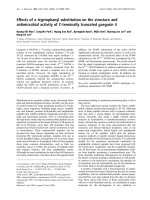
1. Effects of ulinastatin and docetaxel on MDA-MB-231
and MCF-7 cells invasion
Absorbance value at 570 nm reflects the number of cells
penetrated the Matrigel and membrane of the Transwell.
As shown in Figure 1, the invasion rates of cells treated
with ulinastatin , docetaxel and ulinastatin plus docetaxel
were 20.861%, 35.789% and 52.823%, respectively, all
significantly decreased c ompared with that of the con-
trol (p < 0.01).
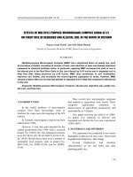
2. Effects of ulinastatin and docetaxel on uPA, uPAR and
ERK mRNA level
As shown in Figure 2(1), uPA and uPAR mRNA levels
in M DA-MB- 231cells treated with ulinastatin as well as
ulinastatin plus docetaxel were significantly decreased
compared with those in control treated cells (p < 0.05).
By contrast, uPA and uPAR mRNA levels were signifi-
cantly enhanced in c ells treated with docetaxel (p <
0.05). I n addition, all treatments had no effects on ERK
mRNA level (p = 0.9). However, ERK mRNA has statis-
tical difference in MCF-7 (p < 0.05). Figure 2(2).
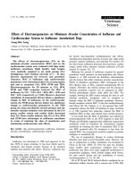
3. Effects of ulinastatin and docetaxel on uPA, uPAR and
phosphorylated ERK1/2 (p-ERK1/2) proteins
Levels of uPA, uPAR and p-ERK1/2 in MDA-MB-231
cells treated with ulinastatin and docetaxel are shown in
Figure 3(1). Treatment of cells with ulinastatin alone or
along with docetaxel significantly decreased uPA, uPAR
and p-ERK1/2 level in MDA-MB-231 cells. By contrast,
treatment of cells with docetaxel significantly augmented
uPA, uPAR and p-ERK1/2 levels Figure 3(2) (p < 0.05).
4. uPA, uPAR and p-ERK1/2 level in exograft of nude mice
Specimens of MDA-MB-231 mouse exograft s were
immunostained for uPA, uPAR and p-ERK. The IOD
Figure 1 Inhibition of ulinastatin and docetaxel on MDA-MB-
231 and MCF-7 cell invasion. Shown are the absorptions at 570
nm of cells treated with ulinastatin, docetaxe and ulinastatin plus
docetaxe for 24 hours, respectively, in the lower chambers of
transwells. Treatment of cells with ulinastatin, docetaxe and
ulinastatin plus docetaxe significantly inhibited MDA-MB-231(1a) and
MCF-7 (1b) cell invasion.
Luo et al. Journal of Experimental & Clinical Cancer Research 2011, 30:71
/>Page 3 of 7
values of the targeted proteins in each group were statis-
tically analyzed. The levels of uPA, uPAR and p-ERK1/2
in ulinastatin group were lower than those of ulinastatin
plus docetaxel group; both groups had significant low er
levels of uPA, uPAR and p-ERK1/2 than the control
group. Figure 4,6. By contrast, the levels of uPA, uPAR
and p-ERK in docetaxel group were significantly higher
than those of the cont rol group (p < 0 .05). The immu-
nohistochemistry result of MCF-7 is same as the result
in MDA-MB-231. Figure 5,7.
Discussion
Proliferation and invasion are import ant biological fea-
tures of breast cancer. Because the development of
breast cancer involves many extremely complicate regu-
latory factors, its treatment is often difficult. Therefore,
the objective of the study is to explore various cytokines’
mechanisms and relationship in regulating tumor cell
proliferation and invasion, and eventually find the corre-
sponding optimal therapeutic measures.
Urokinase-type plasminogen activator (uPA) is the
hub of the plasminogen activator system, also known as
Figure 3 Effects of docetaxe and ulinastatin on expression of uPA, uPAR and p-ERK1/2 in MDA-MB-231 cells.(1)Shownarethe
representative results of western blot of uPA, uPAR and p-ERK1/2 in MDA-MB-231 cells treated with control, ulinastatin, docetaxel, and ulinastatin
plus docetaxel, respectively. (2) Shown are the quantitative results of western blot experiments.
Figure 2 Effects of ulinastatin and doceta xe on mRNA leve l of uPA, uPAR and ERK in MDA-MB-231 cells and MCF-7 cells. (1)Shown
are the RT-PCR results of relative mRNA levels of uPA (a) uPAR (b) and ERK (c) to b-actin in MDA-MB-231 cells treated with ulinastatin, docetaxe
and ulinastatin plus docetaxe for 24 hours, respectively. (2) Shown are the RT-PCR results of relative mRNA levels of uPA (a) uPAR (b) and ERK (c)
to b-actin in MCF-7(a,b,c) cells treated with ulinastatin, docetaxe and ulinastatin plus docetaxe for 24 hours, respectively.
Luo et al. Journal of Experimental & Clinical Cancer Research 2011, 30:71
/>Page 4 of 7
uPA system. As a multifunctional serine protease, in
addition t o its direct contribution to the degradation of
extracellular matrix, uPA also mediates activation of
matrix metalloproteinase[7], thereby promoting cancer
cell invasion and migration. Recent studies have revealed
that uPA is involved in angiongenesis and lymphangio-
gen esis[8] and related to cell proliferati on-related signal
transduction pathway. Binding of uPA to its receptor
uPAR is known to regulate uPAR expression. Therefore,
uPA and uPAR usually are similarly over-expressed in
breast cancer cells[9].
Ulinastatin binds to cells through its domain I, and
exerts its anti-fibrinolytic activity through its domain
II. Our results of real time PCR showed that ulinasta-
tin treatment decreased uPA and uPAR mRNA level,
suggesting that ulinastatin can inhibit uPA at genetic
level and subsequently reducing the expression of
uPAR.
ERK belongs to a class of serine/threonine protein
kinases found in late 80s of the last century and is a
membe r of Ras-Raf-MEK-ERK signal transduction path-
way. Phosphorylated ERK (p-ERK) can promote cell sur-
vival, growth and mitosis by regulating nuclear
transcription factor NF-B activity. The promoter of
uPA gene has NF-B binding sites, therefore, p-ERK can
increases expression of uPA through activation of NF-
B[10]. In addition, a large number of studies in recent
Figure 5 Posit ive immunohistochemical expression of uPA,
uPAR, p-ERK1/2 in MDA-MB-231 exnografts of mice in control
(a), ulinastatin(b), docetaxel(c),ulinastatin plus docetaxel(d)
groups (SP,×400)(1). Positive immunohistochemical expression of
uPA in MDA-MB-231 exnografts of mice in control (a), ulinastatin (b),
docetaxel (c), and ulinastatin plus docetaxel (d) groups (SP, ×400).(2).
Positive immunohistochemical expression of uPAR in MDA-MB-231
exnografts of mice in control (a), ulinastatin (b), docetaxel (c), and
ulinastatin plus docetaxel (d) groups (SP, ×400).(3). Positive
immunohistochemical expression of p-ERK1/2 in MDA-MB-231
exnografts of mice in control (a), ulinastatin (b), docetaxel (c), and
ulinastatin plus docetaxel (d) groups (SP, ×400).
Figure 4 Effectsofdocetaxeandulinastatinonexpressionof
uPA, uPAR and p-ERK1/2 in mouse exografts. Shown are the
quantitative results of uPA, uPAR and p-ERK1/2 expression in
exografts of mice treated with control, ulinastatin, docetaxel, and
ulinastatin plus docetaxel, respectively, in immunohistochemical
experiments.
Figure 6 Effectsofdocetaxeandulinastatinonexpressionof
uPA, uPAR and p-ERK1/2 in mouse exografts. Shown are the
quantitative results of uPA, uPAR and p-ERK1/2 expression in
exografts of mice treated with control, ulinastatin, docetaxel, and
ulinastatin plus docetaxel, respectively, in immunohistochemical
experiments.
Luo et al. Journal of Experimental & Clinical Cancer Research 2011, 30:71
/>Page 5 of 7
years have confirmed[2,3,11-13] that binding of uPA to
uPAR can activate Ras-ERK pathway.
For example, in human breast cancer MCF-7 cells,
when the LDL receptor family members are depolymer-
ized, binding o f endogenous uPA to uPAR can activate
ERK[14,15]. The result shows in MCF-7 cells either, its
ERK decressed obviously. Furthermore, uPAR can also
regulate basal p-ERK level by binding to integrin a5b1
[3,16]. Therefore, uPA-uPAR and ERK can activate each
other through different pathways and form a positive
feedback loop, thereby maintaining high proliferating
and invasive ability of cancer cells.
The basal expression of uPA, uPAR and p-ERK in
breast cancer MDA-MB-231 cells are very high[17,18].
Ulinastatin treatment could significantly decrease uPA
and uPAR protein expression and mRNA level com-
pared with control group (p < 0.05), possibly due to its
inhibitory effect on the translocation of protein kinase C
from the cytoplasm to the membrane and consequent
down-regulation of MEK /ERK/c-Jun pathway, thereby
causing the decline in uPA expression[5]. its mediated-
downregulation of uPA inhibited ERK phosphorylation
Figure 4,5,6,7.
Docetaxel can cause cancer cell mitotic arrest at G2/
M phase by inhibiting tubulin depolymerization and
promoting non-functional microtube formation. Furthe r
studies in recent years have revealed a role of docetaxel
in other mechanisms besides cell toxicity. Our experi-
ments also showed that docetaxel treatment increased
p-ERK1/2 level (p < 0.05), but decreased uPA and uPAR
mRNA and protein levels (p < 0.05), in consistence with
the reports of Yacoub and Mhaidat[19,20]. The specific
mechanism on how docetaxel functions has not yet
been clarified, but probably is related to its role in initia-
tion of cell apoptosis and consequent activation of ERK
pathway and p-ERK-dependent upregulation of uPA
expression. In addition, reports have shown that pre-
treatment of cells with other ERK activity specific inhibi-
tor can markedly promote the effe ct of docetaxel on cell
apoptosis[20,21]. Our study also found that treatment of
cells with ulinastatin along with docetaxel significantly
inhibited uPA, uPAR and ERK1/2, leading to the maxi-
mum cell apoptosis rate among the three treatment
groups (83.254% at 72 hours)[6] . Therefore, the upregu-
lation of these three proteins in response to docetaxel
treatment should be considered as one of the drug-resis-
tance mechanisms of MDA-MB-231 cells, and applica-
tion of inhibitors (such as ulinasta tin) can weaken this
resistance.
This study revealed that uPA, uPAR and p-ERK
expression is obvious ly inhibited by ulinastatin. Because
many factors and mechanisms are involved in cancer
cell proliferation, although t reatment with ulinastatin
alone can inhibit MDA-MB-231 cell proliferation and
exograft growth[6], its effect is not as strong as that
combined with docetaxel. On the other hand, although
docetaxel enhanced the expre ssion of uPA, uPAR and
ERK1/2, its cell toxicity still plays a dominant role, so
when treated with docetaxel alone, the proliferation and
tumor growth of breast cancer cell was inhibited. Com-
bined treatment of ulinastatin plus docetaxel is more
effective in anti-tumor i nvasion. Therefore, the role of
ulinastatin in the antitumor aspect deserves further
study.
Acknowledgements
This work is supported by the Fund of Chongqing Science and Technology
Commission (CSCT, 2008AC5082).
Authors’ contributions
JL did the cell invasion essay and immunohistochemistry, XS did the Cell-
culturing, submitted paper and revised the paper, FG did the medical
statistics, XZ cultured the cell and did PCR, BZ tested the cells in PCR, HW
detected the cells in western blot, ZS designed this experiment and wrote
this paper. All authors read and approved this final draft.
Competing interests
The authors declare that they have no competing interests.
Received: 19 June 2011 Accepted: 29 July 2011 Published: 29 July 2011
Figure 7 Posit ive immunohistochemical expression of uPA,
uPAR, p-ERK1/2 in in MCF-7 exnografts of mice in control(a),
ulinastatin(b), docetaxel(c),ulinastatin plus docetaxel(d) groups
(SP,×400) (1).Positive immunohistochemical expression of uPA in
MCF-7 exnografts of mice in control (a), ulinastatin (b), docetaxel (c),
and ulinastatin plus docetaxel (d) groups (SP, ×400). (2) Positive
immunohistochemical expression of uPAR in MCF-7 exnografts of
mice in control (a), ulinastatin (b), docetaxel (c), and ulinastatin plus
docetaxel (d) groups (SP, ×400). (3). Positive immunohistochemical
expression of p-ERK1/2 in MCF-7 exnografts of mice in control (a),
ulinastatin (b), docetaxel (c), and ulinastatin plus docetaxel (d)
groups (SP, ×400).
Luo et al. Journal of Experimental & Clinical Cancer Research 2011, 30:71
/>Page 6 of 7
References
1. Stillfried GE, Saunders DN, Ranson M: Plasminogen binding and activation
at the breast cancer cell surface: the integral role of urokinase activity.
Breast Cancer Res 2007, 9(1):R14.
2. Nguyen DH, Hussaini IM, Gonias SL: Binding of urokinase-type
plasminogen activator to its receptor in MCF-7 cells activates
extracellular signal-regulated kinase 1 and 2 which is required for
increased cellular motility. J Biol Chem 1998, 273(14):8502-8507.
3. Aguirre GJ, Kovalski K, Ossowski L: Tumor dormancy induced by
downregulation of urokinase receptor in human carcinoma involves
integrin and MAPK signaling. J Cell Biol 1999, 147(1):89-104.
4. Kobayashi H, Shinohara H, Takeuchi K, Itoh M, Fujie M, Saitoh M: Inhibition
of the soluble and the tumor cell receptor-bound plasmin by urinary
trypsin inhibitor and subsequent effects on tumor cell invasion and
metastasis. Cancer Res 1994, 54(3):844-849.
5. Kobayashi H, Suzuki M, Tanaka Y, Hirashima Y, Terao T: Suppression of
urokinase expression and invasiveness by urinary trypsin inhibitor is
mediated through inhibition of protein kinase C- and MEK/ERK/c-Jun-
dependent signaling pathways. J Biol Chem 2001, 276(3):2015-2022.
6. Zhao Xiaoliang, Sun Xin, Gao Feng, Luo Jie, Sun Zhijun: Effects of
ulinastatin and docataxel on breast tumor growth and expression of IL-
6, IL-8, and TNF-a. Journal of Experimental & Clinical Cancer Research 2011,
30:22.
7. Sun ZJ, Yu T, Chen JS: Effects of Ulinastatin and Cyclophosphamide on
the Growth of Xenograft Breast Cancer and Expression of CXCR4 and
MMP-9 in Cancers. The Journal of International Medical Research 2010,
38:967-976.
8. Gao F, Sun ZJ: Progress in the correlation of vascular endothelial growth
factor C and lymphangiogenesis and lymph node metastasis in breast
cancer. Chongqing Medical Journal 2010, , 07: 819-821.
9. Mahanivong C, Yu J, Huang S: Elevated urokinase-specific surface
receptor expression is maintained through its interaction with urokinase
plasminogen activator. Mol Carcinog 2007, 46(3):165-175.
10. Sliva D, Rizzo MT, English D: Phosphatidylinositol 3-kinase and NF-kappaB
regulate motility of invasive MDA-MB-231 human breast cancer cells by
the secretion of urokinase-type plasminogen activator. J Biol Chem 2002,
277(5):3150-3157.
11. Kanse SM, Benzakour O, Kanthou C, Christine K, Roger Lijnen H,
Preissner KT: Induction of vascular SMC proliferation by urokinase
indicates a novel mechanism of action in vasoproliferative disorders.
Arterioscler Thromb Vasc Biol 1997, 17(11):2848-2854.
12. Konakova M, Hucho F, Schleuning WD: Downstream targets of urokinase-
type plasminogen-activator-mediated signal transduction. Eur J Biochem
1998, 253(2):421-429.
13. Tang H, Kerins DM, Hao Q, Inagami T, Vaughan DE: The urokinase-type
plasminogen activator receptor mediates tyrosine phosphorylation of
focal adhesion proteins and activation of mitogen-activated protein
kinase in cultured endothelial cells. J Biol Chem 1998,
273(29):18268-18272.
14. Webb DJ, Nguyen DH, Sankovic M, Gonias SL: The very low density
lipoprotein receptor regulates urokinase receptor catabolism and breast
cancer cell motility in vitro. J Biol Chem 1999, 274(11)
:7412-7420.
15. Webb DJ, Nguyen DH, Gonias SL: Extracellular signal-regulated kinase
functions in the urokinase receptor-dependent pathway by which
neutralization of low density lipoprotein receptor-related protein
promotes fibrosarcoma cell migration and matrigel invasion. J Cell Sci
2000, 113(Pt 1):123-134.
16. Yu W, Kim J, Ossowski L: Reduction in surface urokinase receptor forces
malignant cells into a protracted state of dormancy. J Cell Biol 1997,
137(3):767-777.
17. Seddighzadeh M, Zhou JN, Kronenwett U, Shoshan MC, Auer G, Sten-
Linder M, et al: ERK signalling in metastatic human MDA-MB-231 breast
carcinoma cells is adapted to obtain high urokinase expression and
rapid cell proliferation. Clin Exp Metastasis 1999, 17(8):649-654.
18. Holst-Hansen C, Johannessen B, Hoyer-Hansen G, Romer J, Ellis V,
Brunner N: Urokinase-type plasminogen activation in three human
breast cancer cell lines correlates with their in vitro invasiveness. Clin
Exp Metastasis 1996, 14(3):297-307.
19. Mhaidat NM, Thorne RF, Zhang XD, Hersey P: Regulation of docetaxel-
induced apoptosis of human melanoma cells by different isoforms of
protein kinase C. Mol Cancer Res 2007, 5(10):1073-1081.
20. Yacoub A, Han SI, Caron R, Gilfor D, Mooberry S, Grant S, et al: Sequence
dependent exposure of mammary carcinoma cells to Docetaxel and the
MEK1/2 inhibitor U0126 causes enhanced cell killing in vitro. Cancer Biol
Ther 2003, 2(6):670-676.
21. Davies BR, Logie A, Mckay JS, Martin P, Steele S, Jenkins R, et al: AZD6244
(ARRY-142886), a potent inhibitor of mitogen-activated protein kinase/
extracellular signal-regulated kinase kinase 1/2 kinases: mechanism of
action in vivo, pharmacokinetic/pharmacodynamic relationship, and
potential for combination in preclinical models. Mol Cancer Ther 2007,
6(8):2209-2219.
doi:10.1186/1756-9966-30-71
Cite this article as: Luo et al.: Effects of ulinastatin and docetaxel on
breast cancer invasion and expression of uPA, uPAR and ERK. Journal of
Experimental & Clinical Cancer Research 2011 30:71.
Submit your next manuscript to BioMed Central
and take full advantage of:
• Convenient online submission
• Thorough peer review
• No space constraints or color figure charges
• Immediate publication on acceptance
• Inclusion in PubMed, CAS, Scopus and Google Scholar
• Research which is freely available for redistribution
Submit your manuscript at
www.biomedcentral.com/submit
Luo et al. Journal of Experimental & Clinical Cancer Research 2011, 30:71
/>Page 7 of 7