Báo cáo y học: "Pre- and post-operative gait analysis for evaluation of neck pain in chronic whiplash" pptx
Bạn đang xem bản rút gọn của tài liệu. Xem và tải ngay bản đầy đủ của tài liệu tại đây (820.86 KB, 6 trang )
BioMed Central
Page 1 of 6
(page number not for citation purposes)
Journal of Brachial Plexus and
Peripheral Nerve Injury
Open Access
Research article
Pre- and post-operative gait analysis for evaluation of neck pain in
chronic whiplash
Ake Nystrom*
1,2
, Glen M Ginsburg
1,3
, Wayne Stuberg
3
and Stacey Dejong
3
Address:
1
Department of Orthopaedic Surgery and Rehabilitation, University of Nebraska Medical Center, Omaha NE 68198, USA,
2
Division of
Plastic and Reconstructive Surgery, University of Nebraska Medical Center, Omaha NE 68198, USA and
3
Munroe-Meyer Motion Analysis
Laboratory, University of Nebraska, Lincoln, NE 68588, USA
Email: Ake Nystrom* - ; Glen M Ginsburg - ; Wayne Stuberg - ;
Stacey Dejong -
* Corresponding author
Abstract
Introduction: Chronic neck pain after whiplash is notoriously refractory to conservative
treatment, and positive radiological findings to explain the symptoms are scarce. The apparent
disproportionality between subjective complaints and objective findings is significant for the
planning of treatment, impairment ratings, and judicial questions on causation. However, failure to
identify a symptom's focal origin with routine imaging studies does not invalidate the symptom per
se. It is therefore of a general interest both to develop effective therapeutic strategies in chronic
whiplash, and to establish techniques for objectively evaluation of treatment outcomes.
Methods: Twelve patients with chronic neck pain after whiplash underwent pre- and
postoperative computerized 3D gait analysis.
Results: Significant improvement was found in all gait parameters, cervical range-of-motion, and
self reported pain (VAS).
Conclusion: Chronic neck pain is associated with abnormal cervical spine motion and gait
patterns. 3D gait analysis is a useful instrument to assess the outcome of treatment for neck pain.
Introduction
Serious persistent problems after whiplash trauma to the
neck, sometimes referred to as Whiplash Associated Dis-
orders (WAD)[1] is a common and costly condition; esti-
mates indicate an incidence of over 250,000 in the United
States, at an annual cost in 2002 of $2.7 billion or close to
$10,000 per incident. [2] Although initial symptoms from
acceleration-deceleration trauma to the neck may
improve spontaneously or with physical therapy over the
course of weeks-to-months, [1] chronic and potentially
disabling symptoms persist in a significant percentage of
all cases. [3,4] A complicating factor, which is also a rea-
son for controversy, is the frequent failure of routine clin-
ical laboratory investigative methods including MRI and
electrodiagnostic studies, to objectively identify the cause
of pain and other symptoms. [5,6]
Although not a universal finding, stiffness of the neck and
shoulders is a common sequela of whiplash. [5-10] Using
3D motion analysis techniques, Dall'Alba et al. [11] iden-
tified significant limitations with a particular pattern of
cervical range of motion among patients with WAD, but
also pointed out that their results do not provide an expla-
nation for the loss of neck mobility. In a study where sim-
Published: 17 July 2009
Journal of Brachial Plexus and Peripheral Nerve Injury 2009, 4:10 doi:10.1186/1749-7221-4-10
Received: 22 April 2009
Accepted: 17 July 2009
This article is available from: />© 2009 Nystrom et al; licensee BioMed Central Ltd.
This is an Open Access article distributed under the terms of the Creative Commons Attribution License ( />),
which permits unrestricted use, distribution, and reproduction in any medium, provided the original work is properly cited.
Journal of Brachial Plexus and Peripheral Nerve Injury 2009, 4:10 />Page 2 of 6
(page number not for citation purposes)
ilar techniques were applied, Gargan et al found that
cervical range of motion and psychological scores at three
months were predictive of clinical outcomes at 2 years.
[11] Their findings were confirmed by Tomlinson et al in
a follow-up study on the same cohort, 7.5 years later. [9]
Existing data suggest that neck stiffness in WAD may be an
expression of pain inhibition from soft tissue injury and
painful muscle spasm without pathology of the spine.
Thus, injections of Botox
®
to trigger points in superficial
neck muscles have been shown to provide temporary but
significant decrease in pain and increase in cervical
ROM,[8] with similar effect of short duration from injec-
tions of local anesthetic to myofascial trigger points in the
neck. [12] While rarely a definitive solution to problems
associated with the chronic whiplash syndrome, such
injections may be helpful in identifying focal origin(s) of
soft-tissue pain. [12,13]
3D motion analysis represents the diagnostic gold stand-
ard for conditions that affect the kinematics of the lower
extremities, pelvis and trunk. Using this technology, sev-
eral investigators have confirmed that deviations from
normal gait mechanics also affect the compensatory
movements of the head and neck. [14,15] Other studies
have demonstrated that temporal and spatial changes in
gait are complimented in the neck through input from the
vestibulo-ocular reflex (VOR) for stabilization of gaze dur-
ing angular movements, [16] while head position is con-
trolled by the cervicocollic reflex (CCR), vestibulocollic
reflex (VCR) and optocollic reflexes (OCR) through prop-
rioceptive, vestibular and ocular mechanisms. [14,16]
Whether variations in gait parameters are voluntary (due
to changes in terrain, gait speed, direction, etc.) or repre-
sent deviations from "normal" kinematics (changes in
temporal distance measures of walking or joint move-
ment from disease, injury, or surgery), they will, through
reflex mechanisms, result in adaptive changes in the kine-
matics of the cervical spine.
The effect of lower segment dysfunction on the upper
body kinematics has been previously investigated in nor-
mal controls and in patient groups with musculoskeletal
disorders. [17-19] We have not, however, found any stud-
ies exploring if standard gait parameters are impaired as a
result of upper body dysfunction, The present investiga-
tion was designed for that purpose and, secondly, to
assess the usefulness of computerized 3D gait analysis to
objectively monitor outcomes of treatment for neck pain.
Methods
Subjects
Participants were recruited among patients referred to
University of Nebraska Medical Center for treatment of
chronic neck pain after whiplash (WAD II–III, Table 1).
Inclusion criteria are summarized in Table 2.
The study group consisted of twelve consecutive patients
(10 F, 2 M) ages 26 to 67 (mean 44.9 ± 12.8). All subjects
were able to understand simple commands and ambulate
independently with or without assistive devices.
Treatment
Areas of intense focal tenderness, generally in the lower
cervical paraspinal musculature or horizontal segment(s)
of the trapezius muscle(s), were preoperatively mapped
through diagnostic injections of local anesthetic (Mar-
caine
®
0.25 mg/ml). In a surgical procedure designed to
identify and eliminate focal pain generators, the 'tender
points' were thereafter addressed during an operation that
generally included exploration, neurolysis and decom-
pression of the spinal accessory nerve and/or dorsal sen-
sory branches of cervical nerve roots at their passage
through fibrotic trapezius fascia, and trapezius fasciec-
tomy.[13,20] In order to optimize the outcome of treat-
ment, all patients participated actively with the surgeon in
the operating room to identify focal areas of pain. No
sedation, analgesia or local anesthetic was used during
these key portions of the procedure.
Data collection
Three dimensional motion analyses were carried out
using a six camera Vicon system (60 Hz), Vicon Worksta-
tion and Polygon software, and the Vicon Plug-In-Gait
full body biomechanical model to collect pre- and post-
operative data pertaining to gait (speed, cadance and step
Table 1: Classification of Whiplash Associated Disorders (WAD)
0 No complaints. No objective physical signs
I Pain. No objective physical signs.
II Pain. Objective musculoskeletal signs, e.g. stiffness.
III Pain. Objective neurological signs, e.g. weakness, numbness, absent tendon reflexes.
IV Pain. Radiological evidence of skeletal injury or dislocation.
Journal of Brachial Plexus and Peripheral Nerve Injury 2009, 4:10 />Page 3 of 6
(page number not for citation purposes)
length), and cervical range-of-motion (degrees from rest-
ing position). Pain was assessed with a linear Visual Ana-
logue Scale (VAS) graded 0–1. The evaluations were
performed one week before, and 1–10 weeks (27.7 ± 21.6
days) after surgery.
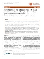
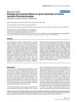
Marker positioning and objective measurements. Four
markers, placed at the left and right temporal and occipi-
tal regions, respectively, defined a 'head' segment. Addi-
tional markers over the sternal notch, xiphoid process,
and spinous processes of C7 and T10, defined a 'thorax'
segment to allow calculation of orthogonal angles
between the two segments. The standard Vicon marker set
was used for the lower extremities with a marker on each
of the anterior iliac spines, centered between the posterior
superior iliac spines, lateral on the thigh and shank, lateral
on the knee joint and lateral malleolus and on the dorsum
of the foot over the head of the second metatarsal. Figure
1. A static trial using a knee-alignment device was used to
estimate knee joint centers.
A standard lower body marker set and Plug-In-Gait mod-
eling software was used for precise calculation of repeated
angle measurements from gait. [21] The precision of angle
measurements for the cervical spine using the Plug-in gait
modeling software has not been determined, but is
assumed to be as valid as measures for the lower body.
Precision of centroid position of the markers has been
demonstrated to be accurate to within a millimeter
(Vicon, Oxford, England).
During data collection, subjects were asked to move the
head along three planes of the neck (flexion-extension,
left-right rotation, left-right lateral flexion) to the point of
maximum ability or tolerance. Angles between the thorax
and head segments were calculated using the Plug-In-Gait
full body model, and the maximum angle for each of
three trials was identified for each direction of movement.
The average of the three trials was used as outcome meas-
ure for maximum active range of motion in each direc-
tion.
Prior to the measurements of cervical mobility, subjects
performed 10 to 15 walking trials at their self selected
usual velocity. Walking speed was calculated for each trial,
and the three trials closest to the subject's average walking
speed were selected for analysis of the temporal distance
parameters. Outcome measures included average walking
speed, cadence, and bilateral step lengths.
Pain assessment. Participants rated their overall pain
before and after each evaluation session, on a linear visual
analog scale (VAS) with 0 representing no pain and 10
representing the most severe pain the subject had ever felt.
Using the same scale, participants also rated their pain in
relation to a typical day during the previous week.
Statistical analysis
Analysis of data was performed using Student's paired t-
test. Statistical significance was set at p < 0.05. Intraclass
correlation coefficient (ICC) was used to assess intra-ses-
sion reliability for each of the six cervical spine motion
measures taken during both pre and post sessions. [22]
The data were compared using ICC (2,1) where time was
modeled as a random effect since we were interested in
Table 2: Inclusion criteria
Age 19 or older
Neck pain precipitated by whiplash trauma
Failure of conservative treatment for more than one year
Absence of gross neurologic signs
Absence of gross radiological (MRI) pathology
Marker placement for computerized 3-D motion analysisFigure 1
Marker placement for computerized 3-D motion
analysis.
Journal of Brachial Plexus and Peripheral Nerve Injury 2009, 4:10 />Page 4 of 6
(page number not for citation purposes)
the reliability between any repeated measurements meas-
ured not on the same time per session.
Results
Excellent reliability of the cervical spine measures were
observed with ICC values consistently above 0.9 as
detailed in Table 3.
The analysis of data confirmed statistically significant (p <
0.005) improvement in cervical range of motion in all six
planes following treatment, with the greatest average
improvements in flexion-extension (54%), followed by
rotation (53.5%). Table 4.
At follow-up, walking speed had increased by an average
of 13.9 centimeters/second, with a 5.2 centimeter average
increase in step length. Table 5.
All patients gave postoperative neck pain ratings that were
significantly lower than before surgery, both for daily
pain, and for how much their pain increased during exer-
tion. Table 6.
No major complications related to treatment were docu-
mented among the participants during surgery or the
postoperative period.
Discussion
Significant improvement in three gait parameters were
documented after treatment for neck pain from whiplash,
a condition that because of a purported lack of diagnostic
laboratory findings has been described by some authors
as a social or emotional disorder in need of no treatment.
[23-25]
Pain-related neck stiffness is a cardinal component of the
chronic whiplash syndrome, but reliable assessment of
cervical range-of-motion is highly dependent on the sub-
ject's voluntary effort. Inclinometer- or observation based
techniques, or even computer-guided three-dimensional
measurement systems are therefore not ideal tools to
objectively confirm or monitor chronic whiplash.[26] In
contrast, gait is a complex but highly automated function
and therefore better suited for standardized analysis.
A clinically validated marker system [27,28] was adopted
for the purpose of this investigation, and the consistency
of cervical range-of-motion was confirmed through
repeated measurements in each participant since kine-
matic reproducibility has been established as a method to
differentiate healthy subjects simulating neck pain from
patients with true whiplash injuries.[7,12,29] With these
precautions, we consider the present findings reliable and
valid.
Various kinematic abnormalities have been reported in
chronic whiplash syndrome, often without conclusive evi-
dence of their underlying cause(s). Thus, even though
imaging evidence of abnormal cervical [30] or craniocer-
vical [31] motion patterns have lead to recommendations
to fuse the cranio-cervical joint complex, [32,33] it has
not been shown that a causative relation exists between
such radiological findings and the clinical whiplash syn-
drome. Other investigators have interpreted patterns of
oculomotor dysfunction in whiplash patients as evidence
of brainstem injury, or "disorganized neck proprioceptive
activity" leading to distortion of the posture control sys-
tem. [34-37] While none of the participants in this inves-
tigation had undergone specific diagnostic studies to
assess brain stem function or cervical stability, the signifi-
cant improvements in pain, cervical range-of-motion, and
temporal-distance gait parameters illustrate that soft tis-
sue surgery may alleviate considerable symptoms after
whiplash in carefully selected patients. The findings also
allow the following conclusions: (1) Upper segment pain,
e.g. in chronic whiplash syndrome, may be expressed as
Table 3: Cervical Spine Measure ICC Values
ICC Value
C-Spine Motion Variables Pre-Session Measure Post-Session Measure
Extension 0.979 0.987
Flexion 0.912 0.956
Left Lateral Flexion 0.983 0.963
Right Lateral Flexion 0.952 0.972
Right Rotation 0.973 0.986
Left Rotation 0.971 0.986
Journal of Brachial Plexus and Peripheral Nerve Injury 2009, 4:10 />Page 5 of 6
(page number not for citation purposes)
Table 4: Maximum Active Neck Range of Motion (degrees)
Pre-op Post-op Mean change Paired t-test
Mean ± SD Mean ± SD Degrees Percent t statistic p-value
Flexion 25.2 ± 11.9 39.6 ± 12.9 14.4 57 -3.61 0.002
Extension 29.3 ± 13.8 44.4 ± 20.2 15.1 52 -4.16 0.0008
L Rotation 36.1 ± 21.0 54.1 ± 18.2 18.0 50 -4.21 0.0007
R Rotation 37.3 ± 16.3 59.1 ± 16.1 21.8 58 -5.78 0.00006
L Lat Flexion 19.4 ± 14.1 25.9 ± 16.2 6.5 34 -3.07 0.005
R Lat Flexion 22.9 ± 1201 32.7 ± 10.2 9.8 42 -4.97 0.0002
Table 5: Temporal-Distance Gait Parameters
Pre-op Post-op Mean Difference Paired t-test
Mean ± SD Mean ± SD Degrees Percent t statistic p-value
Walking speed (cm/sec) 98.5 ± 29.1 112.4 ± 17.4 13.9 14 -2.94 0.007
Cadence (steps/min) 105.9 ± 13.8 112.1 ± 7.6 6.2 6 -2.32 0.02
Step length (cm) 54.5 ± 11.1 59.7 ± 7.9 5.2 10 -2.79 0.009
Table 6: Pain Ratings (Visual-Analog Scale 0–10)
Pre-op Post-op Mean change Paired t-test
Mean ± SD Mean ± SD VAS Percent t statistic p-value
Typical day average 6.2 ± 2.0 2.5 ± 1.8 3.7 -60 3.75 0.002
Increase during test 1.6 ± 2.4 0 ± 1.9 1.6 -100 1.82 0.05
Journal of Brachial Plexus and Peripheral Nerve Injury 2009, 4:10 />Page 6 of 6
(page number not for citation purposes)
gait and posture abnormalities;and (2) Computerized 3D
gait analysis provides objective data for diagnosis or out-
come studies in chronic whiplash.
Competing interests
The authors declare that they have no competing interests.
Authors' contributions
All authors participated in design and planning of the
study, and read/approved the final manuscript. Patient
selection and surgical interventions were performed by
NAN. Data collection was performed by SDJ, and super-
vised by WS and GMG. Statistical analysis by WS.
References
1. Spitzer WO, Skovron ML, Salmi LR, Cassidy JD, Duranceau J, Suissa
S, Zeiss E: Scientific monograph of the Quebec Task Force on
Whiplash-Associated Disorders: redefining "whiplash" and
its management. Spine 1995, 20:1S-73S.
2. United Nations Economic and Social Council Proposal to
Develop a Global Technical Regulation Concerning Head
Restraints. TRANS/WP.29/AC.3/13 2005.
3. Carette S: Whiplash injury and chronic neck pain. N Engl J Med
1994, 330:1083-1084.
4. Rosenfeld M, Seferiadis A, Gunnarsson R: Active intervention in
patients with whiplash-associated disorders improves long-
term prognosis: a randomized controlled clinical trial. Spine
2003, 28:2491-2498.
5. Rauschning W, Jónsson H: Injuries of the cervical spine in auto-
mobile accidents: pathoanatomical and clinical aspects. In
Whiplash injuries. Current concepts in prevention, diagnosis, and treatment
of the cervical whiplash syndrome Edited by: Gunzburb R, Szpalski M.
Philadelphia, PA: Lippincott-Raven Publishers; 1998:33-53.
6. Yoganandan N, Cusick JF, Pintar FA, Rao RD: Whiplash injury
determination with conventional spine imaging and cryomi-
crotomy. Spine 2001, 26:2443-2448.
7. Antonaci F, Bulgheroni M, Ghirmai S, Lanfranchi S, Dalla Toffola E,
Sandrini G, Nappi G: 3D kinematic analysis and clinical evalua-
tion of neck movements in patients with whiplash injury.
Cephalalgia 2002, 22:533-542.
8. Juan FJ: Use of botulinum toxin-A for musculoskeletal pain in
patients with whiplash associated disorders. BMC Musculoskelet
Disord 2004, 5:5.
9. Tomlinson PJ, Gargan MF, Bannister GC: The fluctuation in recov-
ery following whiplash injury 7.5-year prospective review.
Injury 2005, 36:758-761.
10. Gargan MF, Bannister G, Main C, Hollis S: The behavioural
response to whiplash injury. J Bone Joint Surg Br 1997, 79:517-518.
11. Dall'Alba PT, Sterling MM, Treleaven JM, Edwards SL, Jull GA: Cervi-
cal range of motion discriminates between asymptomatic
persons and those with whiplash.
Spine 2001, 26:2090-2094.
12. Freeman MD, Nystrom A, Centeno C: Chronic whiplash and cen-
tral sensitization; an evaluation of the role of a myofascial
trigger points in pain modulation. Brachial Plex Peripher Nerve Inj
2009, 4:2.
13. Duffy MF, Stuberg W, DeJong S, Gold KV, Nystrom NA: Case
Report: Whiplash-Associated Disorder from a low velocity
bumper car collision. History, evaluation, and surgery. Spine
2004, 29:1881-1884.
14. Mulavara AP, Verstraete MC, Bloomberg JJ: Modulation of head
movement control in humans during treadmill walking. Gait
Posture 2002, 16:271-282.
15. Menz HB, Lord SR, Fitzpatrick RC: Acceleration patterns of the
head and pelvis when walking on level and irregular surfaces.
Gait Posture 2003, 18:35-46.
16. Chen KJ, Keshner EA, Peterson BW, Hain TC: Modeling head
tracking of visual targets. J Vestib Res 2002, 12:25-33.
17. Kavanagh JJ, Barrett RS, Morrison S: Upper body accelerations
during walking in healthy young and elderly men. Gait Posture
2004, 20:291-298.
18. Frigo C, Carabalona R, Dalla Mura M, Negrini S: The upper body
segmental movements during walking by young females. Clin
Biomech (Bristol, Avon) 2003, 18:419-425.
19. Bartonek A, Saraste H, Eriksson M, Knutson L, Cresswell AG: Upper
body movement during walking in children with lumbosacral
myelomeningocele. Gait Posture 2002, 15:120-129.
20. Hagert CG, Christenson JT: Hyperpressure in the trapezius
muscle associated with fibrosis. Acta Orthop Scand 1990,
61:263-265.
21. Kadaba MP, Ramakrishnan HK, Wootten ME: Measurement of
lower extremity kinematics during level walking. J Orthop Res
1990, 8:383-390.
22. Shrout PE, Fleiss JL: Intraclass Correlations: Uses in Assessing
Rater Reliabilty.
Psychol Bull 1979, 2:420-428.
23. Ferrari R, Shorter E: From railway spine to whiplash – the recy-
cling of nervous irritation. Med Sci Monit 2003, 9:HY27-37.
24. Ferrari R, Russell AS, Carroll LJ, Cassidy JD: A re-examination of
the whiplash associated disorders (WAD) as a systemic ill-
ness. Ann Rheum Dis 2005, 64:1337-1342.
25. Ferrari R, Kwan O, Russell AS, Pearce JM, Schrader H: best
approach to the problem of whiplash? One ticket to Lithua-
nia, please. Clin Exp Rheumatol 1999, 17:321-326.
26. Schaufele MK, Boden SD: Physical function measurements in
neck pain. Phys Med Rehabil Clin N Am 2003, 14:569-588.
27. Wilk B, Karol LA, Johnston CE 2nd, Colby S, Haideri N: The effect
of scoliosis fusion on spinal motion: a comparison of fused
and nonfused patients with idiopathic scoliosis. Spine 2006,
31:309-314.
28. Engsberg JR, Lenke LG, Uhrich ML, Ross SA, Bridwell KH: Prospec-
tive comparison of gait and trunk range of motion in adoles-
cents with idiopathic thoracic scoliosis undergoing anterior
or posterior spinal fusion. Spine 2003, 28:1993-2000.
29. Berger M, Lechner-Steinleitner S, Hoffmann F, Schönegger J: Akzel-
erations-Dezelerations-Trauma der Halswirbelsäule. Diag-
nose schmerzbedingter und simulierter zervikaler
Bewegungsstörungen. Schmerz 1998, 12:400-405.
30. Kristjansson E, Leivseth G, Brinckmann P, Frobin W: Increased sag-
ittal plane segmental motion in the lower cervical spine in
women with chronic whiplash-associated disorders, grades I-
II: A case-control study using a new measurement protocol.
Spine 2003, 28:2215-2221.
31. Krakenes J, Kaale BR, Moen G, Nordli H, Gilhus NE, Rorvik J: MRI
assessment of the alar ligaments in the late stage of whiplash
injury-a study of structural abnormalities and observer
agreement. Neuroradiology 2002, 44:617-624.
32. Johansson BH: Whiplash injuries can be visible by functional
magnetic resonance imaging. Pain Res Manag 2006, 11:197-199.
33. Volle E, Montazem A: MRI video diagnosis and surgical therapy
of soft tissue trauma to the craniocervical junction. Ear Nose
Throat J 2001, 80:41-4. 46–8
34. Prushansky T, Dvir Z, Pevzner E, Gordon CR: Electro-oculo-
graphic measures in patients with chronic whiplash and
healthy subjects: a comparative study. J Neurol Neurosurg Psychi-
atry 2004, 75:1642-4.
35. Gimse R, Tjell C, Bjørgen IA, Saunte C: Disturbed eye movements
after whiplash due to injuries to the posture control system.
J Clin Exp Neuropsychol 1996, 18:176-186.
36. Kristjansson E, Hardardottir L, Asmundardottir M, Gudmundsson K:
A new clinical test for cervicocephalic kinesthetic sensibility:
"the fly". Arch Phys Med Rehabil 2004, 85:490-495.
37. Hildingsson C, Wenngren BI, Bring G, Toolanen G: Oculomotor
problems after cervical spine injury. Acta Orthop Scand 1989,
60:513-516.