Báo cáo y học: "Structural valve deterioration of a mitral Carpentier-Edwards pericardial bioprosthesis in an 87-year-old woman 16 years after its implantation" doc
Bạn đang xem bản rút gọn của tài liệu. Xem và tải ngay bản đầy đủ của tài liệu tại đây (904.15 KB, 3 trang )
CAS E REP O R T Open Access
Structural valve deterioration of a mitral
Carpentier-Edwards pericardial bioprosthesis in
an 87-year-old woman 16 years after its
implantation
Hiroshi Ito
*
, Kensuke Sakata, Takashi Haruki and Yurio Kobayashi
Abstract
The second-generation pericardial valve, the Carpentier-Edwards perimount bioprosthetic (CEP) valve, shows
dramatically improved durability as compared to the first-generation pericardial valve, and excellent perfo rmance
has been obtained, in both the aortic and mitral positions. Especially in elderly patients with an implanted CEP
valve, reoperation due to structural valve deterioration (SVD) is rarely required. Here, we report the case of an 87-
year-old woman with an explanted CEP valve in the mitral position due to SVD, 16 years after its implantation.
An 87-year-old woman was admitted to our hospital
with acute heart failure, NYHA class IV. An echocardio-
graphy revealed severe mitral regurgitation and heart
failure with pulmonary hypertension. She had been diag-
nosed as having severe mitral stenosis and had under-
gone mitral valve replacement with a 27-mm
Carpentier-Edwards mitral pericardial valve (model
6900) 16 years prior (at 71 years old) to the present
admission. An echocardiography performed 3 months
prior to this admission revealed mild mitral stenosis and
regurgitation; however, there were no associated clinical
symptoms. Prior to the present admission, she was
brought to the hospital with dyspnea of acute onset . A
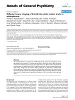
transesophageal echocardiogram revealed severe mitral
regurgitation due to structural valve deterioration (SVD)
of the implanted CEP va lve, moderate TR, and severe
pulmonary hypertension, with a PAP of 93 mm Hg
(Figure 1). She was initially treated with furosemide and
cariperitide, which produced slight improvement of the
heart failure; however, reoperation was found to be
necessary. The reoperation was performed 9 days after
admission via a median sternotomy and under moderate
hypothermic cardiopulmonary bypass with antegrade
cold crystalloid cardiopl egic arrest. The mitral valve was
examined through an incision in the left atrium. A tear
was noted in one of the leaflets of the implanted CEP
mitral valve, which was thought to be the cause of the
severe mitral regurgitation. The cuff of the valve was
covered with thick intima; however, the leaflets were
relatively soft. The valve was resected, and a 27-mm
Mosaic mitral bioprosthesis was implanted i n its place.
Tricuspid valve ring annuloplasty was performed with a
30-mm MC3. The patient was extubated on the day
after the s urgery and disc harged from our hospital on
day 20 after the operation.
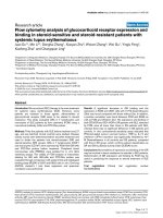
The macroscopic findings of the deteriorated valve
were as follows (Figure 2A, B): The stenosis of the valve
was caused by the host tissue overgrowth restricting the
mobility of the leaflets. A tear was evident in leafl et 1 at
commissure 2, which measured approximately 14 mm,
beginning from the commissure, along the ring of th e
prosthetic valve.
Calcification was detected in the x-ray on leaflets 2
and 3, which w ere covered by a dense layer of host tis-
sue overgrowth (Figure 2C).
Discussion
Marchand et al. reported an actuarial freedom rate from
structural valve deterioration (SVD) in patients receiving
impl antation of the CEP valve, 6900 model, in the mitral
position of 59.2%±6.6% in patients under 60 years of age,
* Correspondence:
Department of Cardiovascular Surgery, Saiseikai Shimonoseki General
Hospital, 8-5-1 Yasuoka, Shimonoseki, 759-6603, Yamaguchi
Ito et al. Journal of Cardiothoracic Surgery 2011, 6:88
/>© 2011 Ito et al; licensee BioMed Cen tral Ltd. This is an Open Access article distributed under the terms of the Creative Commons
Attribution License ( which permits unrestricted use, distribution, and reproduction in
any medium, provided the original work is properly cited.
76.0%±6.3% in patients between 60 and 70 years of age,
and 100% in patients over 70 years of age [1]. A literature
search to the best of our ability revealed no cases that had
undergone CEP valve implantation in the mitral position
at more than 70 years of age, with subsequent SVD and
explantation of the valve. The average life expectancy of
Japanese is quite long, being 79.59 years in males and
86.44 years in females, and the average expected length of
life at 70 years is 15.1 years in males and 19.61 years in
females (Japan Ministry of Health and Welfare 2009
Our
patient had undergone her first implantation of a mitral
bioprosthetic valve at the age of 71 years; her postopera-
tive course had been excellent, and her condition had
remained satisfactory for more than 15 years without war-
farin. Unfortunate ly, she developed SVD suddenly, 16
years after the valve implantation, and needed a reopera-
tion at the age of 87 years. However, she was healthy
enough even at this age to tolerate heart surgery.
Bioprosthetic valve implantation in the mitral position
is usually performed in patients who are more than 60
or 70 years old. As reported here, especially in Japanese
subjects who have a long life expectancy, SVD of an
implanted valve at over 70 years of age may occur , pos-
sibly necessitating reoperation. Notwithstanding, bio-
prosthetic valves must be selected for elderly patients,
considering the risk of thromboembolism, and conse-
quently of hemorrhage associated with the use of war-
farin, in patients with a mechanical valve. Cannegier et
al. reported that the ri sk of hemorrhage in patients over
70 years of age with an implanted mechanical valve was
5.6%/pt-year, which is twice as high as the risk reported
in patients who are less than 70 years old [2]. Holper et
al. reported that the actuarial freedom rate from major
bleeding at 15 years was 88%±4% in patients with an
implanted bioprosthetic valve, and 57%±1.1% in those
with an implante d mechanical valve [3]. In agreement
with this, Marchand et al. reported that the actuarial
freedom rate from major bleeding in patients with an
implanted CEP valve was 86.6%±3.2% at 14 years [1].
These data suggest that a bioprosthetic valve i s superior
to a mechanical valve from the viewpoint of the risk of
major bleeding. As reoperation can also be perf ormed
safely in elderly patients at present, it might be better to
select a bioprosthetic valve fo r elderly patient s, notwith-
standing the risk of reoperation due to SVD more than
10 years later [4].
In regard to the pattern of SVD of a CEP valve, calcifi-
cation (70.4%-73%), valve tear (18.5%-20%), or and both
(7%-11.1%) have been reported [1,5]. The main cause of
SVD in our present patient was a tear of one o f the
valve leaflets, which probably occurred suddenly, causing
severe mitral regurgitation and heat failure. The torn
leaflet showed no calcification on a plain radiograph,
while the other two leaflets showed calcification. The
differential calcification of the leaflets of the same CEP
valve is thought to be related to the different bovine ori-
gin of the component tissues of the valve. The imbal-
ance of calcification in the three leaflets can cause
imbal ance of the tension between these leaflets, increas-
ing the risk of leaflet tear [6,7]. In our patient, the tear
was noted in the leaflet that showed no calcification,
while the other two leaflets showed calcification. An
interesting report on the Mosaic valve, which is of single
porcine origin, indicates excellent durabili ty of the valve
in the mi tral position, with no evidence of S VD at 10
years [8]. If this is true, the different pattern of SVD of
the CEP valve, especially the occurrence of the tear, may
be attributable to its different origin. On the other hand,
the quality of each component could also be different
even in the single porcine valve, hence further investiga-
tion is necessary.
In conclusion, in Japanese patients with a high life
expectancy, SVD of an implanted bioprosthetic valve
can occur in patients undergoing the valve surgery even
Figure 1 Echocardiography revealing prolapse of the
Carpentier-Edwards Perimount bioprosthesis (white arrow) (A)
and severe mitral regurgitation (B).
Figure 2 Explanted Carpentier-Edwards Perimount
bioprosthesis (view from the left ventricule: A, view from the left
atrium: B showing a tear in leaflet 1 (white arrow). X-ray of the valve
showed calcification on leaflets 2 and 3, but not on leaflet 1 (C).
Ito et al. Journal of Cardiothoracic Surgery 2011, 6:88
/>Page 2 of 3
after 70 years of age. While it would be desirable to
implant bioprosthetic valves for elderly patients to avoid
the risk of major bleeding, reoperation may become
necessary in patients living long after the surgery. We
have described the first case of a patient who developed
SVD and explantation of a CEP valve, 16 years after it
was implanted, in a patient who was over 70 years of
age at the time of the surgery.
Authors’ contributions
HI performed the procedure. KS, TH, and YK participated in the procedure.
All authors read and approved the final manuscript.
Competing interests
The authors declare that they have no competing interests.
Received: 29 March 2011 Accepted: 5 July 2011 Published: 5 July 2011
References
1. Marchand MA, Aupart MR, Norton R, et al: Fifteen-year experience with
the mitral Carpentier-Edwards PERIMOUNT pericardial bioprosthesis. Ann
Thorac Surg 2001, 71:S236-9.
2. Cannegieter SC, Rosendaal FR, Wintzen AR, van der Meer FJ,
Vandenbroucke JP, Briët E: Optimal oral anticoagulant therapy in patients
with mechanical heart valves. N Engl J Med 1995, 333:11-7.
3. Holper K, Wottke M, Lewe T, et al: Bioprosthetic and mechanical valves in
the elderly: benefits and risks. Ann Thorac Surg 1995, 60:S443-6.
4. Balsam LB, Grossi EA, Greenhouse DG, et al: Reoperative valve surgery in
the elderly: predictors of risk and long-term survival. Ann Thorac Surg
2010, 90:1195-200.
5. Eric Jamieson WR, Marchand MA, Pelletier CL, et al : Structural valve
deterioration in mitral replacement surgery: comparison of Carpentier-
Edwards supra-annular porcine and perimount pericardial bioprostheses.
J Thorac Cardiovasc Surg 1999, 118:297-304.
6. Misawa Y, Taguchi M, Aizawa K, et al: Twenty-two year experience with
the omniscience prosthetic heart valve. ASAIO J 2004, 50:606-10.
7. Kubota S, Wakasa S, Ooka T, Tachibana T, Shinya N, Matsui Y: A case of
Carpentier-Edwards pericardial bioprosthesis in mitral position explanted
22 years after implantation. J Artif Organs 2010, 13:48-50.
8. Riess FC, Bader R, Cramer E, et al: Hemodynamic performance of the
Medtronic Mosaic porcine bioprosthesis up to ten years. Ann Thorac Surg
2007, 83:1310-8.
doi:10.1186/1749-8090-6-88
Cite this article as: Ito et al.: Structural valve deterioration of a mitral
Carpentier-Edwards pericardial bioprosthesis in an 87-year-old woman
16 years after its implantation. Journal of Cardiothoracic Surgery 2011 6:88.
Submit your next manuscript to BioMed Central
and take full advantage of:
• Convenient online submission
• Thorough peer review
• No space constraints or color figure charges
• Immediate publication on acceptance
• Inclusion in PubMed, CAS, Scopus and Google Scholar
• Research which is freely available for redistribution
Submit your manuscript at
www.biomedcentral.com/submit
Ito et al. Journal of Cardiothoracic Surgery 2011, 6:88
/>Page 3 of 3