Báo cáo y học: "The challenge to verify ceramide’s role of apoptosis induction in human cardiomyocytes a pilot study" doc
Bạn đang xem bản rút gọn của tài liệu. Xem và tải ngay bản đầy đủ của tài liệu tại đây (1.36 MB, 7 trang )
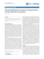
RESEARC H ARTIC L E Open Access
The challenge to verify ceramide’s role of
apoptosis induction in human cardiomyocytes -
a pilot study
Engin Usta
1*
, Migdat Mustafi
2
, Ferruh Artunc
3
, Tobias Walker
2
, Vladimir Voth
2
, Hermann Aebert
4
and
Gerhard Ziemer
1
Abstract
Background: Cardioplegia and reperfusion of the myocardium may be associated with cardiomyocyte apoptosis
and subsequent myocardial injury. In order to establish a pharmacological strategy for the prevention of these
events, this study aimed to verify the reliability of our human cardiac model and to evaluate the pro-apoptotic
properties of the sphingolipid second messenger ceramide and the anti-apoptotic properties of the acid
sphingomyelinase inhibitor amitryptiline during simulated cardioplegia and reperfusion ex vivo.
Methods: Cardiac biopsies were retrieved from the right auricle of patients undergoing elective CABG before
induction of cardiopulmonary bypass. Biopsies were exposed to ex vivo conditions of varying periods of cp/rep
(30/10, 60/20, 120/40 min). Groups: I (untreated control, n = 10), II (treated control cp/rep, n = 10), III (cp/rep +
ceramide, n = 10), IV (cp/rep + amitryptiline, n = 10) and V (cp/rep + ceramide + amitryptiline, n = 10). Fo r
detection of apoptosis anti-activated-caspase-3 and PARP-1 cleavage immunostaining were employed.
Results: In group I the percentage of apoptotic cardiomyocytes was significantly (p < 0.05) low if compared to
group II revealing a time-dependent increase. In group III ceramid increased and in group IV amitryptiline inhibited
apoptosis significantly (p < 0.05). In contrast in group V, under the influence of ceramide and amitryptiline the
induction of apopto sis was partially suppressed.
Conclusion: Ceramid induces and amitryptiline suppresses apoptosis significantly in our ex vivo setting. This
finding warrants further stud ies aiming to evaluate potential beneficial effects of selective inhibition of apoptosis
inducing mediators on the suppression of ischemia/reperfusion injury in clinical settings.
Introduction
Cardioplegia and reperfusion of the myocardium are
essential techniques employed in many cardiac surgical
procedures when a temporarily arrested myocardium is
required. However, as a consequence of exposure to car-
dioplegia and reperfusion apoptosis of cardiomyocyt es
may occur [1]. Apoptosis is the ultimate result of multi-
ple convergent signalling pathways, which are triggered
by events such as nutrient and oxygen deprivation,
intracellular calcium overload and excessive reactive
oxygen species production [1]. In the setting of cardiac
surgery these events can finally result in co ntractile dys-
function of the myocardium [2] and atrial fibrillation
[3]. Apopto sis of cardiac non-myocytes also contributes
to maladaptive remodelling and the transition to decom-
pensated congestive heart failure [4]. Regarding this
potentially impact of apoptosis on clinical outcomes,
there is a demand for pharmacological strategies. Phar-
macological blockade has been shown to reduce apopto-
sis during extra corporeal circulation in an animal model
[5]. In contrast to that we have successfully established
a human cardiac model, which we have presented
recently [6-8].
Our present pilot study was performed just as a sequel
to our recent work [6-8] to further evaluate our presented
human cardiac model during simulated card ioplegia and
* Correspondence:
1
Children’s University Hospital, Div. Congenital & Pediatric Cardiac Surgery;
University Hospital Tübingen, Germany
Full list of author information is available at the end of the article
Usta et al. Journal of Cardiothoracic Surgery 2011, 6:38
/>© 201 1 Usta et al; licensee BioMed Central Ltd. This is a n Open Access article distributed under the terms of the Creative Commons
Attribution License (http://creativecommons.o rg/licenses/by/2.0), which p ermits unrestricted u se, distribution, and reproduction in
any mediu m, prov ided the original work is properly cited.
reperfusion ex vivo respectively the end-points feasibility
and reliability. We conducted this study to clarify if
another pathway of apoptosis induction in cardiomyocytes
exists. Our aim was to evaluate d uring ex vivo simulated
cardioplegia and reperfusion the effect of the sphingolipid
second messenger ceramide and the anti-apoptotic prop-
erties of the sphingomyelinase inhibitor amitryptiline
respectively the end-point apoptosis induction and reduc-
tion in cardiomyocytes which to our knowledge has not
been described in such an experimental setting yet. The
results should c larify if any clinical potential u tilization
could be favoured.
Materials and methods
Ethics declaration
The investigation conforms with the principles outlined
in the Declaration of Helsinki. In addition, approval was
granted by the Ethics Committee of the Faculty of
Medicine of the Eberhard-Karls-University, Tübingen,
Germany (approval reference number 40/2007 V).
Patient characteristics
The study protocol was approved by the ethics commit-
tee of the Faculty of Medicine of the Eberhard-
Karls-University Tübingen. 20 patients undergoing
elective CABG surgery were included in this study and
gave informed consent for study participation. Mean
patient age was 65 years (range 45-70). Mean body mass
index 28 kg/m
2
(range 25-32). Mean left ventricular
ejection fraction 63% (range 55-75). Mean numbe r of
diseased coronary vessels 3 (range 2-3). Mean number
of infarctions 1 (range 1-3) in patients history. The basic
medication of all patients consisted of b-block ers (Beloc
Zok™ 47.5 mg twice per die, angiotensin converting
enzyme inhibitors, statins and diuretics. All patients had
a sinus rhythm.
Material
Human tissue was retrieved from the auricle of the right
atrium of patients before cardiopulmonary-bypass (CPB)
and was processed immediately. Each biopsy was trans-
muraly divided in thirteen pieces with [0.5 to 1 cm
2
]
size, which were placed separately in microperfusion
chambers with continuous perfusion. Cardiac specimens
were outsi de the body before being mounted and tested
in the chamber system for a maximum of 30 min, but
during this period the oxygen supply was maintained
continuously by bubble-oxygenating the Krebs-Henseleit
buffer in the petri dish (Greiner Bio-One, Frickenhausen
Germany).
Chemicals and buffer solutions
The modified Krebs-Henseleit buffer (KH) consisted of
115 mM NaCl, 4.5 mM KCl, 1.18 mM MgCl
2
,1.25mM
CaCl2, 1.23 mM NaH
2
PO
4
,1.19Na
2
SO
4
,80mM
Glucose, and 10 mM HEPES, pH adjusted to 7.4 at 37°C
with NaOH.
Cardioplegic solution
Cardioplegic solution was prepared on the basis of Ca-
free KH consisting of 115 mM NaCl, 4.5 mM KCl,
1.18 mM MgCl
2
, 0.5 mM EGTA, 1.23 mM NaH
2
PO
4
,
1.19 mM Na
2
SO
4
, 80 mM Glucose, and 10 mM HEPES,
pH adjusted to 7.4 at 37°C with NaOH. Furthermore, a
solution containing 20 mM Tris hydroxymethyl-amino-
methane, 60 mmol K
+
and anionic polypeptides to the
isoionic point was added in a 1:4 proportion to Ca-free
KH buffer. This solution served as cardiop legic solution
and was administered at 4°C, in analogy to our clinical
regimen. The resulting K
+
concentration in this mixtur e
was 16.5 mM.
Ceramide
Sphingolipids a re constituents of cellular membranes
and of lipoproteins. The common backbone is the long
chain amino base sphingosine (trans-4-sphingenine),
and the ceramides refer to the N-acyl deriva tives of
sphingosine. For a decade now, ceramides have been
widely studied as regulators of major cellular functions,
i.e., apoptosis, proliferation, or senescence [9-11]. Apop-
tosis induction with short chain ceramide (20-50 μM)
supports the view that ceramides are able to trigger
apoptosis [12]. The concentration of ceramide employed
in this study was 50 μM, similar to previous experimen-
tal settings [12].
Amitryptiline
Amitryptiline (systematic taxonomy: 3-(10,11-dihydro-
5H-dibenzo[[a, d]]cycloheptene-5-ylidene)-N, N-
dimethyl-1-propanamine) is a tricyclic antidepressant.
Besides its known clinical use it has been identified as
an acid sphingomyelinase inhibitor with lowering cera-
mide levels and thus carrying out anti-apoptotic proper-
ties [13,14].
Cell viability
The viability of cardio myocytes in tissue samp les was
assessed by trypan blue exclusion before each experi-
ment. Only samples consisting of ≥ 99% viable cardio-
myocytes were further processed in the experiments of
this study.
Microperfusion chamber
Our self developed, previously described [6-8] microper-
fusion chamber was modified to investigate larger speci-
mens. It consisted of two components (Figure 1). The
first component a temperature-controlled plexiglas
block contained a rectangular cavity forming the
Usta et al. Journal of Cardiothoracic Surgery 2011, 6:38
/>Page 2 of 7
chamber with following dimensions (length × width ×
height, 5.5 × 1.5 × 1.25 cm). The second component
was mounted over the first, and consisted of another
plexiglas block forming the ceiling of the chamber. In
this chamber nylon net with a pore size of 400 μmwas
mounted diagonally. To enable perfusion of the cham-
ber, a thin pipe was introd uced at one e nd of the plexi-
glas component, entered the chamber and exited at the
other end. A thin rubber layer between each component
sealed the microperfusion c hamber. The biopsy was
fixed physically at the nylon net by the laminar flow
(perfusion velocity of 5 ml/min) of the hydrostatic per-
fusion system through the chamber.
Experimental groups
The protocol was designed to simulate clinical routine
procedures administering cardioplegic solution with the
same K
+
concentration (16.5 mM) and temperature
(4°C). Five different groups (I - V) were arranged as fol-
lows: I (untreated control, n = 10), II (treated control
cp/rep, n = 10), III (cp/rep + ceramide, n = 10), IV (cp/
rep + amitryptiline, n = 10) and V (cp/rep + ceramide +
amitryptiline, n = 10). In group III cardiomyocytes were
continuously treated with 50 μMceramid.InIngroup
IV cardiomyocytes were continuously treated with
100 μM amitryptiline. In contrast to that in group V
cardiomyocytes were continuously treated with both
drugs ceramid [50 μM] and amitryptiline [100 μM]. In
general, each assay was carried out with the specimens
of one patient, i.e. specimens of patients were analysed
separately.
Ischemia/reperfusion assay
The cardiac specimens in the microperfusion chambers
were initially equilibrated with KH for 5 min (32°C and
continuously bubble-oxygenated with carbogen (95% O
2
and 5% CO
2
) to attain a PO
2
of 25-30 kPa and pH 7.4.
After that the cardioplegic solution (4°C) was adminis-
tered for 5 min. To induce ischemic injury during the
cardioplegia period the perfus ion of the microperfusion
chamber was st opped and the oxygen supply was dis-
continued. The cardiac specimens were subjected to var-
ious periods of cardioplegia (30, 60 or 120 min) followed
by 1/3 of the chosen cardioplegia time as reperfusion
(10, 20 or 40 min), as in our surgical routine. For reper-
fusion 35°C KH was used. Fina lly, the cardiac specimens
were snap-frozen in liquid nitrogen.
Immunohistochemical apoptosis detection
The slides with the cryosections of the samples (10 μm)
were processed prior to the staining according to the
manufacturer’s recommendation (Epitomics, Inc., Bur-
lingame, CA, USA). The described chemicals were pur-
chased from Biochrom, Berlin Germany. In brief, the
cryosections were immersed into the s taining dish con-
taining the antigen retrieval solution: 9 ml of stock solu-
tion A (0.1 M citric acid solution) and 41 ml of stock
solution B (0.1 M sodium citrate solution) were added
to450mlofdestillatedH
2
OandadjustedtopH6.0.
After warming for 30 min in a rice cooker and cooling
down t he slides were washed with TBST (Tris-Buffered
Salineand0.1%Tween20)for5minonashaker.For
the inactivation of endogenous peroxidases the slides
were covered with 3% hydrogen peroxide for 10 min
and later washed with TBST. After that the slides were
immersed into the blocking solution (PBS (Dulbecco’s
Phosphate Buffered Salts) and 10% bovine serum
albumin) for 1 hour.
Later the cryosections were incubated overnight in a
humidified chamber (4°C) with antibodies against
PARP-1 (Anti-Poly-(ADP-Ribose)-Polymerase)-cleavage
(Epitomics, Inc.). PARP is a zinc-dependent DNA bind-
ing protein that recognizes DNA strand breaks and is
presumed to play a role in DNA repair. PARP is cleaved
in vivo by caspase-3 [15]. The antibody only recognizes
p25 cleaved-form of PARP-1.
On the other hand cryosections were stained with
antibodies against activated Caspase-3 (Epitomics, Inc.),
also. Caspases are a family of cytosolic aspartate-specific
cysteine proteases involved in the initiation and execu-
tion of apoptosis. Caspase-3 (apopain, SCA-1, Yama and
CPP32) is a member of the apoptosis execution func-
tional group of caspases, and is either partially or totally
responsible for the proteolytic cleavage of many key pro-
teins during apoptosis. Caspase-3 is a cytosolic protein
found in cells as an inactive 35 kDa proenzyme. It is
Figure 1 Microperfusion chamber.Theperfusateentersthe
chamber, constructed from plexiglas (2), through the pipe (1) and
fills the rectangular shaped chamber (3). Once laminar flow is
constituted the cardiac tissue is physically fixed before the nylon
net (not featured), which spans in a 135° angle. The fluid exits on
the opposite side (4). Between the bottom and the upper part of
the chamber a rubber layer was placed for sealing and fastened
with 4 screws.
Usta et al. Journal of Cardiothoracic Surgery 2011, 6:38
/>Page 3 of 7
activated by proteolytic cleavage into two active subunits
only when cells undergo apoptosis (3).
Later for detection to each section secondary HRP-
conjugated anti-rabbit antibody (Epitomics, Inc.) diluted
in the blocking solution per manufacturer’s recommen-
dation was applied and incubated for 1 hour at room
temperature.
Fluorescence microscopy
The number of cells on the cryosections was determined
by counting the nuclei of cardiomyocytes after staining
with DAPI (4’,6-Diamidino-2-phenylindole 2 HCl), a dye
known to form fluorescent complexes with natural dou-
ble-stranded DNA, under a fluorescence microscope
(Zeiss, Jena, Germany). In each analysis t hree different
areas of the cryosections were counted using 40-fold
magnification. Apoptotic cells were identified by con-
densation and fragmentation of the nuclei and fluores-
cent conglomerates in the cytoplasm. They were
quantified by c ounting a total of 200 nuclei from each
cryosection an d calculating the percentage of apoptotic
nuclei. After DAPI counterstaining the greater nucl ei of
cardiomyocytes allow their distinction from fibroblasts
with smaller nuclei. In anti-activated caspase-3 positive,
apoptotic cardiomyocytes the cytoplasm reveales an
intensive granular fluorescence (Figure 2). In contrast to
that PARP-1 cleavage positive, apoptotic cardiomyocytes
nuclei feature an intensive granular fluorescence inten-
sity with granular staining of the nucleus.
Fluorescence images (blue) of DAPI loaded cardiac
specimens were obtained at a n excitation wavelength of
360 nm, with an emission wavelength o f 460 nm. DAPI
was purchased from Sigma-Aldrich, Germany.
Statistical Analysis
Analysis of calcium recordings and graphics were
obtained using Sigma Plot software (version 9.0, SPSS
Inc., Chicago, IL). Data are expressed as the mean±
standard error of deviation (SD) and stati stic al analysis
was performed using GraphPad Prism (version 5.0,
GraphPad Software, Inc., CA, USA). Comparison of
groups was performed using repeated measures one-way
ANOVA followed by Tukey’sHSDposthoctest.Ap
value of less than 0.05 was considered to indic ate a sta-
tistically significant difference.
Results
Immunohistochemical apoptosis detection
Anti-activated-caspase-3
Cardiomyocytes in the untreated group I revealed a
significant (p < 0.05) low percentage of apoptotic c ells
(12 ± 5%) in co mparison to the treated control group II
(Figure 3A). There was a significant (p < 0.05) lower
percentage of apoptotic cells in the amitryptiline treat-
ment group IV if compared to group III with ceramide
(Figure 3A).
PARP-1 cleavage
Cardiomyocytes in the untreated group I featured a
significant (p < 0.05) low percentage of apoptotic c ells
(12 ± 4%) in co mparison to the treated control group II
(Figure 3B). There was a significant ( p < 0.05) lower
percentage of apoptotic cells in the amitryptiline treat-
ment group IV if compared to group III with ceramide
(Figure 3B).
Discussion
In the present study our first goal was to apply ceramide
to evaluate the proapoptotic potential during cardiople-
giaandreperfusion[9,16]inanexvivosettingwith
human cardiomyocytes which to our current knowledge
has not been reported yet. Our second goal was to
investigate if the proapoptotic effect of ceramide could
be inhibited by amitryptiline [17]. Our third goal was
just in accordance to our clinical routine to administer
cardioplegia and rep erfusion to simulate the extracor-
poreal circulation in our experimental model and evalu-
ate if the induction or inhibition of apoptosis could be
influenced.
In our experimental model human cardiomyocytes
were kept in their natural envir onment as intac t cardiac
tissue. Otherwise human papillary muscle could
be employed but obtaining it before cardioplegic arrest
is not an imaginable and feasible option during
Figure 2 Representative fluorescent image of cardiomyocytes
treated with ceramide during cardioplegia (60 min) and
reperfusion (20 min) (group III). After DAPI counterstaining the
greater nuclei of cardiomyocytes allow their distinction from
fibroblasts with smaller nuclei. In anti-activated caspase-3 positive,
apoptotic cardiomyocytes the cytoplasm reveales an intensive
granular fluorescence (marked with stars). The exemplary images
represent a single experiment. During the cryosection procedure
artifacts presenting as nuclei conglomerates could not be avoided;
these were excluded from analyses.
Usta et al. Journal of Cardiothoracic Surgery 2011, 6:38
/>Page 4 of 7
clinical routine. The simulation of ischemia in isolated
cardiomyocyte models can provide important insights into
the pathophysiology of myocardial ischemic injury and its
underlying molecular mechanisms as was the subject in
previous studies i n iso lated mammalian cardiomyocytes
[18], isolated papillary muscle preparations [19] or animal
heart models [20]. The distinctive difference of our experi-
mental assay was utilizing human atrial cardiac tissue as a
model for apoptosis studies inducing apoptotis just in
accordance to our clinical rout ine with cardioplegia and
reperfusion without induction of ischemia with N
2
perfu-
sion like in previous studies [21,22]. Like presented above
in our experimental assay the cardioplegia and reperfusion
stimulus proved to be an adequate stimulus for apoptosis
induction and is comparable with those in the literature
[6-8,23].
Further we wanted to enlighten the major mediators
of apoptosis occurring during postischemic reperfusion.
Apoptosis is an important mechanism of active cellular
death that is distinct from necrosis and has been impli-
cated in the pathogenesis of a variety of degenerative
and ischemic human diseases [24]. The family of cas-
pases is key mediator of apoptosis. An extrinsic pathway
involving cell surface death receptors [25] and an intrin-
sic pathway with intracellular and extracellular death
signals which are transmitted to the mitochondria
through memb ers of the Bcl-2 family [26] exist. Several
intracellular stimuli, includi ng oxidative stress, translo-
cate Bax and/or Bak to the mitochondria, leading to
dysfunction of this organelle, the release of pro apoptotic
proteins, and the activation of caspase-9 [27]. Another
important stimulus for apoptosis derive from sphingoli-
pids like ceramides which have been described as sec-
ond messengers for several events like differentiation,
senescence, proliferation and cell death in different cell
lines [9]. Sphingolipids are found in most subcellular
membranes. In the plasma membrane they are predomi-
nantly found in the outer leaflet [28]. The metabolism
of sphingolipids has been proved to be a dynamic pro-
cess and their metabolites (such as ce ramide, sphingo-
sine, and sphingosine 1-phosphate (S1P)) are now
recognized as messengers playing essential roles in cell
growth, survival, as well as cell death [9,29]. Sphingo-
myelin (SM) is a ubiquitous component of animal cell
membranes, where it is by far the most abundant sphin-
golipid. Ceramide can be formed through sphingomyeli-
nases (SMase)-dependent catabolism of SM and by de
novo synthesis. SMases are specialized enzymes with
phospholipase C activity that can hydrolyze the phos-
phodiester bond of SM. It is well known that ceramide
can modulate many different cellular processes.
Ceramide directly regulates protein phosphatase 1 (PP1),
inducing dephosphorylatio n of SR proteins and splicing
of caspase-9 and Bcl-x genes [30]. Interaction of cera-
mide with protein kinase-c can inhibit translocation of
the kinase to the plasma membrane and t herefore inhi-
bits its catalytic acti vity. Finally the intrinsic and extrin-
sic pathways of apoptosis i nduction converge and lead
to the activation of caspases which have been character-
ized as major executioners of apoptosis [31]. During oxi-
dative stress reactive oxygen species trigger the release
of cytochrome c from mitochondria and, s ubsequently,
caspase activation. Active caspases promote cellular
demolition by activating other destructive enzymes, such
Figure 3 Demonstrating the effect of ceramide and
amitryptline on apoptosis in human cardiomyocytes.
Percentage of anti-activated caspase-3 (3A) and anti-PARP-1
cleavage (3B) positive cardiomyocytes. In the treated control group
the time-dependent increase of apoptotic cardiomyoctes is
significant (p < 0.05) if compared to the untreated control group.
Ceramide had a higher impact on apoptosis if compared to the
treated control group. Amitryptiline applied together with ceramide
suppressed the proapoptotic effect of ceramide significantly (p <
0.05) (*). Results shown represent mean±SD of combined results
from n = 10 independent assays.
Usta et al. Journal of Cardiothoracic Surgery 2011, 6:38
/>Page 5 of 7
as DNAses, and by directly targeting key structural
proteins, such as lamin and actin, and regulatory proteins,
thus leading to chromatin margination, DNA fragmenta-
tion, nuclear condensation and collapse [31], which we
could demonstrate in our immunohistochemical assays.
In our experiments, we found that caspase-3 was
already activated at the end of the ischemia, thus sug-
gesting that the mitochondrial pathway of apoptosis is a
very early event in myocardial injury. Caspase-3 has
been shown to cleave the 112 kDa nuclear protein
PARP into an 85 kDa apoptotic fragment [32], and this
cleavage by caspase-3 has been shown to be necessary
for apoptosis [15]. In this regard, the nuclear presence
of proteolytic fragments of PARP has been considered a
hallmark of an apoptotic cell. However, t he role of
PARP-1 in apoptosis remains to be determined because
conflicting data have been repo rted. Some investigators
have shown that neurons or hepatocytes from PARP-
deficient mice do not exhibit any altered sensitivity to
apoptotic stimuli, whereas others have demonstrated
that pharmacological or genetic inhibition may increase
apoptosis in cells subjected to alkylating agents [33,34].
The family of Bcl-2-related proteins constitutes the
most relevant class of apoptotic regulators and, more
specifically, the ratio of anti- or pro-apoptotic pr oteins
determines whether t he cell will survive or die [35,36].
On the other hand, ex pression of Bcl-2 protein prevents
the induction of apoptosis caused by a variety of oxida-
tive stresses, and it can influence the level of caspase
activation [35]
In accordance to this referred data in our presented
study we could demonstrate that apoptosis can be sup-
pressed effectively in our experimental setup. Consider-
ing our immunohistochemical apoptosis detection there
is a significant reductio n of apoptosis in cardiomyocytes
treated with amitryptiline in contrast to the treatment
with ceramide after cardioplegia and succeeding reperfu-
sion. The high apoptosis rate in the treated control
group e specially after 120 min cardioplegia and 40 min
reperfusion should not be extrapolated into the in vivo
situation without any caution as atrial and ventricular
myocardium possess specific cha racteristics that may
influence the susceptibility to ischaemia/reperfusion
injury. One explanation is the reported difference in the
distribution of potassium channels [37], which contri-
butes to the characteristic differences between atrial and
ventricular action potentials and may determine a differ-
ent response to cardioplegia/reperfusion.
Our presented data provide evidence that one of the
key signaling pathways controlling apoptosis could med-
iate, at least in part, ischemia-reperfusion induced
injury. Furthermore, the results of our study suggest
that, although proapoptotic signalling plays an im por-
tant role in the development of reperfusion-induced
damage, acid sphingomyelinase inhibition by amitrypti-
line aside from dose-dependency may not afford alone a
complete protec tion against postischemic damage. This
characteristic has been described in previous studies
[14] and could be an explanation for the partial inhibi-
tion of apoptosis due to the treatment with amitryptiline
like presented in this study.
Limitations
The present study has few potential limitations. First,
clinical ischemia might be quite different from the simu-
lated ischemia we use. Unfortunately, there is currently
no accepted standard that constitutes a clinically rele-
vant “simulated ischemic exposure ” for cells. Simulating
the i schemic environment of the extracellular fluid that
bathes the cells is quite complex due to the fact that
there are alterations in many factors, simulating all of
these events is not currently possible. So, wherea s the
use of si mulated ischemia is not perfect, we believe it
recreates a number of the important components of
clinical ischemia. Further in this study only a single
ceramid and amitryptiline concentration was employed,
but a nalogous to previous studies in a pharmacological
relevant c oncentration [ 38]. Therefore, detailed dose-
response relationships o f neither ceramide nor amitryptiline
on apoptot ic events were no t investigated. Nevertheless,
with the concentration employed in this study, apoptotic
events could be triggered or inhibited considerably.
Furthermore the primary purpose of this study was to
test its effect on apoptotic events in cardiomyocytes in
this new experimental setting rather than to study dose-
response relationships. Our next step would be to verify
our current fi ndings in an animal model. However our
results indicate a definite beneficial effect of amitryptiline
on apoptotic events.
Conclusions
In human cardiomyocytes there is a remarkable i nduc-
tion of apoptosis due to the pro-apoptotic second mes-
senger ceramide.
The treatment of human cardiomyocytes in an ex vivo
experimental setting with si mulated cardioplegia and
reperfusion can result in considerable reduction of
apop totic events by adding amitrypt iline. These findings
warrant further studies in order to evaluate potentially
beneficial effects of acid sphingomyelinase inhibition by
amitryptiline in the in vivo setting of cardioplegia as
employed in cardiac surgery.
Acknowledgements
This work was supported by a research grant (fortüne 1232126.2) of the
Faculty of Medicine of the Eberhard-Karls University Tübingen, Germany.
Author details
1
Children’s University Hospital, Div. Congenital & Pediatric Cardiac Surgery;
University Hospital Tübingen, Germany.
2
Dep. of Thoracic-, Cardiac- and
Usta et al. Journal of Cardiothoracic Surgery 2011, 6:38
/>Page 6 of 7
Vascular Surgery; Tübingen University Hospital, Germany.
3
Dep. of Internal
Medicine IV, Section of Nephrology and Hypertension; Tübingen University
Hospital, Germany.
4
Clinic of Vascular and Thoracic Surgery,
Donaueschingen, Germany.
Authors’ contributions
EU carried out the routine preoperative examinations, patient evaluation and
participated in the study design and coordination. EU performed the
statistical analysis. MM, FA and TW participated in the experiments and data
evaluation. HA and GZ conceived of the study, and participated in its design
and coordination. All authors read and approved the final manuscript.
Competing interests
The authors declare that they have no competing interests.
Received: 29 November 2010 Accepted: 28 March 2011
Published: 28 March 2011
References
1. Bai CX, Namekata I, Kurokawa J, Tanaka H, Shigenobu K, Furukawa T: Role
of nitric oxide in Ca2+ sensitivity of the slowly activating delayed
rectifier K+ current in cardiac myocytes. Circ Res 2005, 96:64-72.
2. Murriel CL, Churchill E, Inagaki K, Szweda LI, Mochly-Rosen D: Protein
kinase Cdelta activation induces apoptosis in response to cardiac
ischemia and reperfusion damage: a mechanism involving BAD and the
mitochondria. J Biol Chem 2004, 279:47985-47991.
3. Ak K, Akgun S, Tecimer T, Isbir CS, Civelek A, Tekeli A, et al: Determination
of histopathologic risk factors for postoperative atrial fibrillation in
cardiac surgery. Ann Thorac Surg 2005, 79:1970-1975.
4. Khoynezhad A, Jalali Z, Tortolani AJ: A synopsis of research in cardiac
apoptosis and its application to congestive heart failure. Tex Heart Inst J
2007, 34:352-359.
5. Zhang S, Sun Z, Liu L, Hasichaonu : Carvedilol attenuates CPB-induced
apoptosis in dog heart: regulationof Fas/FasL and caspase-3 pathway.
Chin Med J (Engl) 2003, 116:761-766.
6. Usta E, Mustafi M, Straub A, Ziemer G: The nonselective beta-blocker
carvedilol suppresses apoptosis in human cardiac tissue: a pilot study.
Heart Surg Forum 2010, 13:E218-E222.
7. Usta E, Mustafi M, Scheule AM, Ziemer G: Suppressing apoptosis with
milrinone simulating extracorporeal circulation: a pilot study. Thorac
Cardiovasc Surg 2010, 58:285-290.
8. Usta E, Renovanz M, Mustafi M, Ziemer G, Aebert H: Human cardiac tissue
in a microperfusion chamber simulating extracorporeal circulation–
ischemia and apoptosis studies. J Cardiothorac Surg 2010, 5:3.
9. Hannun YA: Functions of ceramide in coordinating cellular responses to
stress. Science 1996, 274:1855-1859.
10. Kolesnick RN, Kronke M: Regulation of ceramide production and
apoptosis. Annu Rev Physiol 1998, 60:643-665.
11. Argaud L, Prigent AF, Chalabreysse L, Loufouat J, Lagarde M, Ovize M:
Ceramide in the antiapoptotic effect of ischemic preconditioning. Am J
Physiol Heart Circ Physiol 2004, 286:H246-H251.
12. Zhou H, Summers SA, Birnbaum MJ, Pittman RN: Inhibition of Akt kinase
by cell-permeable ceramide and its implications for ceramide-induced
apoptosis. J Biol Chem 1998, 273:16568-16575.
13. Teichgraber V, Ulrich M, Endlich N, Riethmuller J, Wilker B, De Oliveira-
Munding CC, et al: Ceramide accumulation mediates inflammation, cell
death and infection susceptibility in cystic fibrosis. Nat Med 2008,
14:382-391.
14. Brenner B, Ferlinz K, Grassme H, Weller M, Koppenhoefer U, Dichgans J,
et al: Fas/CD95/Apo-I activates the acidic sphingomyelinase via caspases.
Cell Death Differ 1998, 5:29-37.
15. Tewari M, Quan LT, O’Rourke K, Desnoyers S, Zeng Z, Beidler DR, et al:
Yama/CPP32 beta, a mammalian homolog of CED-3, is a CrmA-
inhibitable protease that cleaves the death substrate poly(ADP-ribose)
polymerase. Cell 1995, 81:801-809.
16. Bielawska AE, Shapiro JP, Jiang L, Melkonyan HS, Piot C, Wolfe CL, et al:
Ceramide is involved in triggering of cardiomyocyte apoptosis induced
by ischemia and reperfusion. Am J Pathol 1997, 151:1257-1263.
17. Gulbins E, Jekle A, Ferlinz K, Grassme H, Lang F: Physiology of apoptosis.
Am J Physiol Renal Physiol 2000, 279:F605-F615.
18. Chanani NK, Cowan DB, Takeuchi K, Poutias DN, Garcia LM, del Nido PJ,
et al: Differential effects of amrinone and milrinone upon myocardial
inflammatory signaling. Circulation 2002, 106:I284-I289.
19. Azuma M, Yamane M, Tachibana K, Morimoto Y, Kemmotsu O: Effects of
epinephrine and phosphodiesterase III inhibitors on bupivacaine-
induced myocardial depression in guinea-pig papillary muscle. Br J
Anaesth 2003, 90:66-71.
20. Fukutomi T, Satoh K, Ogoshi S, Ichihara K: Effects of pimobendan and EGIS
9377, cardiotonic agents, and OG-VI, a nucleoside-nucleotide mixture,
administered during reperfusion after ischemia on stunned myocardium
in dogs. Coron Artery Dis 2000, 11:83-90.
21. Ghosh S, Ng LL, Talwar S, Squire IB, Galinanes M: Cardiotrophin-1 protects
the human myocardium from ischemic injury. Comparison with the first
and second window of protection by ischemic preconditioning.
Cardiovasc Res 2000, 48:440-447.
22. Vanden Hoek TL, Qin Y, Wojcik K, Li CQ, Shao ZH, Anderson T, et al:
Reperfusion, not simulated ischemia, initiates intrinsic apoptosis injury in
chick cardiomyocytes. Am J Physiol Heart Circ Physiol 2003, 284:H141-H150.
23. Miyamoto S, Howes AL, Adams JW, Dorn GW, Brown JH: Ca2+
dysregulation induces mitochondrial depolarization and apoptosis: role
of Na+/Ca2+ exchanger and AKT. J Biol Chem 2005, 280:38505-38512.
24. Communal C, Sumandea M, de TP, Narula J, Solaro RJ, Hajjar RJ: Functional
consequences of caspase activation in cardiac myocytes. Proc Natl Acad
Sci USA 2002, 99:6252-6256.
25. Ashkenazi A, Dixit VM: Death receptors: signaling and modulation. Science
1998, 281:1305-1308.
26. Kubasiak LA, Hernandez OM, Bishopric NH, Webster KA: Hypoxia and
acidosis activate cardiac myocyte death through the Bcl-2 family protein
BNIP3. Proc Natl Acad Sci USA 2002, 99:12825-12830.
27. Danial NN, Korsmeyer SJ:
Cell death: critical control points. Cell 2004,
116:205-219.
28. Koval M, Pagano RE: Intracellular transport and metabolism of
sphingomyelin. Biochim Biophys Acta 1991, 1082:113-125.
29. Prieschl EE, Baumruker T: Sphingolipids: second messengers, mediators
and raft constituents in signaling. Immunol Today 2000, 21:555-560.
30. Chalfant CE, Rathman K, Pinkerman RL, Wood RE, Obeid LM, Ogretmen B,
et al: De novo ceramide regulates the alternative splicing of caspase 9
and Bcl-x in A549 lung adenocarcinoma cells. Dependence on protein
phosphatase-1. J Biol Chem 2002, 277:12587-12595.
31. Villa P, Kaufmann SH, Earnshaw WC: Caspases and caspase inhibitors.
Trends Biochem Sci 1997, 22:388-393.
32. Duan H, Orth K, Chinnaiyan AM, Poirier GG, Froelich CJ, He WW, et al: ICE-
LAP6, a novel member of the ICE/Ced-3 gene family, is activated by the
cytotoxic T cell protease granzyme B. J Biol Chem 1996, 271:16720-16724.
33. Pieper AA, Verma A, Zhang J, Snyder SH: Poly (ADP-ribose) polymerase,
nitric oxide and cell death. Trends Pharmacol Sci 1999, 20:171-181.
34. Oliver FJ, de la RG, Rolli V, Ruiz-Ruiz MC, de Murcia G, Murcia JM:
Importance of poly(ADP-ribose) polymerase and its cleavage in
apoptosis. Lesson from an uncleavable mutant. J Biol Chem 1998,
273:33533-33539.
35. Plas DR, Thompson CB: Cell metabolism in the regulation of programmed
cell death. Trends Endocrinol Metab 2002, 13:75-78.
36. Kroemer G: The proto-oncogene Bcl-2 and its role in regulating
apoptosis. Nat Med 1997, 3:614-620.
37. Amos GJ, Wettwer E, Metzger F, Li Q, Himmel HM, Ravens U: Differences
between outward currents of human atrial and subepicardial ventricular
myocytes. J Physiol 1996, 491:31-50.
38. Relling DP, Hintz KK, Ren J: Acute exposure of ceramide enhances cardiac
contractile function in isolated ventricular myocytes. Br J Pharmacol 2003,
140:1163-1168.
doi:10.1186/1749-8090-6-38
Cite this article as: Usta et al.: The challenge to verify ceramide’sroleof
apoptosis induction in human cardiomyocytes - a pilot study. Journal of
Cardiothoracic Surgery 2011 6:38.
Usta et al. Journal of Cardiothoracic Surgery 2011, 6:38
/>Page 7 of 7