Basics of Blood Management - part 1 ppsx
Bạn đang xem bản rút gọn của tài liệu. Xem và tải ngay bản đầy đủ của tài liệu tại đây (381.4 KB, 40 trang )
BLUKO82-Seeber March 19, 2007 10:12
Basics of Blood Management
i
BLUKO82-Seeber March 19, 2007 10:12
Basics of Blood
Management
Petra Seeber
MD
Department of Anesthesiology
Critical Care Medicine
Pain Management, Emergency Medicine
HELIOS Klinik Blankenhain
Wirthstr. 5
99444 Blankenhain
Germany
Aryeh Shander
MD, FCCM, FCCP, Chief
Department of Anesthesiology, Critical Care Medicine
Pain Management and Hyperbaric Medicine
Englewood Hospital and Medical Center
350 Engle Street
Englewood, NJ 07631
and
Clinical Professor of Anesthesiology, Medicine and Surgery
Mount Sinai School of Medicine, Mount Sinai Hospital,
New York
first edition
iii
BLUKO82-Seeber March 19, 2007 10:12
C
2007 Petra Seeber and Aryeh Shander
Published by Blackwell Publishing
Blackwell Publishing, Inc., 350 Main Street, Malden, Massachusetts 02148-5020, USA
Blackwell Publishing Ltd, 9600 Garsington Road, Oxford OX4 2DQ, UK
Blackwell Publishing Asia Pty Ltd, 550 Swanston Street, Carlton, Victoria 3053,
Australia
The right of the Author to be identified as the Author of this Work has been asserted in
accordance with the Copyright, Designs and Patents Act 1988.
All rights reserved. No part of this publication may be reproduced, stored in a retrieval
system, or transmitted, in any form or by any means, electronic, mechanical,
photocopying, recording or otherwise, except as permitted by the UK Copyright,
Designs and Patents Act 1988, without the prior permission of the publisher.
First published 2007
1 2007
Library of Congress Cataloging-in-Publication Data
Seeber, Petra.
Basics of blood management / Petra Seeber, Aryeh Shander. – 1st ed.
p. ; cm.
Includes bibliographical references and index.
ISBN: 978-1-4051-5131-3
1. Transfusion-free surgery. 2. Blood–Transfusion. 3. Bland banks.
I. Shander, Aryeh. II. Title.
[DNLM: 1. Blood Substitutes–therapeutic use. 2. Blood Banks–organization &
administration. 3. Blood Loss, Surgical–prevention & control.
4. Blood Transfusion. WH 450 S451b 2008]
RD33.35.S44 2008
617–dc22
2007005030
ISBN: 978-1-4051-5131-3
A catalogue record for this title is available from the British Library
Set in 9.25/11.5 Minion by Aptara Inc., New Delhi, India
Printed and bound in Singapore by Fabulous Printers Pte Ltd
Development Editor: Rebecca Huxley
Commissioning Editor: Maria Khan
Editorial Assistant: Jennifer Seward
Production Controller: Debbie Wyer
For further information on Blackwell Publishing, visit our website:
The publisher’s policy is to use permanent paper from mills that operate a sustainable
forestry policy, and which has been manufactured from pulp processed using acid-free
and elementary chlorine-free practices. Furthermore, the publisher ensures that the text
paper and cover board used have met acceptable environmental accreditation standards.
Blackwell Publishing makes no representation, express or implied, that the drug
dosages in this book are correct. Readers must therefore always check that any product
mentioned in this publication is used in accordance with the prescribing information
prepared by the manufacturers. The author and the publishers do not accept
responsibility or legal liability for any errors in the text or for the misuse or
misapplication of material in this book.
iv
BLUKO82-Seeber March 19, 2007 10:12
Contents
Preface to the first edition, vii
Acknowledgments, viii
Introduction, ix
1 History and organization of blood management, 1
2 Physiology of anemia and oxygen transport, 9
3 Anemia therapy I: erythropoiesis stimulating proteins, 21
4 Anemia therapy II (hematinics), 35
5 Growth factors, 50
6 Fluid therapy, 65
7 The chemistry of hemostasis, 77
8 Recombinant blood products, 96
9 Artificial blood components, 110
10 Oxygen therapy, 125
11 Preparation of the patient for surgery, 139
12 Iatrogenic blood loss, 160
13 The physics of hemostasis, 172
14 Anesthesia—more than sleeping, 191
15 The use of autologous blood, 200
16 Cell salvage, 211
17 Blood banking, 227
18 Transfusions. Part I: cellular components and plasma, 243
19 Transfusions. Part II: plasma fractions, 265
20 Law, ethics, religion, and blood management, 287
21 Step by step to an organized blood management program, 299
Appendix A: Detailed information, 322
Appendix B: Sources of information for blood management, 329
Appendix C: Program tools and forms, 334
Appendix D: Teaching aids: research and projects, 346
Appendix E: Address book, 350
Index, 376
v
BLUKO82-Seeber March 19, 2007 10:12
Preface to first edition
The benefit-to-risk ratio of blood products needs con-
stant evaluation. Blood products, as therapeutic agents,
have had the test of time but lack the evidence we ex-
pect from other medicinals. Blood, an organ, is used as
a pharmaceutical agent by the medical profession, due to
the achievements in collection, processing, banking, and
distribution. The fact that the most common risk of blood
transfusion is blood delivery errorsupports the notionthat
blood is handled as a pharmaceutical agent. Over the last
few decades, the risk of blood transfusion and associated
complications has raised concerns about safety of blood
by both the public and health-care providers. At the same
time, experience with patients refusing blood and data on
blood conservation brought to light the real possibility of
other modalities to treat perisurgical anemia and to avoid
it with blood conservation methods. In addition to risks
and complications, data became available demonstrating
the behavioral aspect of transfusion practice versus an
evidence-based practice. In this book, the authors address
many aspects of modern transfusion medicine, known
blood conservation modalities, and new approaches to the
treatment of perisurgical anemia, as well as special clinical
considerations. This approach, now termed “blood man-
agement” by the Society for the Advancement of Blood
Management, incorporates appropriate transfusion prac-
tice and blood conservation to deliver the lowest risk and
highest benefit to the patient. In addition, it brings all
these modalities to the patient’s bedside and above all is a
patient-centered approach. Blood management is a mul-
tidisciplinary, multimodality concept that focuses on the
patient by improving patient outcome, making it one of
the most intriguing and rewarding fields in medicine.
The benefit-to-risk ratio of blood products needs con-
stant evaluation. Blood products, as therapeutic agents,
have had the test of time but lack the evidence we ex-
pect from other medicinals. Blood, an organ, is used as
a pharmaceutical agent by the medical profession, due to
the achievements in collection, processing, banking, and
distribution. The fact that the most common risk of blood
transfusion is blooddelivery error supports the notion that
blood is handled as a pharmaceutical agent. Over the last
few decades, the risk of blood transfusion and associated
complications has raised concerns about safety of blood
by both the public and health-care providers. At the same
time, experience with patients refusing blood and data on
blood conservation brought to light the real possibility of
other modalities to treat perisurgical anemia and to avoid
it with blood conservation methods. In addition to risks
and complications, data became available demonstrating
the behavioral aspect of transfusion practice versus an
evidence-based practice. In this book, the authors address
many aspects of modern transfusion medicine, known
blood conservation modalities, and new approaches to the
treatment of perisurgical anemia, as well as special clinical
considerations. This approach, now termed “blood man-
agement” by the Society for the Advancement of Blood
Management, incorporates appropriate transfusion prac-
tice and blood conservation to deliver the lowest risk and
highest benefit to the patient. In addition, it brings all
these modalities to the patient’s bedside and above all is a
patient-centered approach. Blood management is a mul-
tidisciplinary, multimodality concept that focuses on the
patient by improving patient outcome, making it one of
the most intriguing and rewarding fields in medicine.
vii
BLUKO82-Seeber March 19, 2007 10:12
Acknowledgments
We thank the following individuals for their review
and valuable comments: Philip Battiade, Dr Charles and
Nicole Beard, Prof. Dr Jochen Erhard, Shannon Farmer,
David Grant, Renate Lange, Gregg Lobel, MD, FAAP,
David Moskowitz, MD, Barbara Shackford, CRNA, MS,
Mark Venditti, MD, and Prof. Max Woernhard.
viii
BLUKO82-Seeber March 19, 2007 10:12
Introduction
The benefit-to-risk ratio of blood products needs con-
stant evaluation. Blood products, as therapeutic agents,
have had the test of time but lack the evidence we expect
from other medicinals. Blood, an organ, is used as a phar-
maceutical agent by the medical profession, due to the
achievements in collection, processing, banking, and dis-
tribution. The fact that the most common risk of blood
transfusion is blood delivery error supports the notion
that blood is handled as a pharmaceutical agent. Over the
last few decades, the risk of blood transfusion and asso-
ciated complications has raised concerns about safety of
blood by both the public and health-care providers. At
the same time, experience with patients refusing blood
and data on blood conservation brought to light the
real possibility of other modalities to treat perisurgical
anemia and to avoid it with blood conservation meth-
ods. In addition to risks and complications, data be-
came available demonstrating the behavioural aspect of
transfusion practice versus an evidence-based practice. In
this book, the authors address many aspects of modern
transfusion medicine, known blood conservation (SABM,
www.sabm.org) modalities, and new approaches to the
treatment of perisurgical anemia, as well as special clinical
considerations. This approach, now termed “blood man-
agement” by the Society for the Advancement of Blood
Management (SABM, www.sabm.org), incorporates ap-
propriate transfusion practice and blood conservation to
deliver the lowest risk and highest benefit to the patient. In
addition, it brings all these modalities to the patient’s bed-
side and above all is a patient-centered approach. Blood
management is a multidisciplinary, multimodality con-
cept that focuses on the patient by improving patient out-
come, making it one of the most intriguing and rewarding
fields in medicine.
Blood management requires an understanding of all
elements of blood and transfusions. It includes the
philosophy, biology, physiology, and ethical considera-
tions, as well as demonstrating the practical application of
various techniques. This publication introduces thereader
to blood management and explains how to improve medi-
cal outcomes by avoiding undue blood loss, enhancing the
patient’s own blood, and improving tolerance of anemia
and coagulopathy until any of these underlying conditions
are successfully remedied.
This introduction to blood management is intended for
the training and early practicing clinicians. It is meant to
be both informative and practical and spans many of the
medical specialties that encounter blood and transfusions
as part of their daily practice. It will aid in tailoring indi-
vidual care plans for the different patients. Finally, it ad-
dresses the structure and function of a blood management
program, anovelapproachto blood conservation, and im-
proved patient outcome.
In this book, blood management is considered from
an international perspective, so attention is paid to con-
ditions encountered in developing as well as industrial
countries. Techniques such as cell salvage are performed
differently in economically deprived countries; HIV, hep-
atitis, and malaria may or may not be a threat to the blood
supply, depending on geographic location; oxygen, in-
travenous fluids, and erythropoiesis-stimulating proteins
may be readily available in some countries or inaccessible
in others. The book is intended to broaden the readers’
horizons, discussing working conditions encountered by
blood managers around the world. Many of the clinical
scenarios and the exercise that follow are intended for the
reader to adapt the information to the prevailing circum-
stances in their location.
This book is unique in the fact that it is the first
dedicated in its entirety to the concept of blood man-
agement. The authors hope that this book will stimu-
late the readers to further advance blood management
through shared experience and research. It is intended
to be informative, practical, enjoyable and will stimu-
late debate and discussion as well as help patients in
need.
ix
BLUKO82-Seeber March 14, 2007 15:11
1
History and organization
of blood management
With this introductory chapter the reader will be given
a glimpse into the organization of blood management
and its history—a history that is still extremely active and
changes day to day.
Objectives of this chapter
1 Identify historical developments that led to today’s con-
cept of blood management.
2 Demonstrate the benefits of blood management.
3 Identify blood management as “good clinical” practice.
4 Show that blood management and its techniques
should be used in all cases that qualify.
5 Help understand how a blood management program
works.
Definitions
Bloodless medicine and surgery: Bloodless medicine is a
multimodality, multidisciplinary approach to safe and
effectivepatient care without theuseof allogeneic blood
products. Bloodless medicine and surgery utilizes phar-
macological and technological means as well as medical
and surgical techniques to provide the best possible care
without the use of donor blood.
Transfusion-free medicine and surgery: Since “blood-
less medicine” is kind of a misnomer, the term
“transfusion-free medicine” was coined and is used
instead.
Blood conservation: “Blood conservation is a global con-
cept engulfing all possible strategies aimed at reducing
patient’s exposure to allogeneic blood products” [1].
This concept does not exclude the use of allogeneic
blood entirely.
Blood management: Blood management is the philoso-
phy to improve patient outcomes by integrating all
available techniques to reduce or eliminate allogeneic
blood transfusions. It is a patient-centered, multidisci-
plinary, multimodal, planned approach to patient care.
Blood management is not an “alternative,” it is the stan-
dard of care.
A brief look at history
History of bloodless medicine, transfusion-free
medicine, blood conservation, and blood
management
The term “bloodless medicine” is often associated with
the belief of Jehovah’s Witnesses to refrain from the use of
blood, therefore ruling out the option of blood transfu-
sion. The essence of bloodless medicine, and lately, blood
management, however, is not restricted to the beliefs of a
religious group. To get a better understanding as to what
bloodless medicine and blood management means, let us
go back to the roots of these disciplines.
One is not completely wrong to attribute the origin
of the term “bloodless medicine” to the endeavor of
Jehovah’s Witnesses to receive treatment without resort-
ing to donor blood transfusion. Their attitude toward the
sanctity of blood greatly influences their view of blood
transfusion. This was published as early as 1927 in their
journal The Watchtower (December 15, 1927). Although
the decision to refuse blood transfusion is a completely
religious one, the Witnesses frequently used scientific
information about the side effects of donor blood transfu-
sion. The booklet entitled Blood, Medicine and the Law of
God (published in 1961) addressed issues such as transfu-
sion reactions, transfusion-related syphilis, malaria, and
hepatitis.
Refusing blood transfusions on religious grounds was
not easy. Repeatedly, patients were physically forced to
take donor blood, using such high-handed methods as
1
BLUKO82-Seeber March 14, 2007 15:11
2 Chapter 1
incapacitation by court order, strapping patients to the
bed (even with the help of police officers), and secretly
adding sedatives to a patient’s infusion. In the early
1960s, representatives of Jehovah’s Witnesses started vis-
iting physicians to explain the reasons why transfusions
were refused by the Witness population. Often, during
the same visit, they offered literature which dealt with
techniques that were acceptable to the Witness patients,
informing physicians of the availability of the so-called
transfusion alternatives. After a few years of work, the gov-
erning body of Jehovah’s Witnesses announced the forma-
tion of Hospital Liaison Committees (1979). These con-
tinued to “support Jehovah’s Witnesses in . . . their deter-
mination to prevent their being given blood transfusions,
to clear away misunderstandings on the part of doctors
and hospitals, . . . to establish a more cooperative spirit be-
tween medical institutions and Witness patients” and to
“alert hospital staff to the fact that there are valid alterna-
tives to the infusion of blood” (italics ours). Occasionally,
the Witnesses even went to court to fight for their rights
as patients. In a great number of cases, the Witnesses’ po-
sition was upheld by the courts.
Although many physicians had difficulty with the con-
cept of bloodless medicine, there were some physicians
who took up the challenge to provide the best possi-
ble medical care without the use of blood transfusions.
These were in fact the earliest blood managers. As their
experience in performing “bloodless” surgery increased,
more complex procedures such as open heart surgery,
orthopedic surgery, and cancer surgery could be per-
formed. Even children and newborns could successfully be
treated without transfusing blood. Not before long, those
pioneering physicians published their results with Witness
patients, thereby encouraging other doctors to adopt the
methods used in performing such surgical interventions.
Among the first ones who rose to the challenge was
the heart surgeon Denton Cooley of Texas. In the early
1960s, his team devised methods to treat Witness pa-
tients. Reporting on his early experiences, he published
an article in a 1964 issue of The American Journal of Car-
diology. In the article “Open heart surgery in Jehovah’s
Witnesses” his team described the techniques used. In
1977, Cooley reported his experiences with more than
500 patients [2].
Cooley’s example was followed by many other coura-
geous physicians. For instance, in 1970 Dr Pearce per-
formed bloodless open heart surgery in New Orleans.
His efforts did not go unnoticed. Newspapers reported
on these spectacular cases. Perhaps out of curiosity or
out of the earnest desire to learn, many colleagues visited
Dr Pearce’s team in the operating room to learn how to
do “bloodless hearts.” Dr Jerome Kay, from Los Angeles,
also performed bloodless heart surgery. In 1973 he re-
ported that he is now performing bloodless heart surgery
on the majority of his patients. The call for bloodless treat-
ments spread around the whole world. Dr Sharad Pandey
of the KEM hospital in Mumbai, India, adopted bloodless
techniques from Canada and tailored them to fit Indian
conditions. Centers in Europe and the rest of the world
started adopting those advances as well.
It is understandable that Witness patients preferred the
treatment of physicians who had already proven their will-
ingness and ability to treat them without using donor
blood. The good reputation of such physicians spread
and so patients from far away were transferred to their
facilities. This laid the foundation for organized “blood-
less programs.” One of the hospitals with such a program
was the Esperanza Intercommunity Hospital in Yorba
Linda, California, where a high percentageof patients were
Witnesses. Dr Herk Hutchins, an experienced surgeon and
a Witness himself, was known for his development of an
iron-containing formula for blood-building. Among his
team was the young surgeon Ron Lapin. Later, he was
famed for his pioneering work in the area of bloodless
therapies. Critics labeled him a quack. Nevertheless, he
continued and was later honored for opening one of the
first organized bloodless centers in the world, as well as for
publishing the first journal on this topic, and for his efforts
to teach his colleagues. During his career, he performed
thousands of bloodless surgeries.
All of those pioneers of blood management had to rise
to the challenge of using and refining available techniques,
adjusting them to current needs, and individualizing pa-
tient care. They adopted new technologies as soon as this
was reasonable. Much attention was paid to details of pa-
tient care, thus improving the quality of the whole ther-
apy. They also fought for patients’ rights and upheld those
rights. Many involved in the field of blood management
confirm the good feeling of being a physician in the truest
sense. There is no need to force a particular treatment.
Such an attitude is a precious heritage from the pioneers of
blood management. Now, at the beginning of the twenty-
first century, this pioneer spirit can still be felt at some
meetings dedicated to blood management.
Military use of blood and blood management
Over the centuries, the armies of different nations con-
tributed to what is now available for blood management,
but not on religious grounds. It can actually be said that
BLUKO82-Seeber March 14, 2007 15:11
History and Organization of Blood Management 3
the military made many crucial contributions to blood
management by taking care of the thousands of wounded
operated on before transfusions became feasible. In fact,
every surgery performed before the era of blood trans-
fusion was, strictly speaking, a “bloodless surgery.” Sur-
geons were confronted with blood loss, but had no way to
replaceblood. This meantit was imperative to stop hemor-
rhage promptly and effectively and to avoid further blood
loss. During the centuries, battlegrounds were the places
where surgeons were massively confronted with blood loss
and it was on the battlefield that hemorrhage was recog-
nized as a cause of death. Hemorrhaging victims needed
surgery. It was then that techniques of bloodless medicine
and blood management were invented. The experience of
the early surgeons serving near the battlefield is applicable
in today’s blood management schemes. William Steward
Halsted, a surgeon on the battlefield, described uncon-
trolled hemorrhage [3] and later taught his trainees at
Johns Hopkins the technique of gentle tissue handling,
surgery in anatomic ways, and meticulous hemostasis
(Halstedian principles). His excellent work provides the
basis of the surgical contribution to a blood management
program.
As soon as transfusions became somewhat practical, the
military used them for their purposes. Since war brought
about a deluge of hemorrhaging victims, there was a need
for a therapy. The First World War brought the advent of
blood anticoagulation. This made it possible to transport
blood to the wounded andreduced the use of living donors
in the field. But there were other problems. Storage times
and problems with logistics called for improvements in
blood therapy. During the Second World War, the prob-
lem of storage of blood was partly overcome by the advent
of blood banks. Another development was due to Cohn’s
fractionation of blood, which led to the production of
plasma as a volume expander for war victims. The United
States extensively used plasma for volume expansion in
World War II.
Although the World Wars propelled the development of
transfusion medicine, these simultaneously propelled the
development of alternativetreatments. Tremendousprob-
lems with availability and logistics as well as with compat-
ibility of blood made transfusions near the battlefield dan-
gerous, difficult, and expensive. Those problems, as well
as inherent risks of transfusions, led to the search for other
ways of treatment. Intravenous fluids had been described
in earlier medical literature [4, 5], but the pressing need
to replace lost blood and the difficulties involved in trans-
fusions provided a strong impetus for military medicine
to change practice. In this connection, note the follow-
ing report appearing in the Providence Sunday Journal of
May 17, 1953: “The Army will henceforth use dextran, a
substance made from sugar, instead of blood plasma, for
all requirements at home and overseas, it was learned last
night. An authoritative Army medical source, who asked
not to be quoted by name, said ‘a complete switchover’
to the plasma substitute has been put into effect, after
‘utterly convincing’ tests of dextran in continental and
combat area hospitals during the last few months. This
official said a major factor in the switchover to dextran
was that use of plasma entails a ‘high risk’ of causing a dis-
ease known as serum hepatitis—a jaundice-like ailment.
Not all plasma carries this hazard, he emphasized, but he
added that dextran is entirely free of the hazard. ‘We have
begun to fill all orders from domestic and overseas theaters
with dextran instead of plasma.’”
Efforts to develop another “blood substitute” were in-
tensified by US military in 1985. Major investments sup-
ported research, either by contract laboratories or by mil-
itary facilities themselves [6]. This time, not the search for
a plasma expander but the search for an oxygen carrier
was the driving force behind the army’s efforts.
Promising products in the sector of blood management
were readily introduced to the military. One example is
a cell-saving device. The surgeon Gerald Klebanoff, who
served in the Vietnam War, introduced a device for auto-
transfusion in the military hospitals. Another example is
the recombinant clotting factor VIIa. Although officially
declared to be a product for use in hemophiliacs, the Is-
raeli army discovered its potential to stop life-threatening
hemorrhage and therefore included it in their treatment
of injured victims.
Also, in recent times, the military showed a keen inter-
est in blood management. After the attack on the World
Trade Center in New York on September 11, 2001, physi-
cians of the US military approached the Society for the
Advancement of Blood Management and asked about
blood management. They were aware that a war in a
country like Afghanistan would also require preparation
on the part of the physicians. The high costs of transfu-
sions in war times (up to US $9000 must be calculated for
one unit of red blood cells when transfused in countries
like Afghanistan) and logistic difficulties called for blood-
conserving approaches. Consequently, specialists in the
field of blood management met together with representa-
tivesof the US military, the result of which was an initiative
named STORMACT
r
(strategies to reduce military and
civilian transfusion). The consensus of this initiative was a
blood management concept to be used to treat victims of
war and disaster as well as patients in a preclinical setting.
BLUKO82-Seeber March 14, 2007 15:11
4 Chapter 1
Transfusion specialists support blood
management
Interestingly, right from the beginning of transfusion
medicine, the development of blood transfusion and
transfusion alternatives were closely interwoven. “Alter-
natives” to transfusion are as old as transfusion itself.
The first historically proven transfusions in humans
were performed in the seventeenth century. The physi-
cians were aiming to cure mental disorders rather than
the substitution of lost blood. But the very first transfusion
specialists were in fact also the first people to try infusions
that were later calledtransfusion alternatives. For instance,
it was reported that Christopher Wren was involved in
the first transfusion experiments. He was also the first to
inject asanguinous fluids, such as wine and beer. After two
of Jean Baptiste Denise’s (a French transfusionist) trans-
fused patients died, transfusion experiments were prohib-
ited in many countries. Even the Pope condemned those
early efforts. For a long time, transfusions came to a halt.
In the beginning of the nineteenth century, the physi-
cian James Blundell was looking for a method of pro-
hibiting the death of female patients due to profuse hem-
orrhage related to childbirth. His amazing results with
retransfusion of the women’s shed blood rekindled the in-
terest of the medical community in transfusion medicine.
Due to his work with autotransfusion he was named in
the list of the “fathers of modern transfusion medicine.”
This demonstrates again that transfusion medicine and
alternatives to allogeneic transfusion are closely related.
After Blundell demonstrated that retransfusion of shed
blood saved lives, other physicians followed his example.
This gave new impetus to transfusion medicine, and in
1873 Jennings [7] published a report of about 243 transfu-
sions in humans, of which almost half of the cases died. Al-
logeneic transfusions remained dangerous. Blood groups
were not known at that time. Technical problems with the
transfusion procedure itself resulted in complications and
effective anticoagulants were still unknown. Frustration
around this situation led some researchers to look for al-
ternative treatments in the event of hemorrhage. Barnes
and Little came up with normal saline as a blood substi-
tute [8]. Hamlin tried milk infusions[9]. The use of gelatin
was also experimented with. But soon, normal saline was
introduced into medical practice. One of the advocates of
normal saline, W.T. Bull, wrote in 1884 [10]: “The danger
from loss of blood, even to two-thirds of its whole volume,
lies in the disturbed relationship between the caliber of the
vessels and the quantity of blood contained therein, and
not in the diminished number of red blood corpuscles;
and . . . this danger concerns the volume of the injected
fluids also, it being a matter of indifference whether they
be albuminous or containing blood corpuscles or not.”
In the early 1900, Landsteiner’s discovery of the blood
groups was probably the event that propelled transfusion
medicine to where it is today.Some 10–15 years later, when
Reuben Ottenberg introduced routine typing of blood
into clinical practice, the way was paved for blood transfu-
sions. About that time,technicalproblemshad been solved
by new techniquesand anticoagulation wasin use.Russian
physicians (Filatov, Depp, Yudin) stored cadaver blood.
The groundwork for the first blood bank was laid in 1934
in Chicago by Seed and Fantus [11], and as already men-
tioned, the wars of the first half of the twentieth century
brought about changes in transfusion medicine. After two
World Wars the medical community had a seemingly end-
less and safe stream of blood at their disposal. Adams and
Lundy published an article, suggesting a possible trans-
fusion trigger of a hemoglobin level of 10 mg/dL and a
hematocrit of30. For nearlyfour decades thereafter,physi-
cians transfused to their liking, convinced that the benefits
of allogeneic transfusions outweigh their potential risks.
As time went by, reports about blood-borne diseases
increased. In 1962, when the famous article of J.G. Allen
[12] again demonstrated a connection between transfu-
sion and hepatitis, an era of increased awareness about
transfusion-transmissible diseases began. But the risk of
hepatitis transmission did not concern the general medi-
cal community, and it became an acceptable complication
of banked blood. It was not until the early 1980s that the
medical community and the public became aware of the
risks of transfusions. The discovery that an acquired im-
munodeficiency syndrome was spread by allogeneic trans-
fusion heightened public awareness, and the demand for
safer blood and bloodless medicine increased. Other prob-
lems with allogeneic transfusions such as immunosup-
pression added to the concerns. Again, as in the centuries
before, it was the ones concerned most about transfu-
sion issues who were looking for alternative approaches.
Lessons learned from the work with the Jehovah’s
Witnesses community were ready to be applied on a wider
scale. In the United States, the National Institute of Health
launched a consensus conference on the proper use of
blood. The Adams and Lundy’s 10/30 rule was revised,
and it was agreed upon that a hemoglobin level of 7 mg/dL
would be sufficient in otherwise healthy patients.
With time, the incentives for better blood manage-
ment and blood conservation change. The role of im-
munomodulation with allogeneic blood is controversial
but, nonetheless, offers a reason for blood conservation;
BLUKO82-Seeber March 14, 2007 15:11
History and Organization of Blood Management 5
the incremental increase of blood products is another and
lastly, sporadic but serious blood shortages are all good
reasons to consider effective blood management.
Blood management today and tomorrow
Currently, there are more than 100 organized bloodless
programs in the United States. Many are transitioning to
become blood management programs. This is not unique
to the United States since many more programs have been
established worldwide. Most of them were formed as a
result of the initiatives of Jehovah’s Witnesses. However,
a growing number of those programs have now realized
the benefits that all patients can receive from this care.
The increasing number of patients asking for treatment
without blood demonstrates a growing demand in this
field. Concerns about the public health implications of
transfusion-related hazards have led governmental insti-
tutions, around the globe, to encourage and support the
establishment of these programs.
The growing interest in blood management is reflected
by these activities described herein. Major medical orga-
nizations (e.g., the American Association of Blood Banks,
AABB) are now including blood management issues on
the agenda of their regular meetings. Many transfusion
textbooks and regular medical journals have incorporated
the subject of blood management in their publications.
A growing body of literature invites further investigation
(compare Appendix B). In addition, professional societies
dedicated to furthering blood management were founded
throughout the world. It is their common goal to provide
a forum for the exchange of ideas and information among
professionals engaged in the advancement and improve-
ment of blood management in clinical practice. This is
done by facilitating cooperation among existing and fu-
ture programs for blood conservation, transfusion-free or
bloodless medicine and blood management; also, by re-
inforcing the clinical and scientific aspects of appropriate
transfusion practice, by encouraging and developing ed-
ucational programs for health-care professionals and the
public, and by contributing to the active continuing med-
ical education of its members. Usually, interested persons
from a variety of medical and nonmedical backgrounds
are invited to participate.
Clearly, out of humble beginnings as an outsider spe-
cialty, blood management has evolved to be in the main-
stream of medicine. It improves the outcome for the
patient, reduces costs, and brings satisfaction for the
physician—aclear win–win situation. Blood management
is plainly good medical practice.
What are the future trends in blood management? As
long as there is a need for medical treatment, blood man-
agement will develop. Many new drugs and techniques
are on the horizon. To date, there are many techniques
available to reduce or eliminate the use of donor blood
that it is not necessary to wait for the future. A commit-
ment to blood management is what will change the way
blood is used. The authors of this book hope that the in-
formation provided by its pages will be another piece in
the puzzle that will eventually define future blood man-
agement by a new generation of physicians.
Blood management as a program
The organized approach to blood management is a
program. These programs are named according to the
emphasis each one puts on different facets of blood man-
agement, such as bloodless programs, transfusion-free
programs, blood conservation programs, or global blood
management programs. No matter whata hospital calls its
program, there are some basic features that good quality
programs have in common.
The administration
The basis for establishing a program is not primarily a fi-
nancial investment but rather a great deal of commitment
on the part of the hospital. Administration, physicians,
nurses, and other personnel need to be involved. Only
the sincere cooperation of those involved will make a pro-
gram successful.
The heart and soul of a program is its coordinator
with his/her in-hospital office [13, 14]. As a historical
prospective, coordinators are often members of Jehovah’s
Witnesses. However, as such programs are more widely
accepted, there is an increasing number of coordinators
with other backgrounds. Usually, coordinators are em-
ployed and paid by the hospital.
During the initial phases of development of the pro-
gram, the coordinators may be burdened with significant
workload. Together with involved physicians, the coordi-
nator has to recruit additional physicians who are willing
and able to participate in the program. Since successful
blood management is a multidisciplinary endeavor, spe-
cialists from a variety of fields need to be involved. (What,
for instance, is the use of a dedicated anesthesiologist if
surgeons do not participate?) The coordinator meets with
the heads of the clinical departments and works toward
mutual understanding and cooperation. Each physician
BLUKO82-Seeber March 14, 2007 15:11
6 Chapter 1
willing to participate needs to meet with the coordinator
to affirm the physician’s commitment to the program and
to enhance his/her knowledge of basic ethical and medical
principles involved. To ensure a lasting and dependable
cooperation between physicians and the program, both
parties sign a contract. This contract outlines the points
that are crucial for blood management with its legal, eth-
ical, and medical issues.
The coordinator is also instrumental for the initial and
continuous education of participating and incoming staff.
She/he may use in-service sessions, invite guest speakers,
collect and distribute current literature, get information
on national and international educational meetings, and
help staff interested in hands-on experience in the field of
blood management. Ideally, participating staff members
take care of their education themselves and contribute to
the success of the program.
From the beginning of the program, there needs to be
a set of policies and procedures. Guidelines as to coop-
eration with other staff members need to be worked out.
It is prudent to have the hospital lawyer review all such
documents. Each individual hospital must find a way to
educate patients, document their will, and make sure that
patients are treated according to their will and they are
clearly identifiable. Transfers of patients to and from the
hospital need tobe organized. A mode ofemergency trans-
feral needs to be established. Procedures already in exis-
tence such as storage and release of blood products and
rarely used drugs for emergencies need to be reviewed.
Most probably,there are many medical procedures already
available in the hospital that just need to be adapted to the
needs of the program. Additional blood management pro-
cedures and devices are to be introduced to the hospital
staff. The use of hemodilution, cell salvage, platelet se-
questration, autologous surgical glue, and other methods
needs to be organized. Besides, departments not directly
involved in patient care can contribute to the develop-
ment of policies and procedures. This holds true for ad-
ministrative offices, the blood bank, laboratory, technical
department, pharmacy, and possibly the research depart-
ment. There are also a variety of issues that need legal and
ethical clarification. In keeping with national and inter-
national law, issues involved with pediatric and obstetric
cases need to be clarified well before the first event arises.
Forms need to be developed and a protocol for obtaining
legal consent and/or advance directive must be instituted.
To assure continuing support on the part of the admin-
istration and the public, some measures of quality control
and assurance need implementation. Statistical data from
the time before the establishment of a certain procedure
should be available for comparison with those obtained
after its institution and during the course of its implemen-
tation. This is a valuable instrument to demonstrate the
effectiveness of procedures and their associated costs. It
also serves as an aid in decision making regarding possible
and necessary changes. If records are kept up-to-date, de-
velopments and trends can be used as an effective tool for
quality assurance and for the identification of strong and
weak points in a program. Such records are also helpful
for negotiations with sponsors and financial departments,
discussions with incoming physicians, and for public re-
lations.
The coordinators, and later their staff, need to be well
informed about policies and procedures in their hospital
and the level of care the facility can provide. There may
be times when burden of cases or the severity of a patient’s
condition outsize the faculty’s capacity or capability. In
such cases, a list of alternative hospitals better suited to
perform a certain procedure should be available.
Good communication skills are essential for the daily
activities of the coordinator since he/she is the link be-
tween patients and physicians. The coordinator is in con-
stant contact with the patient and his/her family and is
involved in the development of the plan of care of every
patient in the program. The coordinator informs the staff
involved in the care of the patient about issues pertain-
ing to blood management. In turn, staff members inform
the coordinator about the progress of the patient. Planned
procedures are discussed and any irregular development is
reported. Thus, developing problems can be counteracted
at an early stage, thereby avoiding major mishaps.
There is virtually no limit to the ingenuity of a co-
ordinator. She/he is a pioneer, manager, nurse, teacher,
host, helper, and friend. No successful program is possible
without a coordinator. The last chapter in this book will
further describe how the coordinator can work effectively
for the development of a blood management program.
The physician’s part
Several studies on transfusion practice in relation to cer-
tain procedures demonstrate a striking fact: A great in-
stitutional variability exists in transfusion practice, for
no medical reason. For example, in a study on coronary
bypass surgery the rate of transfusions varied between
27 and 92% [15]. What was the reason? Did physicians
who transfused frequently care for sicker patients? No, the
major differing variable was the institution—and with it
were the physicians. This is in fact good news. If the physi-
cian’s behavior can be modified to appropriately limit the
BLUKO82-Seeber March 14, 2007 15:11
History and Organization of Blood Management 7
transfusion rate, then a blood management program can
effectively reduce transfusions.
Basic andcontinuous education is crucial forphysicians
participating in a blood management program. To start
with, physicians should intercommunicate about cur-
rently available techniques of blood management which
relate to their field of practice and compare their own
knowledge and skills with others. The result of such an
honest comparison identifies the strong and weak areas
in their practice of blood management. Then, new ap-
proaches, techniques, and equipment should be added as
needed. However, remember that not all techniques fit all
physicians and not all physicians fit all techniques. After
all, it is not a sophisticated set of equipment that makes
good blood management—it is a group of skilled physi-
cians. That is why it is desirable that all physicians in a
blood management program be aware of the experiences
and skills of their colleagues, in order to make these avail-
able to the patients.
Another group of professionals that is essential for the
program to succeed are the nurses. Nurses play a vital role
as they contribute much to patient identification, educa-
tion, and care. Nursing staff must therefore also be in-
cluded in the process of initial and continuing education.
Commitment, education, cooperation, and communi-
cation are key factors for a successful blood management
program. To make each treatment a success, it requires
the concerted effort by physicians, coordinators, nurses,
administration, and auxiliary staff on the one side, and
the patient with his/her family on the other.
Key points
r
Blood management is a good clinical practice that
should be applied for all patients.
r
Blood management is best practiced in an organized
program.
r
Blood management improves outcomes, is patient cen-
tered, multidisciplinary, and multimodal.
r
Respect for patients, commitment, education, cooper-
ation, and communication are the cornerstones blood
management builds on.
Questions for review
r
What role did the following play in the development of
modern blood management: Jehovah’s Witnesses, physi-
cians, the military, and transfusion specialists?
r
What do the following terms mean: bloodless medicine,
transfusion-free medicine, blood conservation, blood
management?
r
What are the important facets of a comprehensive blood
management program?
Suggestions for further research
What medical, ethical, and legal obstacles had early blood
managers to overcome? How did they do so? What can be
learned from their experience?
Exercises and practice cases
Read the article of Adams and Lundy that builds the basis
for the 10/30 rule.
Homework
Analyze your hospital and answer the following questions:
What measures are taken to identify patients?
What is done to comply with legal requirements when
it comes to documentation of patients’ preferences for
treatment?
What steps are taken to ensure the patients’ wishes are
heeded?
References
1 Baele, P. and P. Van der Linden. Developing a blood conser-
vation strategy in the surgical setting. Acta Anaesthesiol Belg,
2002. 53(2): p. 129–136.
2 Ott, D.A. and D.A. Cooley. Cardiovascular surgery in Je-
hovah’s witnesses. Report of 542 operations without blood
transfusion. JAMA, 1977. 238(12): p. 1256–1258.
3 Halsted,W.S. Surgical Papers byWilliamSteward Halsted.John
Hopkins Press, Baltimore, MD, 1924.
4 Mudd, S. and W. Thalhimer. Blood Substitutes and
Blood Transfusion, Vol. 1. C.C. Thomas, Springfield, IL,
1942.
5 White, C. and J. Weinstein. Blood Derivates and Substitutes.
Preparation, Storage, Administration and Clinical Results In-
cluding Discussion of Shock. Etiology, Physiology, Pathology
and Treatment, Vol. 1. Williams and Wilkins, Baltimore, MD,
1947.
6 Winslow, R.M.New transfusion strategies: red cellsubstitutes.
Annu Rev Med, 1999. 50: p. 337–353.
BLUKO82-Seeber March 14, 2007 15:11
8 Chapter 1
7 Jennings, C. Transfusion: It’s History, Indications, and Mode of
Application. Leonard & Co., New York, 1883.
8 Diamond, L. A history of blood transfusion. In
Blood, Pure and Eloquent. McGraw-Hill, New York,
1980.
9 Spence, R. Blood substitutes. In L.D., Petz, S., Kleinman, S.N.,
Swisher, and R.K., Spence (eds.) Clinical Practice of Trans-
fusion Medicine. Churchill-Livingstone, New York, 1996.
p. 967–984.
10 Bull, W. On the intravenous injection of saline solutions as
a substitution for transfusion of blood. Med Rec, 1884. 25:
p. 6–8.
11 Fantus, B. Therapy of the Cook County Hospital (blood
preservation). JAMA, 1937. 109: p. 128–132.
12 Allen, J. Serum hepatitis from transfusion of blood. JAMA,
1962. 180: p. 1079–1085.
13 Vernon, S. and G.M. Pfeifer. Are you ready for bloodless
surgery? AmJNurs, 1997. 97(9): p. 40–46; quiz 47.
14 deCastro, R.M. Bloodless surgery: establishment of a pro-
gram for the special medical needs of the Jehovah’s Witness
community—the gynecologic surgery experience at a com-
munity hospital. Am J Obstet Gynecol, 1999. 180(6, Pt 1):
p. 1491–1498.
15 Stover,E.P., et al.Institutional variability in red blood cell con-
servation practices for coronary artery bypass graft surgery.
Institutions of the MultiCenter Study of Perioperative Is-
chemia Research Group. J Cardiothorac Vasc Anesth, 2000.
14(2): p. 171–176.
BLUKO82-Seeber March 14, 2007 16:42
2
Physiology of anemia and
oxygen transport
Tolerance of anemia while it is being treated is one
of the cornerstones of blood management. This chap-
ter explains the physiological and pathophysiological
mechanisms underlying the body’s oxygen transport
and use of oxygen. This will help to understand how
the body deals with states of reduced oxygen delivery
and efforts to increase delivery. Furthermore, it enables
the reader to reflect critically on current and future
therapeutic measures to increase oxygen availability to
tissue.
Objectives of this chapter
1 Review factors that influence oxygen delivery.
2 Learn how to calculate oxygen delivery and
consumption.
3 Identify mechanisms the body uses to adapt to acute
and chronic anemia.
4 Define the vital role of the microcirculation.
5 Describe tissue oxygenation and tissue oxygen
utilization.
Definitions
Anemia: Anemia is a reduction in the total circulating
red blood cell mass, usually diagnosed by a decrease
in hemoglobin concentration. Thresholds for anemia
depend on the age and gender of the patient. Typically,
anemia is said to exist in an adult male when hemoglobin
is below 13.5 g/dL. In adult females, anemia is diagnosed
when the hemoglobin is below 12 g/dL.
Regular physiology
A single equation describes the whole
concept . . .
Let us jump right into the subject, using the well-known
equation where oxygen delivery is simply calculated by
multiplying the cardiac output by arterial oxygen content.
DO
2
= Q ×(Hgb ×1.34 × SaO
2
+ 0.003 × PaO
2
)×10
(1)
where DO
2
,oxygendelivery;Q, flow in L/min; Hgb,
hemoglobin in g/dL; 1.34, Hufner’s number; SaO
2
, oxygen
saturation of hemoglobin in %; 0.003, oxygen solubility in
plasma; PaO
2
, partial pressure of oxygen in arterial blood
in mm Hg.
The equation describes the concept of systemic oxy-
gen transport (macrocirculation), the knowledge of which
constitutes a sound basis for understanding therapeutic
interventions that enhance oxygen delivery.
One of the crucial factors of oxygen transport is the
flow (Q) or cardiac output (CO), which is determined by
the stroke volume (SV) and the heart rate (HR) (CO =
SV × HR). Flow is permanent for oxygen delivery since
neither red cells nor any other blood constituent would
reach their target if sufficient flow were lacking.
Another crucial player in oxygen transport is
hemoglobin. In healthy individuals, most of the oxy-
gen in blood is bound to hemoglobin. One molecule
of hemoglobin can hold a maximum of four oxygen
molecules. In vivo,1ghemoglobinhasthepotentialto
bind approximately 1.34 mL oxygen (Hufner’s number).
In order to know exactly how much oxygen is bound to
9
BLUKO82-Seeber March 14, 2007 16:42
10 Chapter 2
hemoglobin, another variable must be known. This is the
oxygen saturation (SaO
2
), the percentage of hemoglobin
molecules that actually have bound oxygen.
Besides the oxygen bound by hemoglobin, a small
amount of oxygen is physically dissolved in plasma. This
amount is linearly dependent on the partial pressure of
oxygen “above” the plasma, namely the inspiratory oxy-
gen fraction (FiO
2
). The higher the FiO
2
, the more oxygen
is dissolved. The amount of oxygen physically dissolved in
plasma also depends onthe specific Bunsen solubility coef-
ficient ␣of oxygen. A Bunsen solubility coefficient of 0.024
means that there is 0.024 mL oxygen dissolved in 1 mL
blood at normal body temperature (37
◦
C) at a pressure of
1 atm. Using the Henry Dalton equation, it can be calcu-
lated that 0.003 mL O
2
/mL blood is physically dissolved
in normal arterial blood (PO
2
= 95 mm Hg, PCO
2
=
40 mm Hg). Thus, the number 0.003 in eqn. (1) is the
amount of physically dissolved oxygen in the blood under
“normal” conditions. Although the amount of physically
dissolved oxygen might appear insignificant compared
to the amount of oxygen transported by hemoglobin, it
should be borne in mindthat every single molecule of oxy-
gen bound to hemoglobin had to be physically dissolved in
blood before it entered the red cell. Later it will be shown
that the amount of physically dissolved oxygen is crucial
for patients with severe anemia.
A single equation describes the whole
concept . . . does it?
Imagine a patient with a very low serum calcium level.
What treatment should be used? Substituting the body’s
calcium stores sounds reasonable, but a doctor could also
prescribe the patient pebbles and ask him/her to swallow
them. The body’s content of calcium would certainly in-
crease dramatically. Most would object, “But that is com-
plete nonsense,” and they would be right, because it is
obvious that the calcium contained in the pebbles does
not reach the place where it is needed and cannot be used
by the body. On the contrary, it may even cause harm to
the patient.
The same holds true for patients suffering from a lack
of oxygen carrying red cells. Initially the idea of filling
the patient up may sound reasonable. However, the main
point is easily overlooked if only macrocirculatory oxy-
gen delivery is kept in mind; namely, Do I reach the
goal of delivering oxygen to the tissue? And one step fur-
ther: Do I succeed in maintaining aerobic metabolism?
Just increasing the hemoglobin level by transfusing may,
at times, be similar to feeding a stone to a patient with
low calcium level. A number is changed, but the condi-
tion is not improved. For this reason the second half of
oxygen delivery needs to be taken into consideration: the
microcirculation.
How do red cells take up oxygen?
How about accompanying red cells on their journey
through the human body. The trip starts in the capillary
bed of the lungs. Here is where the red cells deliver carbon
dioxide and take up oxygen.
Pulmonary gas exchange is governed by Fick’s law of
diffusion, stating that the flux of diffusing particles (here
oxygen and carbon dioxide) is proportional to their con-
centration gradient. Driven by this gradient, oxygen and
carbon dioxide molecules move across membranes in the
lung, in vessel walls and red cells, as well as through fluids
in random walk. Due to the immense surface of the lung
across which oxygen and carbon dioxide gradients de-
velop, the exchange of oxygen and carbon dioxide is per-
formed rapidly. Hemoglobin molecules further support
the uptake of oxygen by red cells as hemoglobin molecules
diffuse within the cell, also following a gradient. Once
hemoglobin is oxygenated by means of oxygen diffusion
across the red cell membrane, the oxygenated hemoglobin
diffuses into the center of the red cell whereas deoxyhe-
moglobin diffuses toward the cell membrane, ready for
oxygen uptake.
The processes of carbon dioxide release and oxygen
uptake interact closely. As the partial pressure of carbon
dioxide decreases, the affinity of hemoglobin for oxygen
increases (Haldane effect). This effect supports the oxy-
genation of red cells in the lung.
The rate of oxygen uptake by human red cells is ap-
proximately 40 times slower than the corresponding rate
of oxygen combination with free hemoglobin. The reason
being that the hemoglobin in red cells is surrounded by
several layers: cytoplasm, cell membrane, and a fluid layer
adjacent to the red cell membrane. Oxygen therefore has
to diffuse over a long distance before it can penetrate the
cell. In particular the unstirred layers around the red cell
pose a barrier to oxygen uptake. The impact of the red
cell membrane to resist gas exchange is a subject of con-
troversy and may in fact be negligible [1, 2]. The uptake
of oxygen by red cells appears mainly to depend on the
thickness of unstirred fluid layers [1] (and less on pH,
2,3-diphosphoglycerate (2,3-DPG) level, and membrane
resistance).
BLUKO82-Seeber March 14, 2007 16:42
Physiology of Anemia and Oxygen Transport 11
How do red cells reach the microvasculature
in the tissue?
Let us follow the red cells even further. As already de-
scribed, they brought CO
2
for exhalation to the lung and
have taken up oxygen. Now the red cells are ready for their
next mission. They have to travel to the microcirculation
to deliver the oxygen.
As the blood is impelled by the heart, it is urged from
large vessels into the narrower areas of the human vascula-
ture. The red cells thenslow down. Theresulting reduction
in red cell velocity leads to a reduction in hematocrit in a
determined segment of vessel relative to the hematocrit of
blood entering or leaving the tube. This dynamic reduc-
tion of the intravascular hematocrit is called the Farhaeus
effect [3]. The hematocrit in the microcirculation is about
30% of that in the systemic circulation and it remains con-
stant until the systemic hematocrit is lowered to less than
15% [4].
As the red cells travel down the road toward the small-
est capillaries, they tend to aggregate and build “rouleaux”
formations, which look like stacks of coins [3]. This is due
to macromolecular bridging and osmotic water exclusion
from the gap between neighboring red cell membranes.
Red cells line up in the center of the vessel where they have
the maximum velocity. A plasma layer at the vessel wall
works as kind of a lubricant to help the cells pass through
the capillary. This arrangement of cells and plasma leads
to a marked reduction in the viscosity so that blood vis-
cosity in the microcirculation is close to that of plasma
[3]. This effect was described by Farhaeus and Lindqvist
when they wrote, “Below a critical point at a diameter
of about 0.3 mm the viscosity decreases strongly with re-
duced diameter of the tube” [5].
The smallest capillaries have a diameter of less than
3
μ
m, and red cells a diameter of about 7–8
μ
m. Red
cells are therefore much bigger than the roads they have to
travel, but this poses no real problem since red cells are as
soft as a sponge and are easily deformable. They virtually
squeeze through the capillaries. It is obvious, then, that
red cell deformability is essential for the perfusion of the
microcirculation [6].
Now, the red cells have arrived in the microcirculation
and are eager to give their oxygen to the tissues. But where
exactly? Formerly, the Krogh model was used to explain
tissue oxygenation. It described a single capillary with a
surrounding cylinder of tissue. Oxygen gradients between
the tissue and the vasculature were thought to be the driv-
ing forces of tissue oxygenation. Capillaries were the only
structures thought to participate in the oxygen exchange
with the tissue. Tissue sites farthest away from the capil-
lary got the least oxygen. More recent research, however,
has revealed that tissue oxygen distribution is even in all
parts oftissue between vessels. Thecapillary–tissue oxygen
gradient is very low, namely only about 5 mm Hg. Capil-
laries are nearly at equilibrium with the tissue and deliver
oxygen to pericapillary regions only [7]. Thus, capillaries
do not contribute significantly to tissue oxygenation. Most
of the oxygen is delivered to the tissues via arterioles. Oxy-
gen gradients were found to be greatest between arterioles
and the tissue. Significant amounts of oxygen (30%) leave
the vessels already at the arteriolar level. This is surprising
since the tissue surrounding the arterioles does not have
a metabolic demand high enough to justify an uptake of
great oxygen amounts. In fact, only 10–15% of the losses
can be explained by the oxygen consumption of the tissue
surrounding the arterioles. There is no good explanation
of where the other 85–90% of the oxygen remains. Prob-
ably, this oxygen serves the high metabolic demand of the
endothelium [7]. Such demand may be explained by the
enormous amount of endothelial synthesis (e.g., nitric ox-
ide (NO), renin, interleukin, prostaglandins, and prosta-
cyclin, etc.), transformation (of bradykinin, angiotensin,
etc.), and constant work to adjust vascular tone.
Before continuing the trip with the red cells, let us step
back and have a look at the whole microcirculation. Tissue
as a whole depends on a network of capillaries (microvas-
culature), not only for delivery of oxygen but also for re-
moval of metabolites. According to Fick’s law, the size
of the area of diffusion is one main component in the
exchange of oxygen and metabolites. In the microcircu-
lation, the area of diffusion depends on the number of
vessels available for exchange. The term “functional capil-
lary density” (FCD) has been coined to describe the “size”
of the microvasculature. This term refers to the number
of functional, that is, perfused, capillaries per unit tis-
sue volume [8]. Decreased FCD lowers tissue oxygenation
uniformly (without causing oxygenation inhomogeneity)
[7] and is associated with poor outcome.
To maintain tissue survival, adequate FCD is essential.
Several factors modify FCD. The diameter of the capillar-
ies depends on the surrounding tissue and internal pres-
sure. This means that capillaries embedded in tissue can-
not increase their diameter but they can collapse if they
are not properly perfused. Sufficient arterial pressure and
an adequate volume status are therefore needed for cap-
illary perfusion. Another factor that modifies FCD and
capillary blood flow is the metabolic requirement of the
BLUKO82-Seeber March 14, 2007 16:42
12 Chapter 2
0
10
20
30
40
50
60
70
80
90
100
0 10 20 30 40 50 60 70 80 90 100 110 120 130 140 150
Partial pressure of oxygen (mm Hg)
Oxygen saturation (%)
pH decrease
CO
2
increase
2,3-DPG increase
temperature increase
P50
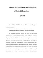
Fig 2.1 Oxygen dissociation curve.
tissue. Demand increases blood flow and excess of oxygen
decreases blood flow. This mechanism is partially medi-
ated by NO. Hemoglobin is able to scavenge NO and,
therefore, constrict vessels. Hence, it seems red cells coun-
teract their own work of delivering oxygen by blocking
(constricting) their own road (vessels). This is not the
case, however. The explanation for this phenomenon lies
in the fact that hemoglobin comes in two different forms:
R (relaxed, with high oxygen affinity) and T (tense, with
low oxygen affinity). In the R form, hemoglobin not only
transports oxygen but can also take up an NO compound
(= S-nitrosylation). When the hemoglobin arrives at pre-
capillary resistance vessels, it loses some oxygen and starts
its transition from R to T. This change liberates the NO
and causes dilatation of the arterioles [9]. With this mech-
anism, oxygen-loaded red cells open their doors to the
tissue in order to deliver oxygen.
How do red cells give oxygen and how is it taken
up by tissue?
The quantity of oxygen released by red cells depends
much on the oxygen affinity of hemoglobin molecules.
It is this affinity that translates oxygen flow into avail-
able oxygen. A common method to depict the behavior
of hemoglobin is the oxygen dissociation curve (Fig. 2.1).
The oxygen dissociation curve is sigmoid-shaped. This
is due to conformational changes in the hemoglobin
molecule that occur when it loads or releases oxygen. The
uptake of each oxygen molecule alters the hemoglobin
conformation and so it enhances the uptake of the next
oxygen molecule. A change in the hemoglobin’s oxygen
affinity profoundly affects oxygen release to the tissue,
whereas the oxygen uptake by hemoglobin is scarcely
affected.
There are several factors that per se can influence the
hemoglobin’s affinity for oxygen (Fig. 2.1). Those factors
include temperature, CO
2
,H
+
, and 2,3-DPG [10]. Red
blood cells deliver oxygen to metabolically active tissues.
Such tissues release CO
2
that diffuses into the red cells.
By carbonic anhydrase, CO
2
and H
2
O react to H
+
and
HCO
−
3
. The resulting HCO
−
3
is exchanged with extracel-
lular Cl
−
, which leads to an intracellular acidification. The
resulting decrease in pH facilitates oxygen dissociation
from hemoglobin. Also, 2,3-DPG, a glycolytic intermedi-
ate, binds to deoxyhemoglobin and stabilizes hemoglobin
in the deoxy form, thus reducing hemoglobin’s oxygen
affinity and supporting oxygen release. In fact, this 2,3-
DPG is so important that no oxygen can be unloaded by
the red cells if it is completely lacking.
After being released from the hemoglobin molecule,
oxygen has to pass several layers until it reaches the tissue.
In contrast to oxygen uptake by red cells, release of oxygen
depends mainly on the affinity of hemoglobin in the red
cell and not so much on the thickness of surrounding un-
stirred fluid layers [1]. Deoxygenation therefore depends
on pH and 2,3-DPG (lowered pH and increased 2,3-DPG
levels facilitate release of oxygen) [1]. Only at very low
hemoglobin concentrations chemical reactions limit re-
lease of oxygen from hemoglobin.
BLUKO82-Seeber March 14, 2007 16:42
Physiology of Anemia and Oxygen Transport 13
After the oxygen is released by the hemoglobin and has
passed all barriers on its way to the tissue, the mitochon-
dria accept the delivered oxygen molecules. Oxygen may
have traveled via free flow or by means of a “coach” to
a myoglobin molecule. The latter is called myoglobin-
facilitated oxygen diffusion. “Deoxymyoglobin captures
oxygen immediately as it crosses the interface: the newly
formed oxymyoglobin diffuses away . . . . The effect is to
make the oxygen pressure gradient from capillary lumen
to the sarcoplasma more steep, thereby enhancing the oxy-
gen flux” [11]. This effect maintains oxygen flow to themi-
tochondria under conditions of low extracellular oxygen
pressure.
Does the tissue use oxygen?
The human body depends on oxygen for adenosine
triphosphate (ATP) generation and maintenance of aer-
obic metabolism. ATP, the body’s main energy source, is
generated in the mitochondria using molecular oxygen.
Only if the mitochondria actually use the oxygen, offered
oxygen can do its job. This means that oxygen utilization
is defined by the mitochondria.
Interestingly, there is a genetic component to the work
of the mitochondria. Inherited or acquired changes in
the enzyme supply determine how effectively oxygen can
be used. Drugs such as propofol, which inhibit oxida-
tive phosphorylation, can influence the mitochondria and
thus the use of delivered oxygen [12]. Other factors influ-
ence tissues’ (and mitochondria’s) use of oxygen as well.
The energy demand of the tissue influences how much
oxygen is used. In turn, factors that influence the tis-
sue’s metabolism also influence the rate of its oxygen use.
NO inhibits mitochondrial respiration, and thus oxygen
consumption. On the other hand, lack of NO increases
metabolism and tissue oxygen consumption [13]. The in-
fluence of the body temperature on oxygen extraction
is well known: Higher body temperature increases oxy-
gen demand, extraction [14], and utilization. There are
many more factors that alter the body’s energy require-
ment and thus the oxygen demand of the tissue: physical
activity, hormones (catecholamines, thyroid hormones),
infections, psychological stress, pain and anxiety, diges-
tion and repair of tissues, just to name a few.
All organs and tissues, with the exception of the cen-
tral nervous system, are able to use the delivered oxygen
to the full, that is, 100%. This is also true for the my-
ocardium [15]. In a healthy individual, however, there is
a wide safety margin. Oxygen delivery in healthy resting
humans exceeds the needs fourfold. The body as a whole
uses only about one in four of the hemoglobin’s oxygen
molecules. The amount of oxygen used is called oxygen
consumption (VO
2
). Another way to express the use of
oxygen is the oxygen extraction ratio O
2
ER. It describes
the percentage of oxygen extracted from the hemoglobin
molecule. The total body’s normal resting O
2
ER equals
about 20–25%. But the organ-specific oxygen extraction
varies. Kidneys extract only 5–10%, and the heart at rest
55% [4]. It can be seen from such numbers that oxygen
delivery is not the only determinant in the body’s oxygen
balance. Only the concerted efforts of all systems included
in oxygen supply and use make aerobic life possible.
Pathophysiology of anemia
The human body is a marvel of creation. It is equipped
with amazing mechanisms to maintain its function and to
ensure that its tissue and organ systems tolerate a broad
variety of conditions. This is also true for diminished
levels of hemoglobin. Oxygen delivery remains sufficient
over a wide range of hemoglobin levels, and even when
hemoglobin levels have decreased markedly, the body can
survive.All this is due to avariety of compensatory mecha-
nisms, some of which are reviewed on the following pages.
The initial adaptation to blood loss is not mainly a reac-
tion to a decrease in oxygen-carrying capacity but rather
the body’s reaction to hypovolemia. If left alone, the hu-
man body initiates a series of changes: first, to restoreblood
volume and second, to restore red cell mass. Within min-
utes, heart rate and stroke volume increase. Theadrenergic
system and the renin-angiotensin-aldosterone system are
stimulated, releasing vasoactive hormones. This leads to
the constriction of vascular sphincters in the skin, skeletal
muscle, kidneys, and splanchnic viscera. The blood flow
is redistributed to high-demand organs, namely the heart
and brain [16]. To restore intravascular volume, fluids are
first shifted from the interstitial space to the vessels, and
later from the intracellular to the extracellular space. Due
to adaptations in renal function, water and electrolytes are
conserved. The liver is stimulated to produce osmotic ac-
tive agents (glucose, lactate, urea, phosphate, etc.), which
results in a net shift of fluid into the vasculature [16] and
thus preload increases. Unless compensatory mechanisms
fail, cardiac output is restored within 1–2 minutes [17].
However, if the body’s compensatory mechanisms fail,
cardiac output and oxygen delivery decrease. At thatpoint,
restoration of blood volume (not red cell volume) is
mandatory. If fluids are infused, cardiac output can be in-
creased and the untoward effects of hypovolemia averted.
BLUKO82-Seeber March 14, 2007 16:42
14 Chapter 2
Blood flow is restored and the body is able to repair dam-
age and replenish the loss of red cell mass.
In the following paragraphs, the trip through the hu-
man body is repeated—this time under anemic, yet nor-
movolemic, conditions. The assumption is that the patient
is already volume-resuscitated and that adaptive mecha-
nisms are mainly due to reduced red cell mass rather than
reduced intravascular volume.
Adaptation of the body: acute is not the same
as chronic
It is not uncommon to meet persons with a hemoglobin
value of less than 4 g/dL doing their normal job—the only
clinically observable effect being reduced exercise toler-
ance. On the other hand, some patients with the same
hemoglobin level are hardly capable of lifting their head.
Responses to blood loss and anemia are obviously not
uniform. How the body responds to anemia depends on
the rapidity of blood loss, the underlying condition of the
patient, drugs taken, preexisting hemoglobin level, etc.
[18]. Some adaptive mechanisms are more pronounced in
acute anemia while others are more common in chronic
anemia.
Adaptive mechanisms to anemia:
macrocirculation
Leonardo da Vinci said: “Movement is the cause of all life.”
This also holds true for blood loss and anemia. In anemia,
increased flow, that is, cardiac output, compensates for
the losses in hemoglobin. In the acute setting, cardiac out-
put increases with increasing levels of volume-resuscitated
anemia. This is mainly due to increases in stroke volume.
The influence of the heart rate in increasing the cardiac
output is a subject of debate. Results conflict depending
on the animal species studied and the patients and their
conditions (anesthetized versus awake, influence of drugs
taken). It seems, however, that an increase in the heart
rate is not the main determinant in increasing the cardiac
output ofacutely anemic, volume-resuscitated individuals
[18, 19].
Two major mechanisms are responsible for increased
cardiacoutput.The most important is a reduction in blood
viscosity. This results in increased venous flow with in-
creased flow to the right heart. Preload increases, resulting
in an enhanced cardiac output. Afterload is reduced by the
decrease in blood viscosity. The other important cause for
the increase in cardiac output is stimulation of the sym-
pathetic nerve system. Via the sympathetic nerve system
and a catecholamine release, myocardial contractility
(and heart rate) increases, and again, the cardiac output
increases.
Both, in acute and chronic anemia cardiac output is
increased by means of viscosity reductionand sympathetic
nerve stimulation. And if anemia is becoming chronic, the
heart adapts to the increased workload withleft ventricular
hypertrophy.
In the discussion of macrocirculatory adaptations to
anemia, special consideration must be given to the heart.
The myocardium requires more oxygen than any other
organ and has a high O
2
ER even at rest. When the heart’s
oxygen demand increases, e.g., by increased cardiac work-
load, the heart can slightly increase its oxygen extraction.
The major increase in oxygen delivery to the heart, how-
ever, is due to vasodilatation of the coronary arteries. Nor-
mally, there is a great reserve in myocardial blood flow
[20]. However, if myocardial blood flow cannot be in-
creased, the heart may not be able to receive the oxygen
it needs for its vital work. On the one hand, increased
myocardial work is beneficial to compensate anemia. But
then, increased myocardial work increases the heart’s oxy-
gen demand. Since an increase in the cardiac output is
a crucial factor in compensating for the loss of oxygen-
carrying capacity, it is vital for the heart to be able to in-
crease its outputon demand. Several factorscan impair the
heart’s ability to increase output. Coronary stenosis, my-
ocardial insufficiency, sepsis, anesthetics, and other drugs
may compromise the work of the heart [21]. In such situa-
tions, the increase in cardiac output may not be sufficient
to compensate for the lost red cell mass. Studies show that
patients with different pathologies of the heart are more
susceptible to ischemia thanother patient populations and
tolerate anemia lessthan the same patients without cardiac
pathology.
Closely related to anemia-induced changes in cardiac
output is the alteration of vascular tone. Again, the sym-
pathetic nerve system plays an important role in this [18].
Increased activity of aortic chemoreceptors has been pos-
tulated to change vasomotor tone, and thus afterload [4].
Part of the reduction in afterload may also be due to hy-
poxic vasodilatation.
An increase in cardiac output and a reduction in vas-
cular tone lead to an increased blood flow. The flow is
directed to high-demand organs, with the brain and heart
first in line to get a major portion of the blood [22]. Even
under normal conditions they extract most of the oxygen
offered by the hemoglobin [4] and are therefore supply
dependent. Redistribution of the blood flow takes place at
the expense of noncritical organs [23], e.g., the skin.
BLUKO82-Seeber March 14, 2007 16:42
Physiology of Anemia and Oxygen Transport 15
Volume-resuscitated anemic patients have a greater
plasma volume than healthy, nonanemic humans. This
volume serves to transport physically dissolved oxygen.
While the portion of physically dissolved oxygen is almost
insignificant in nonanemic patients, it must not be un-
derestimated in severely anemic patients. At times, it may
constitute a major portion of the total oxygen delivered by
the blood [24].
Another adaptive mechanism of anemia tolerance is
increased oxygen extraction. As shown above, given nor-
mal conditions, on average only one out of four oxygen
molecules carried by a hemoglobin molecule is extracted
by the body. Most tissues would be able to extract much
more oxygen from the hemoglobin. In fact, in severe states
of anemia, most tissues can extract nearly100% ofthe oxy-
gen offered. The increase in O
2
ER is thus a valuable tool
to compensate for decreased oxygen carriers.
The extent to which the above-mentioned adaptive
mechanisms are being used by the body differs from pa-
tient to patient: The degree of anemia as well as the con-
dition of the patient, the comorbidities, and the rapid-
ity of the development of anemia play a role. If anemia
becomes a chronic state, systemic vascular resistance re-
turns to normal. Cardiac output is not increased to the
same high degree as in acute anemia. Redistribution of
blood flow from organs, with excess flow to other organs,
takes place [4]. A combination of adaptive mechanisms
enable a chronically anemic body to meet oxygen de-
mand, evenat times when hemoglobin levelsare extremely
low.
As a combined effect of reduction of blood viscosity,
increase in cardiac output, etc., delivery of oxygen in-
creases as the hematocrit starts to decrease. Oxygen de-
livery reaches its maximum at a hematocrit of 25–33%
[4]. At hematocrits above 45 and below 25, oxygen deliv-
ery decreases. Adaptation in cardiac output compensates
for decreased oxygen-carrying capacity. This results in an
almost constant delivery of oxygen to the capillaries, as
long as red cell losses do not exceed about 50% in healthy
persons [13].
Adaptive mechanisms to anemia:
microcirculation
As shown above, the microcirculation plays a crucial role
in oxygen delivery to the tissue. It does not come as a
surprise, then, that on the microcirculatory level there
are also many mechanisms that compensate for decreased
red cell mass. In fact the microcirculation is where the
advantages of hemodilution matter most [19].
How do red cells take up oxygen?
Adaptive mechanisms begin again in the lung, the place
where red cells exchange gases. Despite a reduction of the
blood’s capacity for carrying O
2
and CO
2
,evensevere
anemia is associated with remarkable stability of the pul-
monary gas exchange [25]. Compensatory mechanisms
kick in, ensuring that O
2
transport and CO
2
elimination
are not impaired [26].
In anemia, lung perfusion is altered. Less hemoglobin
molecules are available for interaction with NO, which
preserves the vasodilatatory effect of NO. Resistance to
pulmonary blood flow is thus decreased [27]. This leads
to an increased flow of blood through the pulmonary
vasculature. This flow increases the shear stress in the
endothelium, thus further increasing the production of
vasodilatating NO. Those vasodilatatory effects counter-
act hypoxic pulmonary vasoconstriction [25]. In anemia
there is a tendency toward reduced heterogenicity of pul-
monary blood flow. Selective constriction of pulmonary
vessels diverts blood to better ventilated alveoli [28]. NO
may also alter airway tone, leading to a redistribution of
ventilation. All this results in improved gas exchange in the
lung. Several studies showed that arterial partial pressure
of oxygen in anemic patients may even be greater than in
nonanemic patients.
How do red cells reach their goal, the
microvasculature in the tissue?
There is a clear relationship between hematocrit and vis-
cosity. As the hematocrit decreases, blood viscosity de-
creases and red cells travel at a higher speed. When travel-
ing at this high speed, they do not have sufficient time to
lose oxygen on the way to the tissue. Therefore, in anemic
conditions red cells arrive in the microcirculation with
more oxygen than that in nonanemic conditions [4].
Since FCD is important for tissue survival, several
mechanisms are employed in anemia to recruit capillar-
ies. The blood flow in the capillaries of anemic patients is
more homogenous than in nonanemic ones [19]. Several
mechanisms account for this. Hemoglobin has a very high
affinity for NO. This property is a crucial factor for reg-
ulating the interaction of red cells and the endothelium.
Red cells have the ability to constrict the vascular bed by
scavenging the vasodilatator NO. This effect is concen-
tration dependent; that is, the lower the hematocrit, the
more the vasodilatation and the better tissue perfusion.
A better flow of blood in anemic states results. “Physical
stimuli such as fluid shear stress, pulsatile stretching of
BLUKO82-Seeber March 14, 2007 16:42
16 Chapter 2
the vessel wall, or a low arterial PO
2
, also stimulate the
release of NO above the basal level” [29]. Furthermore, in
anemia, red cells do not aggregate readily and are able to
pass through the narrowest capillary. Arterial/venular dif-
fusional shunting is diminished because of the increased
blood velocity and the decreased red cell residence time in
the vessels [13]. While in the acute anemic setting the body
is only able to recruit available capillaries, in the chronic
anemic setting new vessels develop (neoangiogenesis).
Capillary vessels need arterial pressure to remain open.
As blood viscosity decreases in anemia, flow increases be-
cause less arterial pressure is lost struggling with high
blood viscosity. Thus, the lowered blood viscosity im-
proves capillary perfusion. For this reason hemodilution is
used therapeutically to improve tissue oxygenation. How-
ever, the beneficial effect of reducing the blood viscosity
is only apparent as long as the heart can compensate for
lost hemoglobin by improving flow. At the point where
the heart can no longer compensate for the red cell loss,
this beneficial effect ceases to exist. Hemodilution be-
yond this point reduces viscosity still further, inducing
vasoconstriction and thus reducing FCD. At that point, a
therapeutic maneuver may be employed to improve tis-
sue perfusion again. Vessels are dependent on shear stress
to open. By artificially increasing blood viscosity, shear
stress to the vessels can be exerted, eliciting a vasodi-
latatory response. This may result in recovery of FCD
[8, 13, 30].
In a summary of some interesting findings about the re-
lationship of blood viscosity and transfusions the author
stated: “Microcirculatory studies show that the organism
compensates for reduced blood viscosity only up to reduc-
tions coincident with the conventional transfusion trigger
and that reductions beyond this point lower functional
capillary density. These studies show that the critical limit
for tissue survival at the transfusion trigger is functional
capillary density. Functional capillary density is impor-
tant, primarily, for the extraction of tissue metabolism
byproducts and, secondly, for tissue oxygenation. Thus,
the transfusion trigger signals a condition where the cir-
culation no longer compensates for significantly lowered
viscosity due to hemodilution. Continued hemodilution
with high-viscosity plasma expanders beyond the trans-
fusion trigger is shown to maintain functional capillary
density and improve tissue perfusion, suggesting that the
conventional transfusion trigger is a viscosity trigger . . . ”
[31]. And after a discussion of the benefits of higher blood
viscosity after reaching the “transfusion trigger,” the au-
thor concluded: “It is a corollary to these considerations
that the level of oxygen carrying capacity required to safely
oxygenate the tissue may be much lower than that dictated
by medical experience if microvascular function is main-
tained” [13].
How do the red cells give oxygen and how does
the tissue take up oxygen?
In states of anemia, the oxygen affinity of hemoglobin is
lowered as is reflected in a right shift in the oxygen disso-
ciation curve. This facilitates oxygen release to the tissue.
The right shift in the oxyhemoglobin dissociation curve
is the result of increases of 2,3-DPG in red cells [18]. In
contrast to chronic anemia, however, facilitated O
2
disso-
ciation does not play too big a role in acute anemia where
2,3-DPG levels do not change significantly [20]. Theoret-
ically, in anemia also a shift in pH toward greater acidity
may enhance oxygen release (Bohr effect). However, this
effect is probably not clinically relevant since immense
changes in pH are needed to release significant amounts
of oxygen from red cells [18].
Reserves in oxygen delivery are used in anemia, and
this is reflected in an increased oxygen extraction ratio.
Normally, the tissue extracts only about 20–30% of the
total oxygen delivered. In anemia, the tissue may extract
much more so that body O
2
ER as a whole increases to 50–
60%. As a consequence, the mixed venous oxygen partial
pressure decreases.
In extreme anemia, oxygen uptake by the tissue seems
to be limited. This may be due to the increased flow and
the decreased transit time of red cells in the microvas-
culature. Time available for oxygen diffusion may be in-
sufficient [32]. Also, erythrocyte spacing (increased dis-
tance between adjacent red cells during anemia) and the
diffusion distance may contribute to this phenomenon
[33].
Does the tissue use oxygen?
The last stop on this trip through the anemic body is again
the tissue with the mitochondria, the place where a de-
crease in oxygen delivery should matter most.Intracellular
mechanisms sense the decrease of O
2
in the tissue. In re-
sponse, hypoxia-dependent gene expression is stimulated.
A key factor in this process is the hypoxia-inducible factor
1␣ (HIF1␣). It “induces the expression of genes that influ-
ence angiogenesis and vasodilatation, erythropoiesis and
increased breathing, as well as glycolytic enzyme genes
for anaerobic metabolism” [34]. Interestingly, the basic
BLUKO82-Seeber March 14, 2007 16:42
Physiology of Anemia and Oxygen Transport 17
mechanisms of hypoxia tolerance are shared by different
species, including humans. The following model was de-
scribed for animals.
So, what happens if a cell senses a decrease in oxy-
gen supply? Does the cell die? Not right away. The cell
needs oxygen mainly to produce energy (ATP).So it makes
sense that in anticipation of reduced oxygen (that is, en-
ergy) supply, energy demand is reduced. What does a cell
need energy from ATP for? Almost all energy is needed
for protein synthesis and degradation, maintenance of
ion gradients, and synthesis of glucose and urea. And, in
fact, in an initial defense phase, cells greatly (>90%) and
rapidly suppress their protein, glucose, and urea synthe-
sis. Interestingly, ion gradients over membranes remain
constant, although the pumping activity of ion pumps
is reduced to save energy. The cells use different mecha-
nisms to accomplish this miracle. For instance, liver cells
reduce cell membrane permeability, a process called chan-
nel arrest. Nerve cells reduce the firing frequency (spike
arrest). Employing such measures, many cells can attain
an energy balance at a lower level (ATP demand = AT P
supply). This may ensure long-term survival in hypoxia.
Hypoxia-sensitive cells, however, do not attain a new
balance.
After the defense phase, a second, “rescue” phase, fol-
lows. Cells are now aiming at long-term hypoxia sur-
vival. To that end, cells reactivate some protein biosyn-
thesis to prepare the cell for survival with extremely low
ATP turnover. Hypoxia-dependent expression of key fac-
tors (such as HIF 1) regulates this process. Housekeeping
genes consolidate and stabilize the cell, and enzymes for
anaerobic ATP production are upregulated [34, 35]. With
changes like these, cells can function for a while with a
very low oxygen delivery.
Relationship between oxygen delivery and
oxygen consumption
As mentioned initially, the aim of anemia therapy is to
match the tissue’s demand for oxygen with supply. This
demand is reflected by tissue oxygen consumption VO
2
.
There is a relationship between oxygen delivery and oxy-
gen consumption (Fig. 2.2). Oxygen consumption re-
mains constant over a wide range of delivery. At the point
where oxygen consumption becomes supply dependent,
tissue hypoxia may occur. This point is called “critical oxy-
gendelivery,”DO
∗
2crit
. This, however, is no fixed number,
leaving room for therapeutic interventions.
DO
2
crit
Oxygen delivery (DO
2
)
Oxygen consumption (VO
2
)
Fig 2.2 Relationship between oxygen delivery and oxygen con-
sumption. Continuous line: healthy individuals; dashed line:
pathologic as in severe illness (with a wider range of dependence
of VO
2
on DO
2
and a higher DO
2crit
).
Practical implications
Theoxygendeliveryequation[DO
2
= CO × (Hgb ×
1.34 × SaO
2
+ 0.003 × PaO
2
) × 10] may serve as a
mnemonic for available anemia treatments. Every vari-
able in the equation can be considered, based on which
therapeutic interventions can be evaluated to optimize
oxygen delivery. The cardiac output can be optimized
by administering balanced amounts of intravenous fluids
and removing negative inotropic influences or increas-
ing positive inotropics as tolerated. The hemoglobin level
can be increased, not only by speeding up endogenous
hematopoiesis, but also by avoiding undue hemoglobin
losses. Arterial oxygen partial pressure and oxygen satura-
tion can be increased, using supplemental oxygen and me-
chanical ventilation as indicated. In addition, the amount
of oxygen dissolved in plasma can be increased fur-
ther by increasing the atmospheric pressure (hyperbaric
oxygen).
A more complete picture of anemia therapy, though,
is achieved when not only an increase of oxygen deliv-
ery is aimed at, but also a reduction of oxygen demand is
contemplated. Oxygen delivery and consumption are re-
lated, since VO
2
= DO
2
× O
2
ER. While not many inter-
ventions are available to increase oxygen extraction, there
are quite a few things that can be done to reduce oxy-
gen consumption. “The four pillars of anemia therapy”
(Fig. 2.3) summarize how an understanding of physiol-
ogy and pathophysiology translates into a plan of care.
With the appropriate combination of factors that not only
increase oxygen delivery but also reduce oxygen consump-
tion, even hemoglobin levels way below the supposed