Curcumin modulates dopaminergic receptor, CREB and phospholipase c gene expression in the cerebral cortex and cerebellum of streptozotocin induced diabetic rats potx
Bạn đang xem bản rút gọn của tài liệu. Xem và tải ngay bản đầy đủ của tài liệu tại đây (851.4 KB, 11 trang )
Kumar et al. Journal of Biomedical Science 2010, 17:43
/>Open Access
RESEARCH
BioMed Central
© 2010 Kumar et al; licensee BioMed Central Ltd. This is an Open Access article distributed under the terms of the Creative Commons
Attribution License ( which permits unrestricted use, distribution, and reproduction in
any medium, provided the original work is properly cited.
Research
Curcumin modulates dopaminergic receptor, CREB
and phospholipase c gene expression in the
cerebral cortex and cerebellum of streptozotocin
induced diabetic rats
T Peeyush Kumar, Sherin Antony, G Gireesh, Naijil George and CS Paulose*
Abstract
Curcumin, an active principle component in rhizome of Curcuma longa, has proved its merit for diabetes through its
anti-oxidative and anti-inflammatory properties. This study aims at evaluating the effect of curcumin in modulating the
altered dopaminergic receptors, CREB and phospholipase C in the cerebral cortex and cerebellum of STZ induced
diabetic rats. Radioreceptor binding assays and gene expression was done in the cerebral cortex and cerebellum of
male Wistar rats using specific ligands and probes. Total dopaminergic receptor binding parameter, B
max
showed an
increase in cerebral cortex and decrease in the cerebellum of diabetic rats. Gene expression studies using real time PCR
showed an increased expression of dopamine D1 and D2 receptor in the cerebral cortex of diabetic rats. In cerebellum
dopamine D1 receptor was down regulated and D2 receptor showed an up regulation. Transcription factor CREB and
phospholipase C showed a significant down regulation in cerebral cortex and cerebellum of diabetic rats. We report
that curcumin supplementation reduces diabetes induced alteration of dopamine D1, D2 receptors, transcription
factor CREB and phospholipase C to near control. Our results indicate that curcumin has a potential to regulate
diabetes induced malfunctions of dopaminergic signalling, CREB and Phospholipase C expression in cerebral cortex
and cerebellum and thereby improving the cognitive and emotional functions associated with these regions.
Furthermore, in line with these studies an interaction between curcumin and dopaminergic receptors, CREB and
phospholipase C is suggested, which attenuates the cortical and cerebellar dysfunction in diabetes. These results
suggest that curcumin holds promise as an agent to prevent or treat CNS complications in diabetes.
Introduction
Diabetes mellitus is a heterogeneous disease character-
ized by chronic hyperglycaemia and requires long-term
management. Chronic changes in the antecedent level of
glycaemia induce alterations in brain glucose metabolism
in rodents [1,2]. Chronic hyperglycemia in diabetes can
lead to various complications, affecting the CNS [3]. A
continuous systemic supply of glucose is essential for
normal cerebral metabolism [4].
Controlling blood sugar is essential for avoiding long-
term complications of diabetes like learning and memory.
Although mechanisms leading to cortical and cerebellar
dysfunction associated with diabetic complications are
not completely understood, brain cells are particularly
vulnerable to oxidative stress [5]. Oxidative stress, leading
to an increased production of reactive oxygen species, as
well as lipid peroxidation is increased in diabetes [6-8].
Hyperglycemia causes the autoxidation of glucose, glyca-
tion of proteins, and the activation of polyol metabolism
[9]. These changes accelerate the generation of reactive
oxygen species to increase oxidative modifications of lip-
ids, DNA, and proteins in various tissues. Oxidative
stress is believed to play an important role in the develop-
ment of complications in diabetes associated neuronal
disorders [9]. Greater understanding of CNS (CNS)
involvement could lead to new strategies to prevent or
reverse the damage caused by diabetes mellitus.
* Correspondence:
1
Molecular Neurobiology and Cell Biology Unit, Centre for Neuroscience,
Cochin University of Science and Technology, Cochin- 682 022, Kerala, India
Full list of author information is available at the end of the article
Kumar et al. Journal of Biomedical Science 2010, 17:43
/>Page 2 of 11
Antioxidant agents from diet have a significant thera-
peutic influence on various neurodegenerative disorders
associated with diabetes and oxidative stress [10,11]. Cur-
cuminoids, the main components in Curcuma species,
share a common unsaturated alkyl-linked biphenyl struc-
tural feature and are responsible for their major pharma-
cological effects. The biological and chemical properties
of curcuminoids were reported [12] A number of experi-
mental studies have demonstrated CUR's antioxidant and
neuroprotective potential [13,14] Also curcumin modu-
lates the expression of various molecular targets, such as
transcription factors, enzymes, cytokines, cell cycle pro-
teins, receptors and adhesion molecules [15]. Diabetes
mellitus has also been reported to be accompanied by
behavioural and reduced motor activity [16]. One unify-
ing mechanism which lies behind this neuronal injury is
the excessive free radical generation from the auto oxida-
tion of elevated intracellular glucose levels. Curcumin
may antagonise the deficit of glucose energy metabolism
or oxidative stress related to cognitive impairment associ-
ated with diabetes.
Diabetes is also found to be associated with changes in
somatic sensations which involve the cerebellum, cerebral
cortex and thalamus. Symptoms, like loss of pain,
impaired touch perception and decreased position sense,
have been commonly documented in a diabetic patient
[17]. Dopamine in the CNS is involved in the control of
both motor and emotional behavior [18] and peripherally
modulates insulin secretion in the pancreatic islets [19].
Nafadotride, a preferential antagonist of dopamine D3
receptors administered at low doses directly into the cer-
ebellum, has been shown to activate locomotor activity
[20]. The secretion of insulin by β-cells of the endocrine
pancreas is regulated by glucose and other circulating
nutrients. It is also modulated by several hormones and
neurotransmitters, among which dopamine plays a prom-
inent role.
CREB is a protein that is a transcription factor. It binds
to certain DNA sequences called cAMP response ele-
ments and, thereby, increases or decreases the transcrip-
tion of the downstream genes [21]. Genes whose
transcription is regulated by CREB include: c-fos, BDNF
(Brain-derived neurotrophic factor), tyrosine hydroxylase
and neuropeptides such as somatostatin, enkephalin,
VGF and corticotropin-releasing hormone [21]. In neu-
ronal tissue, CREB regulation by nerve growth factor and
insulin-like growth factor-1 is essential for neuronal plas-
ticity, full axonal development, memory consolidation,
and neuroprotection [22,23]. The Phospholipase C activ-
ity decline in the brain is expected to affect mainly the
18:0/20:4 molecular species of DAG because this is the
principal molecular species of phosphoinositides in the
nervous tissue [24].
Hyperglycaemia is associated with a number of physio-
logical changes, the most profound effects are seen in the
brain, where glucose is the major substrate for energy
metabolism and both local energy store and the supply of
alternate sources are limited. The initiating events in
hyperglycemic encephalopathy still are not understood
completely. But brain injury appears to result from a
number of processes that are initiated when blood glu-
cose concentration is altered. However, the action mech-
anisms of this remain obscure. Therefore, this study was
designed to investigate the beneficial effect of curcumin a
neuroprotective agent, on impairment in dopaminergic
receptors, CREB and phospholipase C in the cerebral cor-
tex and cerebellum of STZ-induced diabetic rats. Our
present study on curcumin dependent regulation of dop-
aminergic receptors, transcription factor CREB and
phosphor lipase C amelioration of cortical and cerebellar
cells will certainly enlighten novel therapeutic possibili-
ties in diabetes treatment.
Materials and methods
Bio chemicals used in the present study were purchased
from Sigma Chemical Co., St. Louis, USA. All other
reagents of analytical grade were purchased locally. [
3
H]
Dopamine were purchased from NEN Life Sciences
Products Inc., Boston, U.S.A. dopamine and curcumin
were from Sigma Chemical Co., USA. Tri-reagent kit was
purchased from MRC, USA. Real Time PCR Taqman
probe assays on demand were from Applied Biosystems,
Foster City, CA, USA.
Male adult Wistar rats of 180-240 g body weight were
used for all experiments. The animals were allowed to
acclimatise for 2 weeks before the experiment. They were
housed individually in separate cages under 12 hour light
and 12 hour dark periods. Rats had free access to stan-
dard food and water ad libitum. All animal care and pro-
cedures were done in accordance with the Institutional
and National Institute of Health guidelines. All efforts
were made to minimize the number of animals used and
their suffering. Diabetes was induced in rats by single
intra femoral vein injection of STZ freshly dissolved in 0.1
M citrate buffer, pH 4.5, under anaesthesia [25]. STZ was
given at a dose of 55 mg/kg body weight [26,27]. Animals
were divided into the following groups: I) Control ii) dia-
betic iii) insulin-treated diabetic iv) curcumin-treated
diabetic rats. Each group consisted of 6-8 animals. The
insulin-treated diabetic group received subcutaneous
injections (1 Unit/kg body weight) of Lente and Plain
insulin (Boots India) daily during the entire period of the
experiment. The last injection was given 24 hours before
sacrificing the rats. Curcumin treated groups received 60
mg/kg suspension of curcumin orally [28] for the entire
period of the experiment. Curcumin was suspended in
0.5% w/v sodium carboxymethylcellulose immediately
Kumar et al. Journal of Biomedical Science 2010, 17:43
/>Page 3 of 11
before administration in constant volume of 5 ml/kg body
weight. Rats were sacrificed on 15th day by decapitation.
The cerebellum was dissected out quickly over ice
according to the procedure of Glowinski and Iversen,
1966 [29] and the tissues collected were stored at -80°C
until assayed.
Estimation of blood glucose
Blood glucose was estimated by the spectrophotometer
method using glucose oxidase-peroxidase reactions.
Blood samples were collected from the tail vein at 0 hour
(Before the start of the experiment), 3rd, 6th, 10th and
14th day and the glucose levels were estimated subse-
quently. Along with this blood samples were collected 3
hrs after the administration of morning dose of insulin
and curcumin. The results were expressed in terms of
milligram per decilitre of blood.
Total Dopamine receptor binding studies in the cerebellum
DA receptor assay was done using [
3
H]DA according to
Madras et al., 1988 [30]. Cerebellum was homogenised in
a polytron homogeniser with 20 volumes of cold 50 mM
Tris-HCl buffer, along with 1 mM EDTA, 0.01%ascorbic
acid, 4 mM MgCl
2
, 1.5 mM CaCl
2
, pH. 7.4 and centri-
fuged at 38,000 × g for 30 min at 4°C. The pellet was
washed twice by rehomogenization and centrifuged twice
at 38,000 × g for 30 min at 4°C. This was resuspended in
appropriate volume of the buffer containing the above
mentioned composition.
Binding assays were done using different concentra-
tions i.e., 0.25 nM-1.5 nM of [
3
H]DA in 50 mM Tris-HCl
buffer, along with 1 mM EDTA, 0.01% ascorbic acid, 1
mM MgCl
2
, 2 mM CaCl
2
, 120 mM NaCl, 5 mM KCl,
pH.7.4 in a total incubation volume of 250 μl containing
200-300 μg of proteins. Specific binding was determined
using 100 μM unlabelled dopamine.
Tubes were incubated at 25°C for 60 min. and filtered
rapidly through GF/B filters (Whatman). The filters were
washed quickly by three successive washing with 5.0 ml
of ice cold 50 mM Tris buffer, pH 7.4. Bound radioactivity
was counted with cocktail-T in a Wallac 1409 liquid scin-
tillation counter. The non-specific binding determined
showed 10% in all our experiments.
Protein determination
The amount of protein was measured by the method of
Lowry et al., 1951 [31] using bovine serum albumin as
standard. The intensity of the purple blue colour formed
was proportional to the amount of protein, which was
read in a spectrophotometer at 660 nm.
Receptor data analysis
The receptor binding parameters were determined using
Scatchard analysis [32]. The specific binding was deter-
mined by subtracting non-specific binding from the total.
The binding parameters, maximal binding (B
max
) and
equilibrium dissociation constant (K
d
), were derived by
linear regression analysis by plotting the specific binding
of the radioligand on X-axis and bound/free on Y-axis
using Sigma plot software (version 2.0, Jandel GmbH,
Erkrath, Germany). The maximal binding is a measure of
the total number of receptors present in the tissue and
the equilibrium dissociation constant is the measure of
the affinity of the receptors for the radioligand. The K
d
is
inversely related to receptor affinity.
Analysis of gene expression by Real-Time PCR
RNA was isolated from the cerebellum of experimental
rats using the Tri-reagent (MRC, USA). Total cDNA syn-
thesis was performed using ABI PRISM cDNA archive kit
in 0.2 ml microfuge tubes. The reaction mixture of 20 μl
contained 0.2 μg total RNA, 10 × RT buffer, 25 × dNTP
mixture, 10 × random primers, MultiScribe RT (50 U/μl)
and RNase free water. The cDNA synthesis reactions
were carried out at 25°C for 10 minutes and 37°C for 2
hours using an Eppendorf Personal Cycler. Real-time
PCR assays were performed in 96-well plates in ABI 7300
real-time PCR instrument (Applied Biosystems). The
primers and probes were purchased from Applied Biosys-
tems, Foster City, CA, USA. The TaqMan reaction mix-
ture of 20 μl contained 25 ng of total RNA-derived
cDNAs, 200 nM each of the forward primer, reverse
primer and TaqMan probe for assay on demand and
endogenous control β-actin and 12.5 μl of Taqman 2×
Universal PCR Master Mix (Applied Biosystems) and the
volume was made up with RNAse free water. The follow-
ing thermal cycling profile was used (40 cycles): 50°C for
2 min, 95°C for 10 min, 95°C for 15 sec and 60°C for 1
min.
Fluorescence signals measured during amplification
were considered positive if the fluorescence intensity was
20-fold greater than the standard deviation of the base-
line fluorescence. The
ΔΔ
CT method of relative quantifi-
cation was used to determine the fold change in
expression. This was done by normalizing the resulting
threshold cycle (CT) values of the target mRNAs to the
CT values of the internal control β-actin in the same sam-
ples (
Δ
CT = CT
Targe t
- CT
β-actin
). It was further normal-
ized with the control (
ΔΔ
CT =
Δ
CT - CT
Control
). The fold
change in expression was then obtained as (2
-ΔΔ
C T) and
the graph was plotted using log 2
-ΔΔ
CT.
Statistics
Statistical evaluations were done by ANOVA, expressed
as mean ± S.E.M using In Stat (Ver.2.04a) computer pro-
gramme.
Kumar et al. Journal of Biomedical Science 2010, 17:43
/>Page 4 of 11
Results
Blood glucose level of all rats before STZ administration
was within the normal range. STZ administration led to a
significant increase (p < 0.001) in blood glucose level of
diabetic rats compared to control rats. Insulin and Cur-
cumin treatment were able to significantly reduce (p <
0.001) the increased blood glucose level to near the con-
trol value compared to diabetic group (Table 1).
Total dopamine receptor analysis
a) Scatchard analysis of [
3
H] dopamine binding against
dopamine in the cerebral cortex and cerebellum of control
and experimental rats
The Scatchard analysis showed that the B
max
and K
d
of the
[
3
H] dopamine receptor binding decreased significantly
(p < 0.001) in the cerebral cortex of diabetic rats com-
pared to control group. In Curcumin and insulin treated
diabetic groups, B
max
reversed to near control value. K
d
of
insulin treated and Curcumin group reversed to near
control. (Table 2, 3)
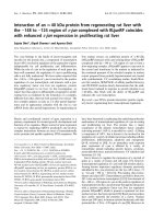
Real Time-PCR Analysis of dopamine D1 Receptor in cerebral
cortex and cerebellum of control and experimental rats
Real Time-PCR analysis showed that the dopamine D1
receptor gene expression was significantly increased (p <
0.001) in the cerebral cortex and decreased (p < 0.001)
cerebellum in diabetic condition. Insulin and curcumin
treatment reversed the altered expression to near control
(Figure 1 and 2).
Real Time-PCR Analysis of dopamine D2 Receptor in cerebral
cortex and cerebellum of control and experimental rats
Real Time-PCR analysis showed that the dopamine D2
receptor gene expression in the cerebral cortex and cere-
bellum was significantly increased (p < 0.001) in diabetic
condition and it reversed to near control value in insulin
and curcumin treated diabetic rats (Figure 3 and 4).
Real Time-PCR Analysis of CREB in the cerebral cortex and
cerebellum of control and experimental rats
Real Time-PCR analysis showed that the CREB gene
expression in the cerebral cortex and cerebellum was sig-
nificantly decreased (p < 0.001) in diabetic condition. In
cerebral cortex, curcumin treatment reversed the altered
expression to near control while insulin treatment shows
no significant reversal. In cerebellum curcumin and insu-
lin treatment reversed the altered expression to near con-
trol value (Figure 5 and 6).
Real Time-PCR Analysis of phospholipase C in the cerebral
cortex and cerebellum of control and experimental rats
Real Time-PCR analysis showed that the phospholipase C
gene expression in the cerebral cortex and cerebellum
was significantly decreased (p < 0.001) in diabetic condi-
tion. In cerebral cortex curcumin and insulin treatment
reversed the altered expression in diabetes to near con-
trol. In cerebellum curcumin treatment reversed the
altered expression to near control while insulin treatment
shows no significant reversal (Figure 7 and 8).
Discussion
There is a complex relationship among diabetes mellitus
and CNS, the present study is an attempt to investigate
the role of curcumin in regularising the altered dopamin-
ergic and second messenger expression in the cerebral
cortex and cerebellum of STZ-induced diabetic rats. Dia-
betic encephalopathy, characterized by impaired cogni-
tive functions and neurochemical and structural
abnormalities, may involve direct neuronal damage.
Therefore, we have assessed the possibility of curcumin
supplementation that target oxidative stress which would
help in preventing and/or delaying the progression of dia-
betes and associated neuronal injury in cerebral cortex
and cerebellum. This study demonstrated for the first
Table 1: Blood glucose (mg/dl) level in Experimental rats
Animal status 0 day (Before
STZ injection)
3rd day (Initial) 6th day 10th day 14th day (Final)
Control 87.2 ± 1.4 86.6 ± 1.2 83.2 ± 1.2 86.3 ± 1.2 85.7 ± 1.5
Diabetic 85.3 ± 1.3 257.3 ± 0.9 318.2 ± 1.6 307.8 ± 1.3 320.5 ± 1.3***
D + I 86.4 ± 0.9 249.8 ± 1.2 303.6 ± 0.8 185.9 ± 1.5 137.0 ± 1.3
ψψψ
ϕϕϕ
D+C 89.3 ± 1.5 259.7 ± 1.8 305 ± 0.9 190 ± 1.7 175.6 ± 1.0
ψψψ
ϕϕϕ
Values are mean ± S.E.M of 4-6 rats in each group. Each group consist of 6-8 rats
*** P < 0.001 when compared to control,
ψψψ
P < 0.001 when compared to diabetic group, ϕϕϕ
p < 0.001 when compared with initial reading
D + I- Insulin treated diabetic rats
D+C- Curcumin treated diabetic rats
Kumar et al. Journal of Biomedical Science 2010, 17:43
/>Page 5 of 11
time that STZ-induced diabetes produces a marked
attenuation of cerebral cortical and cerebellum function
mediated through dopaminergic receptors, phospholi-
pase C activity and transcription factor CREB in the
Wistar rats.
The STZ diabetic rat serves as an excellent model to
study the molecular, cellular and morphological changes
in brain induced by stress during diabetes [33]. In the
present study, STZ-induced rats were used as an experi-
mental model for diabetes, since they provides a relevant
example of endogenous chronic oxidative stress due to
the resulting hyperglycemia [34]. The facts' that increased
blood glucose level and decreased body weight, observed
during diabetes, are similar with previous reports as a
result of the marked destruction of insulin secreting pan-
creatic β-cells by STZ [25]. Previous reports showed that
curcumin has the potential to protect pancreatic islet
cells against STZ-induced death and dysfunction [35] and
increase plasma insulin level in diabetic mice [36]. The
results of this study have demonstrated that insulin and
curcumin treatment to STZ-induced diabetic rats can
have beneficial effects in reducing blood glucose levels to
near control. The central complications of hyperglycemia
also include the potentiation of neuronal damage
observed following hypoxic/ischemic events, as well as
stroke. Glucose utilization is decreased in the brain dur-
ing diabetes [37], providing a potential mechanism for
increased vulnerability to acute pathological events.
Dopamine is the predominant catecholamine neu-
rotransmitter in the mammalian brain, where it controls a
variety of functions including locomotor activity, cogni-
tion, emotion, positive reinforcement, food intake, and
endocrine regulation. This catecholamine also plays mul-
tiple roles in the periphery as a modulator of cardiovas-
cular function, catecholamine release, hormone
secretion, vascular tone, renal function, and gastrointesti-
nal motility [38]. Dopamine receptors are reported to be
increased in diabetes causing significant alterations in
central dopaminergic system [39]. It is hypothesized that
the cerebral cortex participates in the memory, attention,
Table 2: Scatchard analysis of [
3
H] dopamine binding against dopamine in the cerebral cortex of control, and
experimental rats
Animal status
Bmax (fmoles/mg protein) Kd (nM)
Control 23 ± 3.7 2.01 ± 0.05
Diabetic 67 ± 5.7*** 6.17 ± 0.06**
D + I 19.8 ± 4.1
ψψψ
2.4 ± 0.05
ψψ
D + C 21.7 ± 6.6
ψψψ
1.96 ± 0.06
ψψ
Values are mean ± S.E.M of 4-6 separate experiments. Each group consist of 6-8 rats *** P < 0.001 when compared to control,
ψψψ
P < 0.001
when compared to diabetic group **P < 0.01 when compared to control group
ψψ
P < 0.01 when compared to diabetic group.
D + I- Insulin treated diabetic rats
D+C- Curcumin treated diabetic rats
Table 3: Scatchard analysis of [
3
H] dopamine binding against dopamine in the cerebellum of control, and experimental
rats
Animal status
Bmax (fmoles/mg protein) Kd (nM)
Control 112 ± 5.4 3.8 ± 0.14
Diabetic 22 ± 3.6*** 2.3 ± 0.05**
D + I 116 ± 4.3
ψψψ
3.2 ± 0.13
@@
D + C 91.1 ± 3.87
ψψψ
4.0 ± 0.03
@@
Values are mean ± S.E.M of 4-6 separate experiments. Each group consist of 6-8 rats ***P < 0.001 when compared to control,
ψψψ
P < 0.001
when compared to diabetic group **P < 0.01 when compared to control group
@@
P < 0.01 when compared to diabetic group.
D + I- Insulin treated diabetic rats
D+C- Curcumin treated diabetic rats
Kumar et al. Journal of Biomedical Science 2010, 17:43
/>Page 6 of 11
perceptual awareness, thought, language, and conscious-
ness which are necessary for the normal life style. In the
present study the scatchard analysis of total dopamine
receptors in diabetic rats showed an increased receptor
binding or number in cerebral cortex when compared to
control, thus contributing to neurological dysfunctions
associated with cortex. Earlier reports showed significant
alterations in neurotransmitters during hyperglycaemia
and causes degenerative changes in neurons of the CNS
[40]. A converse pattern of the modulation of total dop-
aminergic receptors was obtained in cerebellum, which is
responsible for the coordination of voluntary motor
movement, balance, equilibrium and declarative memory.
Total dopamine receptor density was decreased in the
cerebellum of diabetic rats when compared to control
indicating an unbalance in dopaminergic neural trans-
mission. Furthermore, many behavioural studies have
shown evidence that the dopamine system plays an
important role in regulating exploratory and locomotor
behavior [41,42]. The current data reveal a significant
reversal of this altered binding parameter to near control
in curcumin and insulin treatment. Thus we speculated
Figure 1 Real Time PCR amplification of dopamine D1 receptor
mRNA from the cerebral cortex of control and experimental rats.
Values are mean ± S.D of 4-6 separate experiments. Each group consist
of 6-8 rats Relative Quantification values and standard deviations are
shown in the table. The relative ratios of mRNA levels were calculated
using the
ΔΔ
CT method normalized with β-actin CT value as the inter-
nal control and Control CT value as the calibrator. a p < 0.001 when
compared with control, b p < 0.001 when compared with diabetic
group. D+I - Insulin treated diabetic group. D+C- Curcumin treated di-
abetic group.
Ͳϭ
ͲϬ͘ϱ
Ϭ
Ϭ͘ϱ
ϭ
ϭ͘ϱ
Ϯ
ŽŶƚƌŽů ŝĂďĞƚŝĐ н/ н
Log RQ
a
b
b
Figure 2 Real Time PCR amplification of dopamine D1 mRNA
from the cerebellum of control and experimental rats. Values are
mean ± S.D of 4-6 separate experiments. Each group consist of 6-8 rats
Relative Quantification values and standard deviations are shown in
the table. The relative ratios of mRNA levels were calculated using the
ΔΔ
CT method normalized with β-actin CT value as the internal control
and Control CT value as the calibrator. a p < 0.001 when compared
with control b p < 0.001 when compared with diabetic group. D+I - In-
sulin treated diabetic group. D+C- Curcumin treated diabetic group.
ͲϬ͘ϱ
ͲϬ͘ϰ
ͲϬ͘ϯ
ͲϬ͘Ϯ
ͲϬ͘ϭ
Ϭ
Ϭ͘ϭ
Ϭ͘Ϯ
ŽŶƚƌŽů ŝĂďĞƚŝĐ н/ н
Log RQ
a
b
b
Figure 3 Real Time PCR amplification of dopamine D2 mRNA
from the cerebral cortex of control and experimental rats. Values
are mean ± S.D of 4-6 separate experiments. Each group consist of 6-8
rats Relative Quantification values and standard deviations are shown
in the table. The relative ratios of mRNA levels were calculated using
the
ΔΔ
CT method normalized with β-actin CT value as the internal con-
trol and Control CT value as the calibrator. a p < 0.001 when compared
with control b p < 0.001 when compared with diabetic group. D+I - In-
sulin treated diabetic group. D+C- Curcumin treated diabetic group.
Ͳϭ
ͲϬ͘ϱ
Ϭ
Ϭ͘ϱ
ϭ
ϭ͘ϱ
Ϯ
ŽŶƚƌŽů ŝĂďĞƚŝĐ н/ н
Log RQ
a
b
b
Figure 4 Real Time PCR amplification of dopamine D2 mRNA
from the cerebellum of control and experimental rats. Values are
mean ± S.D of 4-6 separate experiments. Each group consist of 6-8 rats
Relative Quantification values and standard deviations are shown in
the table. The relative ratios of mRNA levels were calculated using the
ΔΔ
CT method normalized with β-actin CT value as the internal control
and Control CT value as the calibrator. a p < 0.001 when compared
with control b p < 0.01 when compared with diabetic group c p <
0.001 when compared with diabetic group. D+I - Insulin treated dia-
betic group. D+C- Curcumin treated diabetic group.
ͲϬ͘ϰ
ͲϬ͘Ϯ
Ϭ
Ϭ͘Ϯ
Ϭ͘ϰ
Ϭ͘ϲ
Ϭ͘ϴ
ϭ
ϭ͘Ϯ
ŽŶƚƌŽů ŝĂďĞƚŝĐ н/ н
Log RQ
a
b
c
Kumar et al. Journal of Biomedical Science 2010, 17:43
/>Page 7 of 11
that curcumin has an ability to modulate dopaminergic
receptors there by ameliorating the impaired cortical per-
formance associated with diabetes. Diabetes mellitus has
been reported to be accompanied by a number of behav-
ioural and hormonal abnormalities, including reduced
locomotor activity [43]. The present experiments further
revealed the effect of curcumin to modulate the dop-
aminergic receptors in the cerebellum by standardising
the altered expression to a normal level.
DA D
1
receptors are highly expressed in basal ganglia
followed by cerebral cortex, hypothalamus and thalamus.
The gene expression studies of dopamine D1 receptors
showed an increase in the cortex of diabetic rats which
confirm and extend our observations of total dopamine
receptors. Dopamine D1 receptor seems to mediate
important actions of dopamine to control movement,
cognitive function and cardiovascular function. The DA
D
1
receptors in the brain are linked to episodic memory,
emotion, and cognition. Diabetes mellitus has been
reported to cause degenerative changes in neurons of the
CNS [44,45,40]. Our study showed that diabetes can reg-
ulate the expression of dopamine D1 receptor which may
reduce the central cortical function. Furthermore, cur-
cumin and insulin exhibited a tendency for decreasing
Figure 5 Real Time PCR amplification of CREB mRNA from the ce-
rebral cortex of control and experimental rats. Values are mean ±
S.D of 4-6 separate experiments. Each group consist of 6-8 rats Relative
Quantification values and standard deviations are shown in the table.
The relative ratios of mRNA levels were calculated using the
ΔΔ
CT
method normalized with β-actin CT value as the internal control and
Control CT value as the calibrator. a p < 0.001 when compared with
control b p < 0.01 when compared with diabetic group. D+I - Insulin
treated diabetic group. D+C- Curcumin treated diabetic group.
ͲϬ͘ϲ
ͲϬ͘ϱ
ͲϬ͘ϰ
ͲϬ͘ϯ
ͲϬ͘Ϯ
ͲϬ͘ϭ
Ϭ
ŽŶƚƌŽů ŝĂďĞƚŝĐ н/ н
Log RQ
a
b
Figure 6 Real Time PCR amplification of CREB mRNA from the cer-
ebellum of control and experimental rats. Values are mean ± S.D of
4-6 separate experiments. Each group consist of 6-8 rats Relative Quan-
tification values and standard deviations are shown in the table. The
relative ratios of mRNA levels were calculated using the
ΔΔ
CT method
normalized with β-actin CT value as the internal control and Control CT
value as the calibrator. a p < 0.001 when compared with control b p <
0.01 when compared with diabetic group. D+I - Insulin treated diabetic
group. D+C- Curcumin treated diabetic group.
Ͳϭ͘ϲ
Ͳϭ͘ϰ
Ͳϭ͘Ϯ
Ͳϭ
ͲϬ͘ϴ
ͲϬ͘ϲ
ͲϬ͘ϰ
ͲϬ͘Ϯ
Ϭ
ŽŶƚƌŽů ŝĂďĞƚŝĐ н/ н
Log RQ
a
b
b
Figure 7 Real Time PCR amplification of phospholipase C mRNA
from the cerebral cortex of control and experimental rats. Values
are mean ± S.D of 4-6 separate experiments. Each group consist of 6-8
rats Relative Quantification values and standard deviations are shown
in the table. The relative ratios of mRNA levels were calculated using
the
ΔΔ
CT method normalized with β-actin CT value as the internal con-
trol and Control CT value as the calibrator. a p < 0.001 when compared
with control b p < 0.001 when compared with diabetic group. D+I - In-
sulin treated diabetic group. D+C- Curcumin treated diabetic group.
Ͳϭ
ͲϬ͘ϴ
ͲϬ͘ϲ
ͲϬ͘ϰ
ͲϬ͘Ϯ
Ϭ
Ϭ͘Ϯ
Ϭ͘ϰ
ŽŶƚƌŽů ŝĂďĞƚĞƐ н/ н
Log RQ
a
b
b
Figure 8 Real Time PCR amplification of phospholipase C mRNA
from the cerebellumof control and experimental rats. Values are
mean ± S.D of 4-6 separate experiments. Each group consist of 6-8 rats
Relative Quantification values and standard deviations are shown in
the table. The relative ratios of mRNA levels were calculated using the
ΔΔ
CT method normalized with β-actin CT value as the internal control
and Control CT value as the calibrator. a p < 0.001 when compared
with control b p < 0.001 when compared with diabetic group. D+I - In-
sulin treated diabetic group. D+C- Curcumin treated diabetic group.
Ͳϭ
ͲϬ͘ϴ
ͲϬ͘ϲ
ͲϬ͘ϰ
ͲϬ͘Ϯ
Ϭ
Ϭ͘Ϯ
Ϭ͘ϰ
ŽŶƚƌŽů ŝĂďĞƚŝĐ н/ н
Log RQ
a
b
Kumar et al. Journal of Biomedical Science 2010, 17:43
/>Page 8 of 11
this altered mRNA expression to near control. Such inter-
ference with the dopaminergic system could explain, at
least in part, the ameliorative effect of curcumin on CNS.
In agreeable with the total dopamine receptor change in
the cerebellum dopamine D1 receptor expression was
down regulated in the diabetic rats when compared to
control. Haloperidol and SCH23390, a selective dop-
amine D1 receptor antagonist, significantly reduced
spontaneous locomotor activity in diabetic mice, but not
in non diabetic mice [46]. In our study, curcumin and
insulin increased the dopamine D1 receptor expression
levels in the cerebellum, which suggests that the cur-
cumin supplementation influenced the functional regula-
tion of these receptors to maintain normal dopaminergic
function and this might also be involved as a mechanism
of preventing cerebellar dysfunctions.
The interest in learning dopamine D2 receptor expres-
sion begins with the hypothesis that dopamine D2 recep-
tors are involved in the pathophysiology of schizophrenia
and in the mechanism of antipsychotic drug action [47].
Also widespread distribution of dopamine D2 receptors
in the cerebral cortex is of considerable clinical signifi-
cance because this may be the site for regulation of cogni-
tive deficits [48]. Thus, our findings should bring
attention to the cortex as a possible site of dysfunction in
diseases like diabetes mellitus. To examine whether dop-
amine D2 receptors are altered in diabetes, we examined
the expression levels of D2 in the cortex, and the cerebel-
lum, because these tissues are regions to which dopamin-
ergic neurons project, and are well known to be related to
memory, attention, perceptual awareness, thought, lan-
guage, consciousness and motor function. The present
study showed that dopamine D2 receptors expression of
cortex and cerebellum in diabetic rats where up regulated
when compared to control. These results may indicate an
alteration of the dopamine system in diabetes, because it
is well known that dopamine is a principal modulator of
higher functions including attention working memory
[49] and motor control [50]. The increase in the central
dopaminergic postsynaptic receptors has been related to
decrease the locomotor and ambulatory activity in STZ-
induced diabetic rats [51,52]. It was reported that injec-
tion of dopamine D2 agonist into lobules 9 and 10 of the
cerebellar cortex, induced balance and motor coordina-
tion disturbances in the rotarod test [53]. It has been sug-
gested that curcumin reverses the effects of diabetes on
dopamine D2 receptors in the cortex and cerebellum to
near control level.
Previous studies from our lab have established the role
of neurotransmitters in maintaining the glucose homeo-
stasis [54-57]. Thus it is evident that the various neu-
rotransmitter systems, including - Dopamine,
acetylcholine, glutamate, GABA; are modulated by diabe-
tes. The coordinated activation and inhibition of different
neurotransmitter systems in control rats are disrupted
during diabetes. The synergistic effect of neurotransmit-
ters receptor alterations results in CNS disorders during
diabetes. Puglisi et al (1995) [58], reported the regulatory
role dopamine D1 and D2 receptors in modulating acetyl-
choline activity. Also hippocampal D2 receptors modu-
late spatial working memory functions, and this effect is
due to the increased acetylcholine release associated with
D2 receptor stimulation [59,60].
The cAMP response element-binding protein (CREB)
plays a pivotal role in dopamine receptor-mediated
nuclear signaling and neuroplasticity [61]. Here we dem-
onstrate the significance of CREB gene expression in the
cerebral cortex and cerebellum of STZ-induced diabetes
rats. Our findings showed a significant down regulation
of CREB in cerebral cortex and cerebellum of diabetic
rats, when compared to control. The study of the dop-
amine receptors expression in relation with CREB phos-
phorylation in diabetes is an important step toward
elucidating the relationship between molecular adapta-
tions and behavioural consequences. CREB proteins in
neurons are thought to be involved in the formation of
long-term memories; this has been shown in the marine
snail Aplysia, the fruit fly Drosophila melanogaster, and
in rats. CREB is necessary for the late stage of long-term
potentiation. CREB also has an important role in the
development of drug addiction [62]. It is therefore impor-
tant to identify the elements that modulate dopaminergic
receptor expressions and phosphorylation of CREB and
there by its expression in the nucleus. Drugs that stimu-
late dopamine receptors have the potential to produce
long-lasting behavioural and neural alterations. The cur-
cumin supplementation significantly modulates the
altered gene expression of CREB in the cerebral cortex
and cerebellum of diabetic rats to near control. In cere-
bral cortex insulin treatment doesn't show any significant
effect in the CREB expression of diabetic rats whereas
cerebellum shows a significant reversal. This study dem-
onstrates that curcumin is having a modulatory effect in
the transcription factor CREB expression which is crucial
in maintaining the normal neuronal function and survival
in diabetes. The dopamine D1 signal transduction path-
way, activation of the transcription factor CREB, and
dopamine-mediated gene expression are critically
involved in memory processing, behavioural responses
and drug addiction [63]. Interruption of this pathway can
interfere with important cognitive performance and
behavioural aspects associated with cerebral cortex and
cerebellum. Dudman et al [64] reported that D2 receptors
activate the cAMP response element-binding protein in
neurons and D1 receptor stimulation leads to phosphory-
lation of the transcription factor Ca
2+
and CREB in the
nucleus by means of NMDA receptor-mediated Ca
2+
sig-
naling. Thus we propose the importance of dopamine
Kumar et al. Journal of Biomedical Science 2010, 17:43
/>Page 9 of 11
receptors in modulating CREB phosphorylation and acti-
vation. Possible interactions of other neurotransmitters
with CREB is also suggested which needs further studies.
The effect of curcumin in interacting with the dopamin-
ergic receptor and CREB in STZ-induced diabetes proves
its potential in managing CNS disorders in diabetes.
Phospholipase C mediates transduction of neurotrans-
mitter signals across membranes via hydrolysis of phos-
phatidylinositol-4,5-bisphosphate, leading to generation
of second messengers inositol- 1,4,5-trisphosphate and
diacylglycerol. In the present study, we determined diabe-
tes-mediated alterations in phospholipase C expression
in the cerebral cortex and cerebellum. Further we
extended the studies to phospholipase C regulation with
curcumin supplementation and insulin treatment a
potential therapeutic drug which can modulate signal
transduction pathway there by contributing in the pre-
vention of CNS dysfunction in diabetes. Our results
showed a decreased expression of phospholipase C in the
cerebral cortex and cerebellum of diabetic rats when
compared to control. The DA D
1
receptors show charac-
teristic ability to stimulate adenylyl cyclase and generate
inositol 1, 4, 5-trisphosphate (IP
3
) and diacylglycerol via
the activation of phospholipase C [65,66]. We considered
that the down regulation of the Phospholipase C in rat
cerebral cortex and cerebellum during diabetes could
contribute to the impaired signal transduction of G-pro-
tein coupled neurotransmitter receptors. Phospolipase C
performs a catalytic mechanism, generating inositol
triphosphate (IP
3
) and diacylglycerol (DAG). Altered
phospholipase C expression fails to modulate the activity
of downstream proteins important for cellular signalling.
Defective expression of phospholipase C results in low
levels of IP3 causing the impaired release of calcium and
bring down the level of intracellular calcium and thus
failed to execute the normal neuronal function in cerebral
cortex and cerebellum. The previous study reports that
phospholipase C-mediated signaling, initiated by growth
factor receptor types, are involved in long-term memory
formation, a process that requires gene expression [67].
Activation of all the G protein coupled receptors includ-
ing Ach, glutamate and dopamine results in second mes-
senger enzyme, phospholipase C expression. These
evidences led us to propose that the enhancement of dia-
betes-mediated phospholipase C gene expression could
impart damage to the central cognitive functions; which
has been found to be effectively protected by curcumin
treatment. Further studies are to be carried out to reveal
the correlation between the expression of phospholipase
C and G protein coupled neurotransmitter receptors.
The possible mechanism of curcumin action in CNS
may be by lowering the blood glucose level which results
in rendering the anti-apoptotic property [68]. Curcumin
could reduce neuronal loss of the ischemic brain tissue,
and inhibit expression of the activated caspase-3, a key
executor of apoptosis [69,70]. Damage to neurons may
occur through oxidative stress and/or mitochondrial
impairment and culminate in activation of an apoptotic
stage. Apoptosis or related phenomena are possibly
involved in secondary cell death in diabetes. These results
imply a potential therapeutic efficacy, i.e., curcumin may
be used clinically as a neuroprotective drug for treatment
of patients suffering from diabetes.
Insulin and sulfonylurea therapy for diabetes mellitus
carries the risk of hypoglycaemic brain injury, and this
risk is a major impediment to optimal glucose regulation
in diabetic patients [71]. Factors that contribute to cogni-
tive deficits as well as the protective factors that reduce
the impact of diabetes on brain functions are still an
enigma. Cerebral cortex and cerebellum are involved in
cognitive, motor, and neuroendocrine activities [72-74];
thus, their affectations during diabetes are relevant in the
pathogenesis of the disease. In addition, curcumin have
recently received considerable attention since they have
been shown to protect neurons against a variety of exper-
imental neurodegenerative conditions. In the present
investigation the generation of unique functional proper-
ties of curcumin via dopamine D1, D2 receptors, CREB
and phospholoipase C interactions may yield a better
understanding of behaviour and CNS disorders induced
by diabetes.
Abbreviations
STZ: Streptozotocin; CREB: Cyclic AMP response element binding protein; CNS:
Central nervous system.
Competing interests
The authors declare that they have no competing interests.
Authors' contributions
TPK and CSP designed research. TPK, SA, GG and NG carried out the experi-
ments and drafted manuscript. All authors read and approved the final manu-
script.
Acknowledgements
This work was supported by grants from DST, DBT, ICMR, Govt. of India, and
KSCSTE, Govt. of Kerala, to Dr. C. S. Paulose. T Peeyush Kumar thanks the
Department of Science and Technology, India for SRF.
Author Details
Molecular Neurobiology and Cell Biology Unit, Centre for Neuroscience,
Cochin University of Science and Technology, Cochin- 682 022, Kerala, India
References
1. McCall , Millington Wr, Wurtman RJ: Metabolic fuel and amino acid
transport into the brain in experimental diabetes mellitus. Proc Nadl
Acad Sci USA 1982, 97:2881-2885.
2. Nagy R, O' Connor A, Kempers S, yeo R, Qualis C: Adaption in brain
glucose uptake following recurrent hypoglycaemia. Proc Acad Sci USA
1994, 91:9352-69356.
3. Brownlee M: Biochemistry and molecular cell biology of diabetic
complications. Nature 2001, 414:813-820.
Received: 13 February 2010 Accepted: 31 May 2010
Published: 31 May 2010
This article is available from: 2010 Kumar et al; licensee BioMed Central Ltd. This is an Open Access article distributed under the terms of the Creative Commons Attribution License ( which permits unrestricted use, distribution, and reproduction in any medium, provided the original work is properly cited.Journa l of Biome dical Scie nce 2010, 17:43
Kumar et al. Journal of Biomedical Science 2010, 17:43
/>Page 10 of 11
4. Pardridge WM: Brain metabolism: a perspective from the blood-brain
barrier. Physiol Rev 1983, 63:1481-1535.
5. Feldman EL, Stevens MJ, Greene DA: Pathogenesis of diabetic
neuropathy. Clin Neurosci 1997, 4:365-370.
6. Auer RN, Siesjo BK: Hypoglycaemia: brain neurochemistry and
neuropathology. Baillieres Clin Endocrinol Metab 1993, 7:611-625.
7. Kamal A, Biessels GJ, Duis SE, Gispen WH: Learning and hippocampal
synaptic plasticity in streptozotocin-diabetic rats: interaction of
diabetes and ageing. Diabetologia 2000, 43:500-506.
8. Ouyang L, Wang J, Zhu X: Diagnostic efficacy of glutamic acid
decarboxylase antibody and islet cell antibody in type I diabetes
mellitus. Zhonghua Nei Ke Za Zhi 2000, 39:674-676.
9. Osawa T, Kato Y: Protective role of antioxidative food factors in
oxidative stress caused by hyperglycemia. Ann N Y Acad Sci 2005,
1043:440-451.
10. Ahmad M, Turkseven S, Mingone CJ, Gupte SA, Wolin MS, Abraham NG:
Heme oxygenase-1 gene expression increases vascular relaxation and
decreases inducible nitric oxide synthase in diabetic rats. Cell Mol Biol
(Noisy-le-grand) 2005, 51:371-376.
11. Ishrat T, Khan MB, Hoda MN, Yousuf S, Ahmad M, Ansari MA, Ahmad AS,
Islam F: Coenzyme Q10 modulates cognitive impairment against
intracerebroventricular injection of streptozotocin in rats. Behav Brain
Res 2006, 171:9-16.
12. Itokawa H, Hirayama F, Funakoshi K, Takeya K: Studies on the antitumor
bisabolane sesquiterpenoids isolated from Curcuma xanthorrhiza.
Chem Pharm Bull 1985, 33:3488-3492.
13. Bala K, Tripathy BC, Sharma D: Neuroprotective and anti-ageing effects
of curcumin in aged rat brain regions. Biogerontology 2006, 7:81-9.
14. Kuhad A, Chopra K: Curcumin attenuates diabetic encephalopathy in
rats: behavioral and biochemical evidences. Eur J Pharmacol 2007,
576:34-42.
15. Shishodia S, Sethi G, Aggarwal BB: Curcumin: getting back to the roots.
Ann N Y Acad Sci 2005, 1056:206-217.
16. Marchall JF, Friedman MI, Heffner TG: Reduced anorexic and locomotor-
stimulant action of d-amphetamine in alloxan-diabetic rats. Brain Res
1976, 111:428-32.
17. Waxman SG, Sabin TD: Diabetic truncal polyneuropathy. Arch Neurol
1981, 38:46-7.
18. Vallone D, Picetti R, Borrelli E: Structure and function of dopamine
receptors. Neurosci Biobehav Rev 2000, 24:125-132.
19. Nogueira CR, Machado UF, Curi R, Carpinelli AR: Modulation of insulin
secretion and 45Ca2+ efflux by dopamine in glucose-stimulated
pancreatic islets. Gen Pharmacol 1994, 25:909-16.
20. Barik S, de Beaurepaire R: Evidence for a functional role of the dopamine
D3 receptors in the cerebellum. Brain Res 1996, 737(1-2):347-350.
21. Slater Lauren: Opening Skinner's Box: Great Psychological Experiments
of the Twentieth Century. New York: W. W. Norton & Company;
2005:86-90.
22. Spaulding SW: The ways in which hormones change cyclic adenosine
3',5'-monophosphate-dependent protein kinase subunits, and how
such changes affect cell behavior. Endocr Rev 1993, 14(5):632-650.
23. Shimomura A, Okamoto Y, Hirata Y, Kobayashi M, Kawakami K, Kiuchi K,
Wakabayashi T, Hagiwara M: Dominant negative ATF1 blocks cyclic
AMP-induced neurite outgrowth in PC12 D cells. J Neurochem 1998,
70(3):1029-34.
24. Whiting PH, Palmano KP, Howthorne JN: Enzymes of myoinositol and
inositol lipid metabolism in rats with streptozotocin induced diabetes.
Biochem J 1979, 179:549-553.
25. Junod A, Lambert AE, Staufferacher W, Renold AE: Diabetogenic action of
Streptozotocin: Relationship of dose to metabolic response. J Clin
Invest 1969, 48:2129-2139.
26. Hohenegger M, Rudas B: Kidney failure in experimental diabetes
mellitus. Wien Z Inn Med 1971, 52(1):36-40.
27. Arison RN, Ciaccio EI, Glitzer MS, Cassaro JA, Pruss MP: Light and electron
microscopy of lesions in rats rendered diabetic with streptozotocin.
Diabetes 1967, 16:51-56.
28. Sharma S, Kulkarni SK, Agrewala JN, Chopra K: Curcumin attenuates
thermal hyperalgesia in a diabetic mouse model of neuropathic pain.
Eur J Pharmacol 2006, 536:256-261.
29. Glowinski J, Iversen LL: Regional studies of catecholamines in the rat
brain. J Neurochem 1966, 13:655-659.
30. Madras BK, Fahey MA, Canfield DR, Spealman RD: D1 and D2 dopamine
receptors in caudate-putamen of nonhuman primates (Macaca
fascicularis). J Neurochem 1988, 51:934-943.
31. Lowry OH, Rosenbrough NH, Farr AL, Randall RJ: Protein measurement
with folin Phenol reagent. J Biol Chem 1951, 193:265-275.
32. Scatchard G: The attraction of proteins for small molecules and ions.
Ann NY Acad Sci 1949, 51:660-72.
33. Aragno M, Parola S, Brignardello E, Mauro A, Tamagno E, Manti R, Danni O,
Boccuzzi G: Dehydroepiandrosterone prevents oxidative injury
induced by transient ischemia/reperfusion in the brain of diabetic rats.
Diabetes 2000, 49:1924-1931.
34. Low PA, Nickander KK, Tritschler HJ: The role of oxidative stress and
antioxidant treatment in experimental diabetic neuropathy. Diabetes
1997, 44:46:38.
35. Meghana K, Sanjeev G, Ramesh B: Curcumin prevents streptozotocin-
induced islet damage by scavenging free radicals: a prophylactic and
protective role. Eur J Pharmacol 2007, 577:183-191.
36. Seo KI, Choi MS, Jung UJ, Kim HJ, Yeo J, Jeon SM, Lee MK: Effect of
curcumin supplementation on blood glucose, plasma insulin, and
glucose homeostasis related enzyme activities in diabetic db/db mice.
Mol Nutr Food Res 2008, 52:995-1004.
37. McCall AL: The impact of diabetes on the CNS. Diabetes 1992,
41:557-570.
38. Missale C, Nash SR, Robinson SW, Jaber MC: Dopamine receptors: From
structure to function. Physiol Rev 1998, 78:189-225.
39. Lozovsky D, Saller CF, Kopin IJ: Dopamine receptor binding is increased
in diabetic rats. Science 1981, 214:1031-1033.
40. Garris : Age diabetes associated alterations in regional brain
norepinephrine concentrations and adrenergic populations in C57BL/
KsL mice. Developmental Brain Research 1990, 51:161-166.
41. Fink JS, Smith GP: Decreased locomotor and investigatory exploration
after denervation of catecholamine terminal fields in the forebrain of
rats. J Comp Physiol Psychol 1979, 93:34-65.
42. Funada M. Suzuki T, Misawa M: The role of dopamine D1-receptor in
morphine induced hyperlocomotion in mice. Neurosci Lett 1994,
169:1-4.
43. Marshall JF, Friedman MI, Heffner TG: Reduced anorexic and locomotor-
stimulant action of D-amphetamine in alloxan-diabetic rats. Brain Res
1976, 111:428-432.
44. Bhattacharya SK, Saraswathi M: Effect of intracerebroventricularly
administered insulin on brain monoamines and acetylcholine in
euglycemic and alloxan- induced hyperglycemic rats. Indian J Exp Biol
1991, 29:1095-1100.
45. Lackovic Z, Salkovic M, Kuci Z, Relja M: Effect of long-lasting diabetes
mellitus on rat and human brain monoamines. J Neurochem 1990,
51:143-147.
46. Kamei J, Saitoh A, Iwamoto Y, Funada M, Suzuki T, Misawa M, Nagase H,
Kasuya Y: Effects of diabetes on spontaneous locomotor activity in
mice. Neurosci Lett 1994, 178:69-72.
47. de Paulis T: The discovery of epidepride and its analogs as highaffinity
radioligands for imaging extrastriatal dopamine D2 receptors in
human brain. Curr Pharm Des 2003, 9:673-696.
48. Verma A, Moghaddam B: NMDA receptor antagonists impair prefrontal
cortex function as assessed via spatial delayed alternation
performance in rats: modulation by dopamine. J Neurosci 1996,
16:373-379.
49. Castellano C, Ventura R, Cabib S, Puglisi-Allegra S: Strain-dependent
effects of anandamide on memory consolidation in mice are
antagonized by naltrexone. Behav Pharmacol 1999, 10:453-457.
50. Zhou QY, Palmiter RD: Dopamine-deficient mice are severely
hypoactive, adipsic, and aphagic. Cell 1995, 83:1197-1209.
51. Kobayashi M, Shigeta Y: Anti-insulin receptor antibody its
measurement and significance. Nippon Rinsho 1990, 48:308-14.
52. Shimomura YSH, Takahashi M, Uehara Y, Kobayashi I, Kobayashi S:
Ambulatory activity and dopamine turnover in streptozotocin-
induced diabetic rats. Exp Clin Endocrinol 1990, 95:385-388.
53. Kolasiewicz W, Maj J: Locomotor hypoactivity and motor disturbances-
behavioral effects induced by intracerebellar microinjections of
dopaminergic DA-D2/D3 receptor agonists. Pol J Pharmacol 2001,
53:509-15.
54. Gireesh G, Balarama Kaimal S, Peeyush Kumar T, Paulose CS: Decreased
muscarinic M1 receptor gene expression in the hypothalamus,
Kumar et al. Journal of Biomedical Science 2010, 17:43
/>Page 11 of 11
brainstem, and pancreatic islets of streptozotocin-induced diabetic
rats. Journal of Neuroscience Research 2008, 86:947-953.
55. Mohanan VV, Kaimal SB, Paulose CS: Decreased 5-HT1A receptor gene
expression and 5HT1A receptor protein in the cerebral cortex and
brain stem during pancreatic regeneration in rats. Neurochemical
Research 2005, 30:25-32.
56. Kaimal SB, George KA, Paulose CS: Gamma-aminobutyric acid A receptor
functional decrease in the hypothalamus during pancreatic
regeneration in rats. Pancreas 2008, 37:e20-30.
57. Anu J, Peeyush Kumar T, Nandhu MS, Paulose CS: Enhanced NMDAR1,
NMDA2B and mGlu5 receptors gene expression in the cerebellum of
insulin induced hypoglycaemic and streptozotocin induced diabetic
rats. Eur J Pharmacol 2010, 630:61-68.
58. Puglisi-Allegra S, Cestari V, Cabib S, Castellano C: Strain-dependent
effects of post-training cocaine or nomifensine on memory storage
involve both D1 and D2 dopamine receptors. Psychopharmacology
1994, 115:157-162.
59. Imperato A, Obinu MC, Gessa GL: Stimulation of both dopamine D1 and
D2 receptors facilitates in vivo acetylcholine release in the
hippocampus. Brain Research 1993, 618:341-345.
60. Umegaki H, Munoz J, Meyer RC, Spangler EL, Yoshimura J, Ikari H, Iguchi A,
Ingram DK: Involvement of dopamine D(2) receptors in complex maze
learning and acetylcholine release in ventral hippocampus of rats.
Neuroscience 2001, 103:27-33.
61. Finkbeiner S: CREB couples neurotrophin signals to survival messages.
Neuron 2000, 25:11-14.
62. Mayr B, Montminy M: Transcriptional regulation by the
phosphorylation-dependent factor CREB. Nat Rev Mol Cell Biol 2001,
2:599-609.
63. Nestler EJ: . Total recall-the memory of addiction. Neurobiology Science
2001, 292:2266-2267.
64. Dudman Joshua JT, Eaton Molly E, Rajadhyaksha Anjali, Taher Wendy Mac
Muffadal, Barczak Amy, Kameyama Kimihiko, Huganir Richard, Konradi
Christine: Dopamine D1 receptors mediate CREB phosphorylation via
phosphorylation of the NMDA receptor at Ser897-NR1. J Neurochem
2003, 87:922-934.
65. Monsma F, Mahan L, McVittie L, Gerfen C, Sibley D: Molecular cloning
and expression of a D1 dopamine receptor linked to adenylyl cyclase
activation. Proc Natl Acad Sci 1990, 87:6723-6727.
66. Sibley DR, Monsma FJ Jr, Shen Y: Molecular neurobiology of
dopaminergic receptors. Int Rev Neurobiol 1993, 35:391-415.
67. Orbana Paul C, Paul FC, Riccardo B: Is the Ras-MAPK signalling pathway
necessary for long-term memory formation? Trends in Neurosciences
1999, 22:38-44.
68. Zhao Jing, Zhao Yong, Zheng Weiping, Lu Yuyu, Feng Gang, Yu Shanshan:
Neuroprotective effect of curcumin on transient focal cerebral
ischemia in rats. Brain Research 2008, 1229:224-232.
69. Ashe PC, Berry MD: Apoptotic signaling cascades. Prog. Neuro-
psychopharmacol. Biol Psychiatry 2003, 27:199-214.
70. Guan QH, Pei DS, Liu XM, Wang XT, Xu TL, Zhang GY: Neuroprotection
against ischemic brain injury by SP600125 via suppressing the
extrinsic and intrinsic pathways of apoptosis. Brain Res 2006,
1092:36-46.
71. Davis EA, Keating B, Byrne GC, Russell M, Jones TW: Impact of improved
glycaemic control on rates of hypoglycaemia in insulin dependent
diabetes mellitus. Arch Dis Child 1998, 78:111-115.
72. Dube MG, Torto R, Kalra SP: Increased leptin expression selectively in the
hypothalamus suppresses inflammatory markers CRP and IL-6 in
leptin-deficient diabetic obese mice. Peptides 2008, 29:593-598.
73. Gao Q, Horvath TL: "Cross-talk between estrogen and leptin signaling in
the hypothalamus,". American Journal of Physiology 2008, 294:E817-E826.
74. Gerozissis K: "Brain insulin, energy and glucose homeostasis; genes,
environment and metabolic pathologies,". European Journal of
Pharmacology 2008, 585:38-49.
doi: 10.1186/1423-0127-17-43
Cite this article as: Kumar et al., Curcumin modulates dopaminergic recep-
tor, CREB and phospholipase c gene expression in the cerebral cortex and
cerebellum of streptozotocin induced diabetic rats Journal of Biomedical Sci-
ence 2010, 17:43