PRINCIPLES OF NEUROLOGY - PART 6 ppt
Bạn đang xem bản rút gọn của tài liệu. Xem và tải ngay bản đầy đủ của tài liệu tại đây (619.3 KB, 57 trang )
CHAPTER 30 / INTRACRANIAL NEOPLASMS 273
TABLE 30-2 Tumors Presenting with Impairment of Mental Function,
Headaches, Seizures, or Focal Neurologic Signs: Increased Intracranial
Pressure a Late Development
Glioblastoma multiforme 20% of all intracranial tumors, 55% of all
(anaplastic astrocytoma) gliomas; mainly cerebral but may affect
all parts of brain and cord, widely
infiltrative; survival about 12 months in most
cases
Astrocytomas 25–30% of cerebral gliomas; in adults,
(low grade) common sites are cerebral hemispheres; in
children, brainstem and cerebellum; slowly
growing, tendency to form cysts; survival
for many years
Oligodendroglioma 5–7% of intracranial gliomas; frontal lobes
are most common sites; slowly growing;
survival for many years if low-grade
Ependymoma Common sites are fourth ventricle
(particularly in children), conus medullaris,
and filum terminale; survival depends on
degree of anaplasia
Meningioma 15% of all primary intracranial tumors;
highest incidence in seventh decade; more
frequent in women; common sites are
sylvian region, superior parasagittal
surfaces, olfactory groove, lesser wing of
sphenoid, tuberculum sellae,
cerebellopontine angle, spinal canal; very
slow growing; symptoms depend on tumor
site
Primary cerebral May arise in any part of the brain
lymphoma (monofocal or multifocal), often near lateral
ventricle, usually in adult life; lymphocytes,
mononuclear and tumor cells often found in
CSF; immunosuppressed patients at risk,
particularly those with AIDS; median
survival less than 30 months
Metastatic carcinoma Three main patterns; (1) skull and dura,
from carcinoma of breast and prostate, and
multiple myeloma; may compress spinal
cord, cranial nerves, and pituitary; (2) brain,
one or several cerebral or other foci, from
lung, breast, melanoma, colon, kidney;
(3) meningeal carcinomatosis or leukemic
infiltration of leptomeninges and cranial and
spinal nerve roots; average survival 3
months with meningeal carcinomatosis;
patients with bony metastases survive
longer
4777 Victor Ch 30 p268-276 6/11/01 3:04 PM Page 273
Each of the tumors that causes hydrocephalus or a specific regional
syndrome requires a special combination of surgical and radiation
therapy.
PARANEOPLASTIC DISORDERS
This is a group of neurologic disorders that occur in patients with car-
cinoma or other types of neoplasia, without invasion or compression of
the nervous system itself. Presumably, tumors that induce these effects
elaborate enzymes, hormones, or antibodies or dispose the patient to a
viral agent capable of invading or cross-reacting with the nervous sys-
tem. The most familiar of these remote effects and the chapters in
which they are discussed are listed below:
1. Polyneuropathy (Chap. 45)
2. Polymyositis or dermatomyositis (Chap. 48)
3. Myasthenic-myopathic syndrome of Lambert-Eaton (Chap. 52)
4. Carcinomatous cerebellar degeneration and myoclonus-opsoclonus
syndrome
274
PART IV / THE MAJOR CATEGORIES OF NEUROLOGIC DISEASE
TABLE 30-3 Tumors Causing Mainly Increased Intracranial Pressure
and Hydrocephalus, Focal or Lateralizing Signs Less Conspicuous
Medulloblastoma and Mainly in children 4 to 8 years; begins with
cystic astrocytoma of listlessness, vomiting, headaches; later,
cerebellum squint, ataxic gait, falling, and
papilledema
Ependymoma and Clinical syndrome similar to
papilloma of choroid medulloblastoma but more protracted;
plexus two-thirds of patients present with increased
ICP, others with vomiting, dysphagia,
paresthesias of extremities, vertigo, head tilt
Hemangioblastoma of Dominant inheritance; retinal angioma
cerebellum (von Hippel– and polycythemia often conjoined; may
Lindau disease) develop multiple spinal cord lesions and
syringomyelia
Pinealoma (includes Onset in adolescence and adulthood;
pineal germinoma symptoms and signs of increased ICP;
and teratoma) paralysis of upward gaze and pupils fixed
to light (Parinaud syndrome)
Colloid (paraphysial) Signs of intermittent or persistent increased
cyst of third ventricle ICP (headache) and hydrocephalus
Craniopharyngioma In children and adolescents, delayed sexual
maturation and growth, diabetes insipidus
combined with visual loss from
chiasmatic–optic nerve lesions; in adults,
visual loss, signs of hydrocephalus, mild
corticospinal and hypothalamic signs
4777 Victor Ch 30 p268-276 6/11/01 3:04 PM Page 274
TABLE 30-4 Distinctive Tumor Syndromes: Local Signs Predominate
and General Cerebral Deficits and Increased ICP Are Late or Absent
Acoustic neuroma Usually solitary; may be part of
(schwannoma) neurofibromatosis, either solitary (type I) or
bilateral (type II, autosomal dominant);
unilateral neurosensory deafness, loss of
balance, facial weakness and loss of
sensation, later ataxia of ipsilateral limbs
and gait and raised intracranial pressure
Carotid body tumor Painless mass at bifurcation of common
carotid, below angle of jaw; grows slowly;
compresses cranial nerves IX to XII and
sympathetics; rarely familial and bilateral
Pituitary adenomas (with enlarged sella, rule out empty-sella syndrome
by CT-MRI). See also page 239.
Prolactinomas Increased incidence with age; headache,
(usually achromatic bitemporal hemianopia, or mixed
chromophobe, chiasmatic–optic nerve changes; sella
sometimes acidophilic turcica expands; hypothyroidism,
adenoma) hypoadrenalism; in females, amenorrhea,
galactorrhea, serum prolactin increased
(Ͼ100 ng/mL); in males, impotence
Acromegaly-gigantism Oversecretion of growth hormone (GH);
(eosinophilic before closure of the epiphyses, gigantism;
adenoma) after closure, acromegaly
Cushing disease Oversecretion of ACTH; sella not enlarged;
(basophil or truncal obesity, striae, hirsutism;
nonbasophil adenoma) hypertension; glycosuria; amenorrhea;
osteoporosis; proximal muscle weakness;
mental changes
Meningioma of Mainly in women, average age 50 years;
sphenoid ridge unilateral exophthalmos, slight temporal
bulge, anosmia, ocular palsies, Tolosa-Hunt
syndrome, monocular blindness
Meningioma of Older adults; anosmia and frontal lobe
olfactory groove signs; high CSF protein
Meningioma of Older adults, mainly women; bitemporal
tuberculum sellae hemianopia with normal-sized sella
Glioma of brainstem Onset mainly in childhood; progressive
cranial nerve and long tract signs;
increased ICP late; prognosis varies with
degree of anaplasia
Glioma of optic Mainly in children and adolescents,
nerve and chiasm sometimes with neurofibromatosis;
progressive loss of vision with optic atrophy
or chiasmal field defect
(continued)
CHAPTER 30 / INTRACRANIAL NEOPLASMS 275
4777 Victor Ch 30 p268-276 6/11/01 3:04 PM Page 275
5. Limbic and brainstem encephalitis (see the Principles, pp 687–688)
6. Progressive multifocal leukoencephalopathy (Chap. 32)
7. Necrotizing myelopathy (Chaps. 35 and 43)
8. Retinopathy
For a more detailed discussion of this topic, see Adams, Victor, and
Ropper: Principles of Neurology, 6th ed, pp 642–694.
ADDITIONAL READING
Dawson DM: Antineoplastic drugs, in Asbury AK, McKhann GM, McDonald WI
(eds): Diseases of the Nervous System, 2nd ed. Philadelphia, Saunders, 1992,
pp 1121–1129.
DeAngelis LM: Current management of primary central nervous system lym-
phoma. Oncology 9:63, 1995.
Glantz MJ, Rottenberg DA: Harmful effects of radiation on the nervous system,
in Asbury AK, McKhann GM, McDonald WI (eds): Diseases of the Nervous
System, 2nd ed. Philadelphia, Saunders, 1992, pp 1130–1143.
Henson RA, Urich H: Cancer and the Nervous System. Oxford, Blackwell, 1982.
Klibanski A, Zervas NT: Diagnosis and management of hormone-secreting pitu-
itary adenomas. New Engl J Med 324:822, 1991.
Levine AJ, Schmidek HH (eds): Molecular Genetics of Nervous System Tumors.
New York, Wiley-Liss, 1993, pp 357–369.
Posner JP: Neurologic Complications of Cancer. Philadelphia, FA Davis, 1995.
Russell DS, Rubinstein LJ: Pathology of Tumors of the Nervous System. 5th ed.
Baltimore, Williams & Wilkins, 1989.
276 PART IV / THE MAJOR CATEGORIES OF NEUROLOGIC DISEASE
TABLE 30-4 Distinctive Tumor Syndromes: Local Signs Predominate and
General Cerebral Deficits and Increased ICP Are Late or Absent (cont.)
Chordoma Common sites are clivus and
sacrococcygeal region; cauda equina
syndrome or successive multiple cranial
nerve signs, with conduction deafness,
facial pain, and ataxia
Nasopharyngeal or Multiple upper cranial nerve abnormalities;
sinus tumors nasopharyngeal mass; erosion base of skull
Tumors of Pain in occiput and posterior neck;
foramen magnum combination of lower cranial nerve, cervical
cord, and cerebellar signs
4777 Victor Ch 30 p268-276 6/11/01 3:04 PM Page 276
31 Nonviral Infections of the Nervous
System (Bacterial, Spirochetal,
Fungal, Parasitic) and Sarcoidosis
BACTERIAL INFECTIONS
The most important members of this group in decreasing order of their
frequency are meningitis, brain abscess, subdural empyema, dural sinus
thrombophlebitis, and focal bacterial encephalitis. In all of these and
other conditions, bacteria reach the brain in one of several ways: by
hematogenous spread (i.e., septicemia or infected emboli), by extension
from infected cranial structures (ears, sinuses, osteomyelitic foci), by
penetrating cranial injuries, or by surgical invasion.
Bacterial Meningitis
Definition This consists essentially of a bacterial infection of the pia
and arachnoid and the cerebrospinal fluid that they enclose. Since the
subarachnoid space is continuous around the brain, spinal cord, and
optic nerves, an infective agent (or blood or tumor cells) gaining entry
to any part of the space spreads to all of it. Thus meningitis is always
cerebrospinal. Infection also reaches the ventricles and their ependymal
lining by reflux from the subarachnoid space. All structures bathed by
the CSF—ependyma, choroid plexuses, intra-arachnoidal portions of
the cranial and spinal nerves, cerebral and cerebellar cortices, and sur-
face veins and arteries—are exposed to the meningeal infection.
Epidemiology Streptococcus pneumoniae, Neisseria meningitidis,
Haemophilus influenzae, and Listeria monocytogenes—the most com-
mon bacteria causing meningitis—have a worldwide distribution and a
more or less even incidence throughout the year. Meningococcal
meningitis tends to occur in epidemics, in roughly 10-year cycles. This
form of meningitis is most frequent in children and adolescents but
occurs throughout adult life. H. influenzae meningitis affects mainly
children between the ages of 2 months and 5 years but is now being
reported in adults over 50 years of age. Pneumococcal meningitis pre-
dominates in the very young and old and has a predilection for patients
with sickle-cell anemia and those who have had a skull fracture or
splenectomy. Escherichia coli, Staphylococcus aureus, group A strep-
tococci, Klebsiella, Proteus, and Listeria monocytogenes are associated
277
4777 Victor Ch 31 p277-289 6/11/01 2:10 PM Page 277
Copyright 1998 The McGraw-Hill Companies, Inc. Click Here for Terms of Use.
with immunodeficiency states, trauma, and neurosurgical procedures,
including ventricular shunts.
Pathogenesis and pathology The usual routes by which bacteria reach
the meninges have been indicated above.
Once bacteria enter the CSF, they excite an acute inflammatory reac-
tion, mainly in the vascular pia. Hyperemia, exudation of blood pro-
teins, and migration of neutrophils occur within hours. This exudate
continues to accumulate for the next few days. Thereafter, lymphocytes
and then plasma cells begin to appear in the pia as part of an immune
response. Veins in the pia may thrombose and cause brain infarction.
As the meningeal exudate blocks the subarachnoid space around the
brainstem and the foramina of Luschka and Magendie, tension hydro-
cephalus develops. There is also an ependymitis at an aqueductal level
which may contribute to the obstruction of CSF flow. Cranial nerve
roots, as they pass through purulent exudate in the subarachnoid space,
may be involved. Although the brain is not invaded by bacteria, their
endotoxins diffuse through the pia and along the Virchow-Robin spaces
and excite a subpial edema and even a superficial focal necrosis. The
thin arachnoid, especially in infants, may be transgressed, with devel-
opment of a subdural inflammatory reaction and a hygroma. If the
meningitis is not treated successfully, arteritis and thrombosis, cerebral
infarction, and hydrocephalus may result.
Clinical features Fever, severe headache, generalized convulsions,
various degrees of drowsiness and confusion, and even coma are the
usual manifestations in adults and older children. Generalized seizures
occur more often in infants and young children. Signs of meningeal irri-
tation—stiffness of the neck on forward flexion, with flexion of the
knees and hips (Brudzinski sign) and inability to completely extend the
legs (Kernig sign)—become evident. In infants and newborns, in whom
meningitis is often lethal, the infection expresses itself by fever and
bulging of the fontanels, vomiting, drowsiness, and, in some instances,
convulsions; stiff neck may not be evident.
Certain clinical clues may betray the type of meningitis:
1. Petechial and purpuric rash and circulatory collapse—meningococ-
cal meningitis with Waterhouse-Friderichsen syndrome (a similar
rash may be seen with certain enteroviral infections)
2. Ventriculoatrial or peritoneal shunt, cranial trauma, or neurosurgical
procedure—coagulase-negative Staphylococcus, other nosocomial
organisms
3. Upper respiratory and ear infections in children—H. influenzae
4. Immunocompromised host—Strep. pneumoniae, L. monocytogenes,
E. coli
5. Infection of ears, sinuses, lung, heart valves—Strep. pneumoniae or
mixed infections, including anaerobic organisms
278
PART IV / THE MAJOR CATEGORIES OF NEUROLOGIC DISEASE
4777 Victor Ch 31 p277-289 6/11/01 2:10 PM Page 278
Ancillary examinations The one indispensable laboratory procedure is
lumbar puncture and examination of the spinal fluid. The CSF is usu-
ally under increased pressure (200 to 400 mmH
2
O), is cloudy owing to
the presence of cells, mainly polymorphonuclear, (a few hundred, or
even less, to 10,000 mm
3
), and contains bacteria seen on Gram stain,
increased protein (100 to 500 mg/dL), and decreased glucose
(Ͻ40 mg/dL or Ͻ40 percent of the blood glucose, which should be
measured simultaneously). The fluid needs to be cultured. The CSF
latex agglutination test and now the polymerase chain reaction (PCR)
for detection of bacterial antigens are especially useful in cases of par-
tially treated meningitis. Also, throat and blood cultures should be
obtained. The peripheral white blood cells are increased with a shift to
the left.
After treatment is underway, films of sinuses and chest are indicated.
Similarly, CT scanning and MRI can be performed to exclude brain
abscess and subdural empyema. Actually, brain abscess rarely compli-
cates meningitis. In infants and children, ultrasound examination is pre-
ferred because anesthesia is not required.
Treatment Bacterial meningitis is a medical emergency. Every hour of
delay in starting antibacterial therapy increases the risk of complica-
tions and permanent neurologic residua. Treatment with broad-spec-
trum antibiotics should be started immediately after the LP, while
identification of the organism is awaited. In Tables 31-1 and 31-2 are
listed the recommended antibiotics at each age and the dosages for dif-
ferent types of meningitis. LP pressure above 400 mmH
2
O warns of
cerebellar herniation and requires treatment with mannitol. The ad-
ministration of dexamethasone to children with meningitis reduces the
incidence of deafness. Treatment should continue for 10 to 14 days.
Persistent and recurrent subdural hygromas usually respond to repeated
aspiration or shunting.
Preventive measures should not be neglected. All household contacts
of patients with meningitis, particularly children, should receive
rifampin, 10 mg/kg q 12h by mouth daily for 2 days. Immunization
against Neisseria meningitidis is effective and should be given during
epidemics. Children after 2 months of age should be vaccinated against
H. influenzae with a new protein-conjugate vaccine.
Bacterial Encephalitis
In acute and subacute bacterial endocarditis (SBE), the brain is seeded
with bacteria-laden emboli; in subacute endocarditis the bacteria are
characteristically of low virulence and do not produce brain abscesses.
Sterile meningeal reactions and small infarcts, some with blood in the
CSF, are the usual complications; mycotic aneurysms may form but are
rare. The emboli of acute bacterial endocarditis do give rise to miliary
CHAPTER 31 / NONVIRAL INFECTIONS OF THE NERVOUS SYSTEM 279
4777 Victor Ch 31 p277-289 6/11/01 2:10 PM Page 279
abscesses, infarcts, small hemorrhages, and bacterial meningitis; large
abscesses are rare. Treatment in both types is directed to the endocardi-
tis and septicemia.
Legionnaire’s disease, Mycoplasma pneumoniae, and L. monocyto-
genes may cause a direct infection of the brain—strictly speaking, a
picture of bacterial encephalitis. The clinical picture may be one of a
confusional state, seizures, brain swelling, cerebellar ataxia, or, in the
case of Listeria, lower cranial nerve palsies coupled with meningitis
(rhomboencephalitis). Lyme disease probably belongs in this category
as well (see p. 286).
Subdural Empyema
This is a purulent infection of the subdural space, stemming usually
from disease of the frontal or ethmoid sinuses or middle ears and mas-
toid cells. Pus accumulates over one cerebral hemisphere (occasionally
interhemispheric). The arachnoid prevents organisms from entering the
subarachnoid space in sufficient numbers to induce a bacterial menin-
gitis. There is, however, a polymorphonuclear pleocytosis (50 to 1000
per mm
3
) and an elevated CSF protein; the glucose is normal.
Meningeal veins that underlie the empyema become thrombosed and
give rise to cortical infarction, which is the cause of the cerebral symp-
toms.
280
PART IV / THE MAJOR CATEGORIES OF NEUROLOGIC DISEASE
TABLE 31-1 Empiric Therapy of Bacterial Meningitis
Age of patient Antimicrobial therapy*
0–4 weeks Cefotaxime plus ampicillin
4–12 weeks Third-generation cephalosporin
†
plus ampicillin
3 months–18 years Third-generation cephalosporin
†
(Ϯ ampicillin); or ampicillin
plus chloramphenicol
18–50 years Third-generation
cephalosporin* (Ϯ ampicillin)
Ͼ50 years Third-generation cephalosporin
†
plus ampicillin
Immunocompromised state Vancomycin plus ampicillin
and ceftazidime
Basilar skull fracture Third-generation cephalosporin
†
Head trauma; neurosurgery Vancomycin plus ceftazidime
CSF shunt Vancomycin plus ceftazidime
*
In communities where highly penicillin resistant pneumococcus is
reported, vancomycin should be added.
†
Cefotaxime and ceftriaxone are currently used.
4777 Victor Ch 31 p277-289 6/11/01 2:10 PM Page 280
Diagnosis is based on the presence of a known sinus or ear infection,
generalized headache and fever, rapid accession of focal seizures,
hemiparesis, hemisensory loss and aphasia, and a sterile CSF under
increased pressure. CT scanning and MRI disclose the extracerebral
accumulation of pus.
Treatment consists of surgical drainage and administration of large
doses of broad-spectrum antibiotics (20 to 24 million units penicillin
per day plus a third generation cephalosporin and metronidazole, mod-
ified according to bacteriologic findings).
Cranial Extradural Abscess
This is usually associated with osteomyelitis of a cranial bone. Local
pain and tenderness, purulent discharge from an ear or sinus, palsies of
cranial nerves V and VI (Gradenigo syndrome), and a normal CSF
(except for a few cells) are the usual manifestations. Staph. aureus is
the most common agent. An intensive course of antibiotics and, later,
surgical removal of the infected bone are the recommended therapeutic
measures.
Spinal epidural abscess is considered in Chap. 43.
CHAPTER 31 / NONVIRAL INFECTIONS OF THE NERVOUS SYSTEM 281
TABLE 31-2 Recommended Dosages of Antimicrobial Agents for
Bacterial Meningitis in Adults with Normal Renal and Hepatic Function*
Antimicrobial Total daily Dosing interval,
agent dose hours
Amikacin
†
15 mg/kg 8
Ampicillin 12 g 4
Cefotaxime 8–12 g 4–6
Ceftazidime 6 g 8
Ceftriaxone 4 g 12–24
Chloramphenicol
‡
4–6 g 6
Gentamicin
†
3–5 mg/kg 8
Nafcillin 9–12 g 4
Oxacillin 9–12 g 4
Penicillin G 24 million units 4
Rifampin
§
600 mg 24
Tobramycin
†
3–5 mg/kg 8
Trimethoprim-
sulfamethoxazole¶ 20 mg/kg 6–12
Vancomycin
†Ƚ
2–3 g 8–12
*Unless indicated, therapy is administered intravenously.
†
Peak and trough serum concentrations must be monitored.
‡
Higher dose recommended for pneumococcal meningitis.
§Oral administration.
¶
Dosage based on trimethoprim component.
Ƚ
CSF concentrations may have to be monitored in severely ill patients.
4777 Victor Ch 31 p277-289 6/11/01 2:10 PM Page 281
Intracranial Thrombophlebitis
The lateral sinus may become thrombosed in the course of an ear infec-
tion and block cerebral venous drainage sufficiently to cause a rise in
CSF pressure. Facial and nasal infections may lead to thrombosis of the
anterior part of the cavernous sinus on one or both sides, manifested by
orbital edema and involvement of cranial nerves III, IV, and VI and
ophthalmic division of V and sometimes, inexplicably, blindness.
Thrombosis of the superior longitudinal (sagittal) sinus and its drain-
ing veins gives rise to headache, seizures, and unilateral or bilateral
paralysis, mainly of the legs. In sagittal and lateral sinus thromboses,
the CSF pressure is greatly elevated and there may be papilledema. The
occurrence of these conditions should always be suspected in the pres-
ence of some other form of intracranial suppuration—meningitis, sinus
or ear infection, subdural empyema, extradural or brain abscess.
Thrombosis of major venous sinuses can often be detected by MRI,
which may also demonstrate an area of hemorrhagic infarction adjacent
to the occluded sinus. The diagnosis can be corroborated by failure of
the superior sagittal or lateral sinuses to fill during the late phase of
carotid arteriography.
Treatment of intracranial thrombophlebitis consists of large doses of
antibiotics, after which surgery of the affected ear or sinus may be nec-
essary. The role of anticoagulation, shown to be of value in aseptic
venous occlusion, is still uncertain.
Brain Abscess
The brain is resistant to abscess formation, but this will occur under
conditions that cause necrosis of tissue with simultaneous bacterial
infection. The disease states that are conducive to the formation of brain
abscess are chronic pulmonary infections (pneumonitis, bronchiectasis,
lung abscess); chronic and recurrent sinusitis, otitis, or mastoiditis; con-
genital heart disease or pulmonary vascular malformation; distant
infection of skin, bone, and kidney; and, rarely, acute bacterial endo-
carditis. In a considerable proportion of cases, the source of the
abscesses cannot be determined.
The abscess, as it forms over a period of several weeks, passes
through several stages—from localized suppurative encephalitis to
complete encapsulation. There may be a solitary abscess or several
abscesses, depending on the cause. Those secondary to ear and sinus
infection are single, with one or more daughter abscesses, and are local-
ized in the part of the brain nearest the source. Thus, with frontal-eth-
moidal sinusitis, the abscess tends to form in the frontal lobe; with
sphenoid sinusitis, in the frontal or anterior temporal lobe; with otitis
media, in the middle or posterior temporal lobe; and with mastoiditis,
in the cerebellum.
282 PART IV / THE MAJOR CATEGORIES OF NEUROLOGIC DISEASE
4777 Victor Ch 31 p277-289 6/11/01 2:10 PM Page 282
The most common organisms causing brain abscess are streptococci,
many of which are anaerobic or microaerophilic; these are often found
in combination with other anaerobes or with enterobacteria.
Clinical manifestations Headache is the most frequent presenting
symptom, followed by drowsiness, confusion, focal or generalized
seizures, and focal motor, sensory, visual field, and language disorders.
The focal signs vary with the location of the abscess. With frontal
abscess, frontal headache, hemiparesis, and unilateral contraversive
seizures are the most prominent manifestations; with temporal lobe
abscess, frontotemporal headache, upper homonymous quadrantanopia,
dysnomia, and other aphasic symptoms if left-sided; and with cerebel-
lar abscess, postauricular headache, ipsilateral ataxia, and paresis of
gaze to the side of the lesion with gaze-paretic nystagmus.
In all types of abscess, the CSF pressure is elevated and there is usu-
ally a pleocytosis with elevated protein but normal glucose. CT scan-
ning and MRI reveal the lesion(s). If the pressure effects are not
controlled, temporal lobe–tentorial or cerebellar herniations may termi-
nate life. Ventricular rupture also proves fatal as a rule.
Treatment Brain abscess in all its forms requires the administration of
a combination of ceftriaxone 4 g IV and metronidazole 2 to 4g daily in
divided doses or 20 to 24 million units penicillin G and 4 to 6 g chlor-
amphenicol daily IV in divided doses. The initial elevation of ICP is
managed by IV mannitol, followed by dexamethasone 6–12 mg every
6 h. A subacute or chronic abscess will usually not respond to these
measures and requires aspiration for precise bacteriologic diagnosis or
open surgical drainage. If the abscess is deep, it should be managed by
aspiration and local injection of antibiotics, which may have to be
repeated, coupled with the IV administration of antibiotics. Multiple
abscesses can be treated only by parenteral antibiotics.
Tuberculous Meningitis
Once frequent, the incidence of tuberculous meningitis (and pulmonary
tuberculosis) decreased steadily and substantially in recent decades in
both the United States and Western Europe. However, beginning in
1985, there was a dramatic surge in the incidence, which increased at a
16 percent annual rate, compared to an average annual decline of 6 per-
cent in the preceding 30 years. In the past 3 to 4 years, the incidence of
tuberculosis has resumed its pre-1985 rate of decline in the United
States—attributable to the intensive public health measures undertaken
by the Centers for Disease Control. In India, sub-Saharan Africa, and
other medically underdeveloped countries, tuberculosis is still very
common.
The causal agent, Mycobacterium tuberculosis, usually reaches the
brain via the bloodstream, the bacteremia occurring intermittently with
CHAPTER 31 / NONVIRAL INFECTIONS OF THE NERVOUS SYSTEM 283
4777 Victor Ch 31 p277-289 6/11/01 2:10 PM Page 283
pulmonary tuberculosis. The meningitis may be a manifestation of mil-
iary tuberculosis or occur in association with one or more tuberculo-
matous foci in the brain, from which infection spreads to the meninges.
Otitic, renal, or vertebral sources are rare.
The pathologic reaction differs from that of other meningitides in
that the meningeal exudate is mainly basal and there are myriads of
small tubercles (foci of caseation, epithelioid cells, and Langhans giant
cells) on the meninges and external surface of the brain and ependyma.
Tension hydrocephalus is usually present. Brain infarction is relatively
frequent because of meningeal arteritis.
Clinical and laboratory features Fever, headache, confusion, and
lethargy evolve less acutely than in other forms of bacterial meningitis,
and cranial nerve palsies are more frequent. Occasionally, the disease
presents with some focal cerebral sign or with signs of increased ICP.
The CSF formula is diagnostic: Increased pressure, pleocytosis
(100 to 500 cells/mm
3
, with lymphocytes predominating after a few
days); protein content increased to 100 to 200 mg/dL, and low glucose
(Ͻ40 mg/dL). When this spectrum of changes is found in a febrile
patient and fungal infections and meningeal carcinomatosis can be
excluded, antituberculous therapy should be instituted at once. Tuber-
cle bacilli are often difficult to find in smears of CSF, and cultures do
not become positive for 3 to 4 weeks or longer. These problems are
being overcome by the use of the polymerase chain reaction, a method
of DNA amplification to detect small amounts of tubercle bacilli. Also,
new culture techniques allow identification of the organism within a
week.
Chest films may demonstrate the source of the infection, and CT
scanning and MRI may reveal hydrocephalus, tuberculomas, gadolin-
ium enhancement of the basal meninges, or zones of infarction.
Treatment If unrecognized and untreated, tuberculous meningitis is
invariably fatal. Treatment consists of administration of a combination
of drugs: (1) isoniazid (5 mg/kg daily for adults and 10 mg/kg for chil-
dren); (2) rifampin (600 mg daily for adults and 15 mg/kg for children);
and (3) a third and sometimes a fourth drug, which may be ethambutol
(15 to 25 mg/kg per day), ethionamide (750 to 1000 mg daily in divided
doses after meals), or pyrazinamide (20 to 35 mg/kg per day). The
drugs need to be given for 18 to 24 months as a rule. Details of admin-
istration, adverse effects, etc., are discussed in the Principles.
Ventricular shunting may be needed for patients who remain stu-
porous with large ventricles.
Sarcoidosis
This disease involves the peripheral or central nervous system in about
5 percent of patients. It may present as a solitary granulomatous mass,
284
PART IV / THE MAJOR CATEGORIES OF NEUROLOGIC DISEASE
4777 Victor Ch 31 p277-289 6/11/01 2:10 PM Page 284
especially in or around the pituitary stalk, or elsewhere. Myelitis and
polyradiculitis are being recognized with increasing frequency. Single
or multiple cranial or peripheral nerves, particularly the facial nerve, are
affected. A relatively common combination of abnormalities consists of
chronic uveitis, parotitis, and facial nerve involvement (uveoparotid
syndrome).
Diagnosis is based on the general medical findings (mediastinal
adenopathy, restrictive lung disease, lesions of the uveal tract, skin, and
bones); blood findings, including hypercalcemia, hyperglobulinemia,
and increased concentration of angiotensin-converting enzyme; and
biopsy of a peripheral lesion (noncaseating granuloma). Contrast-
enhanced CT scanning and MRI may show meningeal involvement
(including dura) and white matter lesions.
Recent onset of symptoms requires treatment with corticosteroids
given over a period of many months (see Principles).
Neurosyphilis
Treponema pallidum is the recognized cause of a wide range of neuro-
logic syndromes, which include acute syphilitic meningitis, meningo-
vascular syphilis, syphilitic meningoencephalitis (general paresis or
paretic neurosyphilis), syphilitic lumbosacral radiculitis (tabes dor-
salis), meningomyelitis, and optic neuritis. The incidence of these late
forms of syphilis has decreased dramatically during the past 3 to 4
decades. However, there has been an increase in reported cases of early
syphilis in recent years in part due to the AIDS epidemic; in the latter
the clinical picture has been altered somewhat from the usual pattern.
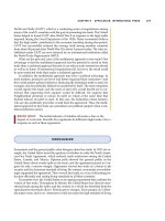
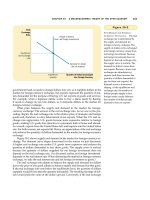
As indicated in Fig. 31-1, all of these syndromes derive from a com-
mon, low-grade, often asymptomatic syphilitic meningitis. In fact, this
is the most chronic of all known forms of meningitis and may be active
for 10 to 15 years. In its more subacute phase (within 2 years of infec-
tion), it may present with headache, drowsiness, and cranial nerve
palsies (meningeal syphilis). After 2 to 10 years, arterial inflammation
may result in a stroke (meningovascular syphilis). General paresis is a
gradual dementing meningoencephalitis appearing 12 to 15 years after
the onset of infection. Tabes dorsalis (literally a wasting of the dorsal
funiculi of the spinal cord secondary to lumbosacral radiculitis) pre-
sents, after 15 to 20 years, with a chronic syndrome of lancinating pains
in the legs, crises of gastric pain, deep sensory loss and ataxia, impo-
tence, hypotonia of the bladder with urinary retention and overflow
incontinence, Charcot joints, and Argyll Robertson pupils (Chap. 14).
Optic neuritis is often added; it consists of unilateral and later bilateral
loss of vision and optic atrophy.
Diagnosis is based on a history of primary or secondary syphilis, the
clinical characteristics of the neurologic syndrome, and the laboratory
testing for reagin and treponemal antibodies (VDRL and FTA-ABS).
CHAPTER 31 / NONVIRAL INFECTIONS OF THE NERVOUS SYSTEM 285
4777 Victor Ch 31 p277-289 6/11/01 2:10 PM Page 285
The CSF is abnormal in all cases of active neurosyphilis (increase in
lymphocytes and mononuclear cells, increased protein, especially
gamma globulin, normal glucose, presence of syphilitis reagin and anti-
bodies).
The treatment of all forms of neurosyphilis consists of administration
of penicillin G, 18 to 24 million units IV daily in six divided doses, for
14 days. Erythromycin and tetracycline, 0.5 g every 6 h, for 20 to 30
days are suitable substitutes in penicillin-sensitive patients. If symp-
toms recede and CSF abnormalities are reversed (disappearance of cells
and reduction in protein, gamma globulin, and serology titers), no fur-
ther treatment is indicated. Relapse, which is revealed by the return of
symptoms and reactivation of the CSF, requires additional treatment.
The CSF should be reexamined at 6 and 12 months after treatment.
Lyme Disease
This disease, known in Europe as erythema chronicum migrans, has
been encountered with increasing frequency during the past decade.
The infective agent is the spirochete Borrelia burgdorferi, and the vec-
tor is the common ixodid tick. The initial manifestation, at the site of
the tick bite, is an enlarging erythematous ring-shaped lesion, some-
times surrounded by satellites. The skin lesion may be overlooked or
disregarded but is followed, weeks to months later, by arthritis (two-
thirds of cases), cardiac manifestations (15 percent), and neurologic
complications (8 percent). The disease is not fatal but can lead to pro-
286
PART IV / THE MAJOR CATEGORIES OF NEUROLOGIC DISEASE
FIG. 31-1 Diagram of the evolution of neurosyphillis in the immune-
competent host.
4777 Victor Ch 31 p277-289 6/11/01 2:10 PM Page 286
longed disability if not recognized and treated. The association of
arthritis and neurologic involvement most often taking the form of a
fluctuating meningoencephalitis (headache, stiff neck, nausea and vom-
iting, chronic fatigue) with cranial or peripheral neuritis, particularly
facial palsy, has long been known in Europe as the Bannwarth syn-
drome. Myelitic and cauda equina syndromes and a polymyositis are
also documented. Meningeal symptoms are associated with a CSF lym-
phocytosis (up to 3000 per mm
3
), an elevated protein content, but nor-
mal glucose.
Diagnostic laboratory tests are the indirect immunofluorescence
assay and the enzyme-linked immunosorbent assay (ELISA). The use
of oral penicillin, tetracycline, or erythromycin in the initial stage of the
disease will prevent the cardiac, arthritic, and neurologic manifesta-
tions. The onset of meningeal symptoms requires high doses of antibi-
otics—penicillin, 20 million units daily IV for 10 days, or probably
better, ceftriaxone, 2 g/day for 30 days. Concomitant administration of
prednisone is said to be helpful.
Fungal Infections of the CNS
These are much less common than bacterial infections. Cryptococcosis,
candidiasis, aspergillosis, mucormycosis, coccidioidomycosis, blasto-
mycosis, and actinomycosis have all been identified, but only the first
three occur with any degree of regularity. Mucormycosis is most often
observed as a complication of diabetes. Candidiasis is associated with
severe burns and other chronic illnesses. Coccidioidomycosis is a com-
mon, influenza-like disease of the southwestern United States, rarely
causing meningitis. These infections may arise without obvious predis-
posing cause, but more often they complicate some other disease
process, such as malignancy or AIDS or other disease that suppresses
the immune responses (opportunistic infections).
Cryptococcosis (formerly called torulosis) is the fungal infection
seen most often in the United States. Its incidence has increased as a
result of AIDS. It gives rise to a subacutely evolving meningitis and
meningoencephalitis, the symptoms of which are much the same as
tuberculous meningitis. The CSF findings are also similar. Some cases
are fatal within a few weeks; others are chronic over months or years,
especially if treated. Specific diagnosis depends upon identifying Cryp-
tococcus neoformans in India ink preparations of the CSF, culturing the
organism on Sabouraud glucose agar, and a positive latex agglutination
test for the cryptococcal polysaccharide antigen in the CSF (90 percent
sensitive in AIDS patients; 50 percent in others). Treatment consists of
IV administration of amphotericin B. After a test dose of 5 mg, the drug
is given in a dosage of 1.0 mg/kg daily or every second day to a total of
2 to 3 g. The addition of flucytosine (150 mg/kg per day) results in
fewer failures and decreased nephrotoxicity, but the mortality is still
CHAPTER 31 / NONVIRAL INFECTIONS OF THE NERVOUS SYSTEM 287
4777 Victor Ch 31 p277-289 6/11/01 2:10 PM Page 287
about 40 percent and the patient must be monitored closely for bone
marrow suppression. Small deep brain infarctions may occur as a result
of basal angiitis, similar to tuberculous meningitis.
Infections Caused by Protozoa and Worms
Of the protozoal infections, only toxoplasmosis is observed with any
frequency in the United States and Europe. Immunocompromised
adults, notably those with AIDS, are particularly vulnerable. In healthy
adults, the infection is usually asymptomatic, but an infected woman
may transmit the disease to her unborn fetus. The disease takes the form
of a multifocal encephalitis with inflammatory necrotic foci, large
enough to be seen by CT scanning and MRI. Diagnosis is established
by elevation of specific serologic titers; it is rare to find the organism in
the CSF. Treatment with sulfadiazine (4 to 6 g daily) and pyrimeth-
amine (50 to 100 mg daily) with folinic acid should be continued for at
least 4 weeks, and lifelong in patients with AIDS. The main differential
diagnostic consideration in AIDS patients is cerebral lymphoma.
Cysticercosis and schistosomiasis are major infections in certain
parts of the world, and involvement of the nervous system greatly wors-
ens the outcome. Cysticercosis (the larval or intermediate stage of
infection with the pork tapeworm Taenia solium) causes focal inflam-
matory lesions in the brain, which become encysted and calcified and
often epileptogenic. Large intraventricular or cerebellar cysts may
cause hydrocephalus. The calcified lesions are readily seen on CT
scans.
In rare instances, the ova of trematodes (schistosomiasis) cause
necrotizing foci in the brain or spinal cord. Treatment of both cysticer-
cosis and schistosomiasis has been greatly enhanced by the use of the
antihelminthic agent praziquantel (50 mg/kg orally for 15 to 30 days) or
albendazole (5 mg tid for 15 to 30 days).
Trichinosis presents essentially as a self-limiting polymyositis,
involving cranial muscles and the heart. Rarely, cerebral emboli com-
plicate the myocarditis (see the Principles).
For a more detailed discussion of this topic, see Adams, Victor, and
Ropper: Principles of Neurology, 6th ed, pp 695–741.
ADDITIONAL READING
Coonrod JD, Dans PE: Subdural empyema. Am J Med 53:85, 1972.
Feigin RD, McCracken GH Jr, Klein JO: Diagnosis and management of meningi-
tis. Pediatr Infect Dis J 11:785, 1992.
Garcia-Monco JC, Benach JL: Lyme neuroborreliosis. Ann Neurol 37:691, 1995.
Leys D, Destee A, Petit H, Warot P: Management of subdural intracranial empye-
mas should not always require surgery. J Neurol Neurosurg Psychiatry 49:635,
1986.
288 PART IV / THE MAJOR CATEGORIES OF NEUROLOGIC DISEASE
4777 Victor Ch 31 p277-289 6/11/01 2:10 PM Page 288
Pomeroy SL, Holmes SJ, Dodge PR, Feigin RD: Seizures and other neurologic
sequelae of bacterial meningitis in children. New Engl J Med 323:1651, 1990.
Quagliarello JJ, Scheld WM: Treatment of bacterial meningitis. New Engl J Med
336:708, 1997.
Reik L: Spirochetal infections of the nervous system, in Kennedy PGE, Johnson
RT (eds): Infections of the Nervous System. Boston, Butterworth, 1987, pp
43–75.
Snider DE, Roper WL: The new tuberculosis. New Engl J Med 326:703, 1992.
Swartz MN: “Chronic meningitis”—Many causes to consider. New Engl J Med
317:957, 1987.
Tyler KL, Martin JB: Infectious Diseases of the Nervous System. Philadelphia, FA
Davis, 1993.
Walsh TJ, Hier DB, Caplan LR: Fungal infections of the central nervous system:
Comparative analysis of risk factors and clinical signs in 57 patients. Neurol-
ogy 35:1654, 1985.
CHAPTER 31 / NONVIRAL INFECTIONS OF THE NERVOUS SYSTEM 289
4777 Victor Ch 31 p277-289 6/11/01 2:10 PM Page 289
32 Viral Infections of the
Nervous System
Viruses enter the body in many ways—via the respiratory passages
(mumps, measles, varicella), by the oral-intestinal route (enteroviruses)
or the genital-mucosal route (herpes), by inoculation (arboviruses,
AIDS), transplacentally (rubella, cytomegalovirus), or along peripheral
nerves (herpes, rabies). Once the nervous system is invaded, the virus
multiplies in selective regions of the brain or spinal cord or in the
choroid plexuses and meninges. Six syndromes are thus induced, occur-
ring with such regularity that if recognized they not only stamp the
infection as viral but also may indicate the identity of the virus. These
syndromes are as follows:
1. Acute aseptic (nonsuppurative) meningitis
2. Acute encephalitis and meningoencephalitis
3. Herpes zoster and simplex ganglionitis
4. Chronic infections due to “slow viruses” and unconventional agents
(prions)
5. Acquired immunodeficiency syndrome (AIDS)
6. Acute anterior poliomyelitis
THE SYNDROME OF ASEPTIC MENINGITIS
The term aseptic meningitis designates a common clinical syndrome
consisting of fever, headache, and other signs of meningeal irritation,
a predominantly lymphocytic pleocytosis with normal CSF glucose and
negative bacterial and fungal cultures. Photophobia and pain on move-
ment of the eyes are other common complaints. Sometimes drowsiness
and confusion are added, making it difficult to distinguish a pure
meningitis from a meningoencephalitis. The CSF reaction is the same
in both—pleocytosis, mainly lymphocytes (typically 100 to 300 per
mm
3
, sometimes more), increase in protein, but normal glucose. Rarely,
the glucose level is reduced slightly.
Most cases of aseptic meningitis are due to viral infections, but there
are important nonviral causes as well. Most cases do not show signs of
a preceding or concomitant respiratory or enteric infection.
Viral Causes of Meningitis
1. Enteroviral infections: echovirus, Coxsackie, enterovirus, and non-
paralytic poliomyelitis. Peak incidence is in August and September.
290
4777 Victor Ch 32 p290-301 6/11/01 2:11 PM Page 290
Copyright 1998 The McGraw-Hill Companies, Inc. Click Here for Terms of Use.
These viruses account for 80 percent of cases of established viral
origin.
2. Mumps: Highest incidence is in late winter and spring. Male-to-
female ratio is 3:1.
3. Herpes simplex, type 2, genital (rarely type 1), Epstein-Barr virus
(EBV), and rarely cytomegalovirus (CMV).
4. Lymphocytic choriomeningitis: Lymphocyte count in CSF may be
1000 per mm
3
or higher. Infection is acquired by contact with
infected hamsters and mice, mainly in late fall and winter.
5. Adenovirus infections.
6. HIV (AIDS) may cause an acute or chronic aseptic meningitis with
a clinical picture like that of infectious mononucleosis (EBV).
Most of these conditions are benign. Specific diagnosis requires viral
isolation or detection of at least a fourfold rise in serum antibody titers
during the acute and convalescent phases of the illness. A specific cause
is not established in one-half or more of cases of presumed viral origin.
The same holds true for many cases of suspected viral encephalitis (see
below).
Nonviral Causes of Aseptic Meningitis
1. Spirochetal infections: The most important are syphilitic meningitis
and Lyme disease, described in Chap. 31. Leptospirosis, with a peak
incidence in August, is acquired by contact with contaminated urine
of rats, dogs, swine, and cattle.
2. Mycoplasma pneumoniae: Cold agglutinins in the serum toward the
end of the first week of illness or detection of the organism by PCR
techniques is diagnostic. Q fever and other rickettsial illnesses may
also give rise to aseptic meningitis and meningoencephalitic syn-
dromes, with atypical pneumonitis.
3. Bacterial infections lying adjacent to the meninges (see Chap. 30).
4. Neoplastic invasion of meninges by lymphoma or carcinoma.
5. Chemical irritation of the meninges by blood, by contents of a cra-
niopharyngioma, or by substances injected intrathecally.
6. Recurrent and chronic inflammatory meningitides of obscure ori-
gin—Vogt-Koyanagi-Harada syndrome (iridocyclitis, depigmenta-
tion of skin, deafness); meningitis with serum sickness and con-
nective tissue disease such as lupus erythematosus; Behçet disease
(relapsing meningitis, iridocyclitis, ulcers of mouth and genitalia);
and so-called Mollaret’s recurrent meningitis (which is probably due
to the herpes simplex virus).
In the diagnosis of aseptic meningitis, it is important to exclude
tuberculosis, cryptococcosis, Lyme disease, syphilis, and inadequately
treated bacterial meningitis, all of which require urgent treatment.
CHAPTER 32 / VIRAL INFECTIONS OF THE NERVOUS SYSTEM 291
4777 Victor Ch 32 p290-301 6/11/01 2:11 PM Page 291
SYNDROME OF ACUTE ENCEPHALITIS
In this class of viral diseases, a febrile illness is expressed by meningi-
tis, to which are added the following neurologic abnormalities in vari-
ous combinations: impairment of consciousness (confusion, stupor, and
coma); seizures; mutism or aphasia; hemiparesis, with asymmetry of
reflexes and Babinski signs; involuntary movements, cerebellar ataxia,
and polymyoclonus; and cranial nerve palsies. The arboviral and some
of the enteroviral encephalitides have a strong seasonal incidence. Viral
encephalitis is in effect a meningoencephalitis, and mild forms of
encephalitis, in which the meningeal symptoms and CSF abnormalities
predominate, cannot be distinguished from viral (aseptic) meningitis, as
mentioned in the preceding section.
Causation
The causes of acute viral meningoencephalitis in their approximate
order of frequency are as follows:
1. Mumps virus
2. Arboviruses: Eastern, Western, and Venezuelan equine; La Crosse,
St. Louis, California, and Colorado tick fever viruses; Japanese B
(outside the United States)
3. Herpes simplex, zoster, CMV, and EBV
4. Lymphocytic choriomeningitis virus
5. Enteroviruses (Coxsackie viruses and echoviruses)
6. Cytomegalovirus
7. Adenoviruses
8. Rabies virus
Herpes Simplex Encephalitis
This, the most serious of the viral encephalitides, occurs sporadically
throughout the year, in patients of all ages, and in all parts of the world.
It is caused by type 1 herpes simplex virus, very rarely by type 2 (gen-
ital herpes).
The symptoms, consisting of fever, headache, confusion, stupor, and
coma, evolve over a period of several days. Additional symptoms in
some patients include olfactory and gustatory hallucinations, temporal
lobe or motor seizures, changes in personality and behavior, and apha-
sia. While a single convulsion or a flurry of seizures is common, status
epilepticus almost never occurs. The latter symptoms betray the pre-
dominant localization of the disease process in the inferior and medial
parts of the temporal lobes and orbital parts of the frontal lobes. The
lesions are characterized by intense inflammation, often hemorrhagic,
and pannecrosis of nearly all tissue elements. Intranuclear eosinophilic
inclusions are found in neurons and glial cells.
292 PART IV / THE MAJOR CATEGORIES OF NEUROLOGIC DISEASE
4777 Victor Ch 32 p290-301 6/11/01 2:11 PM Page 292
The temporal lobe destructive lesions can be seen with CT scanning
and MRI, often asymmetrically in the two hemispheres. The CSF find-
ings are like those of other encephalitides (predominantly mononuclear
pleocytosis, elevated protein, normal glucose), except that in some
cases there may be as many as several thousand red cells. Certain EEG
findings (periodic high-voltage sharp waves and slow-wave complexes
at 2- to 3-s intervals in the temporal leads) should suggest the diagno-
sis. If the diagnosis is reasonably certain, it is best to proceed at once
with treatment. Brain biopsy carries a greater risk than the inappropri-
ate use of antiviral agents. Moreover, there is now a relatively sensitive
polymerase chain reaction technique to detect the virus.
About half the patients with this disease (those who are stuporous or
comatose when first seen) do not survive, and many of those who do are
left with an amnesic state and seizures.
Treatment consists of the administration of acyclovir (30 mg/kg per
day for 14 days). Initiation of treatment early in the illness (before the
onset of stupor and coma) significantly reduces mortality and the sever-
ity of the residual neurologic deficits.
Nonviral Forms of Encephalitis
Numerous bacterial, fungal, parasitic, and noninfectious diseases may
simulate the viral encephalitides and need to be distinguished from
them. These nonviral diseases, many of which require urgent therapeu-
tic intervention, are listed in Table 32-1.
SYNDROME OF HERPES ZOSTER
This well-known disorder (also called zona or “shingles”) is caused by
the varicella-zoster (VZ) virus. It has an overall incidence of three to
five cases per thousand patients per year and is considerably more fre-
quent in the elderly and in those with malignancies, particularly lym-
phoma and Hodgkin disease. Herpes zoster probably represents a
reactivation of varicella virus infection that has been latent in sensory
ganglia following the primary infection with chickenpox.
Clinical features The characteristic manifestations are radicular pain, a
vesicular cutaneous eruption involving one or two dermatomes on one
side of the body, and in some cases sensory and motor deficits in the
segments bearing the skin lesions. The vesicular eruption is preceded
for 3 to 4 days (sometimes as long as 7 days) by dysesthesias in the
involved dermatomes, or there may be severe localized pain suggestive
of pleurisy or an acute abdominal condition.
Any part of the body may be affected, but thoracic lesions are the
most frequent. Involvement of multiple dermatomes should always sug-
gest an underlying immunocompromised state. Involvement of cranial
CHAPTER 32 / VIRAL INFECTIONS OF THE NERVOUS SYSTEM 293
4777 Victor Ch 32 p290-301 6/11/01 2:11 PM Page 293
294 PART IV / THE MAJOR CATEGORIES OF NEUROLOGIC DISEASE
TABLE 32-1 Diseases Simulating Viral Encephalitis
Bacterial
Mycoplasma pneumoniae
Leptospirosis
Lyme disease
Syphilis (secondary or meningovascular)
Listeriosis
Cat-scratch disease (Bartonella henselae)
Brucellosis (particularly Brucella melitensis)
Tuberculosis
Legionella
Typhoid fever
Nocardia
Actinomycosis
Parameningeal infections (epidural, petrositis)
Partially treated bacterial meningitis
Brain abscess
Fungal
Cryptococcosis
Coccidioidomycosis
Histoplasmosis
North American blastomycosis
Candidiasis
Rickettsial
Rocky Mountain spotted fever
Typhus
Q fever
Parasitic
Toxoplasmosis
Cysticercosis
Echinococcosis
Trypanosomiasis
Plasmodium falciparum
Amebiasis (Naegleria and Acanthamoeba)
Neoplastic
Carcinomatous meningitis
Gliomatosis cerebri
Paraneoplastic limbic encephalitis
Vascular
Granulomatous angiitis
Systemic lupus erythematosus
Others
Sarcoid
Behçet syndrome
Oculocephalic syndromes (e.g., Stevens-Johnson, Vogt-Kayanagi-
Harada)
4777 Victor Ch 32 p290-301 6/11/01 2:11 PM Page 294
ganglia is associated with two special syndromes, both with prominent
paralytic features: (1) ophthalmic herpes, with pain and eruption in the
distribution of the first division of the trigeminal nerve, ophthalmople-
gia, and risk of corneal ulceration; and (2) so-called geniculate herpes
(Ramsay Hunt syndrome), with facial paralysis, vertigo, deafness, and
otic-palatal vesiculation (sometimes restricted to a small region of the
concha of the ear). Herpes occipitocollaris, with involvement of palate,
pharynx, neck, and retroauricular region, is caused by herpetic infection
of the ganglia of cranial nerves IX and X and upper cervical roots. The
CSF in all the zoster syndromes contains 10 to 100 cells, mainly lym-
phocytes, and a slightly increased protein. A delayed brainstem arteri-
tis and ischemic stroke complicate some cases.
Pain and dysesthesia last for 1 to 4 weeks in most cases, but in as
many as one-third of patients, pain persists for months or even years
and creates a difficult therapeutic problem.
Pathologically, there is an intense inflammation in two or three dor-
sal root or cranial nerve ganglia and in corresponding posterior and
anterior roots, adjacent meninges, and gray matter of the spinal cord on
one side. The latter lesion is a veritable poliomyelitis, but the neuronal
destruction is more in the posterior than in the anterior horn. Myelitis
and encephalitis are rare complications.
A course of acyclovir (800 mg five times daily for 7 days), if begun
within 48 h after the appearance of the rash, shortens the period of acute
pain and hastens the healing of the vesicles; however, it does not pre-
vent the occurrence of postherpetic neuralgia. In nonimmunosup-
pressed patients, prednisone (45 to 60 mg/day for 7 days, then tapered)
decreases the incidence of postherpetic neuralgia. The latter disorder is
best treated by a combination of carbamazepine or neurontin and
amitriptyline, beginning with small doses that are gradually increased
to 400 to 800 and 75 to 150 mg/day, respectively. Lidocaine or cap-
saicin topical creams and nerve root blocks are effective in some cases.
Herpes Simplex
The most important nervous system complication of herpes simplex
infection is an encephalitis (described above) due usually to the type 1
virus. However, there are other examples of nervous system involve-
ment by the herpes simplex virus, usually type 2—infection of the
facial nerve, perhaps the main cause of Bell’s palsy; localized infection
of the trigeminal ganglion, giving rise to a unilateral facial sensory loss;
genital herpes leading to a unilateral lumbosacral ganglionitis and
radiculopathy; meningitis, sometimes recurrent (Mollaret meningitis);
rare instances of transverse myelitis; and encephalitis (in adults).
In the newborn, herpes simplex infection can be a devastating and
rapidly fatal disease. It is usually contracted in the birth canal from a
mother with type 2 (genital herpes). The results of antiviral treatment
are unclear.
CHAPTER 32 / VIRAL INFECTIONS OF THE NERVOUS SYSTEM 295
4777 Victor Ch 32 p290-301 6/11/01 2:11 PM Page 295
CHRONIC INFECTIONS DUE TO “SLOW VIRUSES” AND
UNCONVENTIONAL AGENTS (PRIONS)
Subacute sclerosing panencephalitis (SSPE) This is a slowly evolv-
ing inflammatory disease appearing in children and adolescents several
years after an attack of measles. It is characterized by dementia, focal
or generalized seizures, ataxia of gait, and polymyoclonus. It evolves
over a period of months to several years and leaves the child virtually
decerebrate. The EEG is typical—periodic bursts of high-voltage slow
waves followed by a flat pattern. Gamma globulin and measles anti-
bodies are greatly elevated in the CSF. Since measles vaccine has come
to be widely used, this neurologic disease has virtually disappeared.
A subacute progressive panencephalitis occurring many years after
congenital rubella has also been identified.
Progressive multifocal leukoencephalopathy (PML) This disease is
usually associated with AIDS, Hodgkin disease, lymphoma, or chronic
leukemia and less often with tuberculosis, sarcoid, or other states of
immunosuppression. It develops over a 3- to 6-month period, with focal
cerebral, brainstem, and cerebellar signs. The lesions are demyelinative
and well delineated by MRI. Inclusion bodies are seen in oligodendro-
cytes, and astrocytes are gigantic and show tumor-like mitoses. A poly-
oma virus—designated JC virus—has been isolated from the lesions.
Remission has occurred in AIDS patients treated with an aggressive
retroviral regimen. There is no effective treatment for the others.
Subacute spongiform encephalopathy (SSE) This disease, also re-
ferred to as Creutzfeldt-Jakob disease, is characterized by a rapidly pro-
gressive dementia in association with cerebellar ataxia, heightened
startle reaction, diffuse myoclonic jerks, and cortical blindness in some
cases. The CSF is normal. Usually, after one or two months of illness,
the EEG is diagnostic—high-voltage slow and sharp waves, occurring
periodically at 1- to 3-Hz intervals, on an increasingly flat background
(“burst suppression”). As the disease advances, the patient becomes
totally unresponsive and the outcome is invariably fatal, usually in less
than a year. A variant illness, contracted from infected cattle (bovine
spongiform encephalopathy), is of concern in Great Britain and western
Europe.
The disease affects principally the cerebral and cerebellar cortices, in
which there is a diffuse loss of neurons, gliosis, and a striking vacuola-
tion of the tissues. Inflammatory changes are absent, and no inclusion
bodies have been observed. The disease is due to an unconventional
agent—a proteinaceous infectious particle, called a prion, which lacks
the structure of a virus and which can be transmitted to chimpanzees,
with an incubation period of more than a year. In more than 90 percent
of cases, the diagnosis can be established by the detection of antibodies
296 PART IV / THE MAJOR CATEGORIES OF NEUROLOGIC DISEASE
4777 Victor Ch 32 p290-301 6/11/01 2:11 PM Page 296
to the prion protein in the CSF; also, the content of enolase in the CSF
is increased. Pathologically and epidemiologically, SSE resembles a
disease first recognized among natives of New Guinea and known as
kuru. Gerstmann-Sträussler disease and fatal familial insomnia are rare
variants of this disease.
There is no known treatment. Precautions need to be taken in the
medical care of these patients, like those recommended for patients
with hepatitis B (see Harrison’s Principles of Internal Medicine).
THE ACQUIRED IMMUNODEFICIENCY SYNDROME (AIDS)
This viral syndrome is characterized by an acquired and unusually pro-
found depression of cell-mediated immunity (cutaneous anergy, lym-
phopenia, reversal of T-helper/T-suppressor cell ratio (CD4/CD8), and
depressed in vitro lymphoproliferative response to various antigens and
mitogens). The causative virus, originally called human T-cell lym-
photropic virus (HTLV-3), is now generally referred to as human
immunodeficiency virus (HIV or HIV-1). The diseases it induces, due
to the effects of the virus itself and a wide array of opportunistic infec-
tions and neoplasms, are designated as AIDS (acquired immunodefi-
ciency syndrome).
Epidemiology AIDS is mainly a disease of homosexual or bisexual
men (56 percent) and of male and female drug users (19 percent). A
smaller group at risk are hemophiliacs (and other patients who receive
transfusions or injections of blood products) and infants born of women
with AIDS. There is a small group of heterosexual men who appear to
have been infected by prostitutes. Four-fifths of the reported cases in
the United States have been from New York, California, New Jersey,
and Florida.
Clinical manifestations These range from the asymptomatic sero-
conversion state to widespread lymphadenopathy, diarrhea, and weight
loss (AIDS-related complex, or ARC) to full-blown AIDS, comprising
some or all of the complications listed in Table 32-2. In approximately
one-third of patients, the CNS or PNS is clinically involved by the time
of death, and on postmortem examination nearly all patients prove to
have CNS lesions.
The neurologic manifestations are too numerous and varied to
describe in detail. They are listed in Table 32-2 and are described in the
appended references as well as in appropriate chapters thorughout the
book.
Laboratory tests Many screening tests are now available, all of them
based on an enzyme-linked immunoassay (ELISA). While highly sen-
CHAPTER 32 / VIRAL INFECTIONS OF THE NERVOUS SYSTEM 297
4777 Victor Ch 32 p290-301 6/11/01 2:11 PM Page 297